Article

Atopobium vaginae and Prevotella bivia Are Able to Incorporate and Influence Gene Expression in a Pre-Formed Gardnerella vaginalis Biofilm

1
Centre of Biological Engineering (CEB), Laboratory of Research in Biofilms Rosário Oliveira (LIBRO), Campus de Gualtar, University of Minho, 4710-057 Braga, Portugal
2
Division of Infectious Diseases, University of Alabama at Birmingham, Birmingham, AL 35294, USA
*
Author to whom correspondence should be addressed.
Pathogens 2021, 10(2), 247; https://doi.org/10.3390/pathogens10020247
Submission received: 31 December 2020 / Revised: 18 February 2021 / Accepted: 19 February 2021 / Published: 20 February 2021
(This article belongs to the Special Issue Vaginal Bacteria from the Genus Gardnerella)

Abstract

:
Bacterial vaginosis (BV) is associated with a highly structured polymicrobial biofilm on the vaginal epithelium where Gardnerella species presumably play a pivotal role. Gardnerella vaginalis, Atopobium vaginae, and Prevotella bivia are vaginal pathogens detected during the early stages of incident BV. Herein, we aimed to analyze the impact of A. vaginae and P. bivia on a pre-established G. vaginalis biofilm using a novel in vitro triple-species biofilm model. Total biofilm biomass was determined by the crystal violet method. We also discriminated the bacterial populations in the biofilm and in its planktonic fraction by using PNA FISH. We further analyzed the influence of A. vaginae and P. bivia on the expression of key virulence genes of G. vaginalis by quantitative PCR. In our tested conditions, A. vaginae and P. bivia were able to incorporate into pre-established G. vaginalis biofilms but did not induce an increase in total biofilm biomass, when compared with 48-h G. vaginalis biofilms. However, they were able to significantly influence the expression of HMPREF0424_0821, a gene suggested to be associated with biofilm maintenance in G. vaginalis. This study suggests that microbial relationships between co-infecting bacteria can deeply affect the G. vaginalis biofilm, a crucial marker of BV.

Graphical Abstract

1. Introduction

Bacterial vaginosis (BV) is the most-common vaginal infection affecting fertile, premenopausal, and pregnant women [1]. It is associated with important adverse outcomes related to pregnancy [2] and infertility [3]. Additionally, it is associated with an increased risk of acquisition of HIV and other sexually transmitted infections (STIs) [4,5,6,7,8]. Microbiologically, BV is a complex polymicrobial infection where beneficial vaginal bacteria, mainly hydrogen peroxide and lactic acid-producing Lactobacillus species, which are usually dominant in vaginal microbiota of healthy women, are replaced by high concentrations of facultative and strict anaerobic bacteria [1,9]. The most prominent of these are Gardnerella spp., facultative anaerobes usually found embedded in a polymicrobial BV biofilm [10,11,12]. However, Gardnerella spp. are also commonly found in asymptomatic or BV-negative women [13]. This has aroused interest in whether genetic differences among G. vaginalis isolates might differentiate pathogenic from commensal organisms [14]. G. vaginalis was the only recognized species in its genus for over four decades, but very recently the Gardnerella taxonomic description was amended based on comparisons of whole-genome sequencing and matrix-assisted laser desorption/ionization time-of-flight (MALDI-TOF) mass spectrometry analysis, resulting in four species (G. vaginalis, G. leopoldii, G. swidsinskii, and G. piotii) and nine ‘genome species’ [15]. It is likely that each named Gardnerella species and ‘genome species’ are specifically associated with either BV or healthy vaginal microbiota due to differences in their virulence potential, as suggested by several studies [16,17,18,19,20,21]. Following this renewed taxonomy of the genus Gardnerella, in this article, the term Gardnerella spp. will be used to address previous publications, since we cannot rule out the fact that other Gardnerella species were involved.
While BV etiology is still a matter of debate [1], a common hypothesis suggests that virulent strains of Gardnerella spp. initiate the formation of the biofilm on vaginal epithelial cells and become a scaffolding to which other BV-associated anaerobes thereafter can attach [9]. Atopobium vaginae is one of the species that is often found associated with Gardnerella spp. biofilms [22]. Evidence suggests that the therapeutic failures and recurrences of BV might be associated with the presence of high loads of A. vaginae, since this species presents specific resistance against standard antibiotics [23,24,25]. Additionally, A. vaginae has been positively associated with vaginal discharge in women with BV, an elevated vaginal pH, and the presence of clue cells [26]. It was also described that high vaginal loads of A. vaginae in combination with Gardnerella spp. are related to late miscarriage and pre-maturity [23].
Recently it has been pointed out that Prevotella bivia might have an important role in the early stage of BV development [9,27]. Using daily vaginal swabs, Muzny and colleagues found that P. bivia was the first BV-associated species to increase in relative abundance above baseline prior to incident BV, followed shortly thereafter by an increase in the relative abundance of Gardnerella spp., suggesting that synergism between P. bivia and Gardnerella spp. might be an important event prior to BV [28]. In fact, an earlier in vitro study demonstrated that Gardnerella spp. and P. bivia can act synergistically [29]. The authors showed that Gardnerella spp. produce amino acids through their metabolism, which can be used by P. bivia as its nutrient source which results in the production of ammonia, which in turn is used by Gardnerella spp. More recently, Gilbert and colleagues established an in vivo BV model in which they coinfected mice with Gardnerella spp. and P. bivia, revealing that Gardnerella facilitates ascension of P. bivia into the uterine horns [30]. The virulence potential of P. bivia is also derived from studies that associated its colonization with preterm birth, endometritis, and other uterine pathologies [31,32,33].
Knowledge of the microbial interactions in the vaginal ecosystem during BV is still scarce since functional microbial studies in polymicrobial biofilms are very limited [34]. We have recently shown that when growing dual-species BV biofilms, distinct microbial interactions can occur, including antagonistic [35] or synergistic biofilm accumulation [35,36], as well as molecular interactions that have an impact on Gardnerella gene expression [37]. Thus, in this study, we aimed to develop and characterize, for the first time, an in vitro model containing G. vaginalis, A. vaginae, and P. bivia, and to investigate the ability of these three species to form a multi-species biofilm, with particular attention to their ability to induce alterations in key genes of interest.

2. Results

2.1. Quantification of the Biomass of Mono-, Dual-, and Triple-Species BV-Associated Biofilms

In the BV-associated vaginal ecosystem, resident microorganisms interact with each other in both synergistic and antagonistic manners, which might affect their ability to form biofilms in this polymicrobial community [35]. Initially, we compared the in vitro biofilm formation ability of each tested species for 24 h and 48 h using optimized [38] in vitro conditions (Figure 1A). In both timepoints tested, G. vaginalis formed a biofilm with the highest total biomass, while P. bivia formed a biofilm with the lowest total biomass. Following the hypothesis that Gardnerella is the early colonizer in BV [9], we then assessed how A. vaginae and P. bivia could incorporate into a 24-h pre-formed G. vaginalis biofilm. Under the tested conditions, all the consortia reached the same level of total biofilm biomass (Figure 1B). Curiously, when compared to the mono-species G. vaginalis 48-h biofilm, none of the consortia provided an added advantage in terms of an increase in total biomass. While it was tempting to compare the 48-h consortia with A. vaginae or P. bivia 48-h mono-species biofilms, it is important to highlight that both species were only allowed to grow for 24 h, after the initial 24-h G. vaginalis biofilm was performed, as described in the methods section.

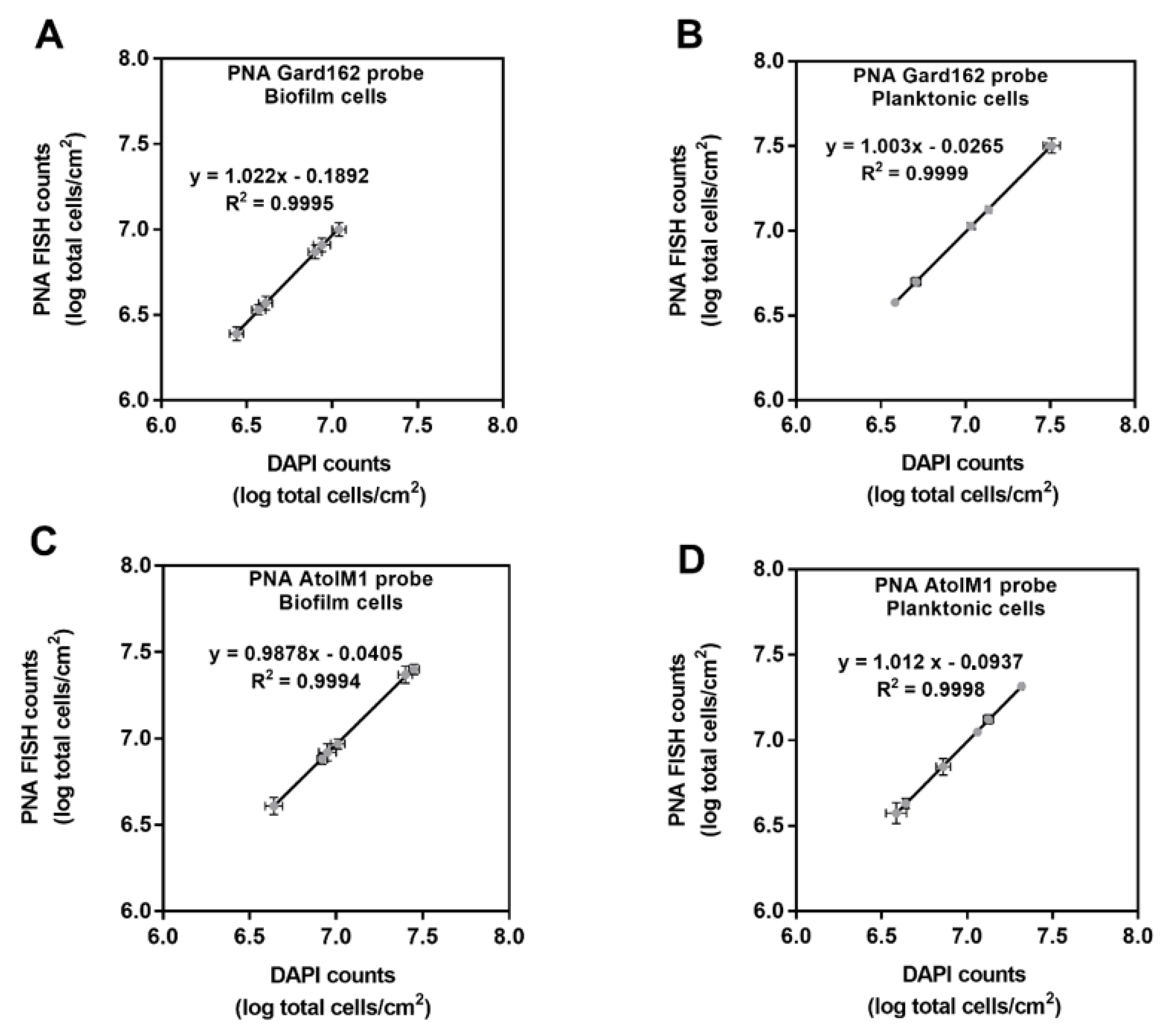
2.2. In Vitro PNA Gard162 and PNA AtoITM1 Probes Specificity and Efficiency

Although the PNA Gard162 [39] and PNA AtoITM1 [40] probes’ specificity had been previously tested for several BV-associated bacteria, we also analyzed these probes’ specificity for the bacterial strains used in this study. Based on our results (Table 1), PNA Gard162 and PNA AtoITM1 probes hybridized with G. vaginalis and A. vaginae, respectively, whereas no hybridization was observed for the other species tested, showing a specificity of 100% as previously reported. Additional details of the specificity of the Gard162 and AtoITM1 probes are shown in Supplementary Figures S1 and S2.
As no P. bivia PNA FISH probe currently exists, the estimation of P. bivia counts could only be assessed indirectly by 4′-6-diamidino-2-phenylindole (DAPI) counterstaining, assuming that all cells with unlabeled PNA probes were P. bivia; however, this needs to be experimentally determined [41]. As such, we compared the data obtained from PNA FISH and DAPI counts for both G. vaginalis and A. vaginae pure-culture biofilms and the planktonic fractions of the biofilm. Not surprisingly, each probe failed to detect 100% of the respective total cells. By performing serial dilutions of each sample, calibration curves were obtained for G. vaginalis biofilm (Figure 2A) or planktonic cells (Figure 2B) and for A. vaginae biofilm (Figure 2C) or planktonic cells (Figure 2D).
Taking into consideration this data, it was possible to calculate the efficiency of each probe and to develop an equation that would correct PNA counts, to prevent overestimation of DAPI counts as P. bivia counts, as listed in Table 2.

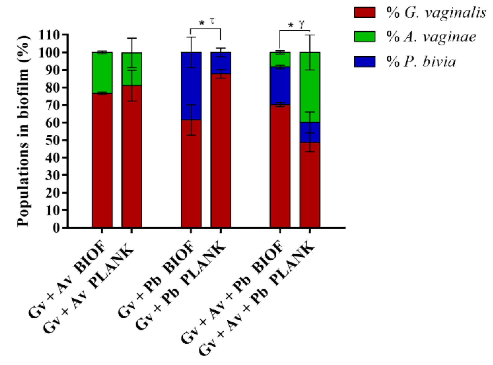
2.3. Quantification and Distribution of Bacterial Populations in Dual- and Triple-Species BV-Associated Biofilms by PNA FISH

Taking advantage of the robustness of the PNA FISH/DAPI approach for the differentiation between G. vaginalis, A. vaginae, and P. bivia, we discriminated the bacterial populations into dual- and triple-species BV-associated biofilms and their planktonic fractions. Similar to what was described before [42], we showed that A. vaginae and P. bivia were able to incorporate in the dual-species biofilms, accounting for up to ∼23% and ∼38% of the total number of cells, respectively (Figure 3). Curiously, in the triple-species biofilms, the relative load of A. vaginae was reduced to ~8.3%, maintaining G. vaginalis as the main species. The lower ability of A. vaginae to integrate the triple-species biofilms is evidence that somewhat specific interactions are established when these three bacterial species act as a consortium, which in our tested conditions promoted the enhanced integration of P. bivia, in depreciation of A. vaginae. When looking at the bacterial populations found on the planktonic fraction of the biofilm, it is noteworthy that significant differences were found both for P. bivia and the triple-species consortia; in contrast the relative composition of both biofilms and planktonic fractions were similar for A. vaginae.
We also analyzed bacterial distribution in the intact structure of the dual and triple-species biofilms by confocal laser scanning microscopy (CLSM). As shown in Figure 4, in both dual- and triple-species biofilms, A. vaginae and P. bivia were found well-distributed across the G. vaginalis biofilm, in small clusters of cells. Details in the orthogonal views of mono-, dual-, and triple- species biofilms are shown in Supplementary Figure S3.

2.4. Impact of A. vaginae and P. bivia on G. vaginalis Virulence

Considering the central role often attributed to Gardnerella in BV etiology [1,43], shifts in the G. vaginalis transcriptome during the establishment of polymicrobial BV-associated biofilms could be a key for unveiling interspecies interactions that enhance the virulence of G. vaginalis. Thus, we focused on deciphering the impact of A. vaginae and/or P. bivia on G. vaginalis virulence. As such, we analyzed the expression of genes related to cytotoxicity, biofilm maintenance, antimicrobial resistance, and evasion of the immune system in cells from mono-, dual-, and triple-species biofilms. Regarding cytotoxicity, G. vaginalis produces the toxin vaginolysin (vly), which might induce lysis in vaginal cells membranes [44,45]. However, as shown in Figure 5A, according to our in vitro conditions, no significant differences were found in the expression of this gene by G. vaginalis between the different biofilm models. We also addressed sialidase (sld) expression, since sialidase appears to contribute to G. vaginalis cytotoxicity by the destruction of the protective mucus layer on the vaginal epithelium [46]. Although we detected a slight decrease in sld transcription, it was not statistically significant in dual- and triple-species biofilms, as compared with mono-species G. vaginalis biofilms (Figure 5B).
Regarding the glycosyltransferases, it has been proposed that they are involved in the transfer of a sugar moiety to a substrate and are thus essential in the biosynthesis of glycoconjugates like exopolysaccharides and glycoproteins, which are important for biofilm maintenance to maximize the full virulence of G. vaginalis [47,48]. Of note, the expression of the HMPREF0424_0821 transcript, which encodes glycosyltransferases type II, was up-regulated in all the tested conditions, being approximately 2.8-fold higher in triple-species than in mono-species biofilms (p < 0.05; Figure 5C). We also analyzed the expression of transcripts encoding antimicrobial-specific resistance proteins belonging to efflux pump families (HMPREF0424_1122 and HMPREF0424_0156). Despite detecting slight changes in the transcription of these genes, these were not statistically significant (Figure 5D,E). Nevertheless, a tendency was observed in the presence of P. bivia, with a downregulation of the tested G. vaginalis genes, similar to what we have previously found [37]. Finally, we analyzed the expression of HMPREF0424_1196 transcript, which encodes a Rib-protein that belongs to the α-like protein (Alf)-family of highly repetitive surface antigens [49], which elicit protective immunity through their inter-strain size variability [48]. Here the most striking difference was in the triple-species biofilm, as compared to the G. vaginalis mono-species biofilm (p < 0.05; Figure 5F).

3. Discussion

The dynamic and complex nature of the vaginal microbiota and the likely role of multiple bacterial species in the pathogenesis of BV have posed major challenges for developing realistic polymicrobial in vitro biofilm models [50,51]. To date, the majority of in vitro studies only address mono- or dual-species biofilms, and are focused on Gardnerella species [37,52]. Herein, we describe, for the first time, an in vitro biofilm composed of G. vaginalis, A. vaginae, and P. bivia, three highly relevant BV-associated species [28,30]. In a polymicrobial community, bacterial species interact extensively with each other and these interactions might also determine the structure and composition of multi-species biofilms [53]. It is, thus, reasonable to assume that in triple-species BV-associated biofilms the microbial interactions become more complex than in dual- or mono-species biofilms [54].
Our experimental model follows the hypothesis that Gardnerella is the early colonizer in BV [9], to which we later allowed co-incubation with A. vaginae and P. bivia. In vitro experimental data supporting the role of some Gardnerella species to be the early colonizers is derived from the fact that some Gardnerella isolates have a significantly higher ability to adhere to epithelial cells than many other BV-associated species [52,55,56] and are also able to displace vaginal lactobacilli [57], a pivotal step in the development of a characteristic multi-species biofilm. It is also important to bear in mind that the recent reclassification of G. vaginalis [15] in multiple Gardnerella species might have important implications in BV etiology, as a particular species might have a different role in BV development, although this still needs to be further explored [19,21,58]. Herein, we selected a type strain of G. vaginalis since its complete genome is available, and thus, it is well genotypically characterized, being used in several studies associated with BV [15,58]. Regarding other BV-associated species, we selected A. vaginae and P. bivia to be included in this study, due to data suggesting they may be more than bystanders [9,32]. Indeed, in a prospective vaginal microbiome study Muzny and colleagues showed that the mean relative abundance of P. bivia, Gardnerella spp., and A. vaginae became significantly higher in cases four days before (P. bivia), three days before (Gardnerella spp.), and on the day of (A. vaginae) incident BV onset [28]. Based on this study, the authors suggested that together with virulent G. vaginalis, P. bivia, and A. vaginae may have potential key roles in the induction and development of incident BV [9]. Nevertheless, knowledge about the microbial relationships between these three bacterial species remains scarce. In order to shed new light on this aspect, we analyzed the effect of A. vaginae and P. bivia on biofilm formation and its impact on G. vaginalis pathogenicity.
Similar to what is described in oral infections, in which the interactions between microbial communities have tremendous importance for the development of oral disease [59], we hypothesize that G. vaginalis, A. vaginae, and P. bivia establish a network of interactions that affect the development of the BV-associated biofilm, a key hallmark of BV. Curiously, under the tested conditions, inoculation or co-inoculation of the different species on the pre-formed G. vaginalis biofilm did not significantly enhance the amount of total biofilm biomass. However, despite widespread utilization of the CV staining method for biofilm quantification used in this study, an inherent limitation of this method is that total biomass comparison between species is not possible, since different species produce distinct biofilm matrices and have different cell sizes [60,61]. Thus, interpretation of these data should be made with reservation. Nevertheless, as shown in our CLSM and PNA-FISH quantitative data, dual- and triple-species biofilms contained significant numbers of each species, with G. vaginalis as the most prominent species, similar to in vivo studies [11,22]. Interestingly, our PNA FISH data suggest that different microbial relationships are established in dual- and triple-species biofilms. This is inferred by the distinct bacterial composition observed in multi-species biofilms, in particular the triple-species biofilm where the relative composition of G. vaginalis and P. bivia increased while A. vaginae decreased. This is even more relevant since P. bivia formed a weak mono-species biofilm and grew slower than A. vaginae and G. vaginalis in New York City III (NYC III) medium, as shown previously [38]. It is important to mention that several factors influenced the bacterial integration in biofilm, including the growth rates of the bacterial species [62] and the ability to adhere to a surface [63,64] and to each other [65,66]. Interestingly, despite the slower growth rate, P. bivia was better fit to grow in the biofilm than A. vaginae in our tested conditions. This suggests that G. vaginalis modifies the local environment, making it more favorable for the growth of P. bivia, which might be a result of an ammonia flow mechanism as proposed by Pybus and colleagues [29].This further demonstrates that different bacterial species influence the growth of other species, and likely have an impact in BV etiology. It is also interesting to observe that the planktonic fraction of the dual- and triple-species biofilm did not always coincide with the biofilm consortia, suggesting that the specific microenvironment of the biofilms provide different competitive advantages, as shown elsewhere [67,68].
It is noteworthy that synergistic effects were, however, observed when we analyzed changes in G. vaginalis gene expression. The expression of the HMPREF0424_0821 transcript, which encodes a glycosyltransferase, was significantly up-regulated in the multi-species consortia. Glycosyltransferases have been described as key enzymes required for biofilm maintenance [48], and may also have a putative role for cell surface glycoconjugates which has been proposed to shape vaginal microbiota–host interactions [69]. This enhancement was only significant in the triple-species biofilm model, highlighting, therefore, the importance of the interplay between multiple bacterial species in the development of BV. Studies to assess the role of glycosyltransferase on biofilm formation and virulence have been conducted for other species, namely for Streptococcus mutans, a bacterium responsible for the initiation of tooth decay [70]. It was shown that glycosyltransferase mediated biofilm matrix dynamics and virulence. Interestingly, the deletion of the glycosyltransferase gene resulted in no change in overall biofilm biomass, however, the mutant strain originated an altered biofilm architecture. Concurrently, the mutant was less virulent in an in vivo rat model of dental caries [70]. Corroborating the relevance of glycosyltransferase in biofilms, we have recently shown that this gene was up-regulated in Gardnerella spp. in 15 different dual-species consortia [37]. Together, these data emphasize the importance of other BV-associated species in G. vaginalis virulence, and consequently, in host infection.
While we have previously shown that P. bivia could enhance G. vaginalis vly expression two-fold, when a dual-species biofilm was grown in supplemented brain-heart infusion (sBHI) medium [37], in NYC III medium vly expression was not changed. Regarding the role of sld, some early studies pointed out that this gene is strongly linked with the development of a biofilm [71,72], however, in our tested conditions, no relevant changes were verified when G. vaginalis was cultivated in mono- or multi-species biofilms, corroborating our previous findings [37]. Such evidence supports a recent study that postulated that sld does not likely have a role in establishing or maintaining the biofilm [73]. It should be pointed out that in this study we chose to use NYC III medium, since sBHI was not appropriate to induce mono-species biofilms from either P. bivia or A. vaginae [38]. Since bacterial gene expression is strongly influenced by media conditions [74,75], we cannot exclude that the optimal growth conditions provided by NYC III could also somehow be affecting gene expression by G. vaginalis. Indeed, by comparing the data from this study with our previous findings [37], we observed that using NYC III, the base level of G. vaginalis vly expression was approximately 10-fold higher than in comparison with sBHI, which could explain the differences observed in both studies. On account of the fact that synergistic effects often occur when one bacterium is providing some advantage to another [76,77], by using an optimal biofilm-inducing media such as NYC III, it might be possible that some synergistic effects were masked. Evidence to support this hypothesis can be observed by the fact that several strains of A. vaginae easily die out when grown in sBHI medium, but maintain viability when co-cultured with G. vaginalis [78]. However, in NYC III, A. vaginae is able to survive alone. Furthermore, for the other tested genes, with exception of sld, we also noted a higher base level (2- to 5-fold) of expression in NYC III as compared with sBHI. Despite these differences, a similar effect on G. vaginalis transcriptomic profile was observed for the remaining tested genes, as compared with our previous dual-species biofilm study [37]. However, further studies are required to elucidate the influence of the growth media in gene expression by G. vaginalis. In triple-species biofilms, we observed a downregulation of these genes when compared to G. vaginalis mono-species biofilms, however, this requires future study to understand the molecular mechanisms involved in antimicrobial resistance and evasion of the immune system. Taken together, our data suggest that microbial relationships between co-infecting bacteria can influence the development of a polymicrobial biofilm, a marker of BV. However, more research is needed to provide a better mechanistic insight into the complex interplay between G. vaginalis, A. vaginae, and P. bivia, and their eukaryotic hosts. While the choice of a very rich medium was used to guarantee that all the tested bacteria were able to grow in vitro in a biofilm phenotype, trying to determine these microbial interactions in conditions more similar to the vaginal environment might provide novel insights, as we previously demonstrated that growing Gardnerella spp. in a medium simulating genital tract secretions and complemented with components of the host immune system had a significant impact on the growth and biofilm formation [79]. Understanding the molecular basis and biological effect of these microbial interactions and microbial–host interactions may provide novel information necessary to define more effective and goal-oriented treatment in BV and improve women’s reproductive health.

4. Materials and Methods

4.1. Bacterial Strains and Culture Conditions

G. vaginalis strain ATCC 14018T, A. vaginae strain ATCC BAA-55T, and P. bivia strain ATCC 29303T were used in this study. Each inoculum was grown in New York City III broth (NYC III) ((1.5% (w/v) Bacto™ proteose peptone no. 3 (BD, Franklin Lakes, NJ, USA), 0.5% (w/v) glucose (Thermo Fisher Scientific, Lenexa, KS, USA), 0.24% (w/v) HEPES (VWR, Sparks, NV, USA), 0.5% (w/v) NaCl (VWR), and 0.38% (w/v) yeast extract (Liofilchem, Roseto degli Abruzz, Italy)) supplemented with 10% (v/v) inactivated horse serum (Biowest, Nuaillé, France) [38] for 24 h at 37 °C under anaerobic conditions (AnaeroGen Atmosphere Generation system, Oxoid, Hampshire, United Kingdom), as we previously showed this to be the optimal condition to grow mono-species biofilms of the selected bacterial species [38].

4.2. Biofilm Formation and Biomass Quantification by the Crystal Violet Method

Dual- and triple-species biofilms were initiated by inoculating a 107 colony-forming units (CFU)/mL bacterial suspension of G. vaginalis into 24-well tissue culture plates (Orange Scientific, Braine L’Alleud, Belgium) and by incubating the plates for 24 h, at 37 °C, and under anaerobic conditions. After 24 h, planktonic cells were removed, and 107 CFU/mL of each bacterial species, A. vaginae or P. bivia (for dual-species biofilms) and A. vaginae, P. bivia (for triple-species biofilms), were inoculated in the pre-formed G. vaginalis biofilms and incubated for another 24 h. Of note, we first adjusted the bacterial concentration of the bacterial suspension to 9 × 10⁷ CFU/mL due to the limit of detection of the microplate reader and then we diluted to 1 × 10⁷ CFU/mL, confirming this concentration by CFUs. At 620 nm, 9 × 10⁷ CFU/mL of G. vaginalis corresponds to an optical density (OD) = 0.15; for A. vaginae an OD = 0.11, and for P. bivia an OD = 0.16. Mono-species biofilms of G. vaginalis were grown as a control for 48 h, in which fresh medium was added to the respective wells after the first 24 h of biofilm formation. In addition, we also performed 24-h mono-species biofilm growth for A. vaginae and P. bivia to examine their individual ability to grow in the tested conditions. To quantify the biomass of mono-, dual-, and triple-species biofilms, we used the crystal violet (CV) method [80]. In brief, after the fixation step with 100% (v/v) methanol (Thermo Fisher Scientific) for 20 min, biofilms were stained with CV solution at 1% (v/v) (Merck, Darmstadt, Germany) for 20 min. Each well was washed twice with phosphate-buffered saline, and bound CV was released with 33% (v/v) acetic acid (Thermo Fisher Scientific). To estimate total biofilm biomass, the OD of the resulting solution was measured at 595 nm. Biofilm assays were repeated three times on separate days, with four technical replicates assessed each time.

4.3. In Vitro PNA Gard162 and AtoITM1 Probes Specificity and Efficiency

Although the specificity of PNA Gard162 [39], and AtoITM1 [40] probes have been previously tested, we also evaluated the probe specificity for the bacterial species used in this study. Thus, we performed an experiment in order to detect any possible cross-hybridization. The evaluation of PNA FISH hybridization was based on a qualitative score, as previously described [39]: (−) absence of hybridization, (++) moderate hybridization, (+++) good hybridization, and (++++) optimal hybridization.
We then carried out an experiment to analyze the efficiency of both PNA probes. As such, we performed several dilutions from pure bacterial suspensions obtained from mono-species biofilms and their planktonic fractions. To determine the efficiency of each probe, the same sample was hybridized with a species-specific probe and then stained with 4′-6-diamidino-2-phenylindole (DAPI, 2.5 μg/mL) to account for nonhybridizing bacteria; defined as double staining. After the double staining, the bacteria were enumerated at two different wavelengths at the same position within the sample. Based on both data, we performed a correlation between the PNA counts and the DAPI counts that allowed us to obtain the equations shown in Figure 2. All experiments were performed in triplicate.

4.4. Quantification of Bacterial Populations in Dual- and Triple-Species Biofilms and Their Planktonic Fraction by PNA FISH

The bacterial population within the 48-h multi-species biofilms and in their planktonic fraction was discriminated using the peptide nucleic acid fluorescence in situ hybridization (PNA FISH) method, as previously described [37]. Briefly, after fixing the biofilm suspension, a PNA probes specific for G. vaginalis (Gard162) and for A. vaginae (AtoITM1) were added to each well of epoxy-coated microscope glass slides (Thermo Fisher Scientific). An additional staining step was done at the end of the hybridization procedure, covering each glass slide with DAPI (2.5 μg/mL). Microscopic visualization was performed using filters capable of detecting the PNA Gard162 probe (BP 530-550, FT 570, LP 591 sensitive to the Alexa Fluor 594 molecule attached to the Gard162 probe), the PNA AtoITM1 probe (BP 470–490, FT500, LP 516 sensitive to the Alexa Fluor 488 molecule attached to the AtoITM1 probe), and DAPI (BP 365–370, FT 400, LP 42). Twenty fields were randomly acquired in each sample. The number of bacteria was counted using ImageJ Software [59]. To reduce any possible overestimation due to the use of DAPI as the probe efficiency was not 100%, we then applied the equations from Table 2 to obtain more accurate relative values. These assays were repeated three times on separate days.

4.5. Confocal Laser Scanning Microscopy Analysis of Biofilm Bacterial Distribution

To analyze the bacterial distribution of dual- and triple-species biofilms, the biofilm structure was evaluated by confocal laser scanning microscopy (CLSM) using the PNA Gard162 and AtoITM1 probes coupled to DAPI staining, as described above. For this experiment, biofilms were formed on an 8-well chamber slide (Thermo Fisher Scientific™ Nunc™ Lab-Tek™, Bohemia, NY, USA) at 37 °C under anaerobic conditions for 48 h with the replacement of NYC III medium at 24 h of growth and the addition of the respective BV-associated bacteria. The CLSM images were acquired in an Olympus™ FluoView FV1000 (Olympus, Tokyo, Japan) confocal scanning laser microscope, using a 40× objective. Microscopic visualization was performed using lasers capable of detecting the PNA Gard162 probe (Laser 559, excitation wavelength 559 nm, emission wavelength 618 nm, BA 575–675, sensitive to the Alexa Fluor 594 molecule attached to the Gard162 probe), the PNA AtoITM1 probe (Laser 488, excitation wavelength 488 nm, emission wavelength 520 nm, BA 505–540, sensitive to the Alexa Fluor 488 molecule attached to the AtoITM1 probe), and DAPI (Laser 405, excitation wavelength 405 nm, emission wavelength 461 nm, BA 430–470). Images were acquired with 640 × 640 resolutions of each surface analyzed. All assays were repeated three independent times with two technical replicates.

4.6. G. vaginalis Gene Expression Quantification in Mono-, Dual-, and Triple-Species Biofilms

Gene expression of six potential Gardnerella virulence genes, specifically vaginolysin (vly), sialidase (sld), glycosyltransferase, type II (HMPREF0424_0821), multidrug ABC transporter (HMPREF0424_1122), bacitracin transport, ATP-binding protein BcrA (HMPREF0424_0156), and a transcript that encodes a Rib-protein (HMPREF0424_1196) was determined in 48-h mono-, dual-, and triple-species biofilms. Total RNA was extracted using an E.Z.N.A.® Bacterial RNA Kit (Omega Bio-tek, Norcross, GA, USA) with minor changes, as previously optimized [81]. Next, genomic DNA was degraded with one step of DNase treatment (Fermentas, Vilnius, Lithuania) following the manufacturer’s instructions. RNA concentration, purity, and integrity were determined, as previously described [82]. The same amount of total RNA (300 ng/μL) was reverse transcribed using the RevertAid™ First Strand cDNA synthesis kit (Fermentas), as previously optimized [83], and gene-specific reverse transcription primers as a priming strategy. Quantitative PCR (qPCR) was prepared by mixing 5 µL of iQ SYBR green supermix (Bio-Rad, Hercules, CA, USA), 2 µL of 1:100 diluted cDNA, 0.5 µL of 5 µM forward and reverse primers (Table S1), and water up to 10 µL. The run was performed in a CFX96TM thermal cycler (Bio-Rad) with the following cycling parameters: 3 min at 95 °C, followed by 45 cycles of 10 s at 95 °C, 10 s at 60 °C, and 15 s at 72 °C. Reaction efficiency was determined by the dilution method [84]. It is of note that at 60 °C, all sets of primers used (Table S1) had similar efficiencies. In addition, the analysis of the melting curves confirmed the presence of a single peak, providing evidence for the specificity of the tested primers. Normalized gene expression was determined by using the delta Ct method (EΔCt), a variation of the Livak method, where ΔCt = Ct (reference gene) − Ct (target gene) and E stands for the reaction efficiency experimentally determined. A non-reverse transcriptase control was included in each reaction. All assays were repeated at least three independent times with three technical replicates.

4.7. Statistical Analysis

The data were analyzed using the statistical package GraphPad Prism version 6 (La Jolla, CA, USA) by paired t-test, two-way ANOVA (Sidak’s multiple comparison test) and Mann–Whitney U test for the data that did not follow a normal distribution according to the Kolmogorov–Smirnov test. Values with p < 0.05 were considered statistically significant.

Supplementary Materials

The following are available online at https://www.mdpi.com/2076-0817/10/2/247/s1, Figure S1: An example of fluorescence microscopy pictures representing the specificity of Gard162 probe, Figure S2: An example of fluorescence microscopy pictures representing the specificity of AtoITM1 probe, Figure S3: An example of orthogonal views of BV-associated biofilms by confocal laser scanning microscopy (CLSM). Table S1: Primers used in qPCR experiments.

Author Contributions

Conceptualization, N.C.; methodology, J.C.; investigation, J.C. and A.S.R.; writing—original draft preparation, J.C. and N.C.; writing—review and editing, A.S.R. and C.A.M.; supervision, N.C.; project administration, C.A.M. and N.C.; funding acquisition, C.A.M. and N.C. All authors have read and agreed to the published version of the manuscript.

Funding

This research was partially funded by the National Institute of Allergy and Infectious Diseases (R01AI146065-01A1). It was also partially funded by the Portuguese Foundation for Science and Technology (FCT), by the research project (PTDC/BIA-MIC/28271/2017), under the scope of COMPETE 2020 (POCI-01-0145-FEDER-028271), and by the strategic funding of unit (UIDB/04469/2020). The funders had no role in study design, data collection and analysis, decision to publish, or preparation of the manuscript.

Conflicts of Interest

Christina A. Muzny, MD, MSPH has received research grant support from Lupin Pharmaceuticals, is a consultant for Lupin Pharmaceuticals and BioFire Diagnostics, and has received honoraria from Elsevier, Abbott Molecular, Cepheid, Becton Dickinson, Roche Diagnostics, and Lupin. She is currently on the scientific advisory board for PhagoMed. The other authors declare no conflict of interest.

References

  1. Rosca, A.S.; Castro, J.; Sousa, L.G.V.; Cerca, N. Gardnerella and vaginal health: The truth is out there. FEMS Microbiol. Rev. 2019, 44, 73–105. [Google Scholar] [CrossRef]
  2. Arif, F. Bacterial Vaginosis: Risk of Adverse Pregnancy Outcome. J. Gynecol. Res. Obstet. 2018, 4, 015–017. [Google Scholar] [CrossRef] [Green Version]
  3. Moragianni, D.; Dryllis, G.; Andromidas, P.; Kapeta-Korkouli, R.; Kouskouni, E.; Pessach, I.; Papalexis, P.; Kodonaki, A.; Athanasiou, N.; Pouliakis, A.; et al. Genital tract infection and associated factors affect the reproductive outcome in fertile females and females undergoing in vitro fertilization. Biomed. Rep. 2019, 10, 231–237. [Google Scholar] [CrossRef]
  4. Brusselaers, N.; Shrestha, S.; van de Wijgert, J.; Verstraelen, H. Vaginal dysbiosis and the risk of human papillomavirus and cervical cancer: Systematic review and meta-analysis. Am. J. Obstet. Gynecol. 2019, 221, 9–18.e8. [Google Scholar] [CrossRef]
  5. Brotman, R.M.; Klebanoff, M.A.; Nansel, T.R.; Yu, K.F.; Andrews, W.W.; Zhang, J.; Schwebke, J.R. Bacterial vaginosis assessed by Gram stain and diminished colonization resistance to incident gonococcal, chlamydial, and trichomonal genital infection. J. Infect. Dis. 2010, 202, 1907–1915. [Google Scholar] [CrossRef]
  6. Abbai, N.S.; Reddy, T.; Ramjee, G. Prevalent bacterial vaginosis infection—A risk factor for incident sexually transmitted infections in women in Durban, South Africa. Int. J. STD AIDS 2016, 27, 1283–1288. [Google Scholar] [CrossRef]
  7. Lokken, E.M.; Balkus, J.E.; Kiarie, J.; Hughes, J.P.; Jaoko, W.; Totten, P.A.; Scott McClelland, R.; Manhart, L.E. Association of Recent Bacterial Vaginosis with Acquisition of Mycoplasma genitalium. Am. J. Epidemiol. 2017, 186, 194–201. [Google Scholar] [CrossRef] [PubMed] [Green Version]
  8. Atashili, J.; Poole, C.; Ndumbe, P.M.; Adimora, A.A.; Smith, J.S. Bacterial vaginosis and HIV acquisition: A meta-analysis of published studies. Aids 2008, 22, 1493–1501. [Google Scholar] [CrossRef] [Green Version]
  9. Muzny, C.A.; Taylor, C.M.; Swords, W.E.; Tamhane, A.; Chattopadhyay, D.; Cerca, N.; Schwebke, J.R. An Updated Conceptual Model on the Pathogenesis of Bacterial Vaginosis. J. Infect. Dis. 2019, 220, 1399–1405. [Google Scholar] [CrossRef]
  10. Swidsinski, A.; Mendling, W.; Loening-Baucke, V.; Swidsinski, S.; Dörffel, Y.; Scholze, J.; Lochs, H.; Verstraelen, H. An adherent Gardnerella vaginalis biofilm persists on the vaginal epithelium after standard therapy with oral metronidazole. Am. J. Obstet. Gynecol. 2008, 198, 1–6. [Google Scholar] [CrossRef]
  11. Swidsinski, A.; Loening-Baucke, V.; Mendling, W.; Dörffel, Y.; Schilling, J.; Halwani, Z.; Jiang, X.F.; Verstraelen, H.; Swidsinski, S. Infection through structured polymicrobial Gardnerella biofilms (StPM-GB). Histol. Histopathol. 2014, 29, 567–587. [Google Scholar] [CrossRef]
  12. Swidsinski, A.; Verstraelen, H.; Loening-Baucke, V.; Swidsinski, S.; Mendling, W.; Halwani, Z. Presence of a Polymicrobial Endometrial Biofilm in Patients with Bacterial Vaginosis. PLoS ONE 2013, 8, 4–8. [Google Scholar] [CrossRef] [Green Version]
  13. Aroutcheva, A.A.; Simoes, J.A.; Behbakht, K.; Faro, S. Gardnerella vaginalis isolated from patients with bacterial vaginosis and from patients with healthy vaginal ecosystems. Clin. Infect. Dis. 2001, 33, 1022–1027. [Google Scholar] [CrossRef] [Green Version]
  14. Bekasiak, A.; Dammann, F.; Nader, C. A rare cause of a scrotal abscess due to the symbiotic infection of Gardnerella vaginalis and Prevotella bivia in an adult male. Pathogens 2020, 9, 93. [Google Scholar] [CrossRef] [Green Version]
  15. Vaneechoutte, M.; Guschin, A.; Van Simaey, L.; Gansemans, Y.; Van Nieuwerburgh, F.; Cools, P. Emended description of Gardnerella vaginalis and description of Gardnerella leopoldii sp. nov., Gardnerella piotii sp. nov. and Gardnerella swidsinskii sp. nov., with delineation of 13 genomic species within the genus Gardnerella. Int. J. Syst. Evol. Microbiol. 2019, 69, 679–687. [Google Scholar] [CrossRef]
  16. Potter, R.F.; Burnham, C.A.D.; Dantas, G. In silico analysis of Gardnerella genomospecies detected in the setting of bacterial vaginosis. Clin. Chem. 2019, 65, 1375–1387. [Google Scholar] [CrossRef] [Green Version]
  17. Pleckaityte, M.; Janulaitiene, M.; Lasickiene, R.; Zvirbliene, A. Genetic and biochemical diversity of Gardnerella vaginalis strains isolated from women with bacterial vaginosis. FEMS Immunol. Med. Microbiol. 2012, 65, 69–77. [Google Scholar] [CrossRef] [Green Version]
  18. Janulaitiene, M.; Gegzna, V.; Baranauskiene, L.; Bulavaitė, A.; Simanavicius, M.; Pleckaityte, M. Phenotypic characterization of Gardnerella vaginalis subgroups suggests differences in their virulence potential. PLoS ONE 2018, 13, 1–20. [Google Scholar] [CrossRef] [PubMed]
  19. Hill, J.E.; Albert, A.Y.K.; VOGUE Research Group. Resolution and cooccurrence patterns of Gardnerella leopoldii, G. swidsinskii, G. piotii, and G. vaginalis within the vaginal microbiome. Infect. Immun. 2019, 87, e00532-19. [Google Scholar] [CrossRef]
  20. Schellenberg, J.J.; Patterson, M.H.; Hill, J.E. Gardnerella vaginalis diversity and ecology in relation to vaginal symptoms. Res. Microbiol. 2017, 168, 837–844. [Google Scholar] [CrossRef]
  21. Khan, S.; Voordouw, M.J.; Hill, J.E. Competition Among Gardnerella Subgroups From the Human Vaginal Microbiome. Front. Cell. Infect. Microbiol. 2019, 9, 1–10. [Google Scholar] [CrossRef] [Green Version]
  22. Swidsinski, A.; Mendling, W.; Loening-Baucke, V.; Ladhoff, A.; Swidsinski, S.; Hale, L.P.; Lochs, H. Adherent biofilms in bacterial vaginosis. Obstet. Gynecol. 2005, 106, 1013–1023. [Google Scholar] [CrossRef] [PubMed] [Green Version]
  23. Menard, J.P.; Mazouni, C.; Salem-Cherif, I.; Fenollar, F.; Raoult, D.; Boubli, L.; Gamerre, M.; Bretelle, F. High vaginal concentrations of Atopobium vaginae and Gardnerella vaginalis in women undergoing preterm labor. Obstet. Gynecol. 2010, 115, 134–140. [Google Scholar] [CrossRef]
  24. Mendling, W.; Palmeira-de-Oliveira, A.; Biber, S.; Prasauskas, V. An update on the role of Atopobium vaginae in bacterial vaginosis: What to consider when choosing a treatment? A mini review. Arch. Gynecol. Obstet. 2019, 300, 1–6. [Google Scholar] [CrossRef] [Green Version]
  25. Ferris, M.J.; Masztal, A.; Aldridge, K.E.; Fortenberry, D.; Fidel, P.L.; Martin, D.H. Association of Atopobium vaginae, a recently described metronidazole resistant anaerobe, with bacterial vaginosis. BMC Infect. Dis. 2004, 4, 1–8. [Google Scholar] [CrossRef] [Green Version]
  26. De Backer, E.; Verhelst, R.; Verstraelen, H.; Alqumber, M.A.; Burton, J.P.; Tagg, J.R.; Temmerman, M.; Vaneechoutte, M. Quantitative determination by real-time PCR of four vaginal Lactobacillus species, Gardnerella vaginalis and Atopobium vaginae indicates an inverse relationship between L. gasseri and L. iners. BMC Microbiol. 2007, 7, 115. [Google Scholar] [CrossRef] [PubMed] [Green Version]
  27. Spiegel, C.; Amsel, R.; Eschenbach, D.; Schoenknecht, F.; Holmes, K. Anaerobic bacteria in nonspecific vaginitis. N. Engl. J. Med. 1980, 303, 601–607. [Google Scholar] [CrossRef]
  28. Muzny, C.A.; Blanchard, E.; Taylor, C.M.; Aaron, K.J.; Talluri, R.; Griswold, M.E.; Redden, D.T.; Luo, M.; Welsh, D.A.; Van Der Pol, W.J.; et al. Identification of Key Bacteria Involved in the Induction of Incident Bacterial Vaginosis: A Prospective Study. J. Infect. Dis. 2018, 218, 966–978. [Google Scholar] [CrossRef]
  29. Pybus, V.; Onderdonk, A.B. Evidence for a commensal, symbiotic relationship between Gardnerella vaginalis and Prevotella bivia involving ammonia: Potential significance for bacterial vaginosis. J. Infect. Dis. 1997, 175, 406–413. [Google Scholar] [CrossRef] [PubMed]
  30. Gilbert, N.M.; Lewis, W.G.; Li, G.; Sojka, D.K.; Lubin, J.B.; Lewis, A.L. Gardnerella vaginalis and Prevotella bivia Trigger Distinct and Overlapping Phenotypes in a Mouse Model of Bacterial Vaginosis. J. Infect. Dis. 2019, 220, 1099–1108. [Google Scholar] [CrossRef]
  31. Gibbs, R.; McDuffi Jr, R.; Kunze, M.; Barr, J.M.; Wolf, D.; Sze, C.-I.; Shikes, R.; Sherman, M. Experimental intrauterine infection with Prevotella bivia in New Zealand White rabbits. Am. J. Obs. Gynecol. 2004, 1082–1086. [Google Scholar] [CrossRef]
  32. Randis, T.M.; Ratner, A.J. Gardnerella and Prevotella: Co-conspirators in the Pathogenesis of Bacterial Vaginosis. J. Infect. Dis. 2019, 220, 1085–1088. [Google Scholar] [CrossRef]
  33. Freitas, A.C.; Bocking, A.; Hill, J.E.; Money, D.M. Increased richness and diversity of the vaginal microbiota and spontaneous preterm birth. Microbiome 2018, 6, 1–15. [Google Scholar] [CrossRef]
  34. Pybus, V.; Onderdonk, A.B. Microbial interactions in the vaginal ecosystem, with emphasis on the pathogenesis of bacterial vaginosis. Microbes Infect. 1999, 1, 285–292. [Google Scholar] [CrossRef]
  35. Castro, J.; Cerca, N. BV and non-BV associated Gardnerella vaginalis establish similar synergistic interactions with other BV-associated microorganisms in dual-species biofilms. Anaerobe 2015, 36, 56–59. [Google Scholar] [CrossRef] [PubMed] [Green Version]
  36. Castro, J.; Machado, D.; Cerca, N. Escherichia coli and Enterococcus faecalis are able to incorporate and enhance a pre-formed Gardnerella vaginal is biofilm. Pathog. Dis. 2016, 74, ftw007. [Google Scholar] [CrossRef] [Green Version]
  37. Castro, J.; Machado, D.; Cerca, N. Unveiling the role of Gardnerella vaginalis in polymicrobial Bacterial Vaginosis biofilms: The impact of other vaginal pathogens living as neighbors. ISME J. 2019, 13, 1306–1317. [Google Scholar] [CrossRef] [PubMed]
  38. Rosca, A.S.; Castro, J.; Cerca, N. Evaluation of different culture media to support in vitro growth and biofilm formation of bacterial vaginosis-associated anaerobes. PeerJ 2020, 8, e9917. [Google Scholar] [CrossRef]
  39. Machado, A.; Almeida, C.; Salgueiro, D.; Henriques, A.; Vaneechoutte, M.; Haesebrouck, F.; Vieira, M.J.; Rodrigues, L.; Azevedo, N.F.; Cerca, N. Fluorescence in situ Hybridization method using Peptide Nucleic Acid probes for rapid detection of Lactobacillus and Gardnerella spp. BMC Microbiol. 2013, 13, 82. [Google Scholar] [CrossRef] [Green Version]
  40. Hardy, L.; Jespers, V.; Dahchour, N.; Mwambarangwe, L.; Musengamana, V.; Vaneechoutte, M.; Crucitti, T. Unravelling the bacterial vaginosis-associated biofilm: A multiplex Gardnerella vaginalis and Atopobium vaginae fluorescence in situ hybridization assay using peptide nucleic acid probes. PLoS ONE 2015, 10, 1–16. [Google Scholar] [CrossRef]
  41. Almeida, C.; Azevedo, N.F.; Santos, S.; Keevil, C.W.; Vieira, M.J. Discriminating multi-species populations in biofilms with peptide nucleic acid fluorescence in situ hybridization (PNA FISH). PLoS ONE 2011, 6. [Google Scholar] [CrossRef] [PubMed] [Green Version]
  42. Machado, A.; Jefferson, K.K.; Cerca, N. Interactions between Lactobacillus crispatus and bacterial vaginosis (BV)-associated bacterial species in initial attachment and biofilm formation. Int. J. Mol. Sci. 2013, 14, 12004–12012. [Google Scholar] [CrossRef] [Green Version]
  43. Schwebke, J.R.; Muzny, C.A.; Josey, W.E. Role of Gardnerella vaginalis in the pathogenesis of bacterial vaginosis: A conceptual model. J. Infect. Dis. 2014, 210, 338–343. [Google Scholar] [CrossRef]
  44. Pleckaityte, M.; Mistiniene, E.; Lasickiene, R.; Zvirblis, G.; Zvirbliene, A. Generation of recombinant single-chain antibodies neutralizing the cytolytic activity of vaginolysin, the main virulence factor of Gardnerella vaginalis. BMC Biotechnol. 2011, 11, 100. [Google Scholar] [CrossRef] [Green Version]
  45. Zilnyte, M.; Venclovas, Č.; Zvirbliene, A.; Pleckaityte, M. The cytolytic activity of vaginolysin strictly depends on cholesterol and is potentiated by human CD59. Toxins 2015, 7, 110–128. [Google Scholar] [CrossRef] [Green Version]
  46. Lopes Dos Santos Santiago, G.; Deschaght, P.; El Aila, N.; Kiama, T.N.; Verstraelen, H.; Jefferson, K.K.; Temmerman, M.; Vaneechoutte, M. Gardnerella vaginalis comprises three distinct genotypes of which only two produce sialidase. Am. J. Obstet. Gynecol. 2011, 204, 450.e1–450.e7. [Google Scholar] [CrossRef]
  47. Breton, C.; Lenka, Š.; Jeanneau, C.; Ko, J.; Imberty, A. Structures and mechanisms of glycosyltransferases. Glycobiology 2006, 16, 29–37. [Google Scholar] [CrossRef]
  48. Yeoman, C.J.; Yildirim, S.; Thomas, S.M.; Durkin, A.S.; Torralba, M.; Sutton, G.; Buhay, C.J.; Ding, Y.; Dugan-Rocha, S.P.; Muzny, D.M.; et al. Comparative genomics of Gardnerella vaginalis strains reveals substantial differences in metabolic and virulence potential. PLoS ONE 2010, 5. [Google Scholar] [CrossRef] [Green Version]
  49. Lindahl, G.; Sta, M.; Areschoug, T. Surface Proteins of Streptococcus agalactiae and Related Proteins in Other Bacterial Pathogens. Clin. Microbiol. Rev. 2005, 18, 102–127. [Google Scholar] [CrossRef] [Green Version]
  50. Onderdonk, A.B.; Delaney, M.L.; Fichorova, N. The Human Microbiome during Bacterial Vaginosis. Clin. Microbiol. Rev. 2016, 29, 223–238. [Google Scholar] [CrossRef] [Green Version]
  51. Pekmezovic, M.; Mogavero, S.; Naglik, J.R.; Hube, B. Host–Pathogen Interactions during Female Genital Tract Infections. Trends Microbiol. 2019, 27, 982–996. [Google Scholar] [CrossRef]
  52. Patterson, J.L.; Stull-Lane, A.; Girerd, P.H.; Jefferson, K.K. Analysis of adherence, biofilm formation and cytotoxicity suggests a greater virulence potential of Gardnerella vaginalis relative to other bacterial-vaginosis-associated anaerobes. Microbiology 2010, 156, 392–399. [Google Scholar] [CrossRef] [Green Version]
  53. Yang, L.; Liu, Y.; Wu, H.; Høiby, N.; Molin, S.; Song, Z.J. Current understanding of multispecies biofilms. Int. J. Oral Sci. 2011, 3, 74–81. [Google Scholar] [CrossRef]
  54. Bai, F.; Cai, Z.; Yang, L. Recent progress in experimental and human disease-associated multi- species biofilms. Comput. Struct. Biotechnol. J. 2019, 17, 1234–1244. [Google Scholar] [CrossRef]
  55. Alves, P.; Castro, J.; Sousa, C.; Cereija, T.B.; Cerca, N. Gardnerella vaginalis Outcompetes 29 other bacterial species isolated from patients with bacterial vaginosis, using in an in vitro biofilm formation model. J. Infect. Dis. 2014, 210, 593–596. [Google Scholar] [CrossRef] [Green Version]
  56. Machado, A.; Salgueiro, D.; Harwich, M.; Jefferson, K.K.; Cerca, N. Quantitative analysis of initial adhesion of bacterial vaginosis-associated anaerobes to ME-180 cells. Anaerobe 2013, 23, 1–4. [Google Scholar] [CrossRef] [Green Version]
  57. Castro, J.; Alves, P.; Sousa, C.; Cereija, T.; França, Â.; Jefferson, K.K.; Cerca, N. Using an in-vitro biofilm model to assess the virulence potential of bacterial vaginosis or non-bacterial vaginosis Gardnerella vaginalis isolates. Sci. Rep. 2015, 5, 11640. [Google Scholar] [CrossRef] [PubMed] [Green Version]
  58. Bulavaitè, A.; Dalgediene, I.; Michailoviene, V.; Pleckaityte, M. Type II Restriction-Modification System from Gardnerella vaginalis ATCC 14018. Pathogens 2020, 9, 703. [Google Scholar] [CrossRef]
  59. Taubman, M.; Ruby, J.; Goldner, M. Nature of Symbiosis in Oral Disease. J. Dent. Res. 2007, 86, 2005–2008. [Google Scholar] [CrossRef]
  60. Haney, E.F.; Trimble, M.J.; Cheng, J.T.; Vallé, Q.; Hancock, R.E.W. Critical Assessment of Methods to Quantify Biofilm Growth and Evaluate Antibiofilm Activity of Host Defence Peptides. Biomolecules 2018, 8, 29. [Google Scholar] [CrossRef] [Green Version]
  61. Flemming, H.; Wingender, J. The biofilm matrix. Nat. Rev. Microbiol. 2010, 8, 623–633. [Google Scholar] [CrossRef] [PubMed]
  62. Melaugh, G.; Hutchison, J.; Kasper, N.; Irie, Y.; Allen, J. Shaping the Growth Behaviour of Biofilms Initiated from Bacterial Aggregates. PLoS ONE 2016, 11, e0149683. [Google Scholar] [CrossRef] [Green Version]
  63. Cerca, N.; Pier, G.B.; Vilanova, M.; Oliveira, R.; Azeredo, J. Quantitative analysis of adhesion and biofilm formation on hydrophilic and hydrophobic surfaces of clinical isolates of Staphylococcus epidermidis. Res. Microbiol. 2005, 156, 506–514. [Google Scholar] [CrossRef] [Green Version]
  64. Roger, T.; Bhakoo, M.; Zhang, Z. Bacterial adhesion and biofilms on surfaces. Prog. Nat. Sci. 2008, 18, 1049–1056. [Google Scholar] [CrossRef]
  65. Dunne, W.M. Bacterial Adhesion: Seen Any Good Biofilms Lately? Clin. Microbiol. Rev. 2002, 15, 155–166. [Google Scholar] [CrossRef] [Green Version]
  66. Giaouris, E.; Heir, E.; Desvaux, M.; Hébraud, M.; Møretrø, T.; Langsrud, S.; Doulgeraki, A.; Nychas, G.-J.; Kacániová, M.; Czaczyk, K.; et al. Intra- and inter-species interactions within biofilms of important foodborne bacterial pathogens. Front. Microbiol. 2015, 6, 1–26. [Google Scholar] [CrossRef]
  67. Létoffé, S.; Chalabaev, S.; Dugay, J.; Stressmann, F.; Audrain, B.; Portais, J.-C.; Letisse, F.; Ghigo, J. Biofilm microenvironment induces a widespread adaptive amino-acid fermentation pathway conferring strong fitness advantage in Escherichia coli. PLoS Genet. 2017, 13, e1006800. [Google Scholar] [CrossRef]
  68. De Weirdt, R.; Van De Wiele, T. Micromanagement in the gut: Microenvironmental factors govern colon mucosal biofilm structure and functionality. NPJ Biofilms Microbiomes 2015, 1, 15026. [Google Scholar] [CrossRef] [Green Version]
  69. Van Der Veer, C.; Hertzberger, R.Y.; Bruisten, S.M.; Tytgat, H.L.P.; Swanenburg, J.; De Kat Angelino-Bart, A.; Schuren, F.; Molenaar, D.; Reid, G.; De Vries, H.; et al. Comparative genomics of human Lactobacillus crispatus isolates reveals genes for glycosylation and glycogen degradation: Implications for in vivo dominance of the vaginal microbiota. Microbiome 2019, 7, 1–14. [Google Scholar] [CrossRef] [Green Version]
  70. Rainey, C.; Michalek, S.M.; Wen, Z.T.; Wu, H. Glycosyltransferase-Mediated Biofilm Matrix Dynamics and Virulence of Streptococcus mutans. Appl. Environ. Microbiol. 2019, 85, 1–15. [Google Scholar] [CrossRef] [Green Version]
  71. Hardy, L.; Jespers, V.; Van den Bulck, M.; Buyze, J.; Mwambarangwe, L.; Musengamana, V.; Vaneechoutte, M.; Crucitti, T. The presence of the putative Gardnerella vaginalis sialidase A gene in vaginal specimens is associated with bacterial vaginosis biofilm. PLoS ONE 2017, 12, e0172522. [Google Scholar] [CrossRef] [Green Version]
  72. Soong, G.; Muir, A.; Gomez, M.I.; Waks, J.; Reddy, B.; Planet, P.; Singh, P.K.; Kanetko, Y.; Wolfgang, M.C.; Hsiao, Y.; et al. Bacterial neuraminidase facilitates mucosal infection by participating in biofilm production. J. Clin. Investig. 2006, 116, 2297–2305. [Google Scholar] [CrossRef] [PubMed] [Green Version]
  73. Kurukulasuriya, S.P.; Patterson, M.H.; Hill, J.E. Slipped strand mispairing in the gene encoding cell wall associated sialidase NanH3 in Gardnerella spp. Infect. Immun. 2020, IAI.00583-20. [Google Scholar] [CrossRef]
  74. França, A.; Cerca, N. Plasma is the main regulator of Staphylococcus epidermidis biofilms virulence genes transcription in human blood. Pathog. Dis. 2016, 74, ftv125. [Google Scholar] [CrossRef] [Green Version]
  75. Smith, A.; Kaczmar, A.; Bamford, R.A.; Smith, C.; Frustaci, S.; Kovacs-Simon, A.; O’Neill, P.; Moore, K.; Paszkiewicz, K.; Titball, R.W.; et al. The culture environment influences both gene regulation and phenotypic heterogeneity in Escherichia coli. Front. Microbiol. 2018, 9, 1–13. [Google Scholar] [CrossRef]
  76. Elias, S.; Banin, E. Multi-species biofilms: Living with friendly neighbors. FEMS Microbiol. Rev. 2012, 36, 990–1004. [Google Scholar] [CrossRef] [PubMed]
  77. Deng, Y.; Wang, S.Y. Synergistic growth in bacteria depends on substrate complexity. J Microbiol. 2016, 54, 23–30. [Google Scholar] [CrossRef] [PubMed] [Green Version]
  78. Castro, J.; Rosca, A.S.; Cools, P.; Vaneechoutte, M.; Cerca, N. Gardnerella vaginalis Enhances Atopobium vaginae Viability in an in vitro Model. Front. Microbiol. 2020, 10, 1–9. [Google Scholar] [CrossRef] [Green Version]
  79. Castro, J.; Jefferson, K.K.; Cerca, N. Innate immune components affect growth and virulence traits of bacterial-vaginosis-associated and non-bacterial-vaginosis-associated Gardnerella vaginalis strains similarly. Pathog. Dis. 2019, 76, 1–7. [Google Scholar] [CrossRef] [Green Version]
  80. Peeters, E.; Nelis, H.J.; Coenye, T. Comparison of multiple methods for quantification of microbial biofilms grown in microtiter plates. J. Microbiol. Methods 2008, 72, 157–165. [Google Scholar] [CrossRef] [Green Version]
  81. França, A.; Freitas, A.I.; Henriques, A.F.; Cerca, N. Optimizing a qPCR gene expression quantification assay for S. epidermidis biofilms: A comparison between commercial kits and a customized protocol. PLoS ONE 2012, 7. [Google Scholar] [CrossRef] [Green Version]
  82. Carvalhais, V.; Delgado-Rastrollo, M.; Melo, L.D.R.; Cerca, N. Controlled RNA contamination and degradation and its impact on qPCR gene expression in S. epidermidis biofilms. J. Microbiol. Methods 2013, 95, 195–200. [Google Scholar] [CrossRef] [PubMed] [Green Version]
  83. Castro, J.; França, A.; Bradwell, K.R.; Serrano, M.G.; Jefferson, K.K.; Cerca, N. Comparative transcriptomic analysis of Gardnerella vaginalis biofilms vs. planktonic cultures using RNA-seq. NPJ Biofilms Microbiomes 2017, 3, 3. [Google Scholar] [CrossRef] [PubMed] [Green Version]
  84. Pfaffl, M. Chapter 3: Quantification strategies in real-time PCR. In A–Z of Quantitative PCR; Ser. 5, Ch. 3; Bustin, S.A., Ed.; Int. Univ. Line: La Jolla, CA, USA, 2004; pp. 87–112. [Google Scholar] [CrossRef]
Figure 1. Total biomass of mono- and multi-species bacterial vaginosis (BV)-associated biofilms was determined by staining with crystal violet (CV). (A) Total biofilm biomass of 24-h and 48-h mono-species biofilms for the three microorganisms of interest. (B) Total biofilm biomass of dual- and triple-species BV-associated biofilms at 48 h. Dual- and triple-species biofilms were initiated by inoculating a bacterial suspension of G. vaginalis into 24-well tissue culture plates in New York City III (NYC III) medium and by incubating the plates for 24 h, at 37 °C under anaerobic conditions. After 24 h, planktonic cells were removed, and each bacterial species, Atopobium vaginae or Prevotella bivia (for dual-species biofilms) and A. vaginae and P. bivia (for triple-species biofilms), were inoculated in the pre-formed G. vaginalis biofilms and incubated for another 24 h. Each data point represents the mean ± s.d. of three independent assays, with four technical replicates assessed each time. * Values were significantly different between 24-h and 48-h mono-species biofilms (paired t-test, p < 0.05). Abbreviations: A. vaginae (Av), G. vaginalis (Gv), and P. bivia (Pb).
Pathogens 10 00247 g001
Figure 2. Correlation between PNA FISH counts and DAPI counts for mono-species biofilms and for their planktonic fraction at different bacterial concentrations. (A) G. vaginalis biofilm cells that were identified indirectly by DAPI coincided with the populations quantified by PNA FISH using PNA Gard162 probe. (B) G. vaginalis planktonic cells that were identified indirectly by DAPI coincided with the populations quantified by PNA FISH using PNA Gard162 probe. (C) A. vaginae biofilm cells that were identified indirectly by DAPI coincided with the populations quantified by PNA FISH using PNA AtoITM1 probe. (D) A. vaginae planktonic cells that were identified indirectly by DAPI coincided with the populations quantified by PNA FISH using PNA AtoITM1 probe. Each data point represents the mean ± s.d. from three independent assays. For each assay, 20 fields were randomly acquired in each sample and the number of bacteria per image was counted using ImageJ Software.
Pathogens 10 00247 g002
Figure 3. Bacterial populations in dual- and triple-species BV-associated biofilms and in their respective planktonic fraction. Biofilms were disrupted and resuspended before quantification was performed, as described in methods section. Percentage of cells detected by PNA FISH for 48-h biofilms and in their planktonic fraction. Each data point represents the mean ± s.d. of three independent assays. For each assay, 20 fields were randomly acquired in each sample and the number of bacteria per image was counted using ImageJ Software. Values were significantly different between the percentage of each bacterial species that integrates the dual- or triple- species biofilm or planktonic fraction, namely, * Gv BIOF vs. Gv PLANK, γ Av BIOF vs. Av PLANK, τ Pb BIOF vs. Pb PLANK (two-way ANOVA test, p < 0.05). Abbreviations: A. vaginae (Av), G. vaginalis (Gv), and P. bivia (Pb); biofilm BIOF; planktonic (PLANK).
Pathogens 10 00247 g003
Figure 4. An example data set on the organization of the dual- and triple-species BV-associated biofilms by confocal laser scanning microscopy (CLSM). Gv and Av cells were differentiated by hybridization with PNA Gard162 (red/purple color when coupled with DAPI) and AtoITM1 probes (green/blue-green color when coupled with DAPI), respectively, while Pb was differentiated by DAPI (blue color). Abbreviations: A. vaginae (Av), G. vaginalis (Gv), and P. bivia (Pb).
Pathogens 10 00247 g004
Figure 5. Quantification of the transcription of known virulence genes in G. vaginalis cultured under mono-, dual-, and triple-species biofilms. (A) Quantification of vaginolysin (vly) transcription. (B) Quantification of sialidase (sld) transcription. (C) Quantification of HMPREF0424_0821 transcript, which encodes type II glycosyl-transferase. (D) Quantification of HMPREF0424_1122 transcript, which encodes a multidrug ABC transporter. (E) Quantification of HMPREF0424_0156 transcript, which encodes bacitracin transport, ATP-binding protein BcrA. (F) Quantification of HMPREF0424_1196 transcript, which encodes a Rib-protein. For qPCR experiments, the bars represent the mean, and the error bars the standard error of the mean (mean ± s.e.m.) of at least three independent assays. * Values are significantly different between the triple-species consortium and the G. vaginalis mono-species biofilm under the same conditions (non-parametric Mann–Whitney U test, p < 0.05). Abbreviations: A. vaginae (Av), G. vaginalis (Gv), and P. bivia (Pb).
Pathogens 10 00247 g005
Table 1. Bacterial species used in peptide nucleic acid fluorescence in situ hybridization (PNA FISH) assays and their specificity with PNA probes a.
StrainsGard162 Probe Specificity AtoITM1 Probe Specificity
G. vaginalis strain ATCC 14018T ++++
A. vaginae strain ATCC BAA-55T++++
P. bivia strain ATCC 29303T
a PNA probes’ (Gard162 and AtoITM1) specificity was tested for each species, with the following hybridization PNA FISH qualitative evaluation: (−) absence of hybridization; (++++); and optimal hybridization.
Table 2. Equations used to quantify the bacterial population in biofilms cells and their planktonic fraction.
ConditionEquationPNA Probe Efficiency (%)
G. vaginalis biofilm G. vaginalis counts = (log (PNA Gard162 probe bacterial counts/area) + 0.1892)/1.02292.08
A. vaginae biofilmA. vaginae biofilm cells counts = (log (PNA AtoITM1 probe bacterial counts/area) + 0.0405)/0.987891.59
Planktonic fraction of G. vaginalis biofilmG. vaginalis planktonic cells counts = (log (PNA Gard162 probe bacterial counts/area) + 0.0265)/1.00398.67
Planktonic fraction of A. vaginae biofilmA. vaginae planktonic cells counts = (log (PNA AtoITM1 probe bacterial counts/area) + 0.0937)/1.012 98.12
Publisher’s Note: MDPI stays neutral with regard to jurisdictional claims in published maps and institutional affiliations.

