
The Oryx Antelope (Oryx gazella): An Unexpected Host for Porcine Circovirus-2 (PCV-2)

 and
and
Abstract
:1. Introduction
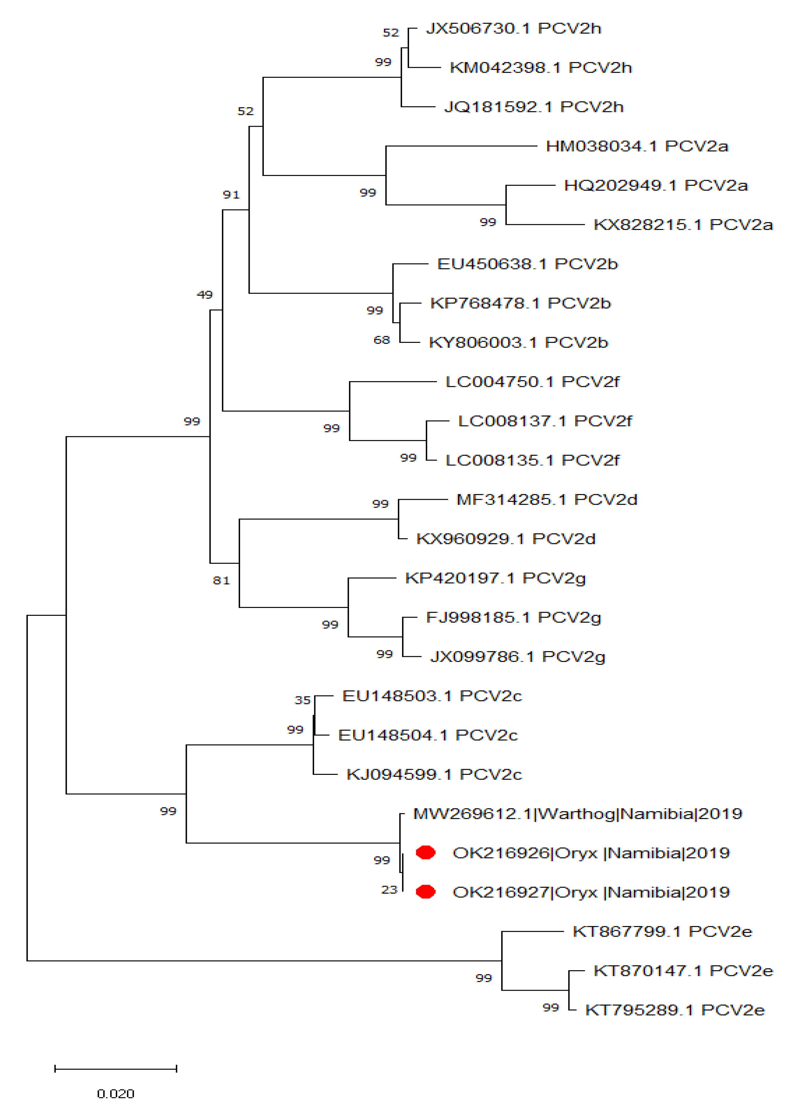
2. Results
3. Discussion
4. Materials and Methods
4.1. Sample Collection and PCV-2 Diagnosis
4.2. Complete Genome Sequencing and Analysis
4.3. Capsid Protein Homology Modeling
5. Conclusions
Author Contributions
Funding
Institutional Review Board Statement
Informed Consent Statement
Data Availability Statement
Acknowledgments
Conflicts of Interest
References
- Tischer, I.; Rasch, R.; Tochtermann, G. Characterization of Papovavirus-and Picornavirus-like Particles in Permanent Pig Kidney Cell Lines. Zentralbl. Bakteriol. Orig. A 1974, 226, 153–167. [Google Scholar]
- Finsterbusch, T.; Mankertz, A. Porcine Circoviruses-Small but Powerful. Virus Res. 2009, 143, 177–183. [Google Scholar] [CrossRef]
- Franzo, G.; Segalés, J. Circoviruses (Circoviridae). In Encyclopedia of Virology; Bamford, D.H., Zuckerman, M., Eds.; Elsevier Science: Amsterdam, The Netherlands, 2020; pp. 182–192. ISBN 9780128145166. [Google Scholar]
- Gillespie, J.; Opriessnig, T.; Meng, X.J.; Pelzer, K.; Buechner-Maxwell, V. Porcine Circovirus Type 2 and Porcine Circovirus-Associated Disease. J. Vet. Intern. Med. 2009, 23, 1151–1163. [Google Scholar] [CrossRef] [PubMed]
- Todd, D. Avian Circovirus Diseases: Lessons for the Study of PMWS. Vet. Microbiol. 2004, 98, 169–174. [Google Scholar] [CrossRef] [PubMed]
- Anderson, A.; Hartmann, K.; Leutenegger, C.M.; Proksch, A.L.; Mueller, R.S.; Unterer, S. Role of Canine Circovirus in Dogs with Acute Haemorrhagic Diarrhoea. Vet. Rec. 2017, 180, 1–5. [Google Scholar] [CrossRef]
- Segalés, J. Best Practice and Future Challenges for Vaccination against Porcine Circovirus Type 2. Expert Rev. Vaccines 2015, 14, 473–487. [Google Scholar] [CrossRef]
- Segalés, J. Porcine Circovirus Type 2 (PCV2) Infections: Clinical Signs, Pathology and Laboratory Diagnosis. Virus Res. 2012, 164, 10–19. [Google Scholar] [CrossRef]
- Franzo, G.; Cortey, M.; Segalés, J.; Hughes, J.; Drigo, M. Phylodynamic Analysis of Porcine Circovirus Type 2 Reveals Global Waves of Emerging Genotypes and the Circulation of Recombinant Forms. Mol. Phylogenet. Evol. 2016, 100, 269–280. [Google Scholar] [CrossRef] [Green Version]
- Franzo, G.; Segalés, J.; Franzo, G. Segalés Joaquim Porcine Circovirus 2 (PCV-2) Genotype Update and Proposal of a New Genotyping Methodology. PLoS ONE 2018, 13, e0208585. [Google Scholar] [CrossRef] [PubMed] [Green Version]
- Wang, Y.; Noll, L.; Lu, N.; Porter, E.; Stoy, C.; Zheng, W.; Liu, X.; Peddireddi, L.; Niederwerder, M.; Bai, J. Genetic Diversity and Prevalence of Porcine Circovirus Type 3 (PCV3) and Type 2 (PCV2) in the Midwest of the USA during 2016–2018. Transbound. Emerg. Dis. 2020, 67, 1284–1294. [Google Scholar] [CrossRef] [PubMed]
- Dupont, K.; Nielsen, E.O.; Bækbo, P.; Larsen, L.E. Genomic Analysis of PCV2 Isolates from Danish Archives and a Current PMWS Case-Control Study Supports a Shift in Genotypes with Time. Vet. Microbiol. 2008, 128, 56–64. [Google Scholar] [CrossRef] [PubMed] [Green Version]
- Grau-Roma, L.; Fraile, L.; Segalés, J. Recent Advances in the Epidemiology, Diagnosis and Control of Diseases Caused by Porcine Circovirus Type 2. Vet. J. 2011, 187, 23–32. [Google Scholar] [CrossRef]
- Xiao, C.T.; Harmon, K.M.; Halbur, P.G.; Opriessnig, T. PCV2d-2 Is the Predominant Type of PCV2 DNA in Pig Samples Collected in the U.S. during 2014–2016. Vet. Microbiol. 2016, 197, 72–77. [Google Scholar] [CrossRef] [PubMed] [Green Version]
- Franzo, G.; Cortey, M.; de Castro, A.M.M.G.; Piovezan, U.; Szabo, M.P.J.; Drigo, M.; Segalés, J.; Richtzenhain, L.J. Genetic Characterisation of Porcine Circovirus Type 2 (PCV2) Strains from Feral Pigs in the Brazilian Pantanal: An Opportunity to Reconstruct the History of PCV2 Evolution. Vet. Microbiol. 2015, 178, 158–162. [Google Scholar] [CrossRef] [PubMed]
- Liu, X.; Wang, F.X.; Zhu, H.W.; Sun, N.; Wu, H. Phylogenetic Analysis of Porcine Circovirus Type 2 (PCV2) Isolates from China with High Homology to PCV2c. Arch. Virol. 2016, 161, 1591–1599. [Google Scholar] [CrossRef] [PubMed]
- Molini, U.; Franzo, G.; Gous, L.; Moller, S.; Hemberger, Y.M.; Chiwome, B.; Marruchella, G.; Khaiseb, S.; Cattoli, G.; Dundon, W.G. Three Different Genotypes of Porcine Circovirus 2 (PCV-2) Identified in Pigs and Warthogs in Namibia. Arch. Virol. 2021, 166, 1723–1728. [Google Scholar] [CrossRef]
- Zhai, S.-L.; Lu, S.-S.; Wei, W.-K.; Lv, D.-H.; Wen, X.-H.; Zhai, Q.; Chen, Q.-L.; Sun, Y.-W.; Xi, Y. Reservoirs of Porcine Circoviruses: A Mini Review. Front. Vet. Sci. 2019, 6, 319. [Google Scholar] [CrossRef] [Green Version]
- Das, S.; Smith, K.; Sarker, S.; Peters, A.; Adriaanse, K.; Eden, P.; Ghorashi, S.A.; Forwood, J.K.; Raidal, S.R. Assessing Circovirus Gene Flow in Multiple Spill-over Events. Virus Genes 2019, 55, 802–814. [Google Scholar] [CrossRef]
- Das, S.; Sarker, S.; Peters, A.; Ghorashi, S.A.; Phalen, D.; Forwood, J.K.; Raidal, S.R. Evolution of Circoviruses in Lorikeets Lags behind Its Hosts. Mol. Phylogenet. Evol. 2016, 100, 281–291. [Google Scholar] [CrossRef]
- Fogell, D.J.; Martin, R.O.; Groombridge, J.J. Beak and Feather Disease Virus in Wild and Captive Parrots: An Analysis of Geographic and Taxonomic Distribution and Methodological Trends. Arch. Virol. 2016, 161, 2059–2074. [Google Scholar] [CrossRef] [Green Version]
- Franzo, G.; Menandro, M.L.; Tucciarone, C.M.; Barbierato, G.; Crovato, L.; Mondin, A.; Libanora, M.; Obber, F.; Orusa, R.; Robetto, S.; et al. Canine Circovirus in Foxes from Northern Italy: Where Did It All Begin? Pathogens 2021, 10, 1002. [Google Scholar] [CrossRef]
- Karuppannan, A.K.; Opriessnig, T. Porcine Circovirus Type 2 (PCV2) Vaccines in the Context of Current Molecular Epidemiology. Viruses 2017, 9, 99. [Google Scholar] [CrossRef] [Green Version]
- Opriessnig, T.; Karuppannan, A.K.; Castro, A.M.M.G.; Xiao, C.T. Porcine Circoviruses: Current Status, Knowledge Gaps and Challenges. Virus Res. 2020, 286, 198044. [Google Scholar] [CrossRef] [PubMed]
- Franzo, G.; Segalés, J. Porcine Circovirus 2 Genotypes, Immunity and Vaccines: Multiple Genotypes but One Single Serotype. Pathogens 2020, 9, 1049. [Google Scholar] [CrossRef]
- Franzo, G.; Tucciarone, C.M.; Cecchinato, M.; Drigo, M. Porcine Circovirus Type 2 (PCV2) Evolution before and after the Vaccination Introduction: A Large Scale Epidemiological Study. Sci. Rep. 2016, 6, 39458. [Google Scholar] [CrossRef] [PubMed]
- Opriessnig, T.; O’Neill, K.; Gerber, P.F.; de Castro, A.M.M.G.; Gimenéz-Lirola, L.G.; Beach, N.M.; Zhou, L.; Meng, X.J.; Wang, C.; Halbur, P.G. A PCV2 Vaccine Based on Genotype 2b Is More Effective than a 2a-Based Vaccine to Protect against PCV2b or Combined PCV2a/2b Viremia in Pigs with Concurrent PCV2, PRRSV and PPV Infection. Vaccine 2013, 31, 487–494. [Google Scholar] [CrossRef]
- Nayar, G.P.; Hamel, A.L.; Lin, L.; Sachvie, C.; Grudeski, E.; Spearman, G. Evidence for Circovirus in Cattle with Respiratory Disease and from Aborted Bovine Fetuses. Can. Vet. J. 1999, 40, 277. [Google Scholar]
- Kappe, E.C.; Halami, M.Y.; Schade, B.; Alex, M.; Hoffmann, D.; Gangl, A.; Meyer, K.; Dekant, W.; Schwarz, B.; Johne, R.; et al. Bone Marrow Depletion with Haemorrhagic Diathesis in Calves in Germany: Characterization of the Disease and Preliminary Investigations on Its Aetiology. Berl. Munch. Tierarztl. Wochenschr. 2010, 123, 31–41. [Google Scholar] [CrossRef]
- Dhindwal, S.; Avila, B.; Feng, S.; Khayat, R. Porcine Circovirus 2 Uses a Multitude of Weak Binding Sites To Interact with Heparan Sulfate, and the Interactions Do Not Follow the Symmetry of the Capsid. J. Virol. 2019, 93. [Google Scholar] [CrossRef] [PubMed] [Green Version]
- Wei, R.; van Renne, N.; Nauwynck, H.J. Strain-Dependent Porcine Circovirus Type 2 (PCV2) Entry and Replication in T-Lymphoblasts. Viruses 2019, 11, 813. [Google Scholar] [CrossRef] [PubMed] [Green Version]
- Lekcharoensuk, P.; Morozov, I.; Paul, P.S.; Thangthumniyom, N.; Wajjawalku, W.; Meng, X.J. Epitope Mapping of the Major Capsid Protein of Type 2 Porcine Circovirus (PCV2) by Using Chimeric PCV1 and PCV2. J. Virol. 2004, 78, 8135–8145. [Google Scholar] [CrossRef] [Green Version]
- Mahé, D.; Blanchard, P.; Truong, C.; Arnauld, C.; le Cann, P.; Cariolet, R.; Madec, F.; Albina, E.; Jestin, A. Differential Recognition of ORF2 Protein from Type 1 and Type 2 Porcine Circoviruses and Identification of Immunorelevant Epitopes. J. Gen. Virol. 2000, 81, 1815–1824. [Google Scholar] [CrossRef]
- Shang, S.B.; Jin, Y.L.; Jiang, X.T.; Zhou, J.Y.; Zhang, X.; Xing, G.; He, J.L.; Yan, Y. Fine Mapping of Antigenic Epitopes on Capsid Proteins of Porcine Circovirus, and Antigenic Phenotype of Porcine Circovirus Type 2. Mol. Immunol. 2009, 46, 327–334. [Google Scholar] [CrossRef]
- Wei, R.; Xie, J.; Theuns, S.; Nauwynck, H.J. Changes on the Viral Capsid Surface during the Evolution of Porcine Circovirus Type 2 (PCV2) from 2009 till 2018 May Lead to a Better Receptor Binding. Virus Evol. 2019, 5. [Google Scholar] [CrossRef]
- An, D.J.; Roh, I.S.; Song, D.S.; Park, C.K.; Park, B.K. Phylogenetic Characterization of Porcine Circovirus Type 2 in PMWS and PDNS Korean Pigs between 1999 and 2006. Virus Res. 2007, 129, 115–122. [Google Scholar] [CrossRef]
- Franzo, G.; Tinello, S.; Grassi, L.; Tucciarone, C.M.; Legnardi, M.; Cecchinato, M.; Dotto, G.; Mondin, A.; Martini, M.; Pasotto, D.; et al. Free to Circulate: An Update on the Epidemiological Dynamics of Porcine Circovirus 2 (PCV-2) in Italy Reveals the Role of Local Spreading, Wild Populations, and Foreign Countries. Pathogens 2020, 9, 221. [Google Scholar] [CrossRef] [PubMed] [Green Version]
- Abascal, F.; Zardoya, R.; Telford, M.J. TranslatorX: Multiple Alignment of Nucleotide Sequences Guided by Amino Acid Translations. Nucleic Acids Res. 2010, 38, W7–W13. [Google Scholar] [CrossRef] [PubMed] [Green Version]
- Kumar, S.; Stecher, G.; Tamura, K. MEGA7: Molecular Evolutionary Genetics Analysis Version 7.0 for Bigger Datasets. Mol. Biol. Evol. 2016, 33, 1870–1874. [Google Scholar] [CrossRef] [PubMed] [Green Version]
- Waterhouse, A.; Bertoni, M.; Bienert, S.; Studer, G.; Tauriello, G.; Gumienny, R.; Heer, F.T.; de Beer, T.A.P.; Rempfer, C.; Bordoli, L.; et al. SWISS-MODEL: Homology Modelling of Protein Structures and Complexes. Nucleic Acids Res. 2018, 46, W296–W303. [Google Scholar] [CrossRef] [PubMed] [Green Version]
- Pettersen, E.F.; Goddard, T.D.; Huang, C.C.; Couch, G.S.; Greenblatt, D.M.; Meng, E.C.; Ferrin, T.E. UCSF Chimera—A Visualization System for Exploratory Research and Analysis. J. Comput. Chem. 2004, 25, 1605–1612. [Google Scholar] [CrossRef] [PubMed] [Green Version]


| Year | Species | Number of Samples Tested | Positive Samples |
|---|---|---|---|
| 2019 | Red hartebeest (Alcelaphus buselaphus caama) | 34 | 0 |
| Kudu (Tragelaphus strepsiceros) | 2 | 0 | |
| Oryx (Oryx gazella) | 36 | 2 | |
| 2020 | Red hartebeest (Alcelaphus buselaphus caama) | 3 | 0 |
| Kudu (Tragelaphus strepsiceros) | 2 | 0 | |
| Oryx (Oryx gazella) | 2 | 0 | |
| 2021 | Red hartebeest (Alcelaphus buselaphus caama) | 7 | 0 |
| Kudu (Tragelaphus strepsiceros) | 6 | 0 | |
| Oryx (Oryx gazella) | 16 | 0 |
Publisher’s Note: MDPI stays neutral with regard to jurisdictional claims in published maps and institutional affiliations. |
© 2021 by the authors. Licensee MDPI, Basel, Switzerland. This article is an open access article distributed under the terms and conditions of the Creative Commons Attribution (CC BY) license (https://creativecommons.org/licenses/by/4.0/).
Share and Cite
Molini, U.; Coetzee, L.M.; Hemberger, M.Y.; Khaiseb, S.; Cattoli, G.; Dundon, W.G.; Franzo, G. The Oryx Antelope (Oryx gazella): An Unexpected Host for Porcine Circovirus-2 (PCV-2). Pathogens 2021, 10, 1402. https://doi.org/10.3390/pathogens10111402
Molini U, Coetzee LM, Hemberger MY, Khaiseb S, Cattoli G, Dundon WG, Franzo G. The Oryx Antelope (Oryx gazella): An Unexpected Host for Porcine Circovirus-2 (PCV-2). Pathogens. 2021; 10(11):1402. https://doi.org/10.3390/pathogens10111402
Chicago/Turabian StyleMolini, Umberto, Lauren Michelle Coetzee, Maria Yvonne Hemberger, Siegfried Khaiseb, Giovanni Cattoli, William G. Dundon, and Giovanni Franzo. 2021. "The Oryx Antelope (Oryx gazella): An Unexpected Host for Porcine Circovirus-2 (PCV-2)" Pathogens 10, no. 11: 1402. https://doi.org/10.3390/pathogens10111402