Review

Microbiologically Influenced Corrosion Mechanism of Ferrous Alloys in Marine Environment

1
Shenzhen Institute of Advanced Technology, Chinese Academy of Sciences, Shenzhen 518000, China
2
College of Materials Science and Engineering, Hunan University, Changsha 410082, China
3
Institute of Metal Research, Chinese Academy of Sciences, Shenyang 110016, China
*
Authors to whom correspondence should be addressed.
Metals 2022, 12(9), 1458; https://doi.org/10.3390/met12091458
Submission received: 22 June 2022 / Revised: 7 August 2022 / Accepted: 26 August 2022 / Published: 30 August 2022

Abstract

:
In marine environments, microbial attacks on metallic materials result in microbiologically influenced corrosion (MIC), which could cause severe safety accidents and high economic losses. To date, MIC of a number of metallic materials ranging from common steels to corrosion-resistant ferrous alloys has been reported. The MIC process has been explained based on (1) bio-catalyzed oxygen reduction; (2) kinetics alternation of the corrosion process by increasing the mass transport of the reactants and products; (3) production of corrosive substances; and (4) generation of auxiliary cathodic reactants. However, it is difficult to have a clear understanding of the MIC mechanism of ferrous alloys due to the interdisciplinary nature of MIC and lack of deep knowledge about the interfacial reaction between the biofilm and ferrous alloys. In order to better understand the effect of the MIC process on ferrous alloys, here we comprehensively summarized the process of biofilm formation and MIC mechanisms of ferrous alloys.

1. Introduction

Microbiologically influenced corrosion (MIC) is the corrosion process initiated and facilitated by microorganisms. It has been reported that 20 to 40% of the corrosion is caused by the direct or indirect involvement of microorganisms [1]. A number of microorganisms such as sulphate-reducing bacteria (SRB), methanogens, fermenters, acetogens, iron-reducing bacteria (IRB), sulphate-reducing archaea (SRA), iron-reducing archaea (IRA), iron-oxidizing bacteria (IOB), acid-producing bacteria (APB), manganese-oxidizing bacteria (MOB), and slime-producing bacteria do exist in biologically active sludge and sea water [2]. By reacting with the surrounding corrosive substance, microorganisms develop a highly aggressive environment for different ferrous alloys. The surfaces of under-deposit carbon steel pipelines in marine environments provide favourable sites for bacteriological activities, resulting in the establishment of biofilm. The biofilm produces extracellular polymeric substances (EPS) and enzymes, which creates very complex array of microenvironments. This process makes the deposits more electroactive, leading to the accumulation of more corrosive substances on the pipeline surfaces [3]. Pseudomonas aeruginosa, a kind of marine bacterium, is well-known for its severe corrosion-causing behaviour. Corrosion induced by P. aeruginosa on different metallic materials ranging from common steels to highly corrosion-resistant materials such as duplex stainless steels and titanium alloys have been reported [4,5,6,7]. Elemental composition of the metals, surface topography, presence or absence of passive oxide film, and mechanical properties such as stress play important roles in determining the rate of the corrosion process. Both carbon steel and stainless steel are iron-based alloys, but MIC behaviour is different on their surfaces. Elements such as iron, chromium, nickel, molybdenum, and copper which constitute iron-based alloys such as carbon steel, stainless steel, etc. strongly affect the microbial attachment and the rate of MIC. Iron has a great influence on MIC and it has been reported that SRB cells in the medium containing a high concentration of iron produced more EPS compared to iron-deficient medium, showing that high concentrations of ferrous ions can significantly speed up corrosion by SRB [8]. Moreover, Bradley observed that lipopolysaccharides situated in the outer membrane of gram-negative bacteria can react with ferrous ions (Fe2+) [9]. Chromium and molybdenum, which are responsible for the formation of passive oxide films, monitor the bacterial cell attachment and biofilm formation on stainless steels surface. Because the passive film comes between the biofilm and substrate, the MIC resistance of stainless steel is much higher than carbon steel [10]. Although chromium oxide passive film makes stainless steel resistant to corrosion, it is still not immune to MIC. Moreover, Shewanella algae (S. algae) is also one of the marine microorganisms enhancing the process of corrosion in marine environment. The synergistic effect of S. algae and chloride ions in simulated marine environments was investigated, and it was found that S. algae decreased the passivity of the oxide film of alloys, leading to the acceleration of chloride ion diffusion into the matrix [11]. Bacterial involvement in the process of corrosion can be classified as either (a) direct, where bacteria directly influence the rate of anodic and cathodic reaction, or (b) indirect, where they generate acidic metabolites which accelerate the corrosion process of the materials [12,13,14]. Bacterial activities cause accumulation of deposits and formation of bacterial biofilm on the ferrous alloys leading to severe corrosion attack [15]. MIC is mainly responsible for localized corrosion such as pitting corrosion and crevice corrosion. Both crevice and pitting corrosion are types of localized corrosion, which means that these forms of corrosion occur in a limited area on the surface, leading to a high corrosion rate compared to the uniform corrosion. The crevice corrosion usually happens in a narrow fissure with a width of a few micrometers. These fissures are generally caused by external agents such as insulation and paint remnants, etc. that form a crevice on the material surface. Pitting corrosion normally begins with microbial colonization and chloride accumulation that rapidly penetrate the protective oxide film covering the metal surface, and these points act as initiation sites for pitting corrosion. In addition, selective dissolution is also a way to initiate pitting corrosion and occurs when one of the components dissolves faster than other components. With the passage of time this localized dissolution leads to the formation of a pit in the metal surface. According to the previous research, the increase in the cathodic reaction rate by bacterial biofilm encouraged the crevice corrosion propagation [16]. Severe crevice corrosion has been reported for stainless steel alloy in the presence of mesophilic microorganisms biofilms [17]. Furthermore, it has been reported that the frequent existence of microorganisms in marine water and their biofilm formation on the material surface increased the risk of pitting corrosion [18,19,20,21]. A huge amount of research has been performed on the interaction of microorganisms and ferrous alloys. The main findings of the researchers working on MIC of ferrous alloys are given in Table 1. In addition, researchers working in this area have proposed different solutions for MIC mitigation. Some of the proposed solutions are given below: (a) killing the iron oxidizing bacteria, (b) eliminating the contact of bacteria with the metal substrate, (c) reinforcing the metal substrate with a non-metallic repair option that is not susceptible to MIC, (d) mechanical cleaning techniques, (e) use of biocides, and (f) incorporation of antibacterial agents such as copper and zinc.
Many studies have been carried out on microbiologically influenced corrosion, including biofilm formation, corrosion behaviour, mitigation strategies of MIC, etc. [22,23,24,25], but less research is concentrated on discussing the different MIC mechanisms of ferrous alloys. Therefore, in this paper we summarized the impact of microorganisms on corrosion and reviewed several important MIC mechanisms of ferrous alloys.
Table 1. The main findings of the researchers in the field of MIC of metallic alloys.
MaterialsAuthorsMicrobesEffects
carbon steelHamza et al. [4]P. aeruginosaBiofilm formation, corrosion damages, high corrosion rate
304 SSHamza et al. [5]P. aeruginosaBiofilm formation, formation of differential aeration cells, pitting corrosion
TitaniumKhan et al. [7]P. aeruginosaBiofilm formation, pitting corrosion
Carbon steelJaved et al. [8]SRBBiofilm formation, EPS secretion, pitting corrosion attacks, high corrosion rate
Iron based oil and gas pipelinesDennis et al. [22]SRBProduction of corrosive hydrogen sulfide, increased deterioration of iron
Low carbon steel (Ship ballast tank)Heyer et al. [26]Slime forming bacteria, sulfur oxidizing (SOB), iron oxidizing bacteria (IOB) and sulphate reducing bacteria (SRB)Increase pitting corrosion, high corrosion rate
316L stainless steelDong et al. [27]Acidithiobacillus caldus SM-1Development of dense biofilm, severe pitting corrosion, high corrosion rate
316L stainless steelTang et al. 28Geobacter sulfurreducens and Geobacter metallireducensDirect electron transfer encouraged the corrosion of stainless steel
304 stainless steelZhang et al. [29]Desulfovibrio vulgarisElectron mediator increased corrosion, weight loss, pitting corrosion

2. Corrosion Induced by the Concentration Cells

Microorganisms establish different types of concentration cells, including oxygen concentration cells and metal concentration cells, that locally attack the steel surface and accelerate pitting corrosion. Figure 1 shows the pitting corrosion of carbon steel caused by SRB. In concentration cells the conditions established for localized corrosion do not need huge numbers of microorganisms or their viability. Bacteria get attached to the steel surface and form biofilm with the help of EPS, which works as a diffusion barrier for corrosive agents like oxygen. However, the development of biofilm and the formation of an EPS layer are not always uniformly distributed on the whole surface of the material. The uneven thickness of biofilm and non-uniform distribution of EPS and the formed corrosion products lead to the formation of oxygen concentration cells. The oxygen concentration cells locally attack the steel surface and accelerate the corrosion process [30]. Corrosion is the process of chemical or electrochemical reaction of the exposed surface of the material with the surrounding aggressive environment [31]. This interaction affects the structure and properties of the materials. There is a natural tendency for almost all metals to oxidize and return to their original form of the ore, where they are thermodynamically stable. For instance, the iron ore called hematite is depicted as α-FeOOH and the common oxides formed by the corrosion reaction of iron are iron oxide-hydroxide (FeOOH), where iron returned to its original form of 3+ oxidation state [26]. In aerated marine water, iron oxidizing bacteria (IOB) develop oxygen concentration cells on the surface of 300 series stainless steels and accelerate under-deposit corrosion [32]. It has been reported that IOB produce highly dense deposits that hinder oxygen diffusion into the material surface, creating anodic sites. In such circumstances the corrosion rate does not depend on the number of bacterial cells present in the deposit, but rather on the metallurgy of the material and the physical/chemical properties of electrolytes such as dissolved oxygen and chloride ions (Cl). Another study reported copper deposition under IOB deposits formed on carbon steel pilings in Duluth-Superior Harbor (DSH) [33]. It was found that the presence of a copper layer leading to the formation of galvanic couple with the iron substratum and the galvanic current was dependant on the iron concentration in the electrolytes instead of the number of bacterial cells. Furthermore, concentration cells can also be formed when metal ions interact with the anionic functional groups such as carboxyle, sulphate, pyruvate, phosphate, and succinate present in EPS secreted by microorganisms.
It is known that biofilm development is not uniform on the metal surface. The metal part underneath the biofilm will work as an anode while the uncovered surface will act as the cathode, resulting in the formation of a galvanic cell, allowing the flow of electrons from anode to the cathodic region of the steel, thus leading to the corrosion of metals. Figure 2 shows the bacterial attachment and the biofilm formation on the metal surface [36]. Figure 2a,b explicates how the microorganisms get attached to the surface, colonize, and secrete extracellular polymeric substances to develop biofilm. Once the biofilm is fully developed and matured it releases the bacteria which move out from the biofilm and try to find a new surface for their attachment. Figure 2c is the CLSM result showing the increase in the biofilm thickness of the marine bacteria with increasing immersion time on the surface of the X80 pipeline steel.

3. Metabolites Induced Corrosion

Microorganisms produce corrosive acidic metabolites including organic and inorganic acids via metabolism. In high concentrations of oxygen, aerobic bacteria usually produce organic acids such as short chain fatty acids. The biofilm hinders the acid’s outwards diffusion to the solution, leading to an acidic environment beneath the biofilm, thus aggravating the steel surface. In an environment lack of oxygen, anaerobic bacteria produce organic acids and sulphides which accelerate the rate of localized corrosion attacks and encourage the process of corrosion. Based on the corrosion mechanism, anaerobic MIC is divided into two classes: metabolite MIC (M-MIC) and extracellular electron transfer MIC (EET-MIC). During M-MIC, microorganisms secrete corrosive metabolites such as organic acids that enhance the corrosion rate of the materials [39]. Acid producing bacteria (ABP) have been considered responsible for M-MIC. As these bacteria have much higher concentrations of sessile cells compared to planktonic cells, the pH measured underneath the biofilm is much lower compared to the bulk solution. Severe localized corrosion of super austenitic stainless steel has been reported in the acidic environment created by Acidithiobacillus caldus SM-1. Furthermore, a high corrosion rate of 316L stainless steel has been observed when it was immersed in A. caldus SM-1 solution [27]. In EET-MIC, the released electrons from the metals cross through the bacterial cell-wall and reduce the non-oxygen oxidants such as nitrate and sulphate in the cytoplasm, thus encouraging the oxidation of the iron simultaneously.
SRB are the most common known anaerobic bacteria deteriorating ferrous alloys in different fields [40]. In 1930, Kurt et al. for the first time proposed cathodic depolarization and metabolite depolarization theory to explain the MIC mechanism of SRB. According to this theory, iron works as an anode and the oxidation of its surface produces iron ions and electrons. The electrons need an electron acceptor because they cannot move into the solution as ions do. In an anaerobic environment, water is electrolysed to produce protons which work as electron acceptors and form hydrogen molecules. SRB consume the molecular hydrogen and reduce the activation energy of the whole reaction by secreting hydrogenase enzymes, and thus accelerate the cathodic depolarization reaction dynamically. These bacteria reduce sulphate into sulphide and accelerate the corrosion process. Cathodic depolarization theory explains the MIC process of SRB, but the mechanism is still not clear and needs to be further explored. The reactions involved during this process are given as follows:
4 Fe 4 Fe 2 + + 8 e
8 H 2 O 8 H + + 8 OH
8 H + + 8 e 8 H ads
8 H ads 4 H 2
8 H 2   8 H + + 8 e
SO 4 2 + 9 H + + 8 e HS + 4 H 2 O
During MIC, SRB get electrons from ferrous alloys such as carbon steel and stainless steel for the reduction of sulphate. The molecular hydrogen produced from the atomic hydrogen depolarizes the cathode and works as an electron carrier [41,42]. The diffusible molecular hydrogen enters the SRB cell in the dissolved form, where it starts oxidation in the cytoplasmic membrane by the biocatalysis of hydrogenase enzyme [43]. Thereafter, hydrogenase enzyme and multihaem cytochromes transfer the electrons through flavodoxin (electron carrier) to the APS (adenosine phosphosulfate) pathway for sulphate reduction in the cytoplasm.

4. MIC Based on Bioenergetics and Bio-Electrochemistry

Although the cathodic depolarization theory explains the mechanisms of MIC caused by SRB to some extent, it has been found that this theory has certain limitations. For example, Gu et al. [44] stated that the traditional corrosion mechanism proposed by cathodic depolarization theory can only explain the MIC behaviour of those SRB that secrete hydrogenase enzyme. On the contrary, not all SRB secrete hydrogenase enzyme, yet they still cause corrosion. Therefore, the traditional cathodic depolarization theory cannot elucidate the corrosion behaviour of all SRB. In order to better comprehend MIC, it is important to study the role and importance of biocatalytic cathodic sulphate reduction (BCSR) theory [45]. BCSR theory proposes the concept of extracellular electron transfer (EET), which explains why and how microorganisms cause metal corrosion from the perspective of bio-electrochemistry and bioenergetics.
As a typical anaerobe, SRB get carbon from the surrounding environment for its metabolic activities [46]. However, the SRB cells close to the surface are unable to access the carbon source due to the diffusion barrier and utilization of carbon by the upper cells. In such circumstances when there is a lack of carbon source, SRB cells get electrons from the steel for anaerobic respiration to maintain their survival. This process of receiving electrons from the steel usually leads to the severe corrosion of ferrous alloys by SRB.
Xu et al. observed an increased corrosion rate of C1018 carbon steel in media 90% and 99% free of carbon sources inoculated with Desulfovibrio vulgaris. It was found that the corrosion rate was increasing with decreasing carbon sources in the medium [47]. Jia et al. immersed C1018 carbon steel in culture media inoculated with P. aeruginosa at different percentages of carbon source. They found that the corrosion rate was highly increased when 90% of the carbon source was removed from the medium [48].
The extracellular electron transfer includes two types: direct electron transfer and indirect electron transfer [44,49]. Direct electron transfer is further divided into two types: (1) bacteria obtain electrons from steel through conductive proteins on cell membrane, and (2) bacteria conduct long-distance electron transfer through conductive appendages called nanowires. Microbial nanowire is an electrically conductive filament observed in microorganisms used for extracellular electron transfer between microorganisms and substrate surface. Indirect electron transfer is carried out by the soluble molecules secreted by the bacteria which work as electrons carriers. A detailed explanation of the direct and indirect electron transfer by microorganisms is given below.

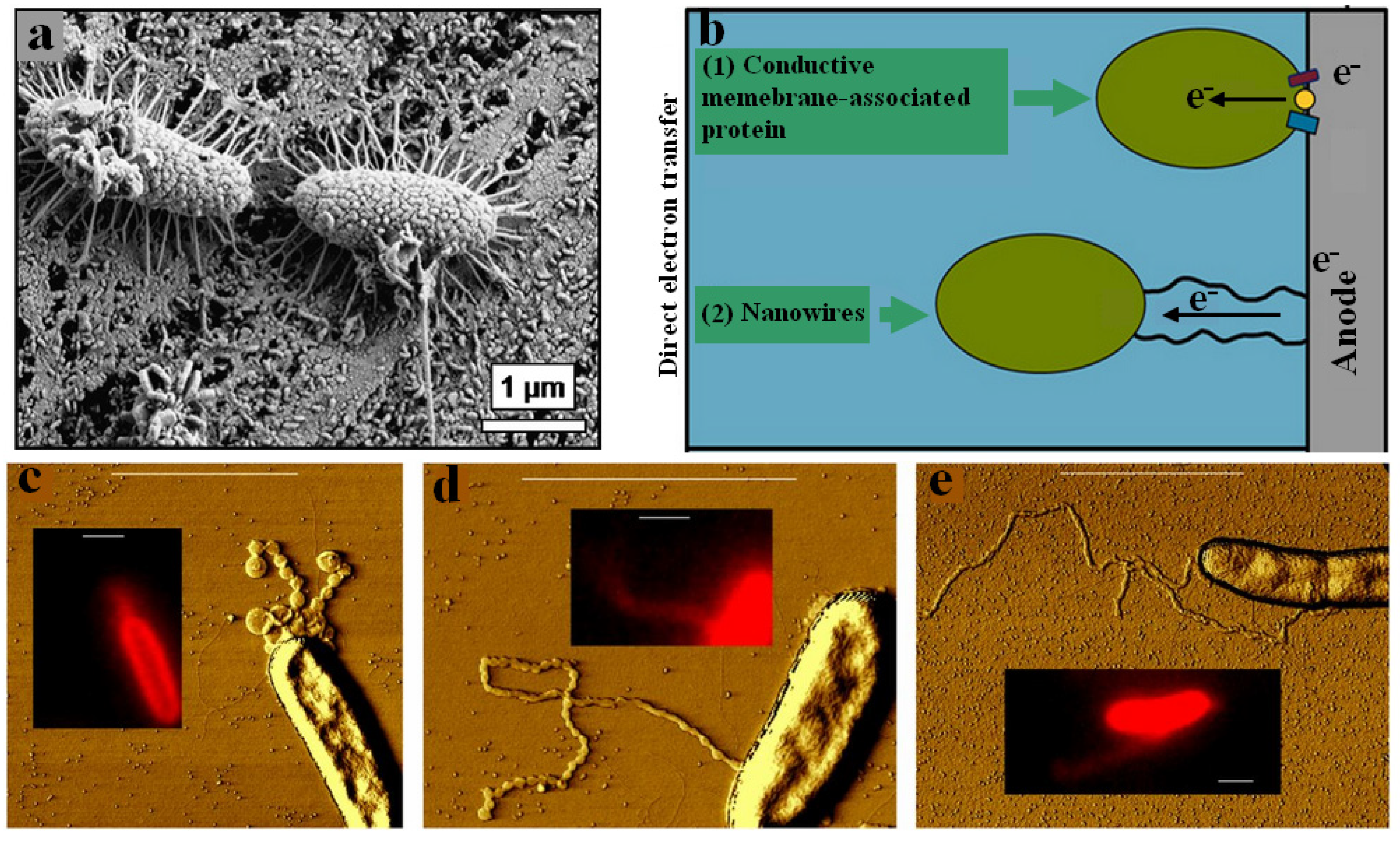
4.1. Direct Electron Transfer

Direct electron transfer between biofilm and metallic surface plays an important role in MIC. Increased uptake of electrons by the biofilm has been observed during the MIC of iron [50,51,52,53,54,55,56,57,58,59,60,61]. In the process of direct electron transfer, the bacterial cells are physically in contact with the steel surface without using any mediator. Direct electron transfer can help to achieve two important physiological functions: (1) getting access to an insoluble electron acceptor; (2) having access to electron donors such as Fe. It has been stated that microorganisms can take electrons for cellular metabolism or donate electrons to an anodic surface, and this process is determined by the redox potential of the surface relative to the corresponding catabolic substrate [62]. Direct electron transfer is based on the concept of electrotrophy, in which anaerobes directly accept electrons from iron electrode for their survival and growth [53]. Direct uptake of electron from Fe(0) for microbes including sulphate-reducing bacteria, methane-producing bacteria, as well as acetogenic microorganisms has been reported [61,63,64,65,66,67]. However, all of these microorganisms also consume H2 which suggests that rather than relying on electrotrophy, these microbes accelerate corrosion because they are effective in enzymatically accelerating H2 production and/or attaching to Fe(0) and consuming H2 as it is released at the surface [68,69,70]. Even though during Fe(0) corrosion H2 works as an electron carrier from metal to microbes, the abiotic production of H2 was not observed during the corrosion of stainless steels, suggesting that these corrosion mechanisms do not elucidate the corrosion of stainless steels [28]. Recently, electrotrophs Geobacter sulfurreducens and Geobacter metallireducens were found to colonize on stainless steel surface. Both of these bacteria have the ability to utilize direct electron transfer from iron to microbes to enhance the corrosion of stainless steel [71]. In green algae Shewanella oneidensis, MR-1 nanowire-like outer membrane and periplasmic extension, in concert with outer membrane multiheme cytochromes MtrC and OmcA, transfer electrons between cell and steel surface [72,73,74], while pili (nanowires) and associated cytochrome to pili are used to transport electrons in Geobacter species [75,76,77,78].
Nanowires are electrically conductive nano-sized appendages produced by the bacteria for electron transfer [73]. Nanowires help bacteria in long-range transfer of electron across the thick biofilm [79]. By interconnection through nanowires, the bacterial cells located in anoxic conditions can still use oxygen as their terminal electron acceptor. For instance, it has been reported that Shewanella develop electrically conductive nanowires when there is a lack of electron acceptors [73]. Figure 3 represents nanowires produced by microorganisms for getting electrons from the iron surface [80]. It has been observed that sessile cells directly receive electrons from the steel surface by using a special type of protein called c-type cytochrome, or they use pili to obtain electrons from the oxidized surface. Furthermore, it has been reported that bacteria use pili only when there is no organic carbon available for their survival [80].

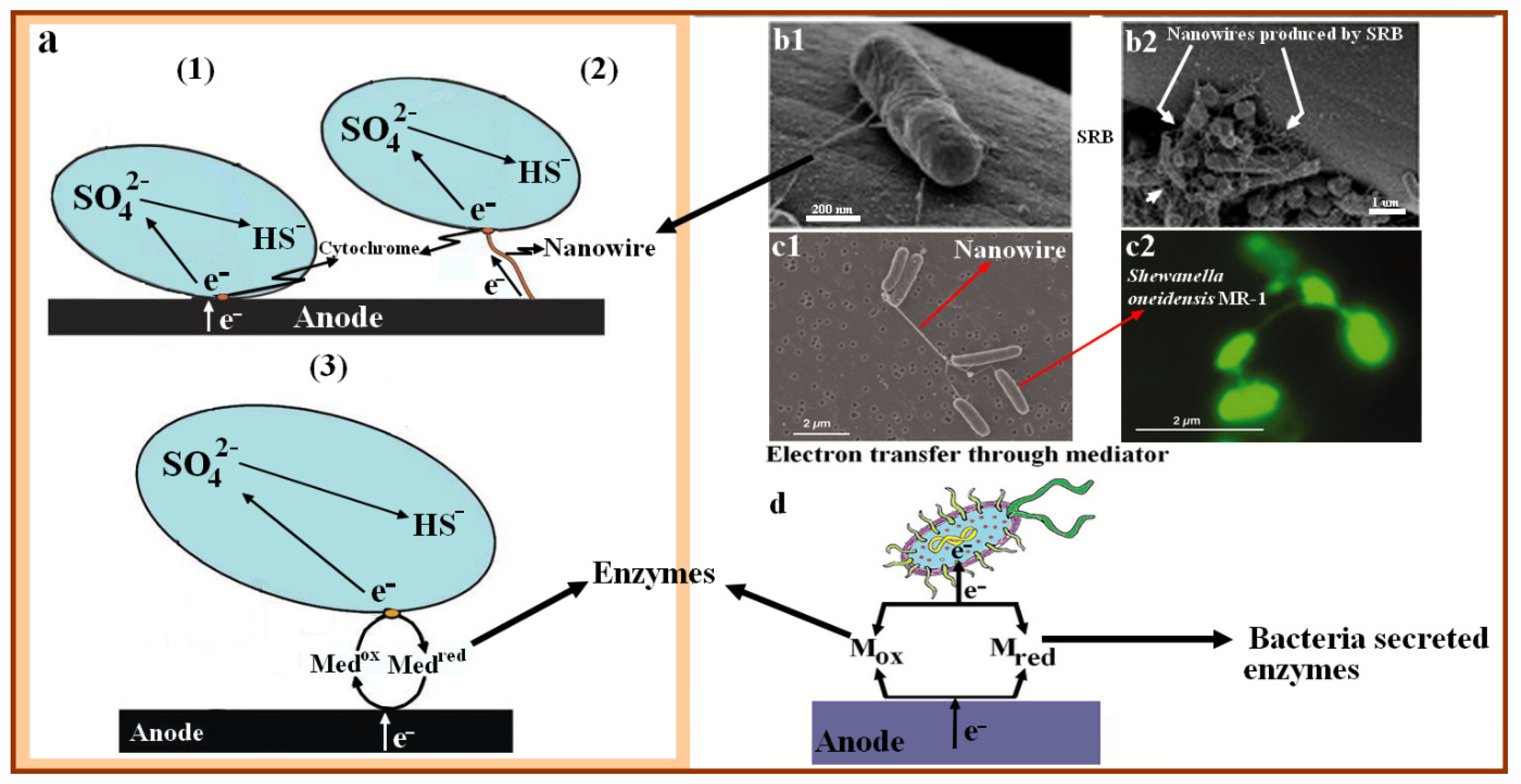
4.2. Electron Mediator

Electron mediators play an important role in transferring electrons from the oxidizing metal surface to the bacterial cells. Electron mediators are also called electron carriers that work as a shuttle, receiving electrons from the anode and donating them to the cathode. Electron carriers are of two types, known as exogenous electron carriers and endogenous electron carriers. Exogenous electron carriers are those added to the system from the outside, such as flavin adenine dinucleotide (FAD) and flavin mononucleotide (FMN), while endogenous electron carriers such as quinones and melanins are enzymes produced by bacterial activities [82,83,84]. It has been reported that electron mediators speed up the MIC process by accelerating the transmission of extracellular electrons. Zhang et al. reported a high corrosion rate of 304 SS in the presence of exogenous electron carriers FAD and FMN [29]. Furthermore, the corrosion resistance of C1018 carbon steel was investigated in a medium containing FMN and FAD inoculated with P. aeruginosa. It was found that the corrosion rate of C1018 carbon steel was highly increased in the presence of FAD and FMN, which were used as electron carriers [85]. Moreover, riboflavin could mediate electron transfer from the metal surface to the bacteria cell [86]. Zhang et al. found that the addition of riboflavin and flavin adenine dinucleotide to the testing medium increased the corrosion rate of stainless steel induced by D. vulgaris [29]. It was found that microorganisms such as Shewanella spp. produced riboflavin and FAD which facilitated their extra-cellular electron transfer activities [87]. In MIC caused by SRB, iron oxidation occurs outside the bacterial cells, while the SRB reduction takes place inside the SRB cytoplasm. This is only possible if an electron mediator is available for carrying electrons from the oxidized surface to the sessile cells [88]. Figure 4 shows the mechanisms of electron transfer between metal surface and bacteria cells.
It has been reported that the favourable oxidation of extracellular metallic iron (Fe0) coupled with the intracellular reduction of sulphate in SRB cytoplasm under biocatalysis caused SRB-induced corrosion [29]. MIC can be of two types: type I MIC and type II MIC. Type I MIC requires electron transport from outside of the cell through the cell wall into the cytoplasm [44,65]. In type II MIC, microorganism-secreted corrosive metabolites (Oxidants) attack iron externally without involving biocatalysis, removing electrons from its surface and causing it to be oxidized [44]. The biofilm developed on the metal surfaces locally secrete high concentration of the oxidants, which increases the rate of localized corrosion. Corrosion caused by hydrogen sulphide released by SRB is an example of type II MIC.
Both Type I MIC and Type II MIC are electrochemical in nature, as two reactions including metal oxidation reaction and sulphate reduction reaction are involved. However, the two basic MIC types are different. For example, in Type II MIC, electrons are exchanged on the steel surface locally without cross-cell wall electron transfer, which is essential in Type I MIC. Type II MIC and conventional chemical corrosion are very similar, except that in type II MIC the oxidants are secreted by microorganisms [59].

5. Conclusions

Analysis of the research work performed by different researchers in the MIC field revealed that microorganisms such as fungi, algae, and bacteria are involved in the deterioration of various metallic alloys in different fields. The collected research works indicate that most of the microorganisms living in marine environments are corrosive, but in some cases it has also been found that bacteria can inhibit the corrosion of the materials [91]. Bacterial biofilm and their metabolic activities contribute to MIC. Microorganisms can induce the corrosion of ferrous alloys. MIC mechanisms including concentration cells, metabolite-induced corrosion, as well as MIC based on bioenergetics and bio-electrochemistry, explain why microorganisms play important roles in the process of corrosion. However, due to the complex and multidisciplinary nature of MIC there is still a lack of systematic investigations of MIC mechanisms. Therefore, further investigations are needed in genetic manipulation of corrosion-causing microorganisms to better understand MIC mechanisms.

Author Contributions

Conceptualization, M.S.K. and Y.Z.; methodology, M.S.K.; software, M.S.K.; validation, T.L., Y.L. and H.L.; formal analysis, H.Z.; investigation, S.G.; resources, Y.S.; data curation, K.Y.; writing—original draft preparation, M.S.K.; writing—review and editing, Y.Z.; visualization, K.Y.; supervision, Y.Z. and H.P.; project administration, H.P.; funding acquisition, Y.Z. All authors have read and agreed to the published version of the manuscript.

Funding

This research was funded by the National Natural Science Foundation of China (51501218), Natural Science Foundation of Guangdong Province (No. 2019A1515110631, 2022A1515011339), and the Science and Technology Innovation Commission of Shenzhen (Nos. JCYJ20210324102013036, JCYJ20210324101809026, JCYJ20180507182239617).

Institutional Review Board Statement

Not applicable.

Informed Consent Statement

Not applicable.

Data Availability Statement

Not applicable.

Conflicts of Interest

The authors declare no conflict of interest.

References

  1. Little, B.J.; Lee, J.S. Microbiologically Influenced Corrosion. In Engineering Materials and Processes; Springer: London, UK, 2015; Volume 2, ISBN 9781119019213. [Google Scholar]
  2. Marciales, A.; Peralta, Y.; Haile, T.; Crosby, T.; Wolodko, J. Mechanistic microbiologically influenced corrosion modeling—A review. Corros. Sci. 2019, 146, 99–111. [Google Scholar]
  3. Suarez, E.M.; Lepkova, K.; Kinsella, B.; Machuca, L.L. Aggressive corrosion of steel by a thermophilic microbial consortium in the presence and absence of sand. Int. Biodeterior. Biodegrad. 2019, 137, 137–146. [Google Scholar] [CrossRef]
  4. Hamzah, E.; Hussain, M.F.; Ibrahim, Z.; Abdolahi, A. Corrosion Behaviour of Carbon Steel in Sea Water Medium in Presence of P. aeruginosa Bacteria. Arab. J. Sci. Eng. 2014, 39, 6863–6870. [Google Scholar] [CrossRef]
  5. Hamzah, E.; Hussain, M.Z.; Ibrahim, Z.; Abdolahi, A. Influence of Pseudomonas aeruginosa bacteria on corrosion resistance of 304 stainless steel. Corros. Eng. Sci. Technol. 2013, 48, 116–120. [Google Scholar] [CrossRef]
  6. Farooq, A.; Zubair, M.; Wadood, H.Z.; Deen, K.M. Effect of Pseudomonas aeruginosa Strain ZK Biofilm on the Mechanical and Corrosion Behavior of 316L Stainless Steel and α-brass. J. Electrochem. Sci. Technol. 2021, 12, 431–439. [Google Scholar] [CrossRef]
  7. Khan, M.S.; Li, Z.; Yang, K.; Xu, D.; Yang, C.; Liu, D.; Lekbach, Y.; Zhou, E.; Kalnaowakul, P. Microbiologically influenced corrosion of titanium caused by aerobic marine bacterium Pseudomonas aeruginosa. J. Mater. Sci. Technol. 2019, 35, 216–222. [Google Scholar] [CrossRef]
  8. Javed, M.A.; Stoddart, P.R.; Wade, S.A. Corrosion of carbon steel by sulphate reducing bacteria: Initial attachment and the role of ferrous ions. Corros. Sci. 2015, 93, 48–57. [Google Scholar] [CrossRef]
  9. Bradley, G.; Gaylarde, C.C. Iron uptake by Desulfovibrio vulgaris outer membrane components in artificial vesicles. Curr. Microbiol. 1988, 17, 189–192. [Google Scholar] [CrossRef]
  10. Javed, M.A.; Neil, W.C.; McAdam, G.; Wade, S.A. Effect of sulphate-reducing bacteria on the microbiologically influenced corrosion of ten different metals using constant test conditions. Int. Biodeterior. Biodegrad. 2017, 125, 73–85. [Google Scholar] [CrossRef]
  11. Li, Z.; Wang, J.; Dong, Y.; Xu, D.; Zhang, X.; Wu, J.; Gu, T.; Wang, F. Synergistic effect of chloride ion and Shewanella algae accelerates the corrosion of Ti-6Al-4V alloy. J. Mater. Sci. Technol. 2021, 71, 177–185. [Google Scholar] [CrossRef]
  12. Alabbas, F.M.; Mishra, B. Microbiologically influenced corrosion of pipelines in the oil & gas industry. In Proceedings of the 8th Pacific Rim International Congress on Advanced Materials and Processing 2013, PRICM 8, Waikoloa, Hawaii, 4–9 August 2013; John Wiley & Sons, Inc.: Hoboken, NJ, USA, 2013; Volume 4, pp. 3441–3448. [Google Scholar]
  13. Zarasvand, K.A.; Rai, V.R. Microorganisms: Induction and inhibition of corrosion in metals. Int. Biodeterior. Biodegrad. 2014, 87, 66–74. [Google Scholar] [CrossRef]
  14. Beech, I.B. Corrosion of technical materials in the presence of biofilms—Current understanding and state-of-the art methods of study. Int. Biodeterior. Biodegrad. 2004, 53, 177–183. [Google Scholar] [CrossRef]
  15. Videla, H.A. Prevention and control of biocorrosion. Int. Biodeterior. Biodegrad. 2002, 49, 259–270. [Google Scholar] [CrossRef]
  16. Zhang, H.-J.; Dexter, S.C. Effect of biofilms on crevice corrosion of stainless steels in coastal seawater. Corrosion 1995, 51, 56–66. [Google Scholar] [CrossRef]
  17. Machuca, L.L.; Bailey, S.I.; Gubner, R.; Watkin, E.L.J.; Ginige, M.P.; Kaksonen, A.H.; Heidersbach, K. Effect of oxygen and biofilms on crevice corrosion of UNS S31803 and UNS N08825 in natural seawater. Corros. Sci. 2013, 67, 242–255. [Google Scholar] [CrossRef]
  18. Machuca, L.L.; Bailey, S.I.; Gubner, R. Microbial Corrosion Resistance of Stainless Steels for Marine Energy Installations. Adv. Mater. Res. 2012, 353, 3591–3596. [Google Scholar] [CrossRef]
  19. Little, B.J.; Lee, J.S.; Ray, R.I. The influence of marine biofilms on corrosion: A concise review. Electrochim. Acta 2008, 54, 2–7. [Google Scholar] [CrossRef]
  20. Beech, I.B.; Sunner, J. Biocorrosion: Towards understanding interactions between biofilms and metals. Curr. Opin. Biotechnol. 2004, 15, 181–186. [Google Scholar] [CrossRef]
  21. Iverson, W.P. Research on the mechanisms of anaerobic corrosion. Int. Biodeterior. Biodegrad. 2001, 47, 63–70. [Google Scholar] [CrossRef]
  22. Enning, D.; Garrelfs, J. Corrosion of iron by sulfate-reducing bacteria: New views of an old problem. Appl. Environ. Microbiol. 2014, 80, 1226–1236. [Google Scholar] [CrossRef]
  23. Anandkumar, B.; George, R.P.; Maruthamuthu, S.; Parvathavarthini, N.; Mudali, U.K. Corrosion characteristics of sulfate-reducing bacteria (SRB) and the role of molecular biology in SRB studies: An overview. Corros. Rev. 2016, 34, 41–63. [Google Scholar]
  24. Loto, C.A. Microbiological corrosion: Mechanism, control and impact—A review. Int. J. Adv. Manuf. Technol. 2017, 92, 4241–4252. [Google Scholar] [CrossRef]
  25. Kip, N.; Van Veen, J.A. The dual role of microbes in corrosion. ISME J. 2015, 9, 542–551. [Google Scholar] [CrossRef]
  26. Heyer, A.; Souza, F.D.; Morales, C.F.L.; Ferrari, G.; Mol, J.M.C.; Wit, J.H.W. De Ship ballast tanks a review from microbial corrosion and electrochemical point of view. Ocean Eng. 2013, 70, 188–200. [Google Scholar] [CrossRef]
  27. Dong, Y.; Li, J.; Xu, D.; Song, G.; Liu, D.; Wang, H.; Saleem Khan, M.; Yang, K.; Wang, F. Investigation of microbial corrosion inhibition of Cu-bearing 316L stainless steel in the presence of acid producing bacterium Acidithiobacillus caldus SM-1. J. Mater. Sci. Technol. 2020. [Google Scholar] [CrossRef]
  28. Tang, H.Y.; Yang, C.; Ueki, T.; Pittman, C.C.; Xu, D.; Woodard, T.L.; Holmes, D.E.; Gu, T.; Wang, F.; Lovley, D.R. Stainless steel corrosion via direct iron-to-microbe electron transfer by Geobacter species. ISME J. 2021. [Google Scholar] [CrossRef]
  29. Zhang, P.; Xu, D.; Li, Y.; Yang, K.; Gu, T. Electron mediators accelerate the microbiologically influenced corrosion of 304 stainless steel by the Desulfovibrio vulgaris biofilm. Bioelectrochemistry 2015, 101, 14–21. [Google Scholar] [CrossRef]
  30. Caldwell, D.E.; Korber, D.R.; Lawrence, J.R. Confocal Laser Microscopy and Digital Image Analysis in Microbial Ecology. In Advances in Microbial Ecology; Springer: Boston, MA, USA, 1992; Volume 12, pp. 1–67. [Google Scholar] [CrossRef]
  31. Alemayehu, T.; Birahane, M. Corrosion and its Protection. IJASR Int. J. Acad. Sci. Res. 2014, 2, 2272–6446. [Google Scholar]
  32. Ray, R.I.; Lee, J.S.; Little, B.J. Iron-oxidizing bacteria: A review of corrosion mechanisms in fresh water and marine environments. In Proceedings of the NACE—International Corrosion Conference Series, San Antonio, TX, USA, 14–18 March 2010. [Google Scholar]
  33. Ray, R.; Lee, J.; Little, B. Factors contributing to corrosion of steel pilings in Duluth-Superior Harbor. Corrosion 2009, 65, 707–717. [Google Scholar] [CrossRef]
  34. Li, Y.; Feng, S.; Liu, H.; Tian, X.; Xia, Y.; Li, M.; Xu, K.; Yu, H.; Liu, Q.; Chen, C. Bacterial distribution in SRB biofilm affects MIC pitting of carbon steel studied using FIB-SEM. Corros. Sci. 2020, 167, 108512. [Google Scholar] [CrossRef]
  35. Blackwood, D. An Electrochemist Perspective of Microbiologically Influenced Corrosion. Corros. Mater. Degrad. 2018, 1, 59–76. [Google Scholar] [CrossRef]
  36. Dong, C.; Bai, X.; Yan, X.; Yuan, C. Research status and advances on tribological study of materials under ocean environment. Tribology 2013, 33, 311–320. [Google Scholar]
  37. Stoodley, P.; Sauer, K.; Davies, D.G.; Costerton, J.W. Biofilms as Complex Differentiated Communities. Annu. Rev. Microbiol. 2002, 56, 187–209. [Google Scholar] [CrossRef] [PubMed] [Green Version]
  38. Verderosa, A.D.; Totsika, M.; Fairfull-Smith, K.E. Bacterial Biofilm Eradication Agents: A Current Review. Front. Chem. 2019, 7, 824. [Google Scholar] [CrossRef] [PubMed]
  39. Little, B.J.; Blackwood, D.J.; Hinks, J.; Lauro, F.M.; Marsili, E.; Okamoto, A.; Rice, S.A.; Wade, S.A.; Flemming, H.C. Microbially influenced corrosion—Any progress? Corros. Sci. 2020, 170, 108641. [Google Scholar] [CrossRef]
  40. Yuan, S.; Liang, B.; Zhao, Y.; Pehkonen, S.O. Surface chemistry and corrosion behaviour of 304 stainless steel in simulated seawater containing inorganic sulphide and sulphate-reducing bacteria. Corros. Sci. 2013, 74, 353–366. [Google Scholar] [CrossRef]
  41. Ćwiek, J. Prevention methods against hydrogen degradation of steel. J. Achiev. Mater. Manuf. Eng. 2010, 43, 214–221. [Google Scholar]
  42. Deckena, S.; Blotevogel, K.H. Growth of methanogenic and sulfate-reducing bacteria with cathodic hydrogen. Biotechnol. Lett. 1990, 12, 615–620. [Google Scholar] [CrossRef]
  43. Gu, T.; Jia, R.; Unsal, T.; Xu, D. Toward a better understanding of microbiologically influenced corrosion caused by sulfate reducing bacteria. J. Mater. Sci. Technol. 2019, 35, 631–636. [Google Scholar] [CrossRef]
  44. Xu, D.; Li, Y.; Song, F.; Gu, T. Laboratory investigation of microbiologically influenced corrosion of C1018 carbon steel by nitrate reducing bacterium Bacillus licheniformis. Corros. Sci. 2013, 77, 385–390. [Google Scholar] [CrossRef]
  45. Gu, T. New understandings of biocorrosion mechanisms and their classifications. J. Microb. Biochem. Technol. 2012, 4, iii–vi. [Google Scholar] [CrossRef]
  46. Lampreia, J.; Fauque, G.; Speich, N.; Dahl, C.; Moura, I.; Truper, H.G.; Moura, J.J.G. Spectroscopic studies on APS reductase isolated from the hyperthermophilic sulfate-reducing archaebacterium Archaeglobus fulgidus. Biochem. Biophys. Res. Commun. 1991, 181, 342–347. [Google Scholar] [CrossRef]
  47. Xu, D.; Gu, T. Carbon source starvation triggered more aggressive corrosion against carbon steel by the Desulfovibrio vulgaris biofilm. Int. Biodeterior. Biodegrad. 2014, 91, 74–81. [Google Scholar] [CrossRef]
  48. Jia, R.; Yang, D.; Xu, J.; Xu, D.; Gu, T. Microbiologically influenced corrosion of C1018 carbon steel by nitrate reducing Pseudomonas aeruginosa biofilm under organic carbon starvation. Corros. Sci. 2017, 127, 1–9. [Google Scholar] [CrossRef]
  49. Lovley, D.R. The microbe electric: Conversion of organic matter to electricity. Curr. Opin. Biotechnol. 2008, 19, 564–571. [Google Scholar] [CrossRef]
  50. Kato, S.; Hashimoto, K.; Watanabe, K. Microbial interspecies electron transfer via electric currents through conductive minerals. Proc. Natl. Acad. Sci. USA 2012, 109, 10042–10046. [Google Scholar] [CrossRef]
  51. Kato, S.; Yumoto, I.; Kamagata, Y. Isolation of acetogenic bacteria that induce biocorrosion by utilizing metallic iron as the sole electron donor. Appl. Environ. Microbiol. 2015, 81, 67–73. [Google Scholar] [CrossRef] [PubMed]
  52. Rabaey, K.; Rozendal, R.A. Microbial electrosynthesis—Revisiting the electrical route for microbial production. Nat. Rev. Microbiol. 2010, 8, 706–716. [Google Scholar] [CrossRef]
  53. Lovley, D.R. Powering microbes with electricity: Direct electron transfer from electrodes to microbes. Environ. Microbiol. Rep. 2011, 3, 27–35. [Google Scholar] [CrossRef]
  54. Nevin, K.P.; Hensley, S.A.; Franks, A.E.; Summers, Z.M.; Ou, J.; Woodard, T.L.; Snoeyenbos-West, O.L.; Lovley, D.R. Electrosynthesis of organic compounds from carbon dioxide is catalyzed by a diversity of acetogenic microorganisms. Appl. Environ. Microbiol. 2011, 77, 2882–2886. [Google Scholar] [CrossRef]
  55. Pfeffer, C.; Larsen, S.; Song, J.; Dong, M.; Besenbacher, F.; Meyer, R.L.; Kjeldsen, K.U.; Schreiber, L.; Gorby, Y.A.; El-Naggar, M.Y.; et al. Filamentous bacteria transport electrons over centimetre distances. Nature 2012, 491, 218–221. [Google Scholar] [CrossRef] [PubMed]
  56. Nielsen, L.P.; Risgaard-Petersen, N.; Fossing, H.; Christensen, P.B.; Sayama, M. Electric currents couple spatially separated biogeochemical processes in marine sediment. Nature 2010, 463, 1071–1074. [Google Scholar] [CrossRef] [PubMed]
  57. Bond, D.R.; Lovley, D.R. Electricity production by Geobacter sulfurreducens attached to electrodes. Appl. Environ. Microbiol. 2003, 69, 1548–1555. [Google Scholar] [CrossRef] [PubMed]
  58. Gregory, K.B.; Bond, D.R.; Lovley, D.R. Graphite electrodes as electron donors for anaerobic respiration. Environ. Microbiol. 2004, 6, 596–604. [Google Scholar] [CrossRef] [PubMed]
  59. Enning, D.; Venzlaff, H.; Garrelfs, J.; Dinh, H.T.; Meyer, V.; Mayrhofer, K.; Hassel, A.W.; Stratmann, M.; Widdel, F. Marine sulfate-reducing bacteria cause serious corrosion of iron under electroconductive biogenic mineral crust. Environ. Microbiol. 2012, 14, 1772–1787. [Google Scholar] [CrossRef]
  60. Lohner, S.T.; Deutzmann, J.S.; Logan, B.E.; Leigh, J.; Spormann, A.M. Hydrogenase-independent uptake and metabolism of electrons by the archaeon Methanococcus maripaludis. ISME J. 2014, 8, 1673–1681. [Google Scholar] [CrossRef]
  61. Dinh, H.T.; Kuever, J.; Mußmann, M.; Hassel, A.W.; Stratmann, M.; Widdel, F. Iron corrosion by novel anaerobic microorganisms. Nature 2004, 427, 829–832. [Google Scholar] [CrossRef]
  62. Deutzmann, J.S.; Sahin, M.; Spormann, A.M. Extracellular Enzymes Facilitate Electron Uptake in Biocorrosion and Bioelectrosynthesis. MBio 2015, 6, e00496-15. [Google Scholar] [CrossRef]
  63. Kato, S. Microbial extracellular electron transfer and its relevance to iron corrosion. Microb. Biotechnol. 2016, 9, 141–148. [Google Scholar] [CrossRef]
  64. Palacios, P.A.; Snoeyenbos-West, O.; Löscher, C.R.; Thamdrup, B.; Rotaru, A.E. Baltic Sea methanogens compete with acetogens for electrons from metallic iron. ISME J. 2019, 13, 3011–3023. [Google Scholar] [CrossRef]
  65. Venzlaff, H.; Enning, D.; Srinivasan, J.; Mayrhofer, K.J.J.; Hassel, A.W.; Widdel, F.; Stratmann, M. Accelerated cathodic reaction in microbial corrosion of iron due to direct electron uptake by sulfate-reducing bacteria. Corros. Sci. 2013, 66, 88–96. [Google Scholar] [CrossRef]
  66. Sharma, M.; Bajracharya, S.; Gildemyn, S.; Patil, S.A.; Alvarez-Gallego, Y.; Pant, D.; Rabaey, K.; Dominguez-Benetton, X. A critical revisit of the key parameters used to describe microbial electrochemical systems. Electrochim. Acta 2014, 140, 191–208. [Google Scholar] [CrossRef]
  67. Vigneron, A.; Head, I.M.; Tsesmetzis, N. Damage to offshore production facilities by corrosive microbial biofilms. Appl. Microbiol. Biotechnol. 2018, 102, 2525–2533. [Google Scholar] [CrossRef] [PubMed]
  68. Philips, J.; Van den Driessche, N.; De Paepe, K.; Prévoteau, A.; Gralnick, J.A.; Arends, J.B.A.; Rabaey, K. A novel Shewanella isolate enhances corrosion by using metallic iron as the electron donor with fumarate as the electron acceptor. Appl. Environ. Microbiol. 2018, 84, e01154-18. [Google Scholar] [CrossRef] [Green Version]
  69. Tang, H.Y.; Holmes, D.E.; Ueki, T.; Palacios, P.A.; Lovley, D.R. Iron corrosion via direct metal-microbe electron transfer. MBio 2019, 10, e00303-19. [Google Scholar] [CrossRef] [PubMed]
  70. Philips, J.; Monballyu, E.; Georg, S.; De Paepe, K.; Prévoteau, A.; Rabaey, K.; Arends, J.B.A. An Acetobacterium strain isolated with metallic iron as electron donor enhances iron corrosion by a similar mechanism as Sporomusa sphaeroides. FEMS Microbiol. Ecol. 2019, 95, fiy222. [Google Scholar] [CrossRef]
  71. Mehanna, M.; Basséguy, R.; Délia, M.L.; Bergel, A. Effect of Geobacter sulfurreducens on the microbial corrosion of mild steel, ferritic and austenitic stainless steels. Corros. Sci. 2009, 51, 2596–2604. [Google Scholar] [CrossRef]
  72. Pirbadian, S.; Barchinger, S.E.; Leung, K.M.; Byun, H.S.; Jangir, Y.; Bouhenni, R.A.; Reed, S.B.; Romine, M.F.; Saffarini, D.A.; Shi, L.; et al. Shewanella oneidensis MR-1 nanowires are outer membrane and periplasmic extensions of the extracellular electron transport components. Proc. Natl. Acad. Sci. USA 2014, 111, 12883–12888. [Google Scholar] [CrossRef]
  73. Gorby, Y.A.; Yanina, S.; McLean, J.S.; Rosso, K.M.; Moyles, D.; Dohnalkova, A.; Beveridge, T.J.; Chang, I.S.; Kim, B.H.; Kim, K.S.; et al. Electrically conductive bacterial nanowires produced by Shewanella oneidensis strain MR-1 and other microorganisms. Proc. Natl. Acad. Sci. USA 2006, 103, 11358–11363. [Google Scholar] [CrossRef]
  74. Okamoto, A.; Nakamura, R.; Hashimoto, K. Electrochimica Acta In-vivo identification of direct electron transfer from Shewanella oneidensis MR-1 to electrodes via outer-membrane OmcA—MtrCAB protein complexes. Electrochim. Acta 2011, 56, 5526–5531. [Google Scholar] [CrossRef]
  75. Reguera, G.; McCarthy, K.D.; Mehta, T.; Nicoll, J.S.; Tuominen, M.T.; Lovley, D.R. Extracellular electron transfer via microbial nanowires. Nature 2005, 435, 1098–1101. [Google Scholar] [CrossRef] [PubMed]
  76. Summers, Z.M.; Fogarty, H.E.; Leang, C.; Franks, A.E.; Malvankar, N.S.; Lovley, D.R. Direct exchange of electrons within aggregates of an evolved syntrophic coculture of anaerobic bacteria. Science 2010, 330, 1413–1415. [Google Scholar] [CrossRef] [PubMed]
  77. Vargas, M.; Malvankar, N.S.; Tremblay, P.L.; Leang, C.; Smith, J.A.; Patel, P.; Synoeyenbos-West, O.; Nevin, K.P.; Lovley, D.R. Aromatic amino acids required for pili conductivity and long-range extracellular electron transport in Geobacter sulfurreducens. MBio 2013, 4, e00105-13. [Google Scholar] [CrossRef]
  78. Tremblay, P.L.; Summers, Z.M.; Glaven, R.H.; Nevin, K.P.; Zengler, K.; Barrett, C.L.; Qiu, Y.; Palsson, B.O.; Lovley, D.R. A c-type cytochrome and a transcriptional regulator responsible for enhanced extracellular electron transfer in Geobacter sulfurreducens revealed by adaptive evolution. Environ. Microbiol. 2011, 13, 13–23. [Google Scholar] [CrossRef] [PubMed]
  79. Reguera, G.; Nevin, K.P.; Nicoll, J.S.; Covalla, S.F.; Woodard, T.L.; Lovley, D.R. Biofilm and nanowire production leads to increased current in Geobacter sulfurreducens fuel cells. Appl. Environ. Microbiol. 2006, 72, 7345–7348. [Google Scholar] [CrossRef] [PubMed]
  80. Shoesmith, D.W.; Mitlin, S.; Power, I.M.; Sherar, B.W.A.; Southam, G.; Keech, P.G. Characterizing the effect of carbon steel exposure in sulfide containing solutions to microbially induced corrosion. Corros. Sci. 2010, 53, 955–960. [Google Scholar] [CrossRef]
  81. Heydorn, R.L.; Engel, C.; Krull, R.; Dohnt, K. Strategies for the Targeted Improvement of Anodic Electron Transfer in Microbial Fuel Cells. ChemBioEng Rev. 2020, 7, 4–17. [Google Scholar] [CrossRef]
  82. Chen, J.J.; Chen, W.; He, H.; Li, D.B.; Li, W.W.; Xiong, L.; Yu, H.Q. Manipulation of microbial extracellular electron transfer by changing molecular structure of phenazine-type redox mediators. Environ. Sci. Technol. 2013, 47, 1033–1039. [Google Scholar] [CrossRef]
  83. Min, D.; Cheng, L.; Zhang, F.; Huang, X.N.; Li, D.B.; Liu, D.F.; Lau, T.C.; Mu, Y.; Yu, H.Q. Enhancing Extracellular Electron Transfer of Shewanella oneidensis MR-1 through Coupling Improved Flavin Synthesis and Metal-Reducing Conduit for Pollutant Degradation. Environ. Sci. Technol. 2017, 51, 5082–5089. [Google Scholar] [CrossRef]
  84. Qiao, Y.J.; Qiao, Y.; Zou, L.; Wu, X.S.; Liu, J.H. Biofilm promoted current generation of Pseudomonas aeruginosa microbial fuel cell via improving the interfacial redox reaction of phenazines. Bioelectrochemistry 2017, 117, 34–39. [Google Scholar] [CrossRef]
  85. Jia, R.; Yang, D.; Xu, D.; Gu, T. Electron transfer mediators accelerated the microbiologically influence corrosion against carbon steel by nitrate reducing Pseudomonas aeruginosa biofilm. Bioelectrochemistry 2017, 118, 38–46. [Google Scholar] [CrossRef]
  86. Huang, L.; Tang, J.; Chen, M.; Liu, X.; Zhou, S. Two modes of riboflavin-mediated extracellular electron transfer in Geobacter uraniireducens. Front. Microbiol. 2018, 9, 2886. [Google Scholar] [CrossRef] [PubMed]
  87. Brutinel, E.D.; Gralnick, J.A. Shuttling happens: Soluble flavin mediators of extracellular electron transfer in Shewanella. Appl. Microbiol. Biotechnol. 2012, 93, 41–48. [Google Scholar] [CrossRef]
  88. Booth, G.H.; Tiller, A.K. Polarization studies of mild steel in cultures of sulphate-reducing bacteria. Trans. Faraday Soc. 1960, 56, 1689–1696. [Google Scholar] [CrossRef]
  89. Schröder, U. Anodic electron transfer mechanisms in microbial fuel cells and their energy efficiency. Phys. Chem. Chem. Phys. 2007, 9, 2619–2629. [Google Scholar] [CrossRef]
  90. Eaktasang, N.; Kang, C.S.; Lim, H.; Kwean, O.S.; Cho, S.; Kim, Y.; Kim, H.S. Production of electrically-conductive nanoscale filaments by sulfate-reducing bacteria in the microbial fuel cell. Bioresour. Technol. 2016, 210, 61–67. [Google Scholar] [CrossRef]
  91. Khan, M.S.; Yang, C.; Zhao, Y.; Pan, H.; Zhao, J.; Shahzad, M.B.; Kolawole, S.K.; Ullah, I.; Yang, K. An induced corrosion inhibition of X80 steel by using marine bacterium Marinobacter salsuginis. Colloids Surf. B Biointerfaces 2020, 189, 110858. [Google Scholar] [CrossRef]
Figure 1. Biofilm formation and pitting corrosion on the surface of carbon steel measured by SEM (a,b). Pit depth measured by confocal laser scanning microscopy CLSM (c). Carbon steel pipeline in water-logged anoxic soil (d), where the arrow sign represents the external pitting corrosion at welding sites. Reprinted with permission from ref. [34]. Copyright 2022 Elsevier. Welding site with corrosion pits (e) where numbers indicate pit depth in millimetres. Higher magnification of corrosion pits from different sites at the same pipeline (f). Reprinted with permission from ref [22]. Copyright 2022 ASM. Schematic diagram showing the mechanism of MIC caused by SRB at iron surface (g). Reprinted with permission from ref. [35]. Copyright 2022 MDPI.
Metals 12 01458 g001
Figure 2. Schematic diagram showing different stages (S-1 to S-5) of biofilm development (a) [37]. Each stage of the biofilm development is also shown by the SEM images (b), where S-1 represents the initial stage of bacterial cells attachment to the surface, S-2 is the second stage which represents the colonization of bacterial cells and EPS secretion, S-3 represents the initial stage of biofilm formation, S-4 represents the well-developed biofilm, and S-5 is the last stage in which the bacterial cells are releasing from the biofilm and are ready to attach a new surface. Reprinted with permission from ref. [37,38]. Copyright 2022 Frontiers in chemistry. (c) represents the CLSM images showing the biofilm thickness measured after 1, 2, and 7 days of immersion in a medium inoculated with marine bacteria Marinobacter aquaeolei.
Metals 12 01458 g002
Figure 3. (a): Nanowires formed by SRB for electron transfer from metal surface to the bacterial cell Reprinted with permission from ref. [80]. Copyright 2022 Elsevier. (b): Schematic diagram showing how direct electron transfer occurs from the metal surface to the bacterial cell. Reprinted with permission from ref. [81]. Copyright 2022 Viley (ce): represent nanowires produced by Shewanella oneidensis MR-1 for extracellular electron transfer. Reprinted with permission from ref. [72]. Copyright 2022 PNAS.
Metals 12 01458 g003
Figure 4. Schematic illustration of electron transfer (a): direct electron transfer through cytochrome (1); electron transfer through nanowires and mediators is shown by (2) and (3) Reprinted with permission from ref. [89]. Copyright 2022 Royal society of chemistry (b2): SRB biofilm and nanowires. (b1): shows the magnified picture of SRB cell where the nanowires are more clearly shown. Reprinted with permission from ref. [90]. Copyright 2022 Elsevier (c1): represents the SEM image of S. oneidensis MR-1 cells, and (c2) represents the epifluorescence micrograph of S. oneidensis MR-1. Reprinted with permission from ref. [73]. Copyright 2022 PNAS (d): Indirect electron transfer where bacteria-secreted enzymes are working as electron mediators. Reprinted with permission from ref. [63]. Copyright 2022 John Wiley & Sons.
Metals 12 01458 g004
Publisher’s Note: MDPI stays neutral with regard to jurisdictional claims in published maps and institutional affiliations.

