Review

Mechanisms of Neuroendocrine Stress Response in Drosophila and Its Effect on Carbohydrate and Lipid Metabolism

by
Margarita A. Bobrovskikh
and
Nataly E. Gruntenko
*
Institute of Cytology and Genetics SB RAS, 630090 Novosibirsk, Russia
*
Author to whom correspondence should be addressed.
Insects 2023, 14(5), 474; https://doi.org/10.3390/insects14050474
Submission received: 24 April 2023 / Revised: 12 May 2023 / Accepted: 15 May 2023 / Published: 17 May 2023
(This article belongs to the Special Issue Insect Hormones, Metabolism, Immunity and Development)

Abstract

:

Simple Summary

The response of living beings to adverse conditions, known as the stress reaction, is a complex mechanism including various signaling pathways and hormones. Some are evolutionarily conserved, such as the insulin signaling pathway, others, such as 20-hydroxyecdysone, adipokinetic or juvenile hormones, are taxon-specific in insects. Here we try to elucidate their interaction on the Drosophila melanogaster model and put together all data on the subject that currently exist in the scientific literature, creating a detailed, coherent picture. We also discuss possible mechanisms which allow stress-related hormones and insulin-like peptides to regulate feeding behavior and carbohydrate and lipid metabolism in D. melanogaster imagoes under heat stress conditions.

Abstract

Response to short-term stress is a fundamental survival mechanism ensuring protection and adaptation in adverse environments. Key components of the neuroendocrine stress reaction in insects are stress-related hormones, including biogenic amines (dopamine and octopamine), juvenile hormone, 20-hydroxyecdysone, adipokinetic hormone and insulin-like peptides. In this review we focus on different aspects of the mechanism of the neuroendocrine stress reaction in insects on the D. melanogaster model, discuss the interaction of components of the insulin/insulin-like growth factors signaling pathway and other stress-related hormones, and suggest a detailed scheme of their possible interaction and effect on carbohydrate and lipid metabolism under short-term heat stress. The effect of short-term heat stress on metabolic behavior and possible regulation of its mechanisms are also discussed here.

1. Introduction

Adverse environmental effects on living beings launch a series of reactions on the cellular, neuroendocrine and behavioral levels, which leads to the activation of defense processes and enhances adaptation. In insects, the neuroendocrine stress reaction is currently considered to include the following elements: the insulin signaling pathway, biogenic amines, dopamine and octopamine, functioning as both neuromediators and neurohormones, the neuropeptide adipokinetic hormone, as well as 20-hydroxyecdysone and the juvenile hormone–two hormones controlling larvae development, metamorphosis and reproduction. Disruption of any of the components of the neuroendocrine stress reaction can influence insect stress resistance. Here, we attempted to present a coherent view of the interplay of these components under stress of varying duration on the Drosophila melanogaster model.

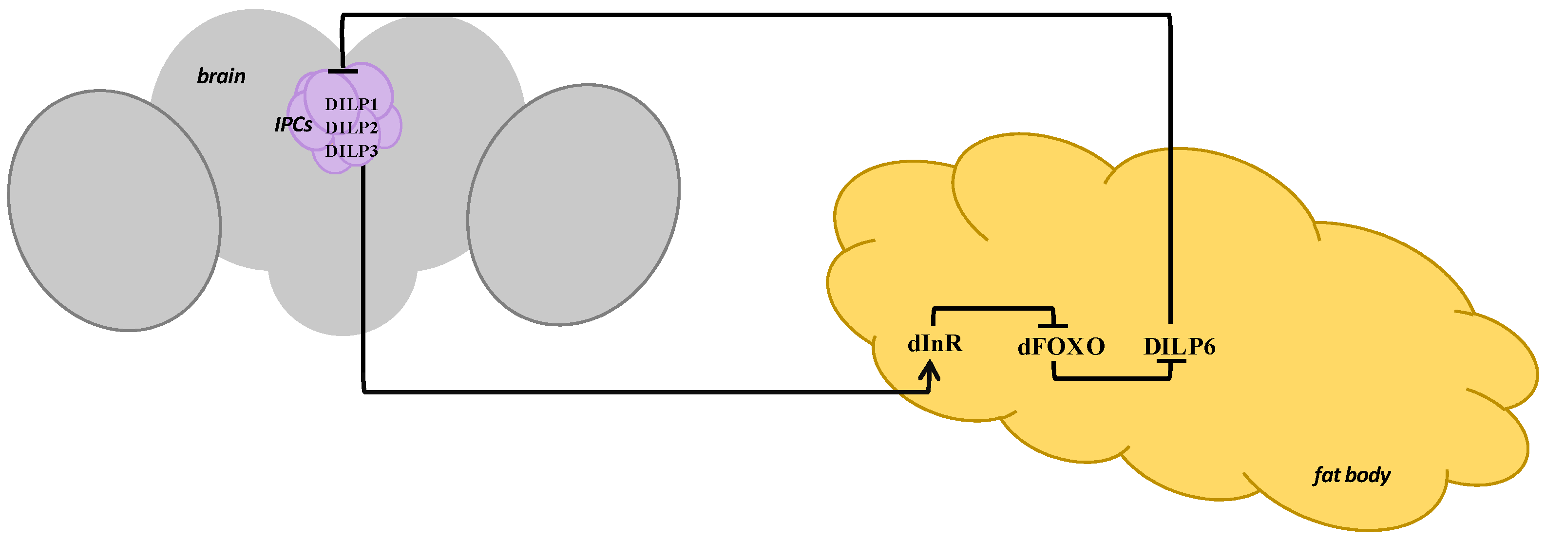
2. Insulin/Insulin-Like Growth Factors Signaling Pathway in Drosophila melanogaster

The insulin/insulin-like growth factors signaling (IIS) pathway is evolutionarily conserved among all metazoans and performs a vital role in the regulation of growth, development, reproduction, longevity, metabolism and stress resistance [1,2]. In D. melanogaster, eight insulin-like peptides (DILP1-8) have been identified: DILP1-5 show significant homology with insulin, DILP6–with insulin-like growth factors, DILP7 and DILP8–with mammalian relaxins [3,4,5,6,7].
DILPs are produced in medial neurosecretory cells or insulin-producing cells (IPCs) of the brain, as well as in the cells of peripheral tissues such as the visceral muscles of the gut, the fat body, which is the main metabolic organ in insects, neurons of the abdominal ganglia, and ovaries in a tissue- and stage-specific way [3,6,8,9,10,11,12]. Neuronal DILPs are secreted into the hemolymph and received by a homolog of the insulin receptor (dInR) for transmitting its signals to target cells [8,13,14,15,16]. DILP7 acts on the Lgr4 receptor bound to G-protein and containing leucine-rich repeats, and DILP8 binds Lgr3 [17,18].
dInR is localized in numerous fly tissues, including the fat body, the endocrine gland corpus allatum (CA) and follicular cells of the ovaries [19,20,21]. Activation of dInR, directly or through an orthologue of the mammalian insulin receptor substrate (CHICO), launches the kinase cascade, and dAkt/PKB (proteinkinase B homolog) inhibits the transfer of the transcription factor of the Drosophila Forkhead box class O family (dFOXO) into the cell nucleus and provokes its return from the nucleus back to the cytoplasm [22]. The main localization of dFOXO in D. melanogaster is the fat body of the head and abdomen [23]. dFOXO plays the role of the main regulator of expression of the downstream genes participating in the metabolism, the cell cycle, the stress response, the control of longevity and apoptosis [24,25,26,27]. It has been shown that a mutant dFOXO lacking dAkt phosphorylation sites does not react to IIS inhibition, remains in the nucleus and is constitutively active [28].

3. Stress-Related Hormones in Drosophila melanogaster

In D. melanogaster, IPCs are similar to vertebrate pancreatic β-cells secreting insulin in response to hyperglycemia, and the role of pancreatic α-cells secreting glucagon in response to hypoglycemia is performed by the cells of the corpus cardiacum (CC) gland, which produce a glucagon-like neuropeptide, the adipokinetic hormone (AKH) [3,29,30,31]. Due to the similarity of their functions to those of α- and β-cells, IPCs and CC, taken together, are seen as the Drosophila analogue of the mammalian pancreatic gland [32].
AKH regulates metabolic response to stress, stimulating catabolic reactions and mobilizing energy stores, especially lipids and trehalose, the latter being the main carbohydrate in insects [33,34]. It has been discovered that AKH deficit leads to obesity and a decrease in the carbohydrate level in D. melanogaster imagoes [35,36], and flies with an Akh mutation have much lower carbohydrate levels in hemolymph, including trehalose, and are resistant to starvation [34]. It has also been shown that Akh expression and AKH content in the cells are under DILP2 regulation in D. melanogaster females [11].
The IIS pathway interacts with other key hormones of the insect neuroendocrine stress reaction: 20-hydroxyecdysone (20E) and juvenile hormone (JH), which play a decisive role in growth, development, molting and metamorphosis in larvae, and perform the function of gonadotropins in imagoes, as well as biogenic amines dopamine (DA) and octopamine (OA) [22].
The central location of the OA and DA synthesis is octopamine- and dopaminergic neurons of the brain, the location of the JH synthesis is the CA gland [37,38]. It has been established that ecdysteroid biosynthesis during development takes place in the prothoracic gland, and the ovaries serve as the main source of ecdysteroids in imagoes [21,37,39]; seminal glands also contain ecdysteroids, but there is no sufficient evidence of an entire de novo biosynthesis pathway in this tissue [39]. In insects, DA and OA play the role of neurotransmitters in the synaptic cleft, neuromodulators within the bounds of one tissue, and neurohormones when being transmitted by the flow of the hemolymph to large distances [40]. It has been shown that they control the activity of the endocrine glands, arousal, desensitization of sensory inputs, various complex behavior forms such as memory and learning, and mobilization of lipids and carbohydrates [40,41,42].
DA is known to participate in JH level regulation, increasing it in young females, and decreasing it in mature ones [43]. Moreover, its regulation has a feedback loop: JH lowers the DA level in young females and increases it in mature ones. DA also regulates the 20E level, increasing it in young females and decreasing it in mature ones; however, no negative relationship has been discovered–20E increases the DA level in young females and decreases it in mature ones. In turn, DA and OA influence the 20E level indirectly through the JH metabolic system. This influence is unidirectional in young females, where it increases the 20E level, and multidirectional in mature ones: OA increases the 20E level, and DA decreases it [43]. Under unfavorable conditions of varying nature, the levels of all these hormones in Drosophila imagoes increase sharply (accompanied by a decrease in the activity of their metabolic enzymes; Table S1), affecting survival, fecundity and longevity [22]. In larvae, stress reaction develops as an inhibition of prothoracicotropic hormone (PTTH) secretion, which leads to a delay in ecdysone secretion and an increase in JH content, resulting in delayed metamorphosis or additional molting and allowing to “wait out” unfavorable conditions [44]. OA and DA levels in larval insects have also been shown to increase under heat stress [44,45].

4. Drosophila IIS Pathway Regulation under Stress

In D. melanogaster, dFOXO transfer to the nucleus is found under oxidative and metabolic stress [46,47]; moreover, there are data implying that under heat stress dFOXO translocation to the nucleus is accompanied by an increase in its gene’s expression level in imagoes [26,48]. Sixty minutes later dFOXO translocates from the nucleus back to the cytoplasm [26].
It has been shown that dFOXO positively regulates dilp6 mRNA level in the fat body of Drosophila imagoes [9]. So, dilp6 expression is not induced in dFOXO-mutant larvae under starvation [49], and dFOXO’s effect on the expression of DILPs produced in IPCs is blocked by simultaneous repression of dilp6 by RNA interference in the fat body [9]. At the same time, an increase in dilp6 expression in flies with mutations in dilp2-3,5 has been demonstrated [4]. Thus, DILP6 seems to connect dFOXO, the fat tissue and the endocrine function of the brain [4,9]. However, it has been shown that although the increase in dilp6 expression observed during pupariation is delayed in larvae with a null mutation of dfoxo, the level of dilp6 transcripts ultimately reaches intermediate levels; i.e., although dFOXO is necessary for timely dilp6 expression during development, its activation can also be caused by other factors [50]. Such a factor could be 20E, as it has been shown to induce dilp6 expression directly in the fat body at the last instar larvae [51].
However, under heat stress, dFOXO leads not to the activation, but to the inhibition of dilp6, which, in turn, results in dInR expression activation in imagoes [48]. This apparent contradiction can be explained by the existence of a feedback loop including other components of the insect neuroendocrine stress reaction, which ensure the increase in dilp6 expression level in response to dfoxo mutations or prolonged exposure to such a factor as starvation. The cardinal difference between IIS pathway regulation under starvation in larvae and under heat stress in imagoes is also demonstrated by the fact that there is a decrease in dilps expression level in larval IPCs under starvation [50], whereas under heat stress there is an increase in the adult DILP3 production [21].
In imagoes, dilp6 has been shown to inhibit the expression of dilps in IPCs and DILP2 secretion into the hemolymph [9], and dilp641 mutants have been found to demonstrate a sharp increase in the intensity of DILP3 production under normal conditions [21], which also agrees with our hypothesis of signal transduction from dFOXO to DILP3 through DILP6 under heat stress. The fact that DILP3 then activates dInR, thus preparing the cell to respond to stress conditions, is supported by our data on the lack of change in dInR expression level under heat stress in flies with dilp6 and dfoxo mutations, in contrast with the w1118 control line, which demonstrates an increase in dInR expression level in response to heat stress [48].
It is notable that not all genes of the IIS pathway participate in the response to short-term heat stress, which implies the complexity of the mechanism of insulin regulation of an organism’s response to adverse conditions. Thus, although it has been shown that in D. melanogaster imagoes dilp6 transcription correlates with a decrease in DILP2 secretion, which leads to reduced insulin signaling under starvation [49], under heat stress the amount of DILP2 in IPCs does not change (unlike DILP3) [21], which indicates that DILP2 is not involved in the response to heat stress in imagoes.
To sum up, a “lesser” feedback loop to DILP6 suppression by dFOXO, which has been activated under heat stress, is formed: decreased DILP6 stops inhibiting the synthesis of DILPs in IPCs, they activate the IIS in the fat body through dInR, which results in dFOXO transduction from the nucleus back to the cytoplasm (Figure 1). At the same time, it can be assumed that there is also a “greater” feedback loop activating under prolonged stress exposure—DILP6 regulation via other stress-related hormones.

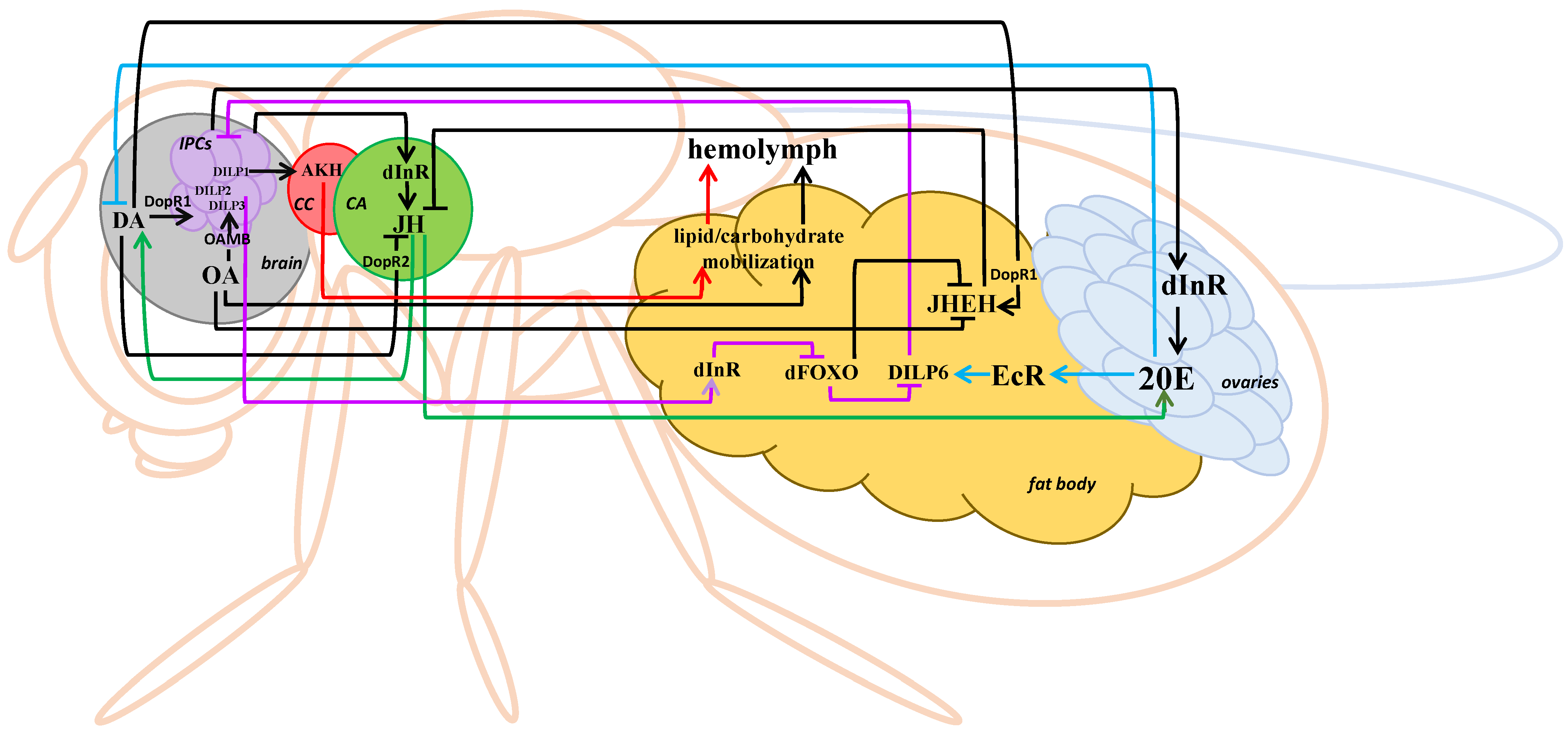
5. Interaction of the IIS Pathway and Stress-Related Hormones in Drosophila melanogaster

It has been discovered that 20E negatively affects general insulin signaling in D. melanogaster larvae, facilitating dFOXO transfer to the nucleus in the cells of the fat body through the activation of the hormone receptor (EcR) [52]. These results agree with the data on the increase in dFOXO transcriptional activity through 20E in the silkworm, Bombyx mori [53]. It has also been demonstrated that dilp6 expression increases under the influence of 20E even in the absence of dFOXO in third instar larva [50]; it can be assumed that dFOXO activation under the influence of 20E is mediated through IIS suppression by DILP6.
In imagoes, 20E, in turn, is regulated by JH [43], which is synthesized in CA under the regulation of DILPs via the activation of dInR [19]. dInR localization in follicular cells of the ovaries also suggests the existence of direct regulation of the 20E synthesis by the IIS pathway [21]. The JH level in D. melanogaster imagoes is also regulated by its degradation enzymes, the most important of which, the JH epoxide hydrolase, is synthesized in the fat body, ovaries and gut [54,55]; it is also, apparently, controlled by dFOXO: a decrease in the enzyme’s activity has been shown both under starvation [56] and in the case of a dfoxo mutation [57]. The influence of this effect on DA metabolism can be negated by JH treatment of the flies [26], which is in agreement with the existence of a feedback loop in DA regulation of the JH level [43]. There is also a feedback loop in the interaction of JH and dFOXO: JH suppresses dFOXO as the activity of the latter in D. melanogaster larvae with an ablated CA increases in comparison to the control [58]. It is possible that this feedback loop is mediated by 20E activation of dFOXO in the fat body [52].
Notably, in imagoes, a decrease in the activity of JH epoxide hydrolase and JH esterase under nutritional stress occurs at least after 6 h of starvation and lasts at least 24 h [56], which, apparently, results in an increase in the 20E level [59] under the influence of the increased JH level and a subsequent positive regulation of DILP6. For this reason, after overnight starvation, similar to the experiments described in [50], the DILP6 level increases, which is the last stage of the “greater” feedback loop of DILP6 expression regulation by dFOXO (Figure 2).
Another two elements of this “greater” loop, DA and OA, the levels of which also increase under stress [22], participate in the activation of DILPs production in IPCs and the regulation of the JH level. In imagoes, OA stimulates IPCs activity by binding with the OAMB receptor [60,61] and suppresses the activity of JH degradation enzymes [62,63]; at the same time, DA activates IPCs through Dopamine Receptor 1 (DopR1) [64] and decreases the JH level via the activation of the corresponding receptors in CA (DopR2) and the fat body (DopR1) in mature females [65]. The opposite action of the two amines on the JH metabolism may normalize the hormone’s level after the stress is over.
It is worth noting that DILPs affect the metabolism of stress-related hormones as well. So, it has been demonstrated that a dInR mutation or a dInR knockdown in CA decreases the 20E and JH synthesis in D. melanogaster imagoes [19,66,67,68] and increases JH degradation [69]. This agrees with the data on insulin injection causing a decrease in JH degradation and an increase in the activity of the first DA synthesis enzyme, the tyrosine hydroxylase, in D. melanogaster females [70].

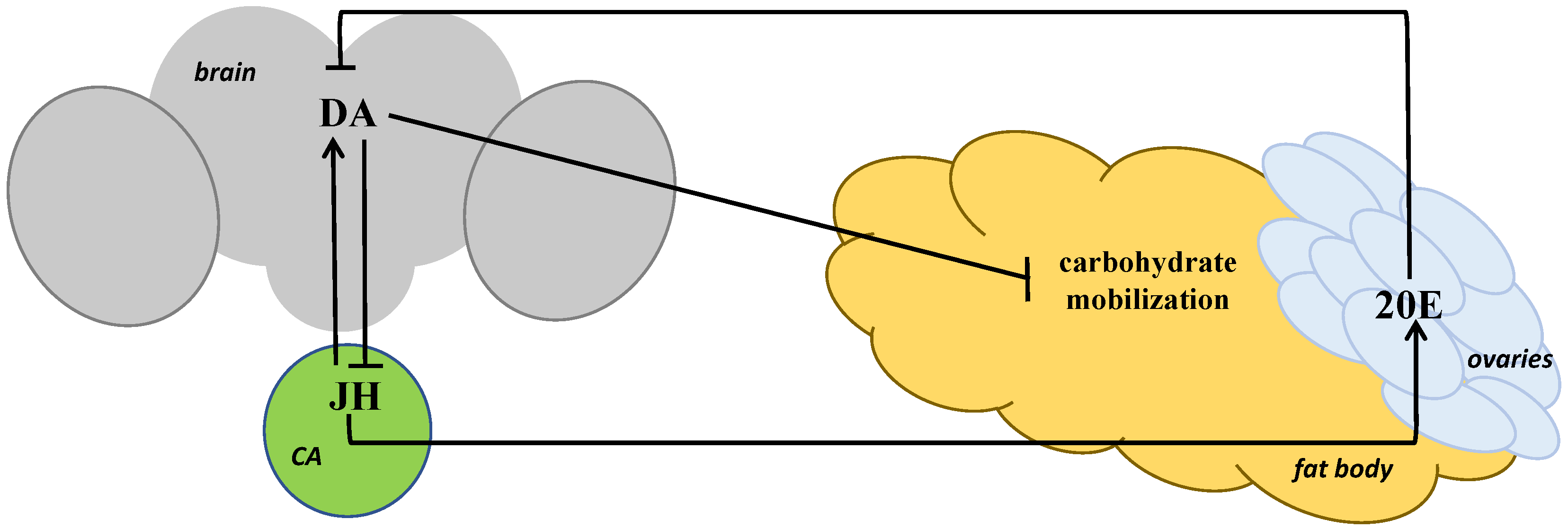
6. A Possible Mechanism of Short-Term Heat Stress Influence on Carbohydrate and Lipid Metabolism in Drosophila melanogaster

It is known that systemic defects in the IIS pathway cause a complex set of phenotypes in D. melanogaster including those connected to metabolism, which usually include an increase in carbohydrate and lipid stores [71]. It has been shown that most viable mutant combinations with a partial loss of function or hypomorphism of the IIS pathway genes have changes in the carbohydrate and lipid level [72]. D. melanogaster imagoes with dilp6 or dfoxo mutations are characterized by elevated levels of glucose and trehalose [73], as well as total lipids [48], and larvae with a dilp6 knockdown have increased levels of triglycerides and glycogen [50].
It has been shown that short-term heat stress causes an increase in both trehalose and glucose content in D. melanogaster females after just 30 min of exposure [73,74]. This agrees with earlier data on the ability of trehalose to increase resistance to temperature stress, demonstrated in the larvae of the Antarctic midge, Belgica antarctica [75]. Total lipid content in D. melanogaster imagoes also changes after heat exposure, although it occurs only 24 h later [48,76].
An immediate effect of short-term heat stress on carbohydrate but not lipid content in D. melanogaster females can be explained by a high solubility of carbohydrates, meaning they can be used for maintaining vital functions in rapidly changing environmental conditions, unlike lipids used by the organism as the last energy reserve under prolonged starvation or other lasting adverse influences [76]. Trehalose is known to be the main fuel for insect flight and the source of energy during nonfeeding periods [77] and thus its increase under stress provides an insect with additional energy, which allows it to avoid and/or survive adverse conditions. The assumption regarding the mobilization of lipid stores under prolonged stress is confirmed by the data on a decrease in total lipid content in D. melanogaster females in 24 h after heat exposure (38 °C for 60 min) [48] as well as a decrease in triglycerides content in D. melanogaster males in 24 h after short-term heat stress (38 °C for 45 min); curiously, this effect lasts for up to 5 days [76]. Moreover, a similar decrease in triglycerides content has been observed in D. melanogaster males in 24 h after short-term cold stress (4 °C, 4 h or 0 °C, 4 h) [76], which signifies a certain universality of this response.
Regarding the possible mechanisms behind the carbohydrate and lipid changes following acute heat stress, it could be assumed that the quick increase in trehalose and glucose levels is a result of increased synthesis, whereas the decrease in total lipid content results from the decrease in food consumption, which also occurs in imagoes only 24 h after the stress exposure [48].
It is worth noting that although dilp6 and dfoxo mutations disturb the total lipid response to heat stress in imagoes, they do not prevent an increase in the carbohydrate level under stress [48,73], which signifies that the IIS pathway is not the only mechanism of the carbohydrate metabolism regulation under stress. OA likely plays the role of an additional regulator in insects when it is released into the hemolymph as a neurohormone and further transported to a target tissue, where it mobilizes lipids and carbohydrates [78,79]; other stress-related hormones, such as JH, 20E and DA, also play this part [74,76]. The largest amount of data on this have been obtained for 20E on different insect species: it stimulates glycolysis increasing the glucose level in hemolymph in the honeybee, Apis mellifera, larvae [80], as well as in pupae of the cotton bollworm, Helicoverpa armigera, [81] and the Chinese oak silk moth, Antheraea pernyi, [82]; it also increases lipolysis and inhibits lipid synthesis in the fall webworm, Hyphantria cunea, larvae [83].
So, an increase in the DA or JH level in D. melanogaster females results in a decrease in trehalose and glucose content [74]; it can be assumed that they take part in normalizing the carbohydrates level after the stress is over (Figure 3). At the same time, 20E has an opposite effect on the content of the two carbohydrates [84]; it can be assumed that 20E effects the carbohydrate metabolism indirectly via DA by lowering its level, as has been shown before [43], and thus increasing the carbohydrate level, because a DA increase leads to the opposite effect on the level of both carbohydrates compared to 20E—their decrease [74]. We also assume DA to mediate the influence on the carbohydrate metabolism of not only 20E but also JH, which promotes an increase in the amine level in mature Drosophila females [46]. The data regarding the increase in trehalose content in Drosophila imagoes caused by JH application to flies [74] agree with the data received on imagoes of the red flour beetle, Tribolium castaneum: a decrease in the expression of genes coding a key enzyme in the JH synthesis, JH acid methyltransferase (JHAMT), and its receptor, methoprene-tolerant (Met), led to an increase in the trehalose content in hemolymph under starvation [85].
To sum up, the data on the DA, JH and 20E effect on the carbohydrate content in D. melanogaster [74,84] and the data on the key role of AKH in triglycerides catabolism and trehalose mobilization from glycogen [11,33,34,35,36] provide evidence that DA, the level of which is regulated positively by JH and negatively by 20E, activates DILPs production in IPCs, they, in turn, stimulate AKH production in CC, and AKH ensures carbohydrate and lipid mobilization in the fat body.

7. The Effect of Short-Term Heat Stress on Feeding Behavior in Drosophila melanogaster

Changes in metabolic behavior can also be considered as a delayed response to heat stress. It has been shown that significant changes in feeding intensity as well as in lipid content in D. melanogaster females occur in 24 h after short-term heat exposure (38 °C, 60 min), resulting in a decrease in food consumption [48]. These data suggest that regulation of feeding behavior under stress may be controlled by means other than the IIS pathway, as the changes in the expression of the pathway’s genes and the increase in the carbohydrates level take place directly after short-term heat exposure, and the behavioral response manifests as a decrease in food consumption 24 h after the exposure, resulting, apparently, in a decrease in lipid content. This conclusion agrees with the idea that feeding regulation and glycaemia control are the result of complex interaction of metabolic, hormonal and neural signals that have not yet been fully elucidated [86].
The regulation of feeding behavior under heat stress seems to occur without the participation of the dilp6 and dfoxo genes as their mutations do not prevent the decrease in the appetite after stress exposure in imagoes [48]. We assume that, in flies with a dfoxo mutation, the IIS pathway feedback loop (see Figure 1) is disrupted, and when in response to food intake the dFOXO signal becomes insufficient for modulating the expression of dilp6 and dInR, the levels of which remain low, and for regulating other genes responsible for changes in feeding behavior as well. The existence of other mechanisms of feeding regulation, besides the IIS pathway, is evidenced by the fact that disruption of the next link of the “lesser” feedback loop, DILP6, causing similar changes in the level of dilp6 and dInR expression, does not prevent the behavioral stress response of a decrease in feeding intensity [48].
DA signaling in the mushroom body, a major Drosophila memory center [87], can potentially be such a mechanism. So, it has been revealed that flies consume more food during long-term memory formation, and RNAi expression against DopR1 in the neurons of the mushroom body results in impairment of long-term memory [88]. In turn, a decrease in sucrose consumption and suppression of proboscis extension in D. melanogaster has been demonstrated under the activation of mushroom body output neurons [89].
A constitutive increase in the appetite of flies with dilp6 and dfoxo mutations could be connected with the increase in DILPs production in IPCs caused by the abovementioned disruptions in the “lesser” feedback loop. This assumption is confirmed by the data on a higher DILP3 level in the mutant dilp641 females with a decreased DILP6 function [21]. The ability of DILP6 to suppress DILPs production in IPCs is also demonstrated by the data on the decrease in both DILP2 secretion and dilp2 and dilp5 expression at the overexpression of dilp6 in the adult fat body [9]. Thus, if DILP6, as a consequence of its mutation or a mutation of dfoxo, is incapable of inhibiting DILPs in IPCs, their levels increase, leading to higher fly appetite and the formation of the diabetic phenotype, including increased carbohydrate and lipid levels. The decrease in dInR expression in flies with a dilp6 mutation is likely caused by constitutionally increased levels of DILPs synthesized in IPCs and plays a compensatory role. Another possible explanation of this phenomenon can be the ability of DILP6 to activate dInR, similarly to DILPs in IPCs, which leads to a decrease in the receptor’s expression at DILP6 dysfunction.

8. Conclusions and Future Perspectives

In recent years, many studies have been performed with the use of modern omics methods, such as transcriptomics, proteomics and metabolomics, undoubtedly providing a plethora of new data regarding the metabolic and other systems of the organism. However, the contribution of studies researching the endocrine regulation of metabolism with the use of classical methods—specific gene mutations and knockouts, pharmacological treatments with various hormones and tissue-specific expression regulation using the UAS-4 system—to elucidate the mechanisms of these systems’ functioning should not be underestimated. Here, we examine results from studies performed almost exclusively with the use of such methods: they reveal the complexity of the mechanism of the D. melanogaster imago neuroendocrine stress reaction, the interaction of its key elements such as biogenic amines (DA and OA), gonadotropins (JH and 20E), AKH and insulin-like peptides, and their role in regulating carbohydrate and lipid metabolism under heat stress conditions. We assume that further investigation in this field will help to unveil the connection of hormonal pathways involved in the stress reaction to elements of cellular stress response, including the c-Jun-N-terminal kinase (JNK) signaling pathway and the heat shock response pathway.

Supplementary Materials

The following supporting information can be downloaded at: https://www.mdpi.com/article/10.3390/insects14050474/s1, Table S1: The time course of changes in the levels of hormones and biogenic amines and the activity of their metabolic enzymes in Drosophila under heat stress (38 °C) [22,90,91,92,93,94,95,96,97].

Author Contributions

Conceptualization, N.E.G.; writing—original draft preparation, M.A.B.; writing—review and editing, N.E.G.; visualization, M.A.B.; supervision, N.E.G.; project administration, N.E.G.; funding acquisition, M.A.B. All authors have read and agreed to the published version of the manuscript.

Funding

This research was funded by the Russian Science Foundation, grant number 23-24-00320.

Data Availability Statement

Not applicable.

Acknowledgments

We thank Darya Kochetova for the translation of our manuscript to English.

Conflicts of Interest

The authors declare no conflict of interest.

References

  1. Garofalo, R.S. 2 Genetic analysis of insulin signaling in Drosophila. Trends Endocrinol. Metab. 2002, 13, 156–162. [Google Scholar] [CrossRef]
  2. Chowański, S.; Walkowiak-Nowicka, K.; Winkiel, M.; Marciniak, P.; Urbański, A.; Pacholska-Bogalska, J. Insulin-like peptides and cross-talk with other factors in the regulation of insect metabolism. Front. Physiol. 2021, 12, 701203. [Google Scholar] [CrossRef] [PubMed]
  3. Brogiolo, W.; Stocker, H.; Ikeya, T.; Rintelen, F.; Fernandez, R.; Hafen, E. An evolutionarily conserved function of the Drosophila insulin receptor and insulin-like peptides in growth control. Curr. Biol. 2001, 11, 213–221. [Google Scholar] [CrossRef] [PubMed]
  4. Grönke, S.; Clarke, D.F.; Broughton, S.; Andrews, T.D.; Partridge, L. Molecular evolution and functional characterization of Drosophila insulin-like peptides. PLoS Genet. 2010, 6, e1000857. [Google Scholar] [CrossRef] [PubMed]
  5. Garelli, A.; Gontijo, A.M.; Miguela, V.; Caparros, E.; Dominguez, M. Imaginal discs secrete insulin-like peptide 8 to mediate plasticity of growth and maturation. Science 2012, 336, 579–582. [Google Scholar] [CrossRef]
  6. Colombani, J.; Andersen, D.S.; Leopold, P. Secreted peptide Dilp8 coordinates Drosophila tissue growth with developmental timing. Science 2012, 336, 582–585. [Google Scholar] [CrossRef]
  7. Álvarez-Rendón, J.P.; Salceda, R.; Riesgo-Escovar, J.R. Drosophila melanogaster as a model for diabetes type 2 progression. BioMed Res. Int. 2018, 2018, 1417528. [Google Scholar] [CrossRef]
  8. Miguel-Aliaga, I.; Thor, S.; Gould, A.P. Postmitotic specification of Drosophila insulinergic neurons from pioneer neurons. PLoS Biol. 2008, 6, e58. [Google Scholar] [CrossRef]
  9. Bai, H.; Kang, P.; Tatar, M. Drosophila insulin-like peptide-6 (dilp6) expression from fat body extends lifespan and represses secretion of Drosophila insulin-like peptide-2 from the brain. Aging Cell 2012, 11, 978–985. [Google Scholar] [CrossRef]
  10. Post, S.; Karashchuk, G.; Wade, J.D.; Sajid, W.; De Meyts, P.; Tatar, M. Drosophila insulin-like peptides DILP2 and DILP5 differentially stimulate cell signaling and glycogen phosphorylase to regulate longevity. Front. Endocrinol. 2018, 9, 245. [Google Scholar] [CrossRef]
  11. Post, S.; Liao, S.; Yamamoto, R.; Veenstra, J.A.; Nässel, D.R.; Tatar, M. Drosophila insulin-like peptide dilp1 increases lifespan and glucagon-like Akh expression epistatic to dilp2. Aging Cell 2019, 18, 12863. [Google Scholar] [CrossRef] [PubMed]
  12. Prince, E.; Kretzschmar, J.; Trautenberg, L.C.; Broschk, S.; Brankatschk, M. DIlp7-producing neurons regulate insulin-producing cells in Drosophila. Front Physiol. 2021, 12, 630390. [Google Scholar] [CrossRef] [PubMed]
  13. Rulifson, E.J.; Kim, S.K.; Nusse, R. Ablation of insulinproducing neurons in flies: Growth and diabetic phenotypes. Science 2002, 296, 1118–1120. [Google Scholar] [CrossRef] [PubMed]
  14. Géminard, C.; Rulifson, E.J.; Léopold, P. Remote control of insulin secretion by fat cells in Drosophila. Cell Metab. 2009, 10, 199–207. [Google Scholar] [CrossRef]
  15. Cognigni, P.; Bailey, A.P.; Miguel-Aliaga, I. Enteric neurons and systemic signals couple nutritional and reproductive status with intestinal homeostasis. Cell Metab. 2011, 13, 92–104. [Google Scholar] [CrossRef]
  16. Nässel, D.R.; Kubrak, O.I.; Liu, Y.; Luo, J.; Lushchak, O.V. Factors that regulate insulin producing cells and their output in Drosophila. Front. Physiol. 2013, 4, 252. [Google Scholar] [CrossRef]
  17. Gontijo, A.M.; Garelli, A. The biology and evolution of the Dilp8-Lgr3 pathway: A relaxin-like pathway coupling tissue growth and developmental timing control. Mech. Dev. 2018, 154, 44–50. [Google Scholar] [CrossRef]
  18. Imambocus, B.N.; Zhou, F.; Formozov, A.; Wittich, A.; Tenedini, F.M.; Hu, C.; Sauter, K.; Varela, E.M.; Herédia, F.; Casimiro, A.P.; et al. A neuropeptidergic circuit gates selective escape behavior of Drosophila larvae. Curr. Biol. 2022, 32, 149–163. [Google Scholar] [CrossRef]
  19. Belgacem, Y.H.; Martin, J.R. Hmgcr in the corpus allatum controls sexual dimorphism of locomotor activity and body size via the insulin pathway in Drosophila. PLoS ONE 2007, 2, e187. [Google Scholar] [CrossRef]
  20. Rauschenbach, I.Y.; Karpova, E.K.; Adonyeva, N.V.; Andreenkova, O.V.; Faddeeva, N.V.; Burdina, E.V.; Alekseev, A.A.; Menshanov, P.N.; Gruntenko, N.E. Disruption of insulin signalling affects the neuroendocrine stress reaction in Drosophila females. J. Exp. Biol. 2014, 217, 3733–3741. [Google Scholar] [CrossRef]
  21. Andreenkova, O.V.; Adonyeva, N.V.; Eremina, M.A.; Gruntenko, N.E.; Rauschenbach, I.Y. The Insulin-like receptor gene expression in the tissues synthesizing gonadotropic hormones at sexual maturation of Drosophila melanogaster females. Russ. J. Genet. 2016, 52, 1342–1344. [Google Scholar] [CrossRef]
  22. Gruntenko, N.E.; Rauschenbach, I.Y. The role of insulin signalling in the endocrine stress response in Drosophila melanogaster: A mini-review. Gen. Comp. Endocrinol. 2018, 258, 134–139. [Google Scholar] [CrossRef] [PubMed]
  23. Zheng, X.; Yang, Z.; Yue, Z.; Alvarez, J.D.; Sehgal, A. FOXO and insulin signaling regulate sensitivity of the circadian clock to oxidative stress. Proc. Natl. Acad. Sci. USA 2007, 104, 15899–15904. [Google Scholar] [CrossRef] [PubMed]
  24. Puig, O.; Matilla, J. Understanding Forkhead box class O function: Lessons from Drosophila melanogaster. Antioxid. Redox Signal. 2011, 14, 635–647. [Google Scholar] [CrossRef] [PubMed]
  25. Wang, Z.; Yu, T.; Huang, P. Post-translational modifications of FOXO family proteins (Review). Mol. Med. Rep. 2016, 14, 4931–4941. [Google Scholar] [CrossRef]
  26. Gruntenko, N.E.; Adonyeva, N.V.; Burdina, E.V.; Karpova, E.K.; Andreenkova, O.V.; Gladkikh, D.V.; Ilinsky, Y.Y.; Rauschenbach, I.Y. The impact of FOXO on dopamine and octopamine metabolism in Drosophila under normal and heat stress conditions. Biol. Open. 2016, 5, 1706–1711. [Google Scholar] [CrossRef]
  27. Ding, K.; Barretto, E.C.; Johnston, M.; Lee, B.; Gallo, M.; Grewal, S.S. Transcriptome analysis of FOXO-dependent hypoxia gene expression identifies Hipk as a regulator of low oxygen tolerance in Drosophila. G3-Genes Genom. Genet. 2022, 12, jkac263. [Google Scholar] [CrossRef]
  28. Puig, O.; Marr, M.T.; Ruhf, M.L.; Tjian, R. Control of cell number by Drosophila FOXO: Downstream and feedback regulation of the insulin receptor pathway. Genes Dev. 2003, 17, 2006–2020. [Google Scholar] [CrossRef]
  29. Ikeya, T.; Galic, M.; Belawat, P.; Nairz, K.; Hafen, E. Nutrient dependent expression of insulin-like peptides from neuroendocrine cells in the CNS contributes to growth regulation in Drosophila. Curr. Biol. 2002, 12, 1293–1300. [Google Scholar] [CrossRef]
  30. Kim, S.K.; Rulifson, E.J. Conserved mechanisms of glucose sensing and regulation by Drosophila corpora cardiaca cells. Nature 2004, 431, 316–320. [Google Scholar] [CrossRef]
  31. Lee, G.; Park, J.H. Hemolymph sugar homeostasis and starvation-induced hyperactivity affected by genetic manipulations of the adipokinetic hormone-encoding gene in Drosophila melanogaster. Genetics 2004, 167, 311–323. [Google Scholar] [CrossRef] [PubMed]
  32. Hughson, B.N. The glucagon-like adipokinetic hormone in Drosophila melanogaster—Biosynthesis and secretion. Front. Physiol. 2021, 12, 710652. [Google Scholar] [CrossRef] [PubMed]
  33. Isabel, G.; Martin, J.-R.; Chidami, S.; Veenstra, J.A.; Rosay, P. AKH-producing neuroendocrine cell ablation decreases trehalose and induces behavioral changes in Drosophila. Am. J. Physiol. Regul. Integr. Comp. Physiol. 2005, 288, 531–538. [Google Scholar] [CrossRef] [PubMed]
  34. Sajwan, S.; Sidorov, R.; Stašková, T.; Žaloudíková, A.; Takasu, Y.; Kodrík, D.; Zurovec, M. Targeted mutagenesis and functional analysis of adipokinetic hormone-encoding gene in Drosophila. Insect Biochem. Mol. Biol. 2015, 61, 79–86. [Google Scholar] [CrossRef]
  35. Gáliková, M.; Diesner, M.; Klepsatel, P.; Hehlert, P.; Xu, Y.; Bickmeyer, I.; Predel, R.; Kühnlein, R.P. Energy homeostasis control in Drosophila adipokinetic hormone mutants. Genetics 2015, 201, 665–683. [Google Scholar] [CrossRef]
  36. Bednářová, A.; Tomčala, A.; Mochanová, M.; Kodrík, D.; Krishnan, N. Disruption of adipokinetic hormone mediated energy homeostasis has subtle effects on physiology, behavior and lipid status during aging in Drosophila. Front. Physiol. 2018, 9, 949. [Google Scholar] [CrossRef]
  37. Toivonen, J.M.; Partridge, L. Endocrine regulation of aging and reproduction in Drosophila. Mol. Cell Endocrinol. 2009, 299, 39–50. [Google Scholar] [CrossRef]
  38. Zhang, X.; Li, S.; Liu, S. Juvenile hormone studies in Drosophila melanogaster. Front. Physiol. 2022, 12, 785320. [Google Scholar] [CrossRef]
  39. Lafont, R.; Dauphin-Villemant, C.; Warren, J.T.; Rees, H.H. Ecdysteroid chemistry and biochemistry. Ref. Mod. Life Sci. 2017, 3, 125–195. [Google Scholar] [CrossRef]
  40. Sasaki, K.; Harano, K.-I. Multiple regulatory roles of dopamine in behavior and reproduction of social insects. Trends Entomol. 2010, 6, 1–13. [Google Scholar]
  41. Farooqui, T. Octopamine-mediated neuromodulation of insect senses. Neurochem. Res. 2007, 32, 1511–1529. [Google Scholar] [CrossRef] [PubMed]
  42. Martin, C.A.; Krantz, D.E. Drosophila melanogaster as a genetic model system to study neurotransmitter transporters. Neurochem. Int. 2014, 73, 71–88. [Google Scholar] [CrossRef] [PubMed]
  43. Gruntenko, N.E.; Rauschenbach, I.Y. Interplay of JH, 20E and biogenic amines under normal and stress conditions and its effects on reproduction. J. Insect Physiol. 2008, 54, 902–908. [Google Scholar] [CrossRef]
  44. Rauschenbach, I.Y. Stress response in insects: Mechanism, genetic control, and role in adaptation. Russ. J. Genet. 1997, 33, 942–949. [Google Scholar]
  45. Hirashima, A.; Nagano, T.; Eto, M. Stress-induced changes in the biogenic amine levels and larval growth of Tribolium castaneum Herbst. Biosci. Biotech. Biochem. 1993, 57, 2085–2089. [Google Scholar] [CrossRef]
  46. Jünger, M.A.; Rintelen, F.; Stocker, H.; Wasserman, J.D.; Végh, M.; Radimerski, T.; Greenberg, M.E.; Hafen, E. The Drosophila forkhead transcription factor FOXO mediates the reduction in cell number associated with reduced insulin signaling. J. Biol. Chem. 2003, 2, 20. [Google Scholar] [CrossRef]
  47. Hwangbo, D.S.; Gershman, B.; Tu, M.P.; Palmer, M.; Tatar, M. Drosophila dFOXO controls lifespan and regulates insulin signalling in brain and fat body. Nature 2004, 429, 562–566. [Google Scholar] [CrossRef]
  48. Eremina, M.A.; Menshanov, P.N.; Shishkina, O.D.; Gruntenko, N.E. The transcription factor dFOXO controls the expression of insulin pathway genes and lipids content under heat stress in Drosophila melanogaster. Vavilov J. Genet. Breed. 2021, 25, 465–471. [Google Scholar] [CrossRef]
  49. Zhang, Y.; Xi, Y. Fat body development and its function in energy storage and nutrient sensing in Drosophila melanogaster. J. Tissue Sci. Eng. 2014, 6, 1. [Google Scholar] [CrossRef]
  50. Slaidina, M.; Delanoue, R.; Grönke, S.; Partridge, L.; Leopold, P. A Drosophila insulin-like peptide promotes growth during nonfeeding states. Dev. Cell 2009, 17, 874–884. [Google Scholar] [CrossRef]
  51. Okamoto, N.; Yamanaka, N.; Yagi, Y.; Nishida, Y.; Kataoka, H.; O’Connor, M.B.; Mizoguchi, A. A fat body-derived IGF-like peptide regulates postfeeding growth in Drosophila. Dev. Cell 2009, 17, 885–891. [Google Scholar] [CrossRef] [PubMed]
  52. Colombani, J.; Bianchini, L.; Layalle, S.; Pondeville, E.; Dauphin-Villemant, C.; Antoniewski, C.; Carré, C.; Noselli, S.; Léopold, P. Antagonistic actions of ecdysone and insulins determine final size in Drosophila. Science 2005, 310, 667–670. [Google Scholar] [CrossRef]
  53. Hossain, M.S.; Liu, Y.; Zhou, S.; Li, K.; Tian, L.; Li, S. 20-Hydroxyecdysone-induced transcriptional activity of FoxO upregulates brummer and acid lipase-1 and promotes lipolysis in Bombyx fat body. Insect Biochem. Mol. Biol. 2013, 43, 829–838. [Google Scholar] [CrossRef]
  54. Khlebodarova, T.M.; Gruntenko, N.E.; Grenback, L.G.; Sukhanova, M.Z.; Mazukov, M.M.; Rauschenbach, I.Y.; Tomas, B.A.; Hammock, B.D. A comparative analysis of juvenile hormone metabolizing enzymes in two species of Drosophila during development. Insect Biochem. Mol. Biol. 1996, 26, 829–835. [Google Scholar] [CrossRef] [PubMed]
  55. Borovsky, D.; Breyssens, H.; Buytaert, E.; Peeters, T.; Laroye, C.; Stoffels, K.; Rougé, P. Cloning and characterization of Drosophila melanogaster juvenile hormone epoxide hydrolases (JHEH) and their promoters. Biomolecules 2022, 12, 991. [Google Scholar] [CrossRef]
  56. Raushenbach, I.Y.; Gruntenko, N.E.; Bownes, M.; Adonieva, N.V.; Terashima, J.; Karpova, E.K.; Faddeeva, N.V.; Chentsova, N.A. The role of juvenile hormone in the control of reproductive function in Drosophila virilis under nutritional stress. J. Insect Physiol. 2004, 50, 323–330. [Google Scholar] [CrossRef]
  57. Rauschenbach, I.Y.; Karpova, E.K.; Gruntenko, N.E. dFOXO transcription factor regulates juvenile hormone metabolism in Drosophila melanogaster females. Russ. J. Genet. 2015, 51, 932–934. [Google Scholar] [CrossRef]
  58. Mirth, C.K.; Tang, H.Y.; Makohon-Moore, S.C.; Salhadar, S.; Gokhale, R.H.; Warner, R.D.; Koyama, T.; Riddiford, L.M.; Shingleton, A.W. Juvenile hormone regulates body size and perturbs insulin signaling in Drosophila. Proc. Natl. Acad. Sci. USA 2014, 111, 7018–7023. [Google Scholar] [CrossRef]
  59. Terashima, J.; Takaki, K.; Sakurai, S.; Bownes, M. Nutritional status affects 20-hydroxyecdysone concentration and progression of oogenesis in Drosophila melanogaster. J Endocrinol. 2005, 187, 69–79. [Google Scholar] [CrossRef]
  60. Crocker, A.; Shahidullah, M.; Levitan, I.B.; Sehgal, A. Identification of a neural circuit that underlies the effects of octopamine on sleep: Wake behavior. Neuron 2010, 65, 670–681. [Google Scholar] [CrossRef]
  61. Luo, J.; Lushchak, O.V.; Goergen, P.; Williams, M.J.; Nässel, D.R. Drosophila insulin-producing cells are differentially modulated by serotonin and octopamine receptors and affect social behavior. PLoS ONE 2014, 9, e99732. [Google Scholar] [CrossRef]
  62. Gruntenko, N.E.; Wilson, T.G.; Monastirioti, M.; Rauschenbach, I.Y. Stress-reactivity and juvenile hormone degradation in Drosophila melanogaster strains having stress-related mutations. Insect Biochem. Mol. Biol. 2000, 30, 775–783. [Google Scholar] [CrossRef]
  63. Gruntenko, N.E.; Karpova, E.K.; Alekseev, A.A.; Chentsova, N.A.; Bogomolova, E.V.; Bownes, M.; Rauschenbach, I.Y. Effects of octopamine on reproduction, juvenile hormone metabolism, dopamine, and 20-hydroxyecdysone contents in Drosophila. Arch. Insect Biochem. Physiol. 2007, 65, 85–94. [Google Scholar] [CrossRef]
  64. Andreatta, G.; Kyriacou, C.P.; Flatt, T.; Costa, R. Aminergic signaling controls ovarian dormancy in Drosophila. Sci. Rep. 2018, 8, 2030. [Google Scholar] [CrossRef]
  65. Gruntenko, N.E.; Laukhina, O.V.; Rauschenbach, I.Y. Role of D1- and D2-like receptors in age-specific regulation of juvenile hormone and 20-hydroxyecdysone levels by dopamine in Drosophila. J. Insect Physiol. 2012, 58, 1534–1540. [Google Scholar] [CrossRef]
  66. Tatar, M.; Kopelman, A.; Epstein, D.; Tu, M.P.; Yin, C.M.; Garofalo, R.S. A mutant Drosophila insulin receptor homolog that extends lifespan and impairs neuroendocrine function. Science 2001, 292, 107–110. [Google Scholar] [CrossRef]
  67. Tu, M.P.; Yin, C.M.; Tatar, M. Impaired ovarian ecdysone synthesis of Drosophila melanogaster insulin receptor mutants. Aging Cell 2002, 1, 158–160. [Google Scholar] [CrossRef]
  68. Tu, M.P.; Yin, C.M.; Tatar, M. Mutations in insulin signaling pathway alter juvenile hormone synthesis in Drosophila melanogaster. Gen. Comp. Endocrinol. 2005, 142, 347–356. [Google Scholar] [CrossRef]
  69. Bogomolova, E.V.; Adonyeva, N.V.; Karpova, E.K.; Gruntenko, N.E.; Rauschenbach, I.Y. Ubiquitous downregulation of InR gene expression affects stress associated hormone metabolism in Drosophila females. Russ. J. Genet. 2013, 49, 778–781. [Google Scholar] [CrossRef]
  70. Karpova, E.K.; Adonyeva, N.V.; Faddeeva, N.V.; Romanova, I.V.; Gruntenko, N.E.; Rauschenbach, I.Y. Insulin affects repro-duction and juvenile hormone metabolism under normal and stressful conditions in Drosophila females. Dokl. Biochem. Biophys. 2013, 452, 264–266. [Google Scholar] [CrossRef]
  71. Mattila, J.; Hietakangas, V. Regulation of carbohydrate energy metabolism in Drosophila melanogaster. Genetics 2017, 207, 1231–1253. [Google Scholar] [CrossRef]
  72. Murillo-Maldonado, J.M.; Sánchez-Chávez, G.; Salgado, L.M.; Salceda, R.; Riesgo-Escovar, J.R. Drosophila insulin pathway mutants affect visual physiology and brain function besides growth, lipid, and carbohydrate metabolism. Diabetes 2011, 60, 1632–1636. [Google Scholar] [CrossRef]
  73. Eremina, M.A.; Karpova, E.K.; Rauschenbach, I.Y.; Pirozhkova, D.S.; Andreenkova, O.V.; Gruntenko, N.E. Mutations in the insulin signaling pathway genes affect carbohydrate level under heat stress in Drosophila melanogaster females. Russ. J. Genet. 2019, 55, 519–522. [Google Scholar] [CrossRef]
  74. Karpova, E.K.; Eremina, M.A.; Pirozhkova, D.S.; Gruntenko, N.E. Stress-related hormones affect carbohydrate metabolism in Drosophila females. Arch. Insect Biochem. Physiol. 2019, 101, e21540. [Google Scholar] [CrossRef]
  75. Benoit, J.B.; Lopez-Martinez, G.; Elnitsky, M.A.; Lee, R.E., Jr.; Denlinger, D.L. Dehydration-induced cross tolerance of Belgica antarctica larvae to cold and heat is facilitated by trehalose accumulation. Comp. Biochem. Physiol. A Mol. Integr. Physiol. 2009, 152, 518–523. [Google Scholar] [CrossRef]
  76. Klepsatel, P.; Gáliková, M.; Xu, Y.; Kühnlein, R.P. Thermal stress depletes energy reserves in Drosophila. Sci. Rep. 2016, 6, e33667. [Google Scholar] [CrossRef]
  77. Arrese, E.L.; Soulages, J.L. Insect fat body: Energy, metabolism, and regulation. Annu. Rev. Entomol. 2010, 55, 207–225. [Google Scholar] [CrossRef]
  78. Roeder, T. Tyramine and octopamine: Ruling behavior and metabolism. Annu. Rev. Entomol. 2005, 50, 447–477. [Google Scholar] [CrossRef]
  79. Farooqui, T. Review of octopamine in insect nervous systems. Open Access Insect Physiol. 2012, 4, 1–17. [Google Scholar] [CrossRef]
  80. Yu, J.; Wang, H.; Chen, W.; Song, H.; Wang, Y.; Liu, Z.; Xu, B. 20-Hydroxyecdysone and receptor interplay in the regulation of hemolymph glucose level in honeybee (Apis mellifera) larvae. Metabolites 2023, 13, 80. [Google Scholar] [CrossRef]
  81. Wang, X.P.; Huang, Z.; Li, Y.-L.; Jin, K.Y.; Dong, D.J.; Wang, J.X.; Zhao, X.F. Krüppel-like factor 15 integrated autophagy and gluconeogenesis to maintain glucose homeostasis under 20-hydroxyecdysone regulation. PLoS Genet. 2022, 18, e1010229. [Google Scholar] [CrossRef]
  82. Li, Y.N.; Lio, Y.-B.; Xie, X.-Q.; Zhang, J.N.; Li, W.L. The modulation of trehalose metabolism by 20-hydroxyecdysone in Antheraea pernyi (Lepidoptera: Saturniidae) during its diapause termination and post-termination period. J. Insect Sci. 2020, 20, 5. [Google Scholar] [CrossRef]
  83. Zhang, S.Y.; Gao, H.; Askar, A.; Li, X.P.; Zhang, G.C.; Jing, T.Z.; Zou, H.; Guan, H.; Zhao, Y.H.; Zou, C.S. Steroid hormone 20-hydroxyecdysone disturbs fat body lipid metabolism and negatively regulates gluconeogenesis in Hyphantria cunea larvae. J. Insect Sci. 2022. Epub ahead of print. [Google Scholar] [CrossRef]
  84. Bobrovskikh, M.A.; Gruntenko, N.E. The role of 20-hydroxyecdysone in the control of carbohydrate levels in Drosophila melanogaster under short-term heat stress. Dokl. Biochem. Biophys. 2023, in press. [Google Scholar]
  85. Xu, J.; Sheng, Z.; Palli, S.R. Juvenile hormone and insulin regulate trehalose homeostasis in the red flour beetle, Tribolium castaneum. PLoS Genet. 2013, 9, e1003535. [Google Scholar] [CrossRef]
  86. Ugrankar, R.; Theodoropoulos, P.; Akdemir, F.; Henne, W.M.; Graff, J.M. Circulating glucose levels inversely correlate with Drosophila larval feeding through insulin signaling and SLC5A11. Commun. Biol. 2018, 1, 110. [Google Scholar] [CrossRef]
  87. Pascual, A.; Préat, T. Localization of long-term memory within the Drosophila mushroom body. Science 2001, 294, 1115–1117. [Google Scholar] [CrossRef]
  88. Plaçais, P.-Y.; Tredern, É.; Scheunemann, L.; Trannoy, S.; Goguel, V.; Han, K.; Isabel, G.; Preat, T. Upregulated energy metabolism in the Drosophila mushroom body is the trigger for long-term memory. Nat. Commun. 2017, 8, 15510. [Google Scholar] [CrossRef]
  89. Chia, J.; Scott, K. Activation of specific mushroom body output neurons inhibits proboscis extension and sucrose consumption. PLoS ONE 2020, 15, e0223034. [Google Scholar] [CrossRef]
  90. Rauschenbach, I.Y.; Serova, L.I.; Timochina, I.S.; Chentsova, N.A.; Schumnaja, L.V. Analysis of differences in dopamine content between two lines of Drosophila virilis in response to heat stress. J. Insect Physiol. 1993, 39, 761–767. [Google Scholar] [CrossRef]
  91. Hirashima, A.; Sukhanova, M.J.; Rauschenbach, I.Y. Biogenic amines in Drosophila virilis under stress conditions. Biosci. Biotechnol. Biochem. 2000, 64, 2625–2630. [Google Scholar] [CrossRef]
  92. Hirashima, A.; Rauschenbach, I.Y.; Sukhanova, M.J. Ecdysteroids in stress responsive and nonresponsive Drosophila virilis lines under stress conditions. Biosci. Biotechnol. Biochem. 2000, 64, 2657–2662. [Google Scholar] [CrossRef]
  93. Gruntenko, N.E.; Khlebodarova, T.M.; Sukhanova, M.J.; Vasenkova, I.A.; Kaidanov, L.Z.; Rauschenbach, I.Y. Prolonged negative selection of Drosophila melanogaster for a character of adaptive significance disturbs stress reactivity. Insect Biochem. Mol. Biol. 1999, 29, 445–452. [Google Scholar] [CrossRef]
  94. Chentsova, N.A.; Gruntenko, N.E.; Rauschenbach, I.Y. Ecdysone 20-monooxygenase activity in Drosophila virilis strains varying in ecdysteroid response to heat stress. Russ. J. Genet. 2007, 43, 829–830. [Google Scholar] [CrossRef]
  95. Sukhanova, M.Z.; Grenback, L.G.; Gruntenko, N.E.; Khlebodarova, T.M.; Rauschenbach, I.Y. Alkaline phosphatase in Drosophila under heat stress. J. Insect Physiol. 1995, 42, 161–165. [Google Scholar] [CrossRef]
  96. Sukhanova, M.Z.; Grenback, L.G.; Gruntenko, N.E.; Khlebodarova, T.M.; Rauschenbach, I.Y. Tyrosine decarboxylase and dopa decarboxylase in Drosophila virilis under heat stress. Biochem. Genet. 1997, 35, 91–103. [Google Scholar] [CrossRef]
  97. Rauschenbach, I.Y.; Sukhanova, M.J.; Shumnaya, L.V.; Gruntenko, N.E.; Grenback, L.G.; Khlebodarova, T.M.; Chentsova, N.A. Role of DOPA decarboxylase and N-acetyltransferase in regulation of dopamine content in Drosophila virilis under normal and stress conditions. Insect Biochem. Molec. Biol. 1997, 27, 729–734. [Google Scholar] [CrossRef]
Figure 1. The scheme of interaction of the key components of the IIS pathway in Drosophila females under short-term heat stress (the “lesser” feedback loop). IPCs—insulin-producing cells, DILP—Drosophila melanogaster insulin-like peptide, dInR—Drosophila melanogaster insulin-like receptor, dFOXO—Drosophila melanogaster Forkhead box O transcription factor.
Insects 14 00474 g001
Figure 2. The scheme of interaction of the key components of the IIS pathway and stress-related hormones in mature Drosophila females under short-term heat stress. Blue arrows—the “lesser” feedback loop, black arrows—the “greater” feedback loop. IPCs—insulin-producing cells, DILP—Drosophila melanogaster insulin-like peptide, dInR—Drosophila melanogaster insulin-like receptor, dFOXO—Drosophila melanogaster Forkhead box O transcription factor, CC—corpus cardiacum, CA—corpus allatum, AKH—adipokinetic hormone, JH—juvenile hormone, JHEH—JH epoxide hydrolase, 20E—20-hydroxyecdysone, EcR—ecdysone receptor, DA—dopamine, DopR1—dopamine receptor mediating excitatory neurotransmission, DopR2—dopamine receptor mediating inhibitory neurotransmission, OA—octopamine, OAMB—octopamine receptor.
Insects 14 00474 g002
Figure 3. The scheme of interaction of the key stress-related hormones in Drosophila females under short-term heat stress. CA—corpus allatum, JH—juvenile hormone, 20E—20-hydroxyecdysone, DA—dopamine.
Insects 14 00474 g003
Disclaimer/Publisher’s Note: The statements, opinions and data contained in all publications are solely those of the individual author(s) and contributor(s) and not of MDPI and/or the editor(s). MDPI and/or the editor(s) disclaim responsibility for any injury to people or property resulting from any ideas, methods, instructions or products referred to in the content.

