Article

An LW-Opsin Mutation Changes the Gene Expression of the Phototransduction Pathway: A Cryptochrome1 Mutation Enhances the Phototaxis of Male Plutella xylostella (Lepidoptera: Plutellidae)

1
State Key Laboratory of Ecological Pest Control for Fujian and Taiwan Crops, Institute of Applied Ecology, Fujian Agriculture and Forestry University, Fuzhou 350002, China
2
Joint International Research Laboratory of Ecological Pest Control, Ministry of Education, Fuzhou 350002, China
3
Ministerial and Provincial Joint Innovation Centre for Safety Production of Cross-Strait Crops, Fujian Agriculture and Forestry University, Fuzhou 350002, China
4
Key Laboratory of Integrated Pest Management for Fujian-Taiwan Crops, Ministry of Agriculture, Fuzhou 350002, China
5
Key Laboratory of Green Pest Control, Fujian Province University, Fuzhou 350002, China
6
Fujian Key Laboratory for Monitoring and Integrated Management of Crop Pests, Institute of Plant Protection, Fujian Academy of Agricultural Sciences, Fuzhou 350013, China
*
Author to whom correspondence should be addressed.
These authors contributed equally to this work.
Insects 2023, 14(1), 72; https://doi.org/10.3390/insects14010072
Submission received: 22 November 2022 / Revised: 27 December 2022 / Accepted: 4 January 2023 / Published: 12 January 2023
(This article belongs to the Section Insect Physiology, Reproduction and Development)

Abstract

:

Simple Summary

The Diamondback moth (DBM, Plutella xylostella) is a worldwide destructive pest with a typical phototaxis, mainly damaging cruciferous vegetables. Our previous study revealed that an LW-opsin mutation causes a defective phototaxis in P. xylostella, but the mechanism behind this phenomenon remains unknown. In this study, the head transcriptomes of the male Geneva 88 strain of P. xylostella (G88) and an LW-opsin mutant were compared to reveal the expression changes of other genes in the phototransduction pathway caused by the mutation of LW-opsin. The results showed that the LW-opsin mutation caused expression changes of the genes in the phototransduction pathway, such as neither inactivation nor afterpotential C (ninaC), retinal degeneration C (rdgC), arrestin1 (arr1), cryptochrome1 (cry1), transient receptor potential (trp), transient receptor potential like (trpl) and inactivation nor afterpotential D (inaD). Considering CRY1 acts as a UV/blue-light photoreceptor, the influence of a cry1 mutation on the phototaxis of P. xylostella was examined, and the results showed that the male cry1 mutant possessed higher phototactic rates to UV and blue lights than the male G88. Our results provide a foundation for further exploration of the phototransduction pathway of P. xylostella.

Abstract

Plutella xylostella is a typical phototactic pest. LW-opsin contributes to the phototaxis of P. xylostella, but the expression changes of other genes in the phototransduction pathway caused by the mutation of LW-opsin remain unknown. In the study, the head transcriptomes of male G88 and LW-opsin mutants were compared. A GO-function annotation showed that DEGs mainly belonged to the categories of molecular functions, biological processes, and cell composition. Additionally, a KEGG-pathway analysis suggested that DEGs were significantly enriched in some classical pathways, such as the phototransduction-fly and vitamin digestion and absorption pathways. The mRNA expressions of genes in the phototransduction-fly pathway, such as Gq, ninaC, and rdgC were significantly up-regulated, and trp, trpl, inaD, cry1, ninaA and arr1 were significantly down-regulated. The expression trends of nine DEGs in the phototransduction pathway confirmed by a RT-qPCR were consistent with transcriptomic data. In addition, the influence of a cry1 mutation on the phototaxis of P. xylostella was examined, and the results showed that the male cry1 mutant exhibited higher phototactic rates to UV and blue lights than the male G88. Our results indicated that the LW-opsin mutation changed the expression of genes in the phototransduction pathway, and the mutation of cry1 enhanced the phototaxis of a P. xylostella male, providing a basis for further investigation on the phototransduction pathway in P. xylostella.

1. Introduction

The visual system is the primary sensory system for insects to perceive light signals. This perception system is realized through the phototransduction pathway, which occurs in specialized neurons known as photoreceptor cells [1]. The Drosophila phototransduction pathway is a paradigm for studying signal transduction. Firstly, the photoisomerization of rhodopsin to meta-rhodopsin activates a heterotrimer of the G protein (Gq) via a GDP–GTP exchange, releasing active Gαq; then, phospholipase C (PLC) is activated, which hydrolyzes phosphatidyl-inositol 4,5 bisphosphate (PIP2) to generate inositol 1,4,5 trisphosphate (InsP3), diacylglycerol (DAG) and a proton. This leads to the opening of the transient receptor potential (TRP) and transient receptor potential-like (TRPL) light-sensitive channels and the Ca2+ and Na+-permeable channels, causing a depolarization of the photoreceptor cell membrane, thereby completing the conversion from optical stimulus to electrical signals [2,3,4,5,6,7]; however, this remains largely unknown on the phototransduction pathways in other insects.
Currently, studies focusing on the phototransduction pathway in other insects are mainly about the expression patterns of homologous genes associated with the Drosophila phototransduction pathway in the insect head or gene-expression changes in insects being stimulated by external light using transcriptomic sequencing technology. For instance, about 20 genes, including LW-opsin, arr1 (arrestin1), arr2, trp and trpl, have been identified in the head of Ptomaphagus hirtus (Tellkampf, 1844) (troglobiont beetle, Coleoptera: Leiodidae) [8]. In Bicyclus anynana (Butler, 1879) (small brown butterfly, Lepidoptera: Nymphalidae), eight genes, such as arr2, BRh (i.e., BL-opsin) and rdgC (retinal degeneration C), adapt to changes in expression with the seasons [9]. The expression levels of genes related to the phototransduction pathway are changed, for example, when Mythimna separata (Walker, 1865) (oriental armyworm, Lepidoptera: Noctuidae) is exposed to different light environments [10]. Additionally, the phototransduction pathway of Lepidoptera is roughly similar to that of Drosophila [11]. The gene expressions between butterflies and moths are different, however, which may be due to different habitats, such as that seen in the expression level of trp in butterflies which is about 50 times higher than that in moths [11].
Opsins belong to the subfamily of G-protein-coupled receptors (GPCR), the leading membrane protein in photoreceptor cells [6,12]. Opsins covalently bind to small molecular chromophores to form light-sensitive photopigments—rhodopsins—which initiate the phototransduction cascade [2,4]. Opsins contribute to phototactic behaviors in insects, such as Drosophila [13], Nephotettix cincticeps (Uhler, 1896) (rice green leafhopper, Hemiptera: Cicadellidae) [14], Spodoptera exigua (Hübner, 1808) (beet armyworm, Lepidoptera: Noctuidae) [15], Diaphorina citri (Kuwayama, 1908) (Asian citrus psyllid, Hemiptera: Liviidae) [16], and Plutella xylostella (Linnaeus, 1758) (diamondback moth, Lepidoptera: Plutellidae) [17]. These studies, however, only focus on the function of opsin, and research is lacking on changes in the expression levels of other genes in the phototransduction pathway caused by an opsin mutation.
Plutella xylostella is a typical phototactic pest, which is one of the most destructive pests of crucifers worldwide. The mutation of LW-opsin results in the defective phototaxis of P. xylostella [17]. The changes in the expression levels of other genes in the phototransduction pathway caused by the opsin mutation remain unknown in P. xylostella. In our study, the head transcriptomes of the G88 and LW-opsin mutation strains were compared to explore further the influence of an LW-opsin mutation on the expression of other genes in the phototransduction pathway. Moreover, the function in the phototaxis of a candidate gene with the expression level influenced by an LW-opsin mutation was also verified. Our study contributes to further exploration of the phototransduction pathway in P. xylostella and provides theoretical guidance for the behavioral regulation of P. xylostella.

2. Materials and Methods

2.1. Insect Strains and Rearing

Three P. xylostella strains were used in the study. Geneva 88 (G88) was employed as a wild type, and the other two mutant strains, namely, LW-13 (a 13-bp deletion in LW-opsin) [17] and Cry1-KO (a 2-bp deletion in cryptochrome1) [18] were developed in a G88 background by using the CRISPR/Cas9 technology. The larvae were reared on an artificial diet (Frontier Scientific Services, Newark, DE, USA), and the adults were fed with a 10% honey solution. The insects were maintained under 26 ± 1 °C, 60% ± 10% relative humidity, and a 14: 10 h (light: dark) photoperiod.

2.2. RNA Extraction and Transcriptome-Sequencing

About 30 heads of 2- to 3-day-old male adults of G88 and LW-13 were collected separately for transcriptome-sequencing, which had been in a totally-dark environment for 2–3 h, and then under a green light (520 ± 5 nm, 2.5 lux) for 10 min. The total RNA was extracted by using the Eastep® Super Total 105 RNA Extraction Kit (Promega, Madison, WI, USA), following the manufacturer’s protocol. The RNA was purified via a phenol/chloroform/isoamyl alcohol (25:24:1) extraction because of the high pigment content in the head. The integrity of the purified products was further examined by agarose gel electrophoresis, and the concentration and purity were examined by a NanoDrop2000 (Thermo Fisher, Waltham, MA, USA). The RNA integrity number (RIN) was further examined using a RNA 6000 Nano Lab Chip Kit and Bioanalyzer 2100 (Agilent Technologies, Santa Clara, CA, USA). The samples (RIN ≥ 8.0) were entrusted to the Shanghai Meiji Biological Company for a library construction and sequencing (Illumina Novaseq 6000, San Diego, CA, USA). All the experiments were repeated three times.

2.3. Bioinformatic Analyses

Adapter trimming and low-quality filtering of the raw reads were performed by using the SeqPrep (https://github.com/jstjohn/SeqPrep, accessed on 21 November 2022) and Sickle (https://github.com/najoshi/sickle, accessed on 21 November 2022). Then, the clean reads were aligned to the P. xylostella genome (http://59.79.254.1/DBM/index.php, accessed on 21 November 2022) by using the Bowtie2 (Version 2.4.1) and HISAT2 (http://ccb.jhu.edu/software/hisat2/index.shtml, accessed on 21 November 2022) for sequence analysis. The mapped reads were assembled and spliced using Cufflinks (http://coletrapnelllab.github.io/cufflinks/, accessed on 21 November 2022) or StringTie (http://ccb.jhu.edu/software/stringtie/, accessed on 21 November 2022) based on the P. xylostella genome. New transcripts were obtained by comparing them with known transcripts using Gffcompare. To avoid omissions caused by annotation errors, the protein sequences of related genes in the photoreceptor pathway of Drosophila that have been published on NCBI were collected and used as the query for the BLASTp to identify the potential genes involved in the phototransduction pathway of P. xylostella.
The differentially-expressed genes (DEGs) between G88 and LW-13 were identified based on transcripts per million reads (TPM) by using RSEM (version 1.3.3) and DESeq2 (version 1.24.0). A Pearson correlation analysis was performed to compare the correlation between the samples. If the Q value was less than 0.05 in the multi-group comparison, and the fold of the up- or down-regulation difference was more than one fold, a significant difference in the expression was considered. The enrichment analysis of the gene ontology (GO) and Kyoto encyclopedia of genes and genomes (KEGG) pathway was performed by using the Omicshare platform (https://www.omicshare.com/, accessed on 21 November 2022). The GO and KEGG pathway were considered as a significant enrichment when the Q value was less than 0.05.

2.4. Validation of Differentially-Expressed Genes by RT-qPCR

The screened DEGs belonging to the phototransduction pathway were selected for a RT-qPCR (Quantitative RT-PCR) to validate the results of the transcriptome sequencing. The amplification of the RT-qPCR was performed by using the GoTaq® qPCR Master Mix (Promega) with an initial step at 95 °C for 10 min followed by 40 cycles at 95 °C for 15 s and 60 °C for 30 s, one cycle at 95 °C for 15 s, and 60 °C for 60 s, and then at 95 °C for 15 s. The gene RPL32 of P. xylostella was used as the reference gene [19]. The gene primers are listed in Table S1. The comparative Ct method (2−ΔCt) was applied to calculate the transcript level [20].

2.5. Phototactic Behavioral Assays

A cry1-knockout strain (Cry1-KO) was used to verify whether a cry1 mutation affects the P. xylostella male phototactic behavior. A UV light (380 nm) and blue light (470 ± 10 nm) were used in the phototactic-behavior assays for CRY1 acting as a UV/blue-light photoreceptor [21,22]. The light intensity was 2.5 lux and the phototactic rate (%) = (the number of months in the illuminated room/total number of moths for the experiment) × 100. The details of the phototactic-behavior assays have been described previously [17]. All the experiments were repeated four times.

2.6. Statistical Analysis

All the statistical analyses were conducted using the SPSS software (version 22.0, IBM, Armonk, NY, USA). An independent t-test was applied to determine the significant differences in the gene expression between the RT-qPCR and transcriptome sequencing. The Mann–Whitney U test was employed to test the significant differences in the phototactic rate between G88 and Cry1-KO.

3. Results

3.1. Transcriptome-Sequencing Data Analysis

Approximately 39.23 Gb of clean data were obtained, and the clean data of each sample was more than 6.13 Gb. In total, 24,186 genes and 37,220 transcripts were detected. The annotated genes and transcripts were 14,590 and 12,938, respectively. About 55.05% of high-quality clean data remained for the assembly and further analysis (Table 1). The low-total mapped ratios in Table 1 might be because the genome of the Fuzhou strain (a heterozygous genome) [23] was used as the reference for analyzing the transcriptomic data from G88 and its mutant. The Q30 of each sample was more than 94.61% (Table 1), indicating that the transcriptome-sequencing data was qualified. The correlation of the samples between the biological replicate samples was more significant than that between the G88 and LW-13, and the Pearson correlation coefficients between the biological replicate samples in each group were all greater than 98.90% (Figure 1), indicating that the transcriptome-sequencing data could be used for the subsequent analysis.

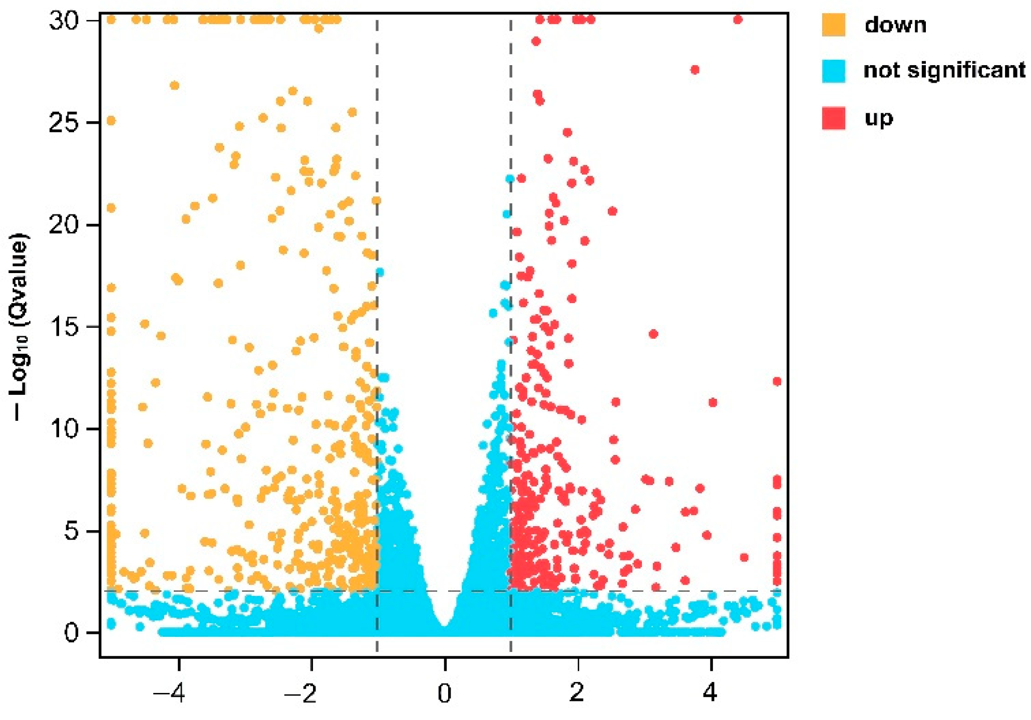
3.2. Identification of DEGs

The head transcriptomes of male G88 and LW-13 were compared, and a total of 1890 (7.81%) DEGs were found. Compared with the G88, 898 genes in the LW-13 were significantly up-regulated, and 992 genes were significantly down-regulated (Figure 2), indicating significant differences in the gene expression between these two P. xylostella strains.

3.3. Gene Ontology of DEGs

The GO cluster diagram of the DEGs showed that the DEGs were mainly enriched in 16 biological processes, such as the metabolic process, cellular process, and localization; in 14 cellular components, such as the cell part, membrane part, and membrane; and in 7 molecular functions, such as in binding, catalytic activity, and transporter activity (Figure S1).
A top 20 GO term enrichment analysis showed that the DEGs mainly belonged to: the categories of catalytic activity in molecular functions (Q < 0.05, Figure 3), such as hydroxymethyl-, formyl- and related transferase activity (GO: 0016742, Q = 0.037), oxidoreductase activity (GO: 0016491, Q = 0.037), and enzyme inhibitor activity (GO: 0004857, Q = 0.045); the categories of metabolic and cellular processes in biological processes (Q < 0.05, Figure 3), such as regulation of the immune system process (GO: 0002682, Q < 0.001), regulation of the defense response (GO: 0031347, Q < 0.001), and actin filament organization (GO: 0007015, Q = 0.018); and the categories of cell components and membrane components in cell composition (Q < 0.05, Figure 3), including the extracellular region (GO: 0005576, Q < 0.001) and extracellular space (GO: 0005615, Q = 0.010).

3.4. KEGG Pathway of DEGs

The KEGG pathways analysis suggested that some classical pathways, such as the phototransduction-fly (KO04745, Q = 0.01), one carbon pool by folate (KO00670, Q = 0.04), ovarian steroidogenesis (KO04913, Q = 0.04), phagosome (KO04145, Q = 0.04), vitamin digestion and absorption (KO04977, Q = 0.04), and toll and Imd signaling pathway (KO04624, Q = 0.04), were significantly enriched (Figure 4).

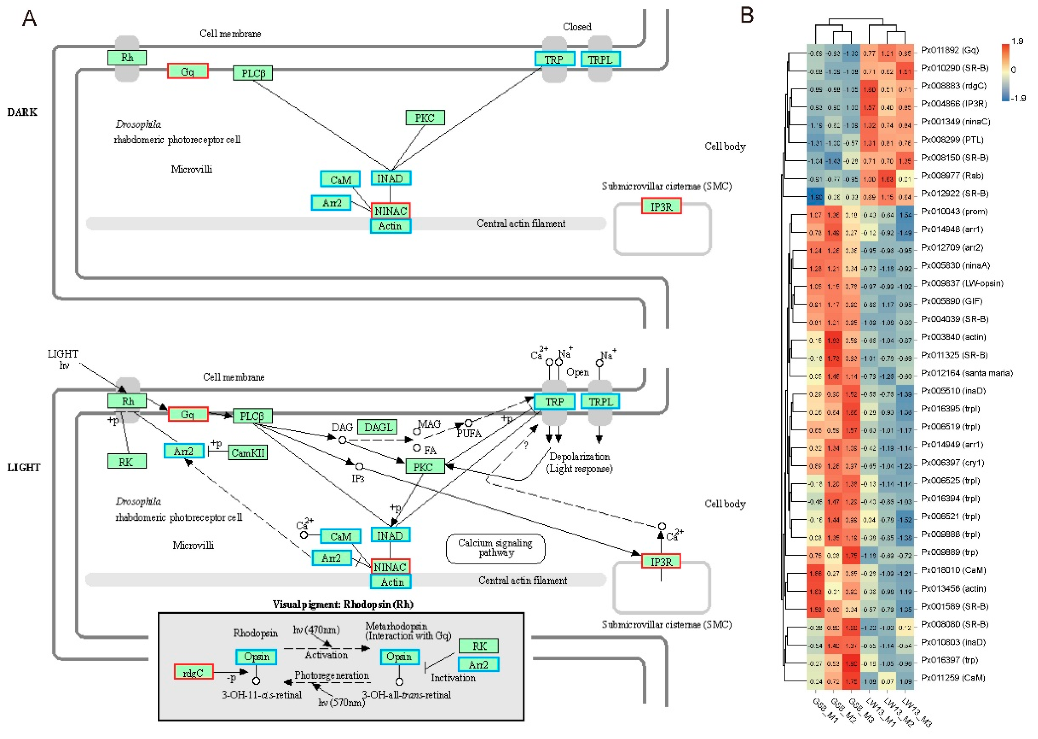
3.5. Influences of LW-Opsin Mutation on the Expression of Genes in the Phototransduction Pathway

Further analysis of the phototransduction-fly pathway revealed that the mRNA expression of the key elements of Gq, IP3R (InsP3 receptor), ninaC (neither inactivation nor afterpotential C) and rdgC were significantly up-regulated, that the mRNA expression of the key elements of arr2, trp, trpl, CaM (calmodulin), actin and inaD (inactivation nor afterpotential D) were significantly down-regulated, and that the mRNA expression of the key elements of PLC, PKC (protein kinase C), CaMKII (CaM kinase II), and DAGL (diacylglycerol lipase) were not affected (Figure 5A,B). Moreover, other genes that participated in the phototransduction-fly pathway, such as cry1, ninaA and arr1, were also significantly down-regulated (Figure 5B).

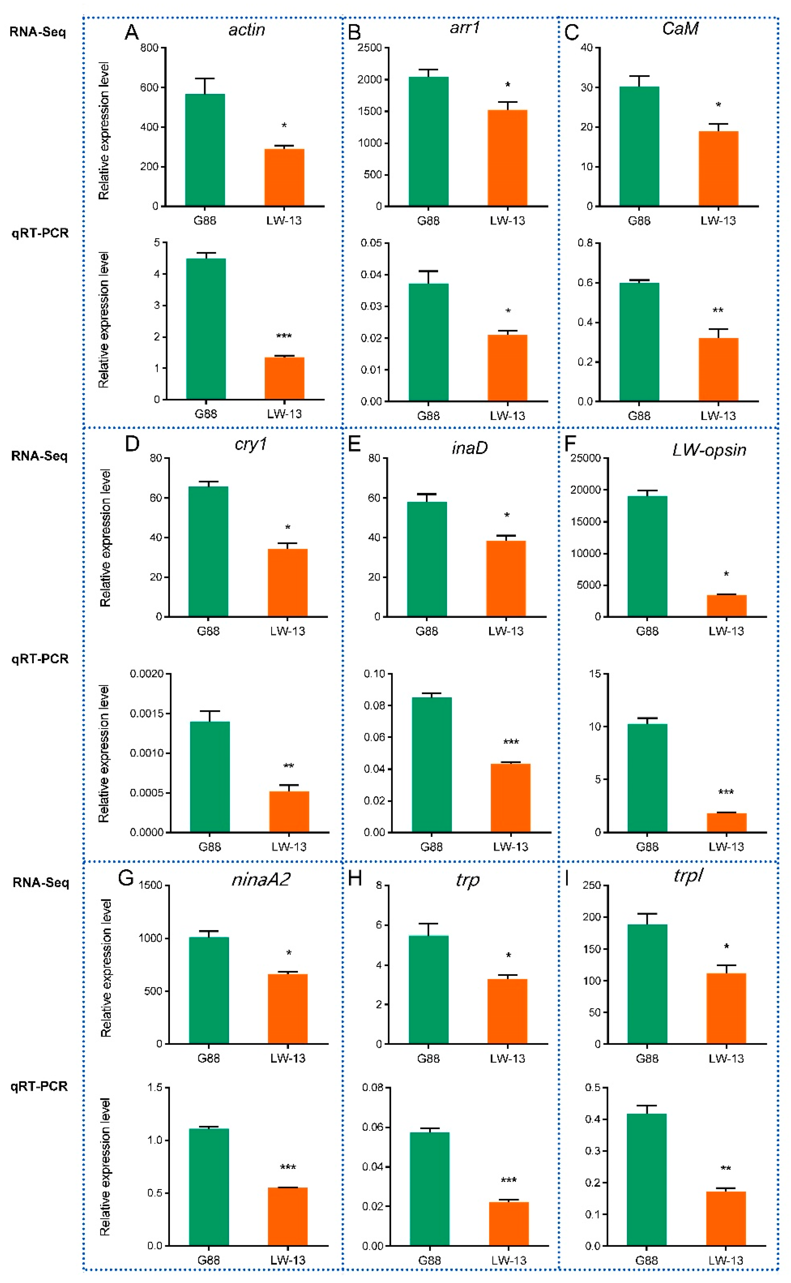
3.6. RT-qPCR Validation of DEGs in the Phototransduction Pathway

The gene expression levels of the nine genes of actin, arr1, CaM, cry1, inaD, LW-opsin, ninaA, trp and trpl in the phototransduction pathway by the RT-qPCR were consistent with the transcriptome-sequencing results in G88 and LW-13 (Figure 6; r = 0.9136, p = 0.0006). It was worth noting that the LW-opsin mutation caused a remarkably lower mRNA expression of mutant LW-opsin, indicating the reliability of our transcriptomic data.

3.7. The Effects of cry1 Mutation on Phototactic Behavior of P. xylostella

The male P. xylostella with a 2-bp deletion in cry1 (Cry1-KO) showed higher phototactic rates to UV and blue lights than the male G88 (Figure 7).

4. Discussion

LW-opsin is a gene with an absolute dominance expression compared with other opsins in P. xylostella [17]. Differentially-expressed genes caused by an LW-opsin mutation in P. xylostella mainly enriched in the GO terms, such as the actin filament organization, extracellular region, and extracellular space, confirming that opsins are required to maintain photoreceptor integrity [24,25,26]. Further work on observing the internal morphological characteristics of photoreceptors using a transmission electron microscope (TEM) will be needed. Although opsins are known as light sensors, some of them (Rh1 and Rh6) also contribute to locomotion in Drosophila [27]. In our study, DEGs were also enriched in the locomotion GO term (Figure S1), and this function had been preliminarily verified in our previous work [17], although this result needs to be confirmed by a proprioceptor dissection.
The phototransduction-fly pathway was significantly enriched after an LW-opsin mutation, indicating the P. xylostella phototransduction pathway was similar to that in Drosophila. In P. xylostella, the LW-opsin mutation affected the mRNA expression of some key elements, such as arr2, inaD, rdgC, trp and trpl; but the mRNA expression of some other vital elements, such as PLC and PKC, were not affected. The results suggest that there are variations to accommodate the visual requirements for P. xylostella compared with Drosophila, which might be due to the different lifestyles and eye morphologies between the two insect species. The functions of these critical elements have been well demonstrated in Drosophila. For instance, Gq is an essential effector in the activation of PLC, and the mutant shows a decreased light sensitivity [28]. Arr2 is an inhibitory protein responsible for inactivation of rhodopsin and plays an essential role in terminating the phototransduction response, and a mutation of arr2 results in retinal degeneration [29]. Additionally, INAD is a scaffold protein containing five PDZ structural domains, which can directly bind PKC, PLC, TRP, TRPL, and NINAC, and a mutation of inaD results in terminating the phototransduction response [30,31,32,33]. Finally, TRP and TRPL are cation channels, and a mutation of trp and trpl results in a decreased Ca2+ influx and transient response to light [34,35]. The function of these key elements in P. xylostella will be further verified by using RNAi or CRISPR/Cas9 technology.
The vitamin digestion and absorption pathway was also significantly enriched, which might have been because vitamin A is a critical substrate in chromophore biogenesis [26,36,37,38,39]. Whether an LW-opsin mutation in P. xylostella disrupts chromophore production will be further explored by using HPLC-MS (high-performance liquid chromatography-mass spectrometry). Moreover, vitamin A is essential for an immune response [40,41], which might be the reason for the DEGs’ enrichment in GO terms, such as regulation of the immune system process and the KEGG pathway–Toll and Imd signaling pathway.
Higher phototaxis responses to the UV and blue lights were observed in the male P. xylostella cry1 mutants than in the G88. It has previously been assumed that the ability of an insect to sense and respond to UV light is regulated by UV-opsin [21]; however, UV-opsin does not participate in sensing UV light in P. xylostella [17]. Insects express other non-opsin photoreceptors, including the UV/blue-light sensitive flavoprotein, CRY [21,22]. The dCRY is a significant modulator of Drosophila behavioral responses to UV light, containing both attraction and avoidance behaviors. Drosophila shows avoidance behaviors to high-intensity UV light (400 μW/cm2), while showing attraction behaviors to low-intensity UV light (10 μW/cm2) [42]. Conversely, mutant Drosophila lacking CRY showed attraction behaviors instead of avoidance behaviors to high-intensity UV light and more attraction to low-intensity UV light than wild-type control flies [21,42]. Although further studies are needed to verify whether P. xylostella shows avoidance behaviors to high-intensity UV light or blue light, our results suggested that CRY1 might be a significant modulator of the P. xylostella behavioral responses to low-intensity UV light (380 nm and 2.5 lux ≈ 60 μW/cm2) and blue light (470 nm and 2.5 lux ≈ 4 μW/cm2).
Interestingly, the phototaxis of female P. xylostella cry1 mutants to UV and blue lights showed no difference with the G88 (Figure S2). A gender difference in the phototaxis behavior was also observed in the P. xylostella LW-opsin mutant [17]. Moreover, the P. xylostella males exhibited a better locomotor circadian rhythm (induced by light) than the females [43]. These results suggest that the phototransduction mechanism might be diverse between P. xylostella male and females.
In Drosophila, the dCRY C-terminal interacts with NINAC through INAD in a light-dependent manner [44]. In our study, the LW-opsin mutation resulted in lower expressions of cry1 and inaD, and a higher expression of ninaC. These results suggested that the behavioral response to the UV light mediated by CRY1 might be similar between Drosophila and P. xylostella males; however, the expression of inaD and ninaC in Cry1-KO will need further verification.

5. Conclusions

In conclusion, an LW-opsin mutation causes gene expressions in the phototransduction pathway, such as arr1, cry1, trp, trpl and inaD, and cry1 plays an essential role in enhancing the phototaxis of P. xylostella males. Our results provide a preliminary system-level understanding of the influences of the LW-opsin mutation on the expression of genes in P. xylostella phototransduction and they provide a foundation for further exploration of the phototactic mechanisms in P. xylostella.

Supplementary Materials

The following supporting information can be downloaded at: https://www.mdpi.com/article/10.3390/insects14010072/s1, Figure S1: GO cluster diagram of DEGs; Figure S2: The influence of cry1 mutation on the phototaxis of P. xylostella female; Table S1: RT-qPCR primers.

Author Contributions

Conceptualization, G.Y. and S.-P.C.; methodology, S.-P.C.; validation, S.-P.C., X.-L.L., R.-Z.Q. and M.-X.C.; data curation, G.Y., S.-P.C., R.-Z.Q. and M.-X.C.; writing—original draft preparation, S.-P.C. and X.-L.L.; writing—review and editing, G.Y. and S.-P.C.; visualization, S.-P.C. and X.-L.L.; supervision, G.Y.; funding acquisition, G.Y. All authors have read and agreed to the published version of the manuscript.

Funding

This research was funded by the National Key R & D Program of China, grant number 2017YFD0200400, the Special Key Project of Fujian Province, grant number 2018NZ01010013, and a fund of the “111” program, grant number KRA16001A.

Institutional Review Board Statement

Not applicable.

Informed Consent Statement

Not applicable.

Data Availability Statement

The data presented in this study are available on request from the corresponding author.

Acknowledgments

We thank Muhammad Asad for language improvements on the manuscript.

Conflicts of Interest

The authors declare no conflict of interest.

References

  1. Fain, G.L.; Hardie, R.; Laughlin, S.B. Phototransduction and the evolution of photoreceptors. Curr. Biol. 2010, 20, 114–124. [Google Scholar] [CrossRef] [PubMed] [Green Version]
  2. Hardie, R.C.; Juusola, M. Phototransduction in Drosophila. Curr. Opin. Neurobiol. 2015, 34, 37–45. [Google Scholar] [CrossRef] [Green Version]
  3. Katz, B.; Minke, B. The Drosophila light-activated TRP and TRPL channels—Targets of the phosphoinositide signaling cascade. Prog. Retin. Eye Res. 2018, 66, 200–219. [Google Scholar] [CrossRef] [PubMed]
  4. Landry, C.R.; Castillo-Davis, C.I.; Ogura, A.; Liu, J.S.; Hartl, D.L. Systems-level analysis and evolution of the phototransduction network in Drosophila. Proc. Natl. Acad. Sci. USA 2007, 104, 3283–3288. [Google Scholar] [CrossRef] [Green Version]
  5. Montell, C. Drosophila visual transduction. Trends Neurosci. 2012, 35, 356–363. [Google Scholar] [CrossRef] [Green Version]
  6. Paulsen, R.; Schwemer, J. Biogenesis of blowfly photoreceptor membranes is regulated by 11-cis-retinal. Eur. J. Biochem. 1983, 137, 609–614. [Google Scholar] [CrossRef] [PubMed]
  7. Yau, K.W.; Hardie, R.C. Phototransduction motifs and variations. Cell 2009, 139, 246–264. [Google Scholar] [CrossRef] [PubMed] [Green Version]
  8. Friedrich, M.; Chen, R.; Daines, B.; Bao, R.; Caravas, J.; Rai, P.K.; Zagmajster, M.; Peck, S.B. Phototransduction and clock gene expression in the troglobiont beetle Ptomaphagus hirtus of Mammoth cave. J. Exp. Biol. 2011, 214, 3532–3541. [Google Scholar] [CrossRef] [Green Version]
  9. Macias-Munoz, A.; Smith, G.; Monteiro, A.; Briscoe, A.D. Transcriptome-wide differential gene expression in Bicyclus anynana butterflies: Female vision-related genes are more plastic. Mol. Biol. Evol. 2016, 33, 79–92. [Google Scholar] [CrossRef] [Green Version]
  10. Duan, Y.; Gong, Z.J.; Wu, R.H.; Miao, J.; Jiang, Y.L.; Li, T.; Wu, X.B.; Wu, Y.Q. Transcriptome analysis of molecular mechanisms responsible for light-stress response in Mythimna separata (Walker). Sci. Rep. 2017, 7, 45188. [Google Scholar] [CrossRef]
  11. Macias-Muñoz, A.; Rangel Olguin, A.G.; Briscoe, A.D.; Li, W.H. Evolution of phototransduction genes in lepidoptera. Genome Biol. Evol. 2019, 11, 2107–2124. [Google Scholar] [CrossRef] [PubMed] [Green Version]
  12. Feuda, R.; Marletaz, F.; Bentley, M.A.; Holland, P.W.H. Conservation, duplication, and divergence of five opsin genes in insect evolution. Genome Biol. Evol. 2016, 8, 579–587. [Google Scholar] [CrossRef] [PubMed] [Green Version]
  13. Yamaguchi, S.; Desplan, C.; Heisenberg, M. Contribution of photoreceptor subtypes to spectral wavelength preference in Drosophila. Proc. Natl. Acad. Sci. USA 2010, 107, 5634–5639. [Google Scholar] [CrossRef] [PubMed] [Green Version]
  14. Wakakuwa, M.; Stewart, F.; Matsumoto, Y.; Matsunaga, S.; Arikawa, K. Physiological basis of phototaxis to near-infrared light in Nephotettix cincticeps. J. Comp. Physiol. A 2014, 200, 527–536. [Google Scholar] [CrossRef] [PubMed]
  15. Liu, Y.J.; Yan, S.; Shen, Z.J.; Li, Z.; Zhang, X.F.; Liu, X.M.; Zhang, Q.W.; Liu, X.X. The expression of three opsin genes and phototactic behavior of Spodoptera exigua (Lepidoptera: Noctuidae): Evidence for visual function of opsin in phototaxis. Insect Biochem. Mol. Biol. 2018, 96, 27–35. [Google Scholar] [CrossRef] [PubMed]
  16. Li, C.; Tian, F.; Lin, T.; Wang, Z.; Liu, J.; Zeng, X. The expression and function of opsin genes related to the phototactic behavior of Asian citrus psyllid. Pest Manag. Sci. 2020, 76, 1578–1587. [Google Scholar] [CrossRef] [PubMed]
  17. Chen, S.P.; Liu, Z.X.; Chen, Y.T.; Wang, Y.; Chen, J.Z.; Fu, S.; Ma, W.F.; Xia, S.; Liu, D.; Wu, T.; et al. CRISPR/Cas9-mediated knockout of LW-opsin reduces the efficiency of phototaxis in the diamondback moth Plutella xylostella. Pest Manag. Sci. 2021, 77, 3519–3528. [Google Scholar] [CrossRef]
  18. Chen, S.P.; Wang, D.F.; Ma, W.F.; Lin, X.L.; Yang, G. Knockout of cryptochrome1 disturbs the locomotor circadian rhythm and development of Plutella xylostella. Insect Sci. 2022. [Google Scholar] [CrossRef]
  19. Ma, X.L.; He, W.Y.; Wang, P.; You, M.S. Cell lines from diamondback moth exhibiting differential susceptibility to baculovirus infection and expressing midgut genes. Insect Sci. 2019, 26, 251–262. [Google Scholar] [CrossRef]
  20. Livak, K.J.; Schmittgen, T.D. Analysis of relative gene expression data using real-time quantitative PCR and the 2−ΔΔCT method. Methods 2001, 25, 402–408. [Google Scholar] [CrossRef]
  21. Baik, L.S.; Fogle, K.J.; Roberts, L.; Galschiodt, A.M.; Chevez, J.A.; Recinos, Y.; Nguy, V.; Holmes, T.C. CRYPTOCHROME mediates behavioral executive choice in response to UV light. Proc. Natl. Acad. Sci. USA 2017, 114, 776–781. [Google Scholar] [CrossRef] [PubMed] [Green Version]
  22. Bouly, J.P.; Schleicher, E.; Dionisio-Sese, M.; Vandenbussche, F.; Van Der Straeten, D.; Bakrim, N.; Meier, S.; Batschauer, A.; Galland, P.; Bittl, R.; et al. Cryptochrome blue light photoreceptors are activated through interconversion of flavin redox states. J. Biol. Chem. 2007, 282, 9383–9391. [Google Scholar] [CrossRef] [PubMed] [Green Version]
  23. You, M.; Yue, Z.; He, W.; Yang, X.; Yang, G.; Xie, M.; Zhan, D.; Baxter, S.W.; Vasseur, L.; Gurr, G.M.; et al. A heterozygous moth genome provides insights into herbivory and detoxification. Nat. Genet. 2013, 45, 220–225. [Google Scholar] [CrossRef] [PubMed] [Green Version]
  24. Colley, N.J.; Cassill, J.A.; Baker, E.K.; Zuker, C.S. Defective intracellular transport is the molecular basis of rhodopsin-dependent dominant retinal degeneration. Proc. Natl. Acad. Sci. USA 1995, 92, 3070–3074. [Google Scholar] [CrossRef] [Green Version]
  25. Kumar, J.P.; Ready, D.F. Rhodopsin plays an essential structural role in Drosophila photoreceptor development. Development 1995, 121, 4359–4370. [Google Scholar] [CrossRef]
  26. Wang, T.; Montell, C. Phototransduction and retinal degeneration in Drosophila. Pflug. Arch. Eur. J. Physiol. 2007, 454, 821–847. [Google Scholar] [CrossRef] [Green Version]
  27. Zanini, D.; Giraldo, D.; Warren, B.; Katana, R.; Andrés, M.; Reddy, S.; Pauls, S.; Schwedhelm-Domeyer, N.; Geurten, B.R.H.; Göpfert, M.C. Proprioceptive opsin functions in Drosophila larval locomotion. Neuron 2018, 98, 67–74. [Google Scholar] [CrossRef] [Green Version]
  28. Scott, K.; Becker, A.; Sun, Y.; Hardy, R.; Zuker, C. Gqα protein function in vivo: Genetic dissection of its role in photoreceptor cell physiology. Neuron 1995, 15, 919–927. [Google Scholar] [CrossRef] [Green Version]
  29. Dolph, P.J.; Ranganathan, R.; Colley, N.J.; Hardy, R.W.; Socolich, M.; Zuker, C.S. Arrestin function in inactivation of G protein-coupled receptor rhodopsin in vivo. Science 1993, 260, 1910–1916. [Google Scholar] [CrossRef]
  30. Chevesich, J.; Kreuz, A.J.; Montell, C. Requirement for the PDZ domain protein, INAD, for localization of the TRP store-operated channel to a signaling complex. Neuron 1997, 18, 95–105. [Google Scholar] [CrossRef]
  31. Li, H.S.; Montell, C. TRP and the PDZ protein, INAD, form the core complex required for retention of the signalplex in Drosophila photoreceptor cells. J. Cell Biol. 2000, 150, 1411–1421. [Google Scholar] [CrossRef] [PubMed] [Green Version]
  32. Wes, P.D.; Xu, X.Z.S.; Li, H.S.; Chien, F.; Doberstein, S.K.; Montell, C. Termination of phototransduction requires binding of the NINAC myosin III and the PDZ protein INAD. Nat. Neurosci. 1999, 2, 447–453. [Google Scholar] [CrossRef] [PubMed]
  33. Xu, X.Z.S.; Choudhury, A.; Li, X.; Montell, C. Coordination of an array of signaling proteins through homo- and heteromeric interactions between PDZ domains and target proteins. J. Cell Biol. 1998, 142, 545–555. [Google Scholar] [CrossRef] [PubMed]
  34. Hardie, R.C.; Minke, B. The trp gene is essential for a light-activated Ca2+ channel in Drosophila photoreceptors. Neuron 1992, 8, 643–651. [Google Scholar] [CrossRef]
  35. Leung, H.T.; Geng, C.; Pak, W.L. Phenotypes of trpl mutants and interactions between the transient receptor potential (TRP) and TRP-like channels in Drosophila. J. Neurosci. 2000, 20, 6797–6803. [Google Scholar] [CrossRef] [Green Version]
  36. Dewett, D.; Lam-Kamath, K.; Poupault, C.; Khurana, H.; Rister, J. Mechanisms of vitamin A metabolism and deficiency in the mammalian and fly visual system. Dev. Biol. 2021, 476, 68–78. [Google Scholar] [CrossRef]
  37. Dewett, D.; Labaf, M.; Lam-Kamath, K.; Zarringhalam, K.; Rister, J. Vitamin A deficiency affects gene expression in the Drosophila melanogaster head. G3 Genes Genomes Genet. 2021, 11, jkab297. [Google Scholar] [CrossRef]
  38. Harris, W.; Ready, D.; Lipson, E.; Hudspeth, A.; Stark, W. Vitamin A deprivation and Drosophila photopigments. Nature 1977, 266, 648–650. [Google Scholar] [CrossRef]
  39. Wang, T.; Jiao, Y.; Montell, C. Dissection of the pathway required for generation of vitamin A and for Drosophila phototransduction. J. Cell Biol. 2007, 177, 305–316. [Google Scholar] [CrossRef] [Green Version]
  40. Lane, M.A.; Bailey, S.J. Role of retinoid signalling in the adult brain. Prog. Neurobiol. 2005, 75, 275–293. [Google Scholar] [CrossRef]
  41. Travis, G.H.; Golczak, M.; Moise, A.R.; Palczewski, K. Diseases caused by defects in the visual cycle: Retinoids as potential therapeutic agents. Annu. Rev. Pharmacol. Toxicol. 2007, 47, 469–512. [Google Scholar] [CrossRef] [PubMed] [Green Version]
  42. Baik, L.S.; Recinos, Y.; Chevez, J.A.; Au, D.D.; Holmes, T.C. Multiple phototransduction inputs integrate to mediate UV light-evoked avoidance/attraction behavior in Drosophila. J. Biol. Rhythm. 2019, 34, 391–400. [Google Scholar] [CrossRef] [PubMed]
  43. Wang, D.; Yang, G.; Chen, W. Diel and circadian patterns of locomotor activity in the adults of diamondback moth (Plutella xylostella). Insects 2021, 12, 727. [Google Scholar] [CrossRef] [PubMed]
  44. Mazzotta, G.; Rossi, A.; Leonardi, E.; Mason, M.; Bertolucci, C.; Caccin, L.; Spolaore, B.; Martin, A.J.M.; Schlichting, M.; Grebler, R.; et al. Fly cryptochrome and the visual system. Proc. Natl. Acad. Sci. USA 2013, 110, 6163–6168. [Google Scholar] [CrossRef]
Figure 1. Correlation coefficient heatmap of gene expressions between different samples of P. xylostella. The green bar outside the heatmap represents the LW-13 group; the red bar outside the heatmap represents the G88 group; the values in square frames with red and blue color represent the Pearson correlation coefficients of LW-13 and G88, respectively; the darker color indicates the bigger value.
Insects 14 00072 g001
Figure 2. Volcanic map of DEGs in LW-13 compared with G88.
Insects 14 00072 g002
Figure 3. Top 20 GO term enrichment of DEGs in LW-13 compared with G88. From the outside to the inside, the first circle represents the enriched GO classification where the different colors represent different classifications, and the numbers outside the circle represent the coordinate ruler of the number of genes. The second circle represents the number of genes corresponding to the classification and the Q value, where the longer the bar indicates a higher number of genes, and the darker red color indicates a smaller Q value. The third circle represents the ratio of up- and down-regulated genes, where dark purple represents the up-regulated genes, light purple represents the down-regulated genes, and the numbers of the corresponding bottom represent the up-regulated genes number or down-regulated genes number. The fourth circle represents the rich factor value of each GO term, and each cell represents 0.1.
Insects 14 00072 g003
Figure 4. Top 20 of the KEGG enrichment of DEGs in LW-13 compared with G88.
Insects 14 00072 g004
Figure 5. DEGs in LW-13 compared with G88 based on the phototransduction-fly pathway and their heatmap. (A) DEGs in LW-13 compared with G88 strains based on the phototransduction-fly pathway. Red and blue frames represent up-regulated and down-regulated DEGs, respectively. (B) Heatmap of related DEGs. The value in the box represents the value of the gene’s TPM normalized by the Z-score.
Insects 14 00072 g005
Figure 6. Verification of the expressions of 9 DEGs in LW-3 compared with G88 by RT-qPCR. (A) actin (gene ID, Px003840); (B) arr1 (gene ID, Px014948); (C) CaM (gene ID, Px018010); (D) cry1 (gene ID, Px006397); (E) inaD (gene ID, Px005510); (F) LW-opsin (gene ID, Px009837; the LW-opsin expression in LW-13 is mutant LW-opsin); (G) ninaA (gene ID, Px005830); (H) trp (gene ID, Px009889); (I) trpl (gene ID, Px009888). “*” indicates P < 0.05; “**” indicates 0.01 < P < 0.05; “***” indicates P < 0.01.
Insects 14 00072 g006
Figure 7. The influence of cry1 mutation on the phototaxis of P. xylostella male. “*” represents P < 0.05.
Insects 14 00072 g007
Table 1. Summary of transcriptome-sequencing results generated from G88 and LW-13.
SampleQ30 (%)Total MappedMultiple MappedUniquely Mapped
G88 M194.6123,206,466 (55.63%)1,350,166 (3.24%)21,856,300 (52.39%)
G88 M294.6725,289,426 (55.93%)1,448,410 (3.20%)23,841,016 (52.72%)
G88 M394.8625,923,219 (56.01%)1,521,146 (3.29%)24,402,073 (52.72%)
LW-13 M194.6722,971,282 (54.15%)1,367,482 (3.22%)21,603,800 (50.92%)
LW-13 M294.8123,454,846 (54.49%)1,393,748 (3.24%)22,061,098 (51.25%)
LW-13 M394.7626,024,544 (54.10%)1,496,406 (3.11%)24,528,138 (50.99%)
Disclaimer/Publisher’s Note: The statements, opinions and data contained in all publications are solely those of the individual author(s) and contributor(s) and not of MDPI and/or the editor(s). MDPI and/or the editor(s) disclaim responsibility for any injury to people or property resulting from any ideas, methods, instructions or products referred to in the content.

