Article

Unexpected High Species Diversity of Mesolycus Gorham (Coleoptera, Lycidae) from China, with a Preliminary Investigation on Its Phylogenetic Position Based on Multiple Genes †

1
Key Laboratory of Zoological Systematics and Application, School of Life Science, Institute of Life Science and Green Development, Hebei University, Baoding 071002, China
2
Key Laboratory of Zoological Systematics and Evolution, Institute of Zoology, Chinese Academy of Sciences, Beijing 100101, China
*
Authors to whom correspondence should be addressed.
This published work and the nomenclatural acts it contains have been registered in ZooBank, the Online Registration System for the ICZN (International Code of Zoological Nomenclature). The LSID (Life Science Identifier) for This Publication is: LSID urn:lsid:zoobank.org:pub:ADBEC6C2-5FD6-4182-9AC4-D1717348E1AE.
Insects 2022, 13(12), 1171; https://doi.org/10.3390/insects13121171
Submission received: 17 November 2022 / Revised: 13 December 2022 / Accepted: 14 December 2022 / Published: 17 December 2022
(This article belongs to the Section Insect Systematics, Phylogeny and Evolution)

Abstract

:

Simple Summary

The East Palaearctic fauna of net-winged beetles is very rich, but poorly studied in China. As an important faunistic component of the East Palaearctic fauna, the detailed taxonomic studies of Lycidae from China are necessary for understanding the evolution of this important biodiversity hotspot. More importantly, it is a region where some lycid lineages occur only there and in an adjacent part of the Oriental Region, such as Mesolycus Gorham, 1883. The genus remains controversial in its systematic placements but has never been rigorously tested due to unavailability of sampling material. In the present study, the genus Mesolycus was reviewed, with an emphasis on the species diversity from the Chinese fauna. Meanwhile, multiple genetic data of this genus were obtained and included in the phylogeny of lycid beetles for the first time. As a result, it is quite surprising that the number of the Chinese Mesolycus species is increased from 6 to 13, which makes China stand out in the species diversity on a world scale. Meanwhile, the statuses of some previously known species were clarified, and the systematic placement of Mesolycus was verified. This study makes us better understand the species diversity and phylogenetic position of Mesolycus.

Abstract

The lycid genus Mesolycus Gorham, 1883 is mainly distributed in East Palaearctic and Indochinese regions, but poorly studied in China; moreover, its phylogenetic placement remains controversial but has never been rigorously tested. In this study, Mesolycus was reviewed and its placement within Lycidae was tested based on a multilocus phylogeny (cox1, nad5, cox2 and Lrna) by both ML and BI analyses. The reconstructed phylogenies show that Mesolycus is a consistently recovered sister to Dilophotes Waterhouse, 1879, and they form a monophyletic clade which is well supported. This suggests that Mesolycus definitely belongs to Dilophotini rather than to Macrolycini of Lycinae. Besides, three species originally described or placed in Dilophotes are transferred to Mesolycus, including M. atricollis (Pic, 1926) comb. n., M. particularis (Pic, 1928) comb. n. and M. pacholatkoi (Bic, 2002) comb. n. Four new species are discovered in China, including M. shaanxiensis sp. n., M. dentatus sp. n., M. breviplatus sp. n. and M. varus sp. n. Two species, M. murzini Kazantsev, 2004 and M. rubromarginatus Kazantsev, 2013, are recorded from China for the first time. A key for the identification of all Mesolycus species is provided. China was revealed as the region with the highest species diversity of this genus.

1. Introduction

Lycidae, or net–winged beetles, is a moderately large beetle family, with about 4600 species hitherto known in the world [1,2]. Many species occur in small ranges and their diversity has been commonly underestimated [3,4,5].
The East Palaearctic fauna of net–winged beetles is very rich [6], but poorly studied in China. It is a region where some lineages occur only there and in an adjacent part of the Oriental Region, such as Macrolycini and Lyponiini [1], which have the highest diversity in East Palaearctic, and very probably expanded their ranges to the Oriental region [7]. Meanwhile, some lineages, such as Dilophotini, which has more species in the Oriental region than those of the East Palearctic, may be present because they are in a converse dispersal route [2,8,9,10]. Therefore, as an important faunistic component of the East Palaearctc fauna, the detailed taxonomic studies of the Lycidae from China are necessary for understanding the evolution of this important biodiversity hotspot [11].
The lycid genus Mesolycus Gorham, 1883 was proposed for the Japanese species Eros atrorufus Kiesenwetter, 1879. Although Nakane [6] has already shown the close relationship between Mesolycus and Dilophotes Waterhouse, 1879, he did not formally synonymize them, which was done later by Bocak and Bocakova [12]. Until Kazantsev [9], who examined the type species for each genus, revalidated Mesolycus from synonym with Dilophotes, he meanwhile synonymized Flabellodilophotes Pic, 1921 and Dilophotes (Biphilodes) Kazantsev, 2000 with Mesolycus. Up to date, a total of 30 species of Mesolycus were recorded from the East Palaearctic and Oriental regions, of which six species occurred in China [8,9,10,13]. However, since it was redefined by Kazantsev [9], no systematic work has been done on this genus, and some species remain unclear in their placements; moreover, multiple species remain unknown particularly in China.
The genus Mesolycus was redefined and the diagnostic characteristics were summarized well by Kazantsev [9], which is recognizable by a blunt angle between the vertex and frons planes, serrated antennae hardly reaching over the elytral middle in both sexes, absence of wedge cells in metathoracic wings, plantar pads usually being present on tarsomeres 1 and 2 (absent in M. shelfordi (Bourgeois, 1906)), and with symmetrical aedeagus including the inner sac structures.
Due to the poorly resolved taxonomy of the group the systematic placement of Mesolycus is uncertain. This genus, once a junior synonym of Dilophotes, was placed in the tribe Macrolycini Kleine, 1929 [8,10,12,14]. The name Dilophotini was erected for Dilophotes by Kleine [15] and synonymized to Macrolycini by Bocak and Bocakova [16], but the former was revalidated as a subtribe of the latter by Kazantsev based on a morphologically phylogenetic analysis [9]. In the latter study, Mesolycus together with Macrolycus Waterhouse, 1878 and Calopteron Kazantsev, 2004 were placed in Macrolycina, and Dilophotes was solely included in Dilophotina, which was soon resurrected into a separate tribe based on another hypothesis of morphological phylogeny [17]. After that, Mesolycus was noted to be a member of Dilophotini rather than Macrolycini by Bocak & Bocakova [1], but no material was included in their multigene phylogenetic analysis. However, this placement was not agreed by Kazantsev [13], who insisted that Mesolycus was a member of Macrolycini. Although another molecular phylogeny of Lycidae [18] was reconstructed recently, no Mesolycus sample was included in the analysis. Therefore, the systematic status of Mesolycus remains controversial at present.
In our recent study, we obtained some material of Mesolycus from China. After our examination, we discovered some new species and describe them herein. Given the narrow geographic distribution of most lycid beetles, it seems plausible that many species are vanishing without ever having been collected due to habitat loss. In light of the biodiversity crisis and accelerating rates of biological extinction, taxonomic work and targeted collecting (with specimens suitable for both morphological and molecular work) remain essential for the future of systematic studies. Luckily, we successfully sequenced the mitochondrial genomes of two species, which allowed us to investigate the phylogenetic position of Mesolycus. The present study is another contribution to the knowledge of Mesolycus, with an emphasis on exploring the species diversity of this lycid group in the Chinese fauna.

2. Materials and Methods

2.1. Materials

The studied material is preserved in the Institute of Zoology, Chinese Academy of Sciences, Beijing, China (IZAS) and the Museum of Hebei University, Baoding, China (MHBU). The sampling material of two Mesolycus species was preserved in 100% ethanol at −20 °C and catalogued in the voucher collection (Nos. CAN-25 and 2CA179) at MHBU. As these species were considered non–protected invertebrates, no permits were required for this study.

2.2. Morphological and Geographical Studies

The specimens were softened in water, and the male genitalia were dissected, then cleared in 10% NaOH solution, examined and photographed in glycerol, and finally glued on a paper card for permanent preservation. Habitus photos were taken by a Canon 7D camera and aedeagi by Leica M205A stereomicroscope; multiple layers were stacked using Combine ZM (Helicon Focus 5.3) and edited in Adobe Photoshop CS3.10.0.1. The aedeagi was illustrated and colored in Inkscape 1.0.2-2 (e86c870879, 2021-01-15).
The measurements were taken with Image J 1.50i (NIH, USA). Body length was measured from the anterior margin of the clypeus to the elytral apex, and the width across the humeral part of elytra. Pronotal length was measured from the middle of the anterior margin to the middle of the posterior margin and the width across the widest part of the pronotum. Eye diameter was measured at the widest point and the interocular distance was taken at the point of the minimum.
The distribution information was collected from the original publications and the material studied in this study (Table S1). The distribution map was prepared by the ArcMap 10.8 and edited in Adobe Photoshop CS3.10.0.1.

2.3. DNA Extraction, Sequencing and Annotation

Total DNA was extracted from the thoracic muscle using a DNeasy Blood & Tissue kit (QIAGEN, Beijing, China) following the manufacturer’s instructions. DNA was stored at −20 °C until molecular analyses. Unlike the previous studies (e.g., [18]), the gene fragments were extracted from the complete mitogenomes, not sequenced separately. The mitogenomes were sequenced at Berry Genomics, Beijing, China, using an Illumina Novaseq 6000 platform (Illumina, San Diego, CA, USA) with 150 bp paired-end reads. High-quality reads were assembled using an iterative De Bruijn graph de novo assembler, the IDBA–UD toolkit [19], with a similarity threshold of 98% and k values of 40–160 bp. The gene COI was amplified through polymerase chain reaction using universal primers (Table S2, [20]) as ‘reference sequence’ to target mitochondrial scaffolds and acquire the best fit using IDBA–UD, which achieved a minimum of 98% similarity. Clean reads were manually mapped to the produced mitochondrial scaffolds to specify the precision of the assembly using Geneious 2019.2 [21]. The assembled mitogenome sequences were manually annotated and adjusted using Geneious 2019.2 [21] with a sequence of Platerodrilus sp. as a reference [22]. Protein-coding genes (PCGs) were determined by identifying open reading frames, and the secondary structure and positions of 22 tRNAs were predicted using MITOS WebServer (http://mitos.bioinf.uni-leipzig.de/index.py, accessed on 19 September 2022) [23]. Before cox1, cox2, nad5, and Lrna were extracted using Geneious 2019.2 [21], the four gene sequences were checked by comparing with sequences from GenBank using a Blast search. The gene fragments of the two Mesolycus species were deposited at the NCBI database with accession nos. OP735348-49, OP729892-93, OP803896-99 (Table S3).

2.4. Phylogenetic Analyses

A total of 32 species of the Lycinae were chosen as ingroups, and one species from Lyropaeinae was selected as an outgroup (Table S3). Except the newly sequenced gene fragments of two Mesolycus species, all others were downloaded from the GenBank (Table S3, [4,5,18,24,25,26,27]). Four mitochondrial gene markers (cox1, cox2, nad5, and Lrna) were aligned using MAFFT v7.313 [28] with the default strategy integrated in PhyloSuite v1.2.2 [29]. After removing gaps and ambiguous sites using trimAI with default parameters [30], the separately aligned fragments were concatenated using PhyloSuite v1.2.2 [29] in a combined dataset. The optimal partition schemes and substitution models for the combined dataset were predicted using PartitionFinder2 [31] with the “greedy” algorithm. The “link” estimated branch lengths and the Bayesian information criterion were applied (Table S4).
Phylogenies were deduced by Bayesian inference (BI) and maximum likelihood (ML) methods. IQ-Tree v.1.6.8 [32] and MrBayes v3.2.6 [33] were employed for ML and BI analyses, respectively. An ML tree was constructed with ultrafast 5000 bootstrap replicates. The BI tree was produced with four chains of two parallel runs for 10 million Markov Chain Monte Carlo (MCMC) generations and was terminated with an average standard deviation of split frequencies < 0.01, in which trees were sampled every 1000 generations. The initial 25% of sampled trees were discarded as burn-in, and the majority rule consensus tree was computed to estimate Bayesian posterior probabilities of each node. Phylogenetic trees were visualised and annotated using FigTree 1.4.4 [34].

3. Results

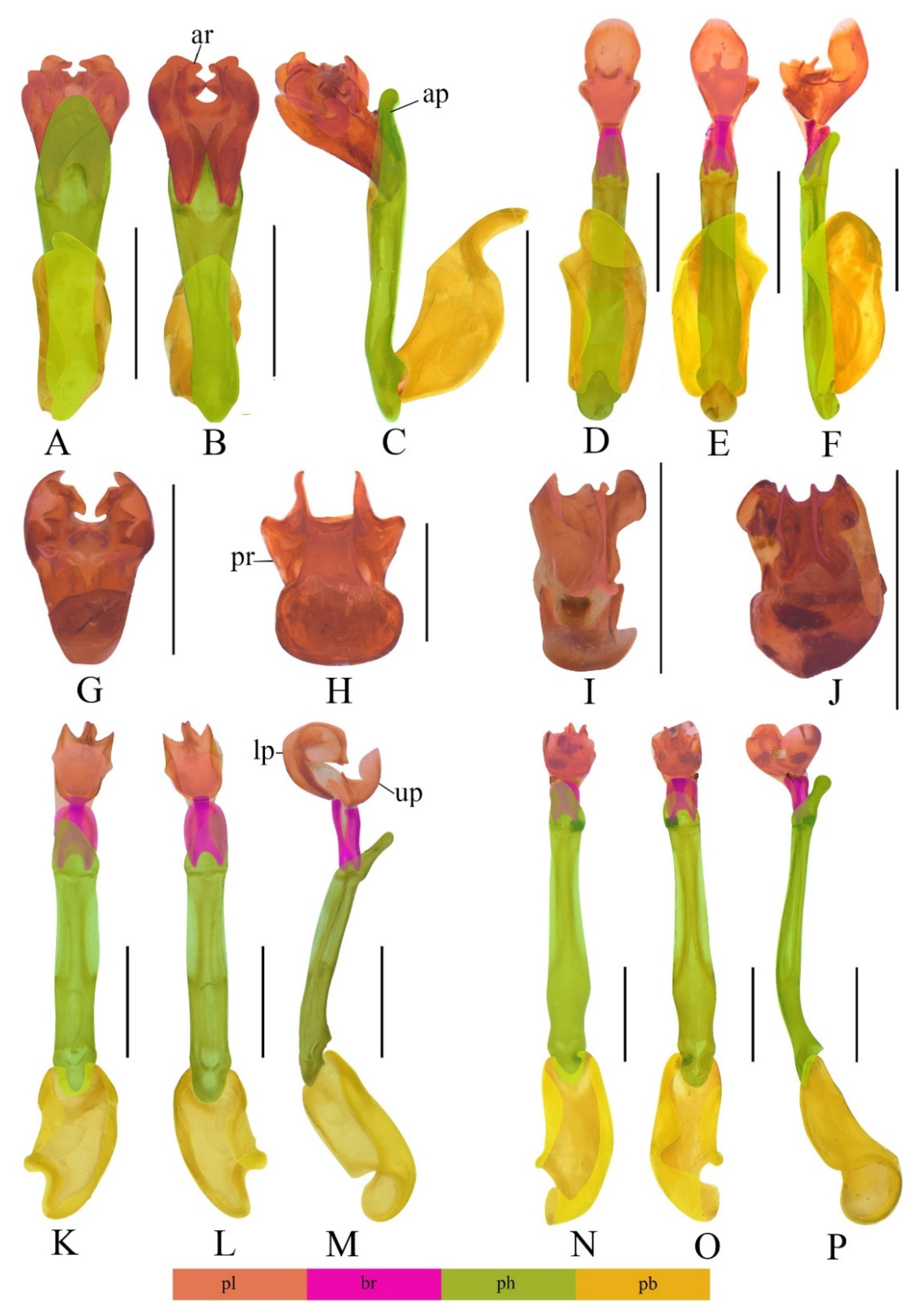
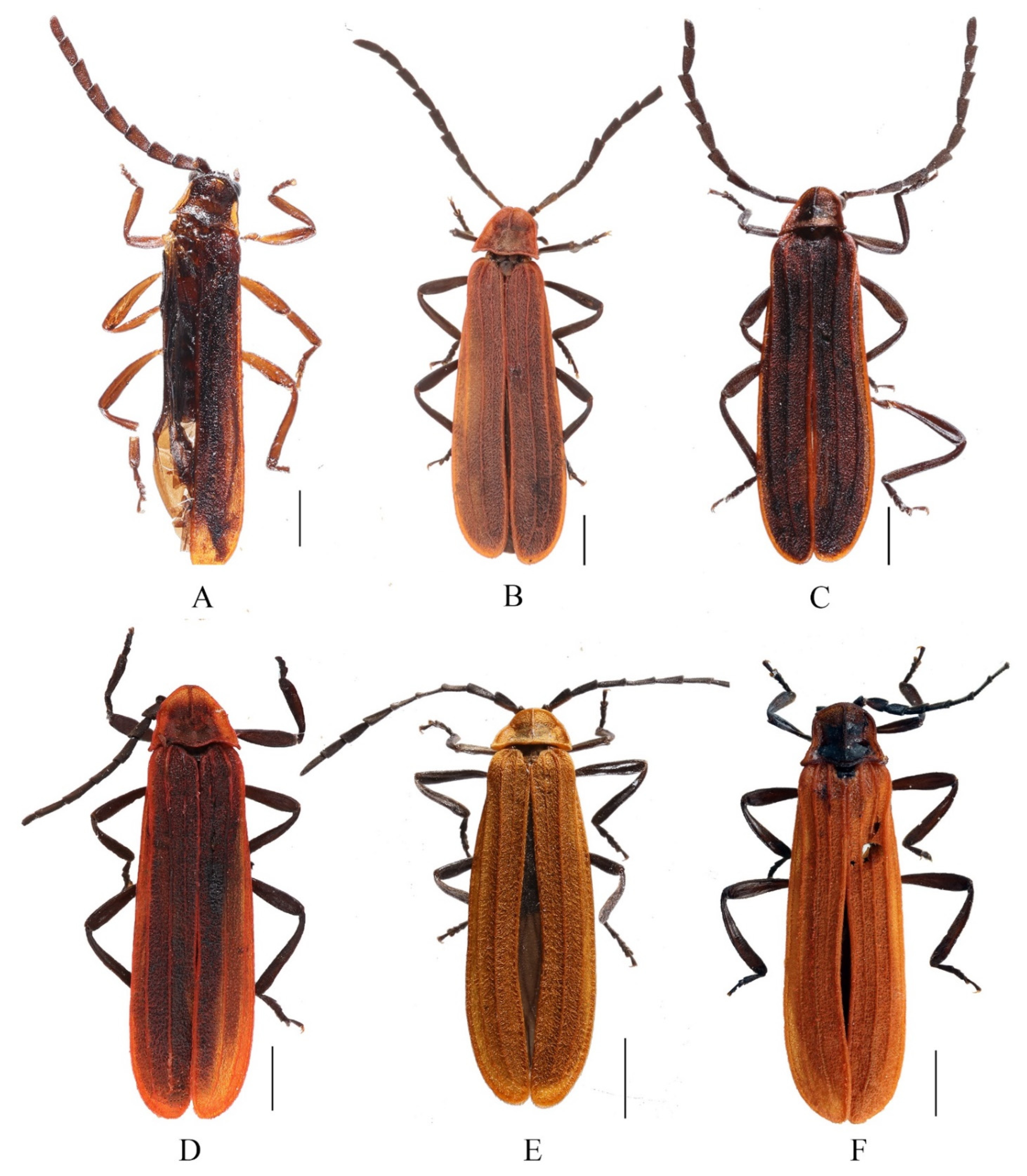
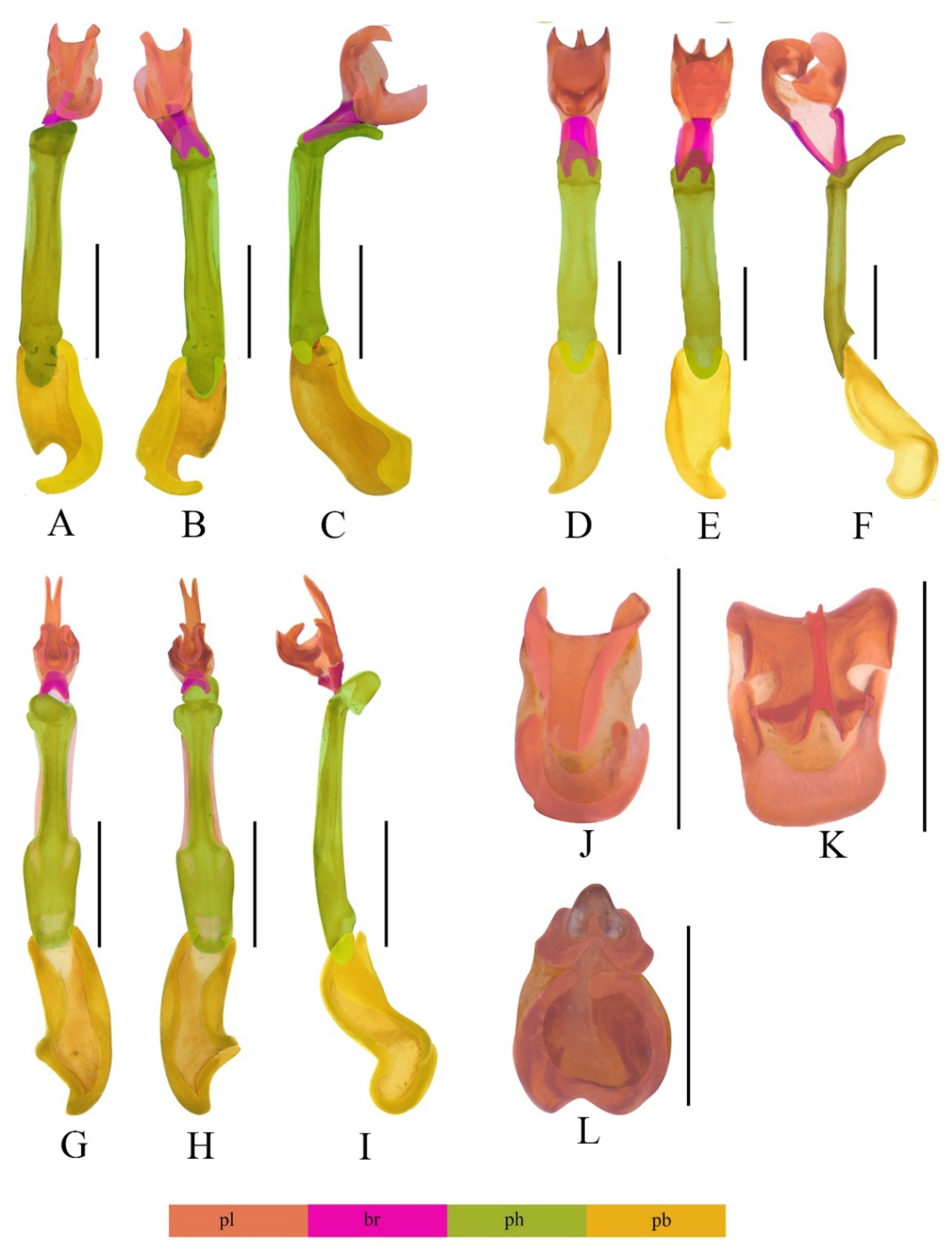
3.1. Descriptions of the Species

Mesolycus rubromarginatus Kazantsev, 2013
Mesolycus rubromarginatus Kazantsev, 2013 [13]: 249.
Figure 1A–C,G.
Material examined. China—2♂♂ (IZAS), Yunnan, Menglongbanna, Mengsong, 1600 m, 23.IV.1958, S. Y. Wang leg.; 1♀ (IZAS), same locality, 26.IV.1958, Y. R. Zhang leg.
Descriptive notes. Male. Phallus (Figure 1A,B) stout and uniformly sclerotized, in a horizontal line with apical hood (Figure 1C). Internal sac relatively large, about half length of phallus (Figure 1A–C). Internal sac absent with upper plate, lower plate relatively large and complex, with four conspicuous teeth on dorsal side, apical ring sub–circular (Figure 1G).
Distribution. N. Laos, China (new record: Yunnan).
Remark. This species is recorded from China for the first time. The male habitus was provided by Kazantsev [13], so it is not present here. The aedeagus is provided with the macrophotographs for the first time to better know its characters and make comparison with others.
Mesolycus dentatus Y. Yang, Liu et X. Yang, sp. n.
Figure 1D–F,H and Figure 2A
Type material. Holotype: ♂ (IZAS), China, Yunnan, Menglongbanna, Mengsong, 1600 m, 24.IV.1958, X. W. Meng leg.
Description. Male (Figure 2A). Length 7.1 mm, width at humeri 1.5 mm.
Body brown. Pronotum reddish brown and brown in lateral margin of disc. Elytra brick red, mixed with dark patches. Surface covered with sparsely inconspicuous red pubescence.
Head medium size, as wide as anterior margin of pronotum. Vertex flat, without median line. Eyes relatively large, interocular distance about 1.1 times greater than eye diameter. Antennae serrate, overlapping two-thirds length of elytra when inclined. Antennomeres II compressed, shorter than wide of apices, III-X subequal in length, XI longest, nearly parallel-sided, pointed at apex.
Pronotum trapezoidal, 0.7 times as long as wide, disc present with a median longitudinal keel extending from anterior margin to middle part. Anterior margin round, lateral margins diverging posteriorly, posterior margin bisinuate; anterior angles confluent with anterior margin and posterior angles sharply projecting. Scutellum trapezoidal, feebly emarginate at apex.
Elytra slender, 4.4 times longer than humeral width, with outer margins subparallel, inner margins dehiscent feebly behind the middle. Elytral costae I weak but complete and visible, II and III strong.
Phallus (Figure 1D,E) slender and uniformly sclerotized, at an angle of 150 degrees with apical hood (Figure 1F). Internal sac relatively large, about half length of phallus (Figure 1D–F). Upper plate (Figure 1F) longer than lower plate, almost straight basally, with internal margin largely emarginate and external margin simple. Lower plate (Figure 1F) short, present with a stout ventro-apical process, having four teeth but without laminae on apical surface, of which ventral teeth obtuse, while dorsal ones sharp at apices (Figure 1H).
Female. Unknown.
Distribution. China (Yunnan).
Diagnosis. The new species resembles M. nanensis Kazantsev, 2004 in the shape of aedeagus, but differs from the latter in the internal sac, of which lower plate is wider than upper plate, and ventral teeth of lower plate are obtuse, while dorsal ones are sharp (Figure 1H). In contrast, in the internal sac of M. nanensis, upper plate and lower plate are equal in width, dorsal teeth of lower plate are truncate, while dorsal ones are sharp at apices ([9]: Figures 44 and 45).
Etymology. The specific name is derived from the Latin dentatus (toothed, having teeth), referring to its lower plate of inner sac of aedeagus with four teeth.
Mesolycus qinlinganus (Kazantsev, 2000)
Dilophotes qinlinganus Kazantsev, 2000 [8]: 330; Bic, 2002 [10]: 5.
Mesolycus qinlinganus: Kazantsev, 2004 [9]: 19.
Figure 1K–M,I, and Figure 2B.
Material examined. China—3♂♂5♀♀ (IZAS), Gansu, Wenxian, Qiujiaba, 2000–2100 m, 1. VII. 1998; S. Y. Wang leg.; 4♂♂4♀♀ (IZAS), same data as the proceeding, W.Y. Zhou leg.; 3♂♂4♀♀ (IZAS), Gansu, Zhouqu, Shatan Forestry, 2400 m, 17. VII. 1999; H. J. Wang leg.; 1♂1♀ (IZAS), Gansu, Wenxian, Fanba, 800 m, 25. VI. 1998; X. K. Yang leg.; 5♂♂5♀♀ (MHBU), Ningxia, Jingyuan, Erlonghe Forestry, X. P. Wang, H. F. Ran & Q. Q. Wu leg.
Descriptive notes. Male (Figure 2B). Phallus (Figure 1K,L) slender and uniformly sclerotized, at an angle of 150 degrees with apical hood (Figure 1M). Internal sac relatively large, about half length of phallus (Figure 1K–M). Upper plate shorter than lower plate (Figure 1M). Upper plate (Figure 1M) curved basally, with internal margin largely emarginate and external margin simple. Lower plate (Figure 1M) widely curved ventrally, external margin slightly curled dorsally, present with a slender ventro-apical process, having a pair of modified laminae merged together in middle of apical surface (Figure 1I).
Distribution. China (Ningxia, Gansu, Sichuan, Shaanxi, Hubei, Yunnan).
Remark. This species is widely distributed, and its coloration of pronotum and elytra has obvious geographical differences. As noted by Bic [10], they are either red or dark red individuals. It is the first time this species has been recorded in Gansu province, and all of the specimens are uniformly dark red both at low and high altitudes. The pronotum and elytra are always concolor.
Mesolycus shaanxiensis Y. Yang, Liu et X. Yang, sp. n.
Figure 1N–P,J and Figure 2C.
Type material. Holotype: ♂ (IZAS), China, Shaanxi, Ningshan, Huoditang, 18.VIII.1998, 1580 m, D. C. Yuan leg. Paratype: 1♂ (IZAS), Shaanxi, Zhouzhi, Houzhenzi, 23.VI.1999, 1320 m, Y. W. Zhang leg.
Description. Male (Figure 2C). Length 6.3–6.5 mm, width at humeri 1.3 mm.
Body brown to black. Pronotum reddish brown and black in center of disc. Elytra brick red, and all margins bright red. Surface covered with sparsely inconspicuous red pubescence, which is brown on middle of pronotal disc and mixed with long black hairs on antennae.
Head small, narrower than anterior margin of pronotum. Vertex flat, without median line. Eyes relatively small, interocular distance 1.3 times greater than eye diameter. Antennae serrate, overlapping two-fifths length of elytra when inclined. Antennomeres II compressed, shorter than wide of apices, III–X subequal in length, XI longest and tapered apically.
Pronotum trapezoidal, 0.7 times as long as wide, disc present with a median longitudinal keel extending from anterior margin to middle part. Anterior margin extremely round, lateral margins obliquely diverging posteriorly, posterior margin straight; anterior angles confluent with anterior margin and posterior angles moderately projecting. Scutellum trapezoidal, feebly emarginate at apex. Elytra slender, 3.9 times longer than humeral width, with outer margins slightly diverging posteriorly, inner margins dehiscent behind the middle. Elytral costae I weak but complete and visible, II and III strong.
Phallus (Figure 1N,O) slender and uniformly sclerotized, at an angle of 150 degrees with apical hood (Figure 1P). Internal sac relatively small, about one-third the length of phallus (Figure 1N–P). Upper plate (Figure 1P) as long as lower plate, almost straight basally, with internal margin feebly emarginate and external margin simple. Lower plate (Figure 1P) slightly curved ventrally, present with a stout ventro-apical process, having a pair of simple laminae separated on both sides of apical surface (Figure 1J).
Female. Unknown.
Distribution. China (Shaanxi).
Diagnosis. This new species is most alike M. hubeicus Kazantsev, 2004, but differs from the latter in the lower plate of internal sac, whose ventro-apical process is stout and present with a pair of simple laminae separated on both sides of apical surface (Figure 1J). In contrast, the ventro-apical process of M. hubeicus is slender and absent with laminae on apical surface ([9]: Figure 43).
Etymology. The specific name is derived from the type locality, Shaanxi prov., China.
Mesolycus varus Y. Yang, Liu et X. Yang, sp. n.
Figure 2D and Figure 3A–C,J.
Type material. Holotype: ♂ (IZAS), China, Yunnan, Weixi, Pantiange, 2800 m, 21.VII.1981, S. Y. Wang leg. Paratypes: 1♂1♀ (IZAS), same as the holotype.
Description. Male (Figure 2D). Length 6.5–6.8 mm, width at humeri 1.5 mm.
Body brown to black. Pronotum red, with a square black patch in the center of the disc. Elytra reddish brown and all margins bright red. Surface covered with decumbent red pubescence.
Head relatively small, narrower than anterior margin of pronotum. Vertex flat, with median line. Eyes relatively small, interocular distance 1.5 times greater than eye diameter. Antennae serrated, overlapping elytral mid–length when inclined. Antennomeres II compressed, shorter than wide of apices, III teardrop–shaped, IV–X subequal in length, XI longest, parallel–sided, pointed at apex.
Pronotum trapezoidal, 0.7 times as long as wide, disc present with a median longitudinal keel extending from anterior margin to middle part. Anterior margin extremely round, lateral margins diverging posteriorly and posterior margin feebly bisinuate; anterior angles confluent with anterior margin and posterior angles sharply projecting. Scutellum trapezoidal, feebly emarginate at apex.
Elytra slender, 3.8 times longer than humeral width, outer margins subparallel and inner margins dehiscent behind the middle. Elytral costae I weak and reaching to two-thirds length of elytra, II and III strong.
Phallus (Figure 3A,B) relatively stout and uniformly sclerotized, at an angle of 120 degrees with apical hood (Figure 3C). Internal sac relatively large, about half length of phallus (Figure 3A–C). Upper plate (Figure 3C) as long as lower plate, curved basally, feebly emarginate at internal margin and external margin simple. Lower plate (Figure 3C) slightly curved ventrally, absent with ventro-apical process or teeth, external margin obviously curled dorsally (Figure 3J).
Female. Similar to male, but antennae close to filamentous.
Diagnosis. This species is different from all others of Mesolycus by the aedeagus, whose lower plate is simple and absent with ventro-apical process (Figure 3J), in contrast with other species of which the lower plate is present with ventro-apical process.
Etymology. The specific name is derived from the Latin varus (bent outwards, turned awry), referring to its internal sac with upper plate obviously curled dorsally.
Distribution. China (Yunnan).
Mesolycus breviplatus Y. Yang, Liu et X. Yang, sp. n.
Figure 2E and Figure 3D–F,K.
Type material. Holotype: ♂ (IZAS), CHINA, Yunnan, Dimaluo-Biluoxueshan, 6.VII.2019, Y. D. Chen & Q. L. Lei leg. Paratype: 1♀ (IZAS), same data to the holotype.
Description. Male (Figure 2E). Length 5.2 mm, width at humeri 0.8 mm.
Body brown to black. Pronotum and elytra orange. Surface covered with dense and decumbent orange pubescence.
Head small, narrower than anterior margin of pronotum. Vertex flat, with median line. Eyes relatively small, interocular distance about 1.7 times greater than eye diameter. Antennae serrated, overlapping two-thirds length of elytra when inclined. Antennomeres II compressed, shorter than width of apices, III–X sub-equal in length, XI longest and parallel-sided, pointed at apex.
Pronotum trapezoidal, about half as long as wide, disc present with a median longitudinal keel extending from anterior margin to three-fourth part. Anterior margin round, lateral margins diverging posteriorly and posterior margin straight; anterior angles round and posterior angles moderately projecting. Scutellum trapezoidal, obviously emarginate at apex.
Elytra slender, 4.1 times longer than humeral width, with outer margins slightly diverging posteriorly, inner margins dehiscent along whole length. Elytral costae I very weak, extending to mid-length of elytral, II stronger than III.
Phallus (Figure 3D–E) slender and uniformly sclerotized, at an angle of 135 degrees with apical hood (Figure 3F). Internal sac relatively large, about half length of phallus (Figure 3D–F). Upper plate (Figure 3F) as long as lower plate, curved basally, with internal margin largely emarginate and external margin simple. Lower plate (Figure 3F) slightly curved ventrally, present with a relatively stout ventro-apical process, having a pair of modified laminae merged together in middle of apical surface (Figure 3K).
Female. Similar to male, but body longer and wider.
Distribution. China (Yunnan).
Diagnosis. This species is most similar to M. vitalisi in the structures of aedeagus, but differs from the latter by the lower plate whose external margin is slightly curled dorsally and the upper plate whose external margin is simple (Figure 3K). In contrast, the lower plate of M. vitalisi whose external margin is obviously curled dorsally and external margin of the upper plate is concave in the middle ([9]: Figures 38 and 39).
Etymology. The specific name is derived from the Latin brevis (short) and plata (plate), referring to its relatively short lower plate of internal sac of aedeagus.
Mesolycus murzini Kazantsev, 2004.
Mesolycus murzini Kazantsev, 2004: 18 [9].
Figure 2F and Figure 3G–I,L.
Material examined. CHINA– 5♂♂5♀♀ (MHBU), Xizang, Zayu, Shang Zayu, 14.IV. 2015, A. M. Shi leg.
Descriptive notes. Male (Figure 2F). Phallus (Figs 3G,H) slender and uniformly sclerotized, membranous at subterminal margin, at an angle of 135 degrees with apical hood (Figure 3I). Internal sac relatively large, about mid-length of phallus (Figure 3G–I). Upper plate (Figure 3I) longer than lower plate, almost straight basally, feebly emarginate at internal margin, present with two dorso-apical teeth and an elongated bifurcated protrusion in the middle. Lower plate (Figure 3I) curved, present with two ventro-apical teeth, but without ventro-apical process and laminae (Figure 3L).
Distribution. Myanmar, China (new record: Xizang).
Remarks. This species is recorded from China for the first time. Although the aedeagus of this species was illustrated in the original publication [9], the male habitus and aedeagus are provided with the macrophotographs here for the first time to better know its characters and make comparisons with others.

3.2. Taxonomic Notes

Mesolycus atrorufus group
Remarks. Bic [8] proposed the atricollis group to denote an aggregate of species characterized by serrate male antennae and a slender phallus with a characteristically sclerotized internal sac. However, Kazantsev [9] applied the atrorufus group for this aggregate’s name. The Principle of Priority applies to such names, according to the Article 23.3.3 of ICZN [35]. Due to M. atrorufus (Kiesenwetter, 1879) being the oldest one in this aggregate of species, we adopted the atrorufus group as the valid name in the present study.
Mesolycus ilyai group
Remarks. Bic [10] proposed the moxiensis group to denote the other aggregate of Mesolycus species characterized by the shape of the internal sac, which has the form of a complex plate armed with processes and bent margins. However, Dilophotes moxiensis Bic, 2002 was synonymized with M. ilyai (Kazantsev, 2000) by Kazantsev [9]. Accordingly, the name of this aggregate of species should be changed into the ilyai group, in terms of the Principle of Priority, Article 23.3.5 of ICZN [35].
Mesolycus atrorufus (Kiesenwetter, 1879).
Eros atrorufus Kiesenwetter, 1879: 305 [36].
Mesolycus puniceus Gorham, 1883: 399 [37]; Kleine, 1933: 6 [14]; Kazantsev, 2004:10. [9].
Dilophotes atrorufus: Kleine, 1942 [38]; Nakane, 1969 [6]; Bocak & Bocakova, 1988: 444 [12]; Bic, 2002: 8 [10]; Kazantsev, 2000: 329 [8].
Mesolycus atrorufus: Lewis, 1895: 407 [39]; Kazantsev, 2004: 12 [9].
Dilophotes atrorufus: Bocakova & Bocak, 2007: 212 [40].
Mesolycus atrorufus: Matsuda, 2012:1 [41].
Distribution. Japan.
Remarks. Mesolycus puniceus Gorham, 1883 was synonymized with M. atrorufus (Kiesenwetter, 1879) by Kazantsev [9]. However, this taxonomic change was neglected by Bocakova & Bocak [40] in the Palaearctic Catalogue. Besides, the citation of this species was confusing in the past decades, either included in Dilophotes or Mesolycus by different taxonomists. It definitely belongs to Mesolycus (sensu Kazantsev [9]) due to its symmetrical aedeagus including median lobe and internal sac [10].
Mesolycus atricollis (Pic,1926) comb. n.
Dilophotes atricollis Pic, 1926: 68 [42]; Bic, 2002: 8 [10]; Bocakova & Bocak, 2007: 212 [40].
Distribution. China (Taiwan).
Remarks. Based on the similarity of its aedeagus to M. qinlinganus [10], we know that this species is a member of Mesolycus (sensu Kazantsev [9]).
Mesolycus particularis (Pic, 1928) comb. n.
Dilophotes particularis Pic, 1928: 32 [43]; Bic, 2002: 13 [10].
Distribution. Vietnam, Nepal.
Remarks. Based on the similarity of its aedeagus to M. qinlinganus [10], we suggest it be attributed to Mesolycus (sensu Kazantsev [9]).
Mesolycus pacholatkoi (Bic, 2002) comb. n.
Dilophotes pacholatkoi Bic, 2002: 17 [10].
Distribution. China (Yunnan, Guizhou), N. Vietnam, N. Laos, NE. Thailand
Remarks. The symmetrical aedeagus [10] makes it as a member of Mesolycus (sensu Kazantsev [9]). However, this species may have been neglected by Kazantsev [9] when he redefined Mesolycus, although the remaining species were clearly indicated with generic changes (comb. nov.).

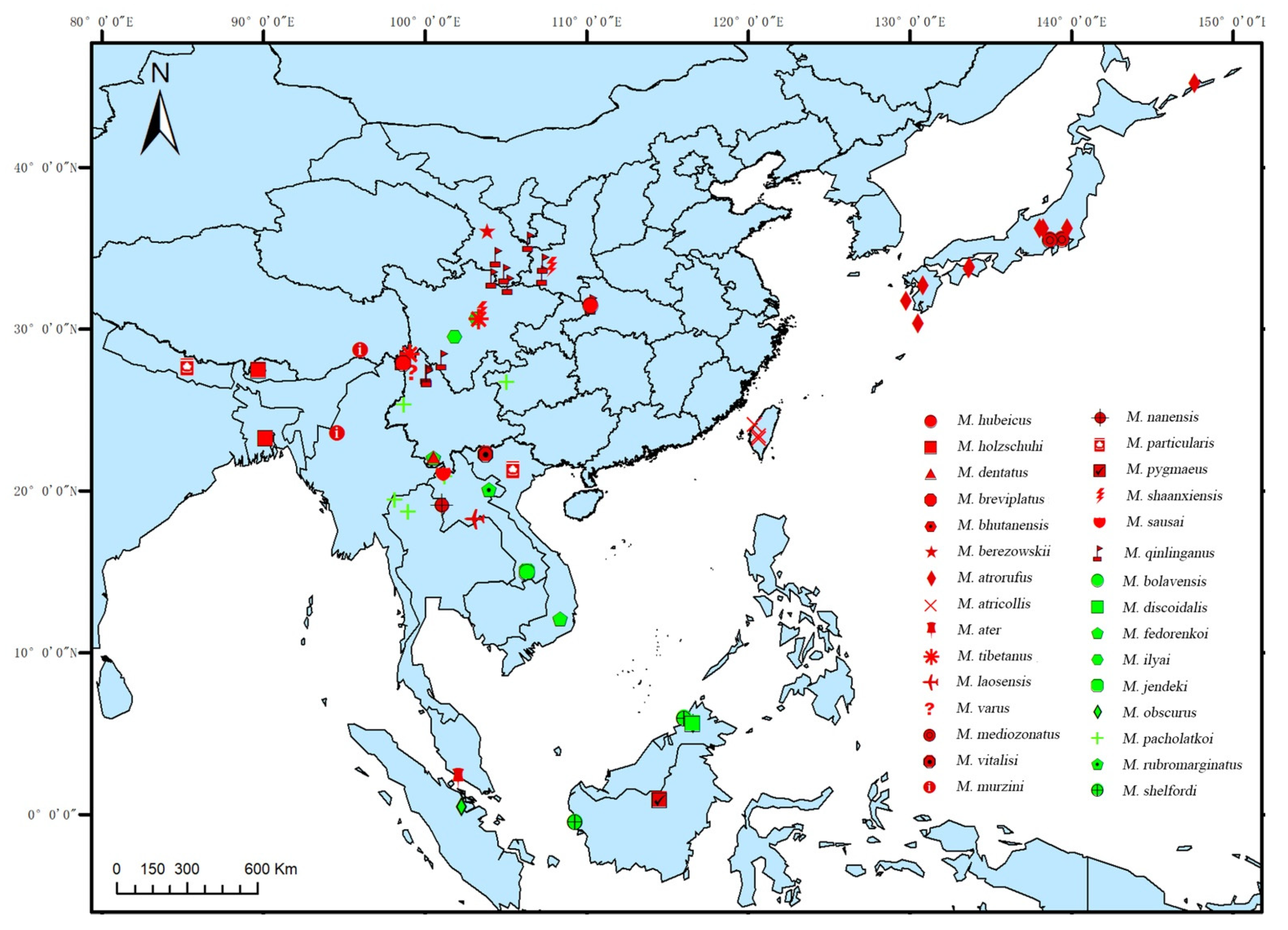
3.3. Distribution Pattern

A total of 30 Mesolycus species are known at present, of which 70% (21 species) belong to the atrorufus group, and the remainder (9 species) to the liyai group. Generally, the species of the atrorufus group and the ilyai group overlap in their distribution, but the range of the former is much larger than that of the latter.
In overall view, the Mesolycus species are widely distributed in the East Palaearctic and Oriental regions, −0.40–45.20° N, 85.32–147.60° E (Figure 4). Within this region, 13 species are distributed in China (10 species are endemic), accounting for 43.33% of the total number worldwide; six species in Laos; four species in Borneo; three species in Vietnam; and one or two species in the other countries (including Japan, Bhutan, India, Nepal, Myanmar, Thailand, Malacca and Sumatra), respectively.
The species are narrowly distributed, and most of them are restricted to one or a few localities, except for M. pacholatkoi (occurring in SW China, N. Laos, N. Thailand and N. Vietnam) and M. qinlinganus (restricted to China) covering a relatively large distribution range. Even some species are sympatric, such as M. hubeicus and M. qinlinganus (China, Hubei prov., Dashennongjia Natur. Res.), M. dentatus sp. n. and M. rubromarginatus (China, Yunnan prov., Mengsong), M. bolavensis and M. jendeki (Laos, Attapu prov., Bolvan Plataeu, Nong Lom lake env.), M. holzschuhi and M. bhutanensis (Bhutan, Thimphu Distr., Menshunang).

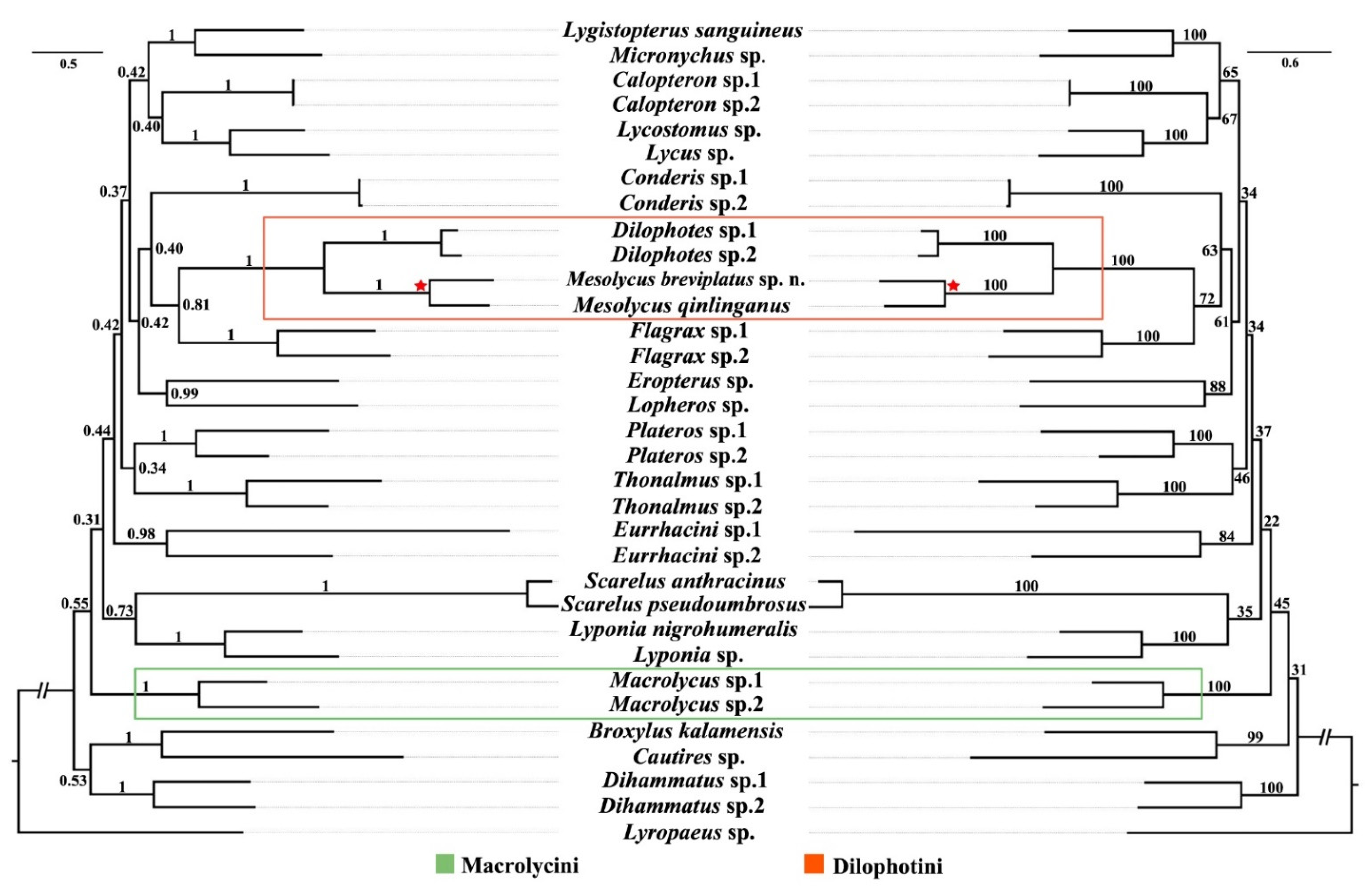
3.4. Phylogenetic Analyses

Both ML and BI analyses produced almost congruent topologies (Figure 5). The monophyly of all tribes (each with two representative species) as morphologically defined [1,16] was recovered in both analyses, and each of them was well supported (BS = 84–100, PP = 0.98–1). However, the relationships among the tribes of Lycinae got low support (BS = 22–72, PP = 0.31–0.81).
Macrolycini (Macrolycus) was a recovered sister to the remaining tribes except for Metriorrhynchini (Broxylus and Cautires) and Dihammatini (Dihammatus), albeit with low supporting values (BS = 45, PP = 0.31). Mesolycus was solidly grouped with Dilophotes into a monophyletic clade (BS = 100, PP = 1), which is recovered as a sister to Slipinskiini (Flagrax), and had moderate supports (BS = 72, PP = 0.81).

4. Discussion

4.1. Separate Status of the New Species

The variability in general appearance sometimes prevents reliable identification, and therefore the delineations of species are regularly based on the shape of the male genitalia, which are provided here in the key. As noted, M. pygmaeus Waterhouse, 1879 (Borneo) and M. ater Pic, 1943 (Malacca) were excluded from the key because only female types [9] are hitherto known and because of a poor description in the original publications.
Key to the Males of Mesolycus Species
  • Phallus very slender, internal sac attached to the phallus by two basal rods and additionally formed by two apical sclerotized complex plates (the atrorufus group, e.g., Figure 1D–F,H–P and Figure 3A–L)
2
-
Phallus robust, considerably shorter, internal sac attached to the phallus only by a complex stiffness plate (the ilyai group, e.g., Figure 1A–C,G)
20
2.
Phallus at least three times longer than internal sac (e.g., Figure 1N–P)
3
-
Phallus at most twice longer than internal sac (e.g., Figure 3A–C)
6
3.
Phallus curved apically ([9]: Figures 40 and 41), Japan
M. mediozonatus (Nakane, 1955)
-
Phallus almost straight
4
4.
Lower plate of internal sac straight basally ([10]: Figure 13), Bhutan, SE India
M. holzschuhi (Bic, 2002)
-
Lower plate of internal sac curved extensively
5
5.
Ventro-apical process of lower plate stout (Figure 1J), China (Shannxi)
M. shaanxiensis sp. n.
-
Ventro-apical process of lower plate slender ([9]: Figure 43), China (Hubei)
M. hubeicus Kazantsev, 2004
6.
The upper and lower plates subequal in length (e.g., Figure 3A–F)
7
-
The upper and lower plates obviously different in length (e.g., Figure 1D–F,K–M and Figure 3G–I)
11
7.
Lower plate of internal sac simple, without ventro-apical process (Figure 3J), China (Yunnan)
M. varus sp. n.
-
Lower plate of internal sac present with ventro-apical process (e.g., Figure 1H,I and Figure 3K)
8
8.
Upper plate of internal sac dentate at external margin (e.g., [10]: Figures 12 and 15)
9
-
Upper plate of internal sac simple at external margin (e.g., Figure 3K)
10
9.
Dorso-apical part of external margins of upper plate round ([10]: Figure 15), N. Laos
M. sausai (Bic, 2002)
-
Dorso-apical part of external margins of upper plate straight ([10]: Figure 12), Bhutan
M. bhutanensis (Bic, 2002)
10.
Lower plate of internal sac wider than upper plate ([9]: Figures 38 and 39), Vietnam
M. vitalisi (Pic, 1923)
-
Lower plate and upper plate of internal sac subequal in width (Figure 3K), China (Yunnan)
M. breviplatus sp. n.
11.
Upper plate of internal sac longer than lower plate (e.g., Figure 1D–F and Figure 3G–I)
12
-
Upper plate of internal sac shorter than lower plate (e.g., Figure 1K–M)
15
12.
Internal sac almost as wide as phallus (e.g., Figure 3G–I)
13
-
Internal sac obviously wider or narrower than phallus (e.g., Figure 1D–F)
14
13.
Upper plate of internal sac narrow, with elongate narrow upper portion (Figure 3G–I), Myanmar, China (Xizang)
M. murzini Kazantsev, 2004
-
Upper plate of internal sac wide, moderately curved apically ([10]: Figure 11), Japan
M. atrorufus (Kiesenwetter, 1879)
14.
Upper plate of internal sac and lower plate equal in width, dorso–apical teeth of lower plate truncate at apices ([9]: Figures 44 and 45), Thailand
M. nanensis Kazantsev, 2004
-
Lower plate of internal sac wider than upper plate, dorso–apical teeth of lower plate sharp at apices (Figure 1H), China (Yunnan)
M. dentatus sp. n.
15.
Lower plate of internal sac twice length of upper plate (e.g., [10]: Figure 14)
16
-
Lower plate of internal sac slightly longer than upper plate (e.g., Figure 1I)
17
16.
Internal margin of lower plate of internal sac dentate ([8]: Figure 5), China (Gansu)
M. berezowskii (Kazantsev, 2000)
-
Internal margin of lower plate of internal sac simple ([10]: Figure 14), Laos
M. laosensis (Bic, 2002)
17.
Pronotum dark red with infuscate middle, elytra dark red, and sometimes slightly infuscate basally, Vietnam, Nepal
M. particularis (Pic, 1928) comb. n.
-
Pronotum concolour, black or red
18
18.
Pronotum black, with very short inconspicuous brown pubescence, elytra brightly orange red, China (Taiwan)
M. atricollis (Pic, 1926) comb. n.
-
Pronotum and elytra concolour, dark red to brownish black, and always covered with red pubescence
19
19.
Lower plate of internal sac with a pair of modified laminae merged together on apical surface of ventro-apical process [Figure 1I], China (Sichuan, Shaanxi, Hubei, Yunnan, Gansu, Ningxia)
M. qinlinganus (Kazantsev, 2000)
-
Lower plate of internal sac with a pair of simple laminae separate on apical surface of ventro-apical process ([8]: Figure 3), China (Gansu, Yunnan)
M. tibetanus (Kazantsev, 2000)
20.
Tarsomeres I and II absent with plantar pads
M. shelfordi (Bourgeois, 1906)
-
Tarsomeres I and II present with plantar pads
21
21.
Internal sac extends beyond apex of phallus by at least mid-length of phallus (e.g., Figure 1A)
22
-
Internal sac not or slightly exceeding apex of phallus (e.g., [9]: Figure 37)
26
22.
Internal sac slender, as wide as or narrower than the apical part of the phallus ([9]: Figures 32–35), Borneo, Sumatra
M. obscurus (Pic, 1912)
-
Internal sac robust, distinctly wider than apical part of phallus
23
23.
Apical ring twice as wide as long ([10]: Figure 18), China (Sichuan)
M. ilyai (Kazantsev, 2000)
-
Apical ring nearly as long as wide (e.g., Figure 1B, [10]: Figure 20)
24
24.
Eyes small, interocular distance 1.36 times maximum eye diameter, China (Yunnan, Guizhou), N. Vietnam, N. Laos, NE. Thailand
M. pacholatkoi (Bic, 2002) comb. n.
-
Eyes large, interocular distance at most 1.1 times greater than eye diameter
25
25.
Internal sac present with three teeth on ventral side ([13]: Figures 17 and 18), Laos, China (Yunnan)
M. fedorenkoi Kazantsev, 2013
-
Internal sac present with four teeth on ventral side (Figure 1G), Vietnam
M. rubromarginatus Kazantsev, 2013
26.
Internal sac simple on ventral side ([9]: Figures 36 and 37), Borneo
M. discoidalis (Pic, 1912)
-
Internal sac with complex structures on ventral side
27
27.
Phallus 3 times longer than internal sac ([10]: Figure 17), Laos
M. jendeki (Bic, 2002)
-
Phallus 5 times longer than internal sac ([10]: Figure 16), Laos
M. bolavensis(Bic, 2002)

4.2. Species Diversity of Chinese Mesolycus

The number of the Chinese Mesolycus species is increased from six to 13 through this study, which is unexpected. On the world scale, the total species diversity of Mesolycus is highest in China, accounting for nearly a half of the total number. In southern China, there are vast mountainous areas and an usually rich spectrum of vegetation types, numerous geographical and ecological isolations [44], thereby accelerating the speciation of Mesolycus there.
The endemism of Mesolycus in China is also high (ca. 77%). The high endemism may result from a lack of comprehensive taxonomic studies in the group previously. The widely distributed genus throughout China and surrounding countries is evidence enough that these beetles are, in fact, very well capable of flying and dispersing. This makes it particularly necessary for us to conduct extensive and thorough field surveys in order to fully understand the species diversity of Mesolycus. Sympatric distribution is another phenomenon in Mesolycus, since two or more species occur at the same locality. The highly disparate morphologies in male genitalia may account for effective reproductive isolation among these sympatric congeneric species.
Despite high endemism, the species-rich Chinese fauna has certain faunal connections with adjacent regions, including the Palaearctic part of East Asia, Indochina, Indo-Malaysia, and the southern Qinghai-Tibet Plateau, by sharing several common species or similar species, such as M. pacholatkoi, M. murzini and M. rubromarginatus. As a transitional zone between Palaearctic and Oriental regions, China plays a critical role in studying the biogeographic fauna. More specifically, Chinese fauna cannot be neglected in tracking the spatial origin and dispersal of Mesolycus or higher grades. Thus, the taxonomy of net-winged beetles remains essential, or even a priority, for the future of systematics, biogeography, ecology, and conservation, etc.

4.3. Phylogenetic Position of Mesolycus

In the present study, the sampling of Mesolycus was included in the molecular phylogenetic analyses for the first time. In terms of our results, Mesolycus was suggested to be a sister group of Dilophotes and that they together form a monophyletic Dilophotini. This was consistent with the opinion of Bocak & Bocakova [1], while it conflicts with that of Kazantsev [8,9,17], who hypothesized that Mesolycus was more closely related to Macrolycus attributed to Macrolycini. Although a tentative apomorphy (presence of wedge cell) was presumed for Dilophotes [9], some more common characters were found between the latter and Mesolychus, which are the diagnosis to define the Dilophotini [1].
Further, the produced topology demonstrated that Dilophotini was rooted in a distant position from Macrolycini, which was congruent with the molecular phylogenetics of Bocak & Bocakova [1] and Masek et al. [18]. In contrast, a sister relationship was recovered between the two taxa by Kazantsev [8,9,17], based on the analyses of morphological data. Kazantsev [9] proposed the reason for this hypothesis was that they shared some morphological features, as opposed to the bulk of the Lycidae, i.e., presence of the coronal and sub-antennal sutures, presence of the median longitudinal pronotal carina, bifid claws of all tarsi and an asymmetrical phallobase. However, only the bifid claws were acknowledged to be unique for them and hypothesized to be their synapomorphy [9]. However, this character was different between the two tribes, with all claws bifid in both sexes of Macrolycini (Macrolycus s. l., [11]) or only in males, while tooted basally in females (Dilophotes and Mesolycus, [9]) of Dilophotini. Furthermore, the male genitalia is usually considered as a more stable and dependable structure in the taxonomy and classification, as well as in inferring the relationships of Lycidae [16]. Dilophotini (including Dilophotes and Mesolycus) often have considerably sclerotized and exposed internal sacs, which sometimes form a very complex structure, and a long phallobase [10], while Macrolycini (only Macrolycus s. l.) has a simple, slender, membranous internal sac with at most its apical part exposed and a small and rounded phallobase [16]. Moreover, they differ in the number of longitudinal elytral costae, present with three (Dilophotini) or four costae (Macrolycini) [1]. Therefore, these differences are probably adequate to separate Dilophotini and Macrolycini in distant positions. Nevertheless, more DNA markers should be explored to confirm this result due to the low supporting values in inferring the relationships among tribes.

5. Conclusions

In the present study, the lycid genus Mesolycus is reviewed, with an emphasis on the Chinese fauna. As a result, three species are transferred from Dilophotes, including M. atricollis (Pic, 1926) comb. n., M. particularis (Pic, 1928) comb. n. and M. pacholatkoi (Bic, 2002) comb. n. Four new species are discovered in China, including M. shaanxiensis sp. n., M. dentatus sp. n., M. breviplatus sp. n. and M. varus sp. n. Two species, M. murzini Kazantsev, 2004 and M. rubromarginatus Kazantsev, 2013, are recorded in China for the first time. It is quite surprising that the number of Chinese Mesolycus species is increased from six to 13, which is much higher than other countries in the species diversity. In addition, the aedeagi are photographed for the previously inadequately illustrated species and a key for the identification of the Mesolycus species globally is provided. Moreover, the reconstructed phylogenies of Lycinae based on multiple genes (cox1, nad5, cox2 and Lrna) by both ML and BI analyses show that Mesolycus is a consistently recovered sister to Dilophotes, and they together form a monophyletic clade which is well supported, suggesting that Mesolycus definitely belongs to Dilophotini rather than to Macrolycini of Lycinae. This study is another contribution to the Chinese lycid beetles and helps us better understand the species diversity and phylogenetic position of Mesolycus.

Supplementary Materials

The following supporting information can be downloaded at: https://www.mdpi.com/article/10.3390/insects13121171/s1, Table S1: The distribution information of Mesolycus species; Table S2: The primers of COI gene used for PCR; Table S3: Information on the mitochondrial gene fragments of the Lycidae species used in this study; Table S4: The optimal partition schemes and the best-fit substitute models for the ML and BI analysis of gene segments dataset.

Author Contributions

Conceptualization, H.L., X.Y. and Y.Y.; Methodology, R.D., Y.K., X.G. and Y.Y.; Investigation, R.D., Y.K. and Y.Y.; Data curation, X.G., Y.K. and R.D.; Formal analysis: R.D., Y.K. and Y.Y.; Visualization: R.D. and Y.K.; Validation, R.D., Y.K., H.L. and Y.Y.; Writing—Original Draft Preparation, R.D., Y.K. and Y.Y.; Writing—Review and Editing, H.L, R.D., Y.K., X.Y. and Y.Y.; Funding acquisition, Y.Y. and H.L. All authors have read and agreed to the published version of the manuscript.

Funding

This study was financially supported by the National Natural Science Foundation of China (No. 32270491), the Natural Science Foundation of Hebei Province (No. C2022201005), and Excellent Youth Scientific Research and Innovation Team of Hebei University (No. 605020521005) to Y.Y., also the Interdisciplinary Research Program of Natural Science of Hebei University (No. DXK202103) to H.L.

Institutional Review Board Statement

Not applicable.

Informed Consent Statement

Not applicable.

Data Availability Statement

The sequence generated in this study is deposited in GenBank with accession numbers (OP735348-49, OP729892-93, OP803896-99).

Acknowledgments

We are grateful to Sergey V. Kazantsev (Moscow, Russia) for his kind help in sending us some literature. Also, thanks are given to Han-Qing Lin for her help in taking the photos. Moreover, we are indebted to the two anonymous reviewers for their valuable suggestions in improving our original manuscript.

Conflicts of Interest

The authors declare no conflict of interest.

References

  1. Bocak, L.; Bocakova, M. Phylogeny and classification of the family Lycidae (Insecta: Coleoptera). Ann. Zool. 2008, 58, 695–720. [Google Scholar] [CrossRef]
  2. Motyka, M.; Kampova, L.; Bocak, L. Phylogeny and evolution of Müllerian mimicry in aposematic Dilophotes: Evidence foradvergence and size–constraintsin evolution of mimetic sexual dimorphism. Sci. Rep. 2018, 8, 3744. [Google Scholar] [CrossRef] [PubMed]
  3. Bocak, L.; Yagi, T. Evolution of mimicry patterns in Metriorrhynchus (Coleoptera: Lycidae): The history of dispersal and speciation in South East Asia. Evolution 2010, 64, 39–52. [Google Scholar] [CrossRef] [PubMed]
  4. Malohlava, V.; Bocak, L. Evidence of extreme habitat stability in a southeast Asian biodiversity hotspot based on the evolutionary analysis of neotenic net–winged beetles. Mol. Ecol. 2010, 19, 4800–4811. [Google Scholar] [CrossRef]
  5. Sklenarova, K.; Chesters, D.; Bocak, L. Phylogeography of poorly dispersing net–winged beetles: A role of drifting India in the origin of Afrotropical and Oriental fauna. PLoS ONE 2013, 8, e67957. [Google Scholar] [CrossRef] [Green Version]
  6. Nakane, T. Lycidae (Insecta: Coleoptera). In Fauna Japonica; Academic Press of Japan: Tokyo, Japan, 1969; 224 p. [Google Scholar]
  7. Bocak, L.; Matsuda, K. Review of the immature stages of the family Lycidae (Insetcta: Coleoptera). J. Nat. Hist. 2003, 37, 1463–1507. [Google Scholar] [CrossRef]
  8. Kazantsev, S.V. On Dilophotes Waterhouse, 1879 and Mimodilophotes Pic, 1939 (Coleoptera, Lycidae). Russ. Entomol. J. 2000, 9, 329–332. [Google Scholar]
  9. Kazantsev, S.V. Contribution to the knowledge of Macrolycini with description of Calcaeron, new genus (Coleoptera, Lycidae). Zootaxa 2004, 493, 1–32. [Google Scholar] [CrossRef]
  10. Bic, V. Review of the genus Dilophotes Waterhouse (Coleoptera: Lycidae) of the Palaearctic region and Indochina. Zootaxa 2002, 59, 1–26. [Google Scholar] [CrossRef]
  11. Li, Y.; Bocak, L.; Pang, H. New species of Macrolycus Waterhouse, 1878 from China and Laos, with a check–list of the genus (Coleoptera: Lycidae). Zootaxa 2012, 3232, 44–61. [Google Scholar] [CrossRef]
  12. Bocak, L.; Bocakova, M. Taxonomic notes on East–Asiatic Lycidae (Coleoptera). Acta Entomol. Bohemoslov. 1988, 85, 444–446. [Google Scholar]
  13. Kazantsev, S.V. New net–winged beetles (Coleoptera: Lycidae) from Indochina, with synonymic and taxonomic notes. Cauc. Entomol. Bull. 2013, 9, 247–252. [Google Scholar] [CrossRef]
  14. Kleine, R. Lycidae. In Coleopterorum Catalogus; Pars 123; Junk, W., Schenkling, S., Eds.; W. Junk: Berlin, Germany, 1933; 145 p. [Google Scholar]
  15. Kleine, R. Neue Indische Lycidae nebst faunistischen Bemerkungen. Indian For. Rec. 1928, 13, 221–268. [Google Scholar]
  16. Bocak, L.; Bocakova, M. Revision of the supergeneric classification of the family Lycidae (Coleoptera). Pol. Pismo Entomol. 1990, 59, 623–676. [Google Scholar]
  17. Kazantsev, S.V. Morphology of Lycidae with some considerations on evolution of the Coleoptera. Elytron 2005, 17, 49–226. [Google Scholar]
  18. Masek, M.; Motyka, M.; Kusy, D.; Bocek, M.; Li, Y.; Bocak, L. Molecular Phylogeny, Diversity and Zoogeography of Net–Winged Beetles (Coleoptera: Lycidae). Insects 2018, 9, 154. [Google Scholar] [CrossRef] [Green Version]
  19. Peng, Y.; Leung, H.C.; Yiu, S.M.; Chin, F.Y. IDBA–UD: A de novo assembler for single–cell and metagenomic sequencing data with highly uneven depth. Bioinformatics 2012, 28, 1420–1428. [Google Scholar] [CrossRef] [Green Version]
  20. Hebert, P.D.N.; Ratnasingham, S.; de Waard, J.R. Barcoding animal life: Cytochrome c oxidase subunit I divergences among closely related species. Proceedings of the Royal Society of London. Ser. B Biol. Sci. 2003, 270, 96–99. [Google Scholar] [CrossRef] [Green Version]
  21. Kearse, M.; Moir, R.; Wilson, A.; Stones–Havas, S.; Cheung, M.; Sturrock, S.; Buxyon, S.; Cooper, S.; Duran, C.; Thierer, T.; et al. Geneious Basic: An integrated and extendable desktop software platform for the organization and analysis of sequence data. Bioinformatics 2012, 28, 1647–1649. [Google Scholar] [CrossRef] [Green Version]
  22. Uribe, J.E.; Gutiérrez–Rodríguez, J. The complete mitogenome of the trilobite beetle, Platerodrilus sp. (Elateroidea: Lycidae). Mitochondrial DNA Part B 2016, 1, 658–659. [Google Scholar] [CrossRef] [Green Version]
  23. Bernt, M.; Donath, A.; Jühling, F.; Externbrink, F.; Florentz, C.; Fritzsch, G.; Pütz, J.; Middendorf, M.; Stadler, P.F. MITOS: Improved de novo metazoan mitochondrial genome annotation. Mol. Phylogenetics Evol. 2013, 69, 313–319. [Google Scholar] [CrossRef] [PubMed]
  24. Motyka, M.; Masek, M.; Bocak, L. Congruence between morphology and molecular phylogeny: The reclassification of Calochromini (Coleoptera: Lycidae) and their dispersal history. Zool. J. Linn. Soc. 2017, 180, 147–165. [Google Scholar] [CrossRef]
  25. Bocakova, M.; Bocak, L.; Hunt, T.; Teraväinen, M.; Vogler, A.P. Molecular phylogenetics of Elateriformia (Coleoptera): Evolution of bioluminescence and neoteny. Cladistics 2007, 23, 477–496. [Google Scholar] [CrossRef]
  26. Sklenarova, K.; Kubecek, V.; Bocak, L. Subtribal classification of Metriorrhynchini (Insecta, Coleoptera, Lycidae): An integrative approach using molecular phylogeny and morphology of adults and larvae. Arthropod Syst. Phylogeny 2014, 72, 37–54. [Google Scholar]
  27. Levkanicova, Z.; Bocak, L. Identification of net-winged beetle larvae (Coleoptera: Lycidae) using three mtDNA fragments: A comparison of their utility. Syst. Entomol. 2009, 34, 210–221. [Google Scholar] [CrossRef]
  28. Katoh, K.; Standley, D.M. MAFFT multiple sequence alignment software version 7: Improvements in performance and usability. Mol. Biol. Evol. 2013, 30, 772–780. [Google Scholar] [CrossRef]
  29. Zhang, D.; Gao, F.; Jakovlić, I.; Zou, H.; Zhang, J.; Li, W.X.; Wang, G.T. PhyloSuite: An integrated and scalable desktop platform for streamlined molecular sequence data management and evolutionary phylogenetics studies. Mol. Ecol. Resour. 2020, 20, 348–355. [Google Scholar] [CrossRef]
  30. Capella–Gutiérrez, S.; Silla–Martínez, J.M.; Gabaldón, T. TrimAI: A tool for automated alignment trimming in large–scale phylogenetic analyses. Bioinformatics 2009, 25, 1972–1973. [Google Scholar] [CrossRef] [Green Version]
  31. Lanfear, R.; Calcott, B.; Ho, S.Y.; Guindon, S. PartitionFinder: Combined selection of partitioning schemes and substitution models for phylogenetic analyses. Mol. Biol. Evol. 2012, 29, 1695–1701. [Google Scholar] [CrossRef] [Green Version]
  32. Nguyen, L.T.; Schmidt, H.A.; Von–Haeseler, A.; Minh, B.Q. IQ–TREE: A fast and effective stochastic algorithm for estimating maximum–likelihood phylogenies. Mol. Biol. Evol. 2015, 32, 268–274. [Google Scholar] [CrossRef]
  33. Ronquist, F.; Huelsenbeck, J.P. MrBayes 3: Bayesian phylogenetic inference under mixed models. Bioinformatics 2003, 19, 1572–1574. [Google Scholar] [CrossRef] [PubMed] [Green Version]
  34. Rambaut, A. FigTree v1.4.3 Software; Institute of Evolutionary Biology, University of Edinburgh: Edinburgh, UK, 2016. [Google Scholar]
  35. ICZN [International Commission on Zoological Nomenclature]. International Code of Zoological Nomenclature, 4th ed.; The International Trust for Zoological Nomenclature: London, UK, 1999. [Google Scholar]
  36. Kiesenwetter, H. Coleoptera Japoniae collecta a Domino Lewis et aliis. Dtsch. Entomol. Zaitschrift 1879, 23, 305–320. [Google Scholar] [CrossRef]
  37. Gorham, H.S. Revision of the genera and species of Malacoderm Coleoptera of the Japanese fauna. Trans. R. Entomol. Soc. Lond. 1883, 31, 393–411. [Google Scholar] [CrossRef]
  38. Kleine, R. Bestimmungstabelle der Lycidae. In Bestimmungstabellen der europäischen Coleopteren; 123 Heft; Edmund Reitter: Troppau, Czech Republic, 1942; 90p. [Google Scholar]
  39. Lewis, G. On the lamellicorn Coleoptera of Japan, and notices of others. Ann. Mag. Nat. Hist. 1895, 16, 374–408. [Google Scholar] [CrossRef] [Green Version]
  40. Bocakova, M.; Bocak, L. Lycidae. In Catalogue of Palaearctic Coleoptera; Elateroidea—Derodontoidea—Bostrichoidea—Lymexyloidea—Cleroidea—Cucujoidea; Löbl, I., Smetana, A., Eds.; Apollo Books: Stenstrup, Demarks, 2007; Volume 4, pp. 211–224. [Google Scholar]
  41. Matsuda, K. Identification manual of the lycid–beetles in Japan, IV. Sayabane N. S. 2012, 7, 1–6. [Google Scholar]
  42. Pic, M.H. Sauter’s Formosa Ausbeute. Mordellidae, Lycidae, Cantharidae. Entomol. Mitt. 1926, 15, 67–69. [Google Scholar]
  43. Pic, M. Nouveautés diverses. Mélanges Exot. Entomol. 1928, 52, 1–32. [Google Scholar]
  44. Li, D.; Aspöck, H.; Aspöck, U.; Liu, X.-Y. Mining the Species Diversity of Lacewings: New Species of the Pleasing Lacewing Genus Dilar Rambur, 1838 (Neuroptera, Dilaridae) from the Oriental Region. Insects 2021, 12, 451. [Google Scholar] [CrossRef]
Figure 1. Aedeagi of Mesolycus species: (AC,G). M. rubromarginatus Kazantsev, 2013; (DF,H). M. dentatus sp. n.; (KM,I). M. qinlinganus Kazantsev, 2000; (NP,J). M. shaanxiensis sp. n.: (A,D,K,N)—ventrally; (B,E,L,O)—dorsally; (C,F,M,P)—laterally; (GJ)—apically. Scales: (AG,IP), 0.5 mm; (H), 0.2 mm. pl—plate; br—basal rods; ph—phallus; pb—phallobase; ar—apical ring; ap—apical hood; lp—lower plate; up—upper plate; pr—ventro-apical process.
Insects 13 01171 g001
Figure 2. Male habitus of Mesolycus species: (A). M. dentatus sp. n.; (B). M. qinlinganus (Kazantsev, 2000); (C). M. shaanxiensis sp. n.; (D). M. varus sp. n.; (E). M. breviplatus sp. n.; (F). M. murzini Kazantsev, 2004. Scales: 1.0 mm.
Insects 13 01171 g002
Figure 3. Aedeagi of Mesolycus species: (AC,J). M. varus sp. n.; (DF,K). M. breviplatus sp. n.; (GI,L). M. murzini Kazantsev, 2004. (A,D,G)—ventrally; (B,E,H)—dorsally; (C,F,I)—laterally; (JL)—apically. Scales: (AI,K), 0.5 mm; (J,L), 0.2 mm. pl—plate; br—basal rods; ph—phallus; pb—phallobase.
Insects 13 01171 g003
Figure 4. Distribution map of Mesolycus in the world scale (The red represents the atrorufus group and the green denotes the ilyai group).
Insects 13 01171 g004
Figure 5. Phylogenetic tree of Lycinae produced from the ML (right) and BI (left) analyses based on the cox1, nad5, cox2 and Lrna genes. The numbers on the branches represent bootstrap (BS) (right) and posterior probability (PP) (left), respectively. Dilophotini is circled in orange and Macrolycini in green box. The pentagram represents Mesolycus species used in this study.
Insects 13 01171 g005
Publisher’s Note: MDPI stays neutral with regard to jurisdictional claims in published maps and institutional affiliations.

