Article

Expression of Immunity- and Stress-Related Genes during an Intermolt Period in the Colorado Potato Beetle

Institute of Systematics and Ecology of Animals, Siberian Branch of Russian Academy of Sciences, Frunze Str. 11, 630091 Novosibirsk, Russia
*
Author to whom correspondence should be addressed.
Insects 2022, 13(12), 1168; https://doi.org/10.3390/insects13121168
Submission received: 9 November 2022 / Revised: 7 December 2022 / Accepted: 14 December 2022 / Published: 16 December 2022
(This article belongs to the Special Issue Insect Hormones, Metabolism, Immunity and Development)

Abstract

:

Simple Summary

During ontogeny, many insects change habitats, which are characterized by specific communities of opportunistic and pathogenic microorganisms. Therefore, different developmental stages may be subject to different directions of natural selection that cause alterations in immunity. We analyzed changes in humoral immunity during the intermolt period of last-instar larvae of the Colorado potato beetle under laboratory conditions. We observed an increase in basal expression of immunity- and stress-related genes at the stages of finishing feeding and migration into soil. Changes in gene expression in response to fungal infection were also examined. The results suggest that the larvae develop strong defenses against soil pathogens (especially against fungi) before migration into soil. The study is also promising in terms of a gene knockdown for enhancing the susceptibility of the Colorado potato beetle to native or introduced fungal pathogens in potato agrosystems.

Abstract

Different developmental stages of insects may be dissimilar in immunity functioning. Additionally, the stages often inhabit diverse environments with specific microbial communities. In the Colorado potato beetle, a strong increase in resistance to entomopathogenic fungi is observed during the intermolt period of last-instar larvae, but mechanisms of this change are insufficiently understood. We studied changes in the expression of immunity- and stress-related genes in the fat body and integument during this intermolt period by quantitative PCR. By the end of the instar, there was upregulation of transcription factors of Toll, IMD, and Jak–Stat pathways as well as genes encoding metalloprotease inhibitors, odorant-binding proteins, and heat shock proteins. Nonetheless, the expression of gene LdRBLk encoding β-lectin did not change during this period. Most of the aforementioned genes were upregulated in response to Metarhizium robertsii topical infection. The expression alterations were more pronounced in recently molted larvae than in finishing feeding larvae and in the integument compared to the fat body. We believe that upregulation of immune-system- and stress-related genes at the end of the intermolt period is an adaptation caused by migration of larvae into soil, where the probability of encountering entomopathogenic fungi is high.

1. Introduction

Insect immune reactions and susceptibility to pathogenic microorganisms change during ontogeny. It is well known that larvae of different instars, pupae, and adults demonstrate different levels of immunity and resistance to pathogens [1,2]. An important role in the formation of these reactions can be determined by metamorphosis and the habitat in which it occurs. Each habitat (e.g., a plant, leaf litter, or soil) contains a specific community of microorganisms, including pathogens. Habitats can be switched by insects within one larval instar. For holometabolous herbivores, this is often the last larval instar when the insects finish feeding on a plant and migrate into soil to pupate. Soil is the richest reservoir of microorganisms, including entomopathogenic fungi, capable of penetrating the host through the integuments. The density of entomopathogenic ascomycetes such as Beauveria and Metarhizium in soil reaches substantial concentrations: 104–105 colony-forming units per gram [3,4]. Infection of insects by these fungi in natural and anthropogenic ecosystems most often occurs in the leaf litter and soil [3]. From this point of view, changes in the host’s immunity at stages of completion of feeding and burrowing into the soil are of interest.
The Colorado potato beetle (CPB) Leptinotarsa decemlineata Say (Coleoptera, Chrysomelidae) is one of the most dangerous potato pests in the world. It is known that entomopathogenic fungi can have a significant impact on the natural regulation of its populations and are used for the inundative biological control of this pest (reviewed by [5,6]). Natural epizootics caused by fungi are extremely rare among feeding CPB larvae; however, high mortality from fungal infections can be observed at the stages inhabiting soil: the prepupa, pupa, and hibernating adults (reviewed by [7]). At the same time, the resistance to fungi at CPB soil stages is higher than in the feeding larvae. In particular, we have studied changes in susceptibility to entomopathogenic fungi during the intermolt period of the last larval instar [8] and observed a gradual increase in resistance to the fungi at the end of this period. During development within the last instar, along with thickening of the cuticle, there was a decrease in the level of adhesion of conidia to the cuticle, which correlated with a decrease in the amount of epicuticular hydrocarbons. In addition, during this period, significant elevation of the encapsulation activity and of the total hemocyte count was observed [8]. Thus, the change in habitats from a plant to soil is accompanied by an increase in cuticular and cellular defense systems associated with resistance to fungal infections. Nevertheless, alterations of molecular mechanisms such as regulation of immune signaling pathways during this period have remained unexplored.
Among insect immune signaling pathways, the most important are Toll, IMD, and Jak–Stat. The Toll cascade is related to defense against Gram-positive bacteria and fungi, whereas the IMD pathway is mainly related to defense against Gram-negative bacteria and Jak–Stat against viruses [9]. Nonetheless, IMD and Jak–Stat pathways are also upregulated in insects in response to fungal infections, and silencing or chemical inhibition of IMD and Jak–Stat cascades leads to increased susceptibility to fungi [10,11]. On the one hand, this phenomenon may indicate synergy between different pathways against fungal invaders. On the other hand, different bacteria may often be involved in the process of mycosis, thereby aggravating the pathogenesis and accelerating mortality (e.g., [12]).
Insect metalloproteinase inhibitors (IMPIs) are some of the key antifungal compounds inactivating the fungal proteinases [13]. Ricin-like β lectins (RBLs) are related to C-type lectins and may function in insect defenses against fungal infections [14] and against neonicotinoid insecticides [15]. Odorant-binding proteins (OBPs) function in a wide range of physiological processes and may change expression in response to fungal infections [16]. Heat shock proteins (HSPs) are stress-induced proteins. HSP70 and HSP90 are cochaperones and work as adaptors and folding proteins [17]. Expression of HSPs is dependent on insect development [18], and induction of HSPs in response to different infections, including a fungal invasion, was also reported (e.g., [19]). It should be noted that the research into activities of CPB immunity during development various pathologies is “far from exhaustive” [20]. At the same time, understanding the mechanisms that work against fungal pathogens in the CPB may allow for improved biological control of this pest.
In the present work, we studied changes in the expression of CPB genes related to immune signaling pathways (Toll, IMD, and Jak–Stat) as well as to the production of IMPIs, OBPs, HSPs, and C-type lectins in the fat body and integument during the intermolt period of the last larval instar. Changes in the expression of these genes in response to fungal infection (Metarhizium robertsii) at different stages of the period were also analyzed.

2. Materials and Methods

2.1. Insects and Fungi

Third-instar larvae of L. decemlineata were collected in potato kitchen gardens (Solanum tuberosum) near Karasuk town, Western Siberia (53°43′05.4″ N 77°38′14.4″ E). The larvae were maintained in a laboratory in 350 mL ventilated plastic containers (10 larvae per container) at 25 °C, 20% relative humidity, under a photoperiod of 14:10 light:dark. Foliage of S. tuberosum was used as feed.
Strain P-72 of M. robertsii (GenBank accession no. KP172147.2) was used, taken from the collection of microorganisms at the Institute of Systematics and Ecology of Animals, the Siberian Branch of the Russian Academy of Sciences. To obtain conidia, twice-autoclaved millet was inoculated with a 4-day-old submerged culture of P-72 and incubated for 10 days at 25 °C in the dark. Then, the culture was dried for 12 days at room temperature, and conidia were collected using a soil sieve and stored at 4 °C for 20 days until infection. For infecting the CPB, the conidia were suspended in an aqueous solution of Tween 20 (0.05%). Concentrations of the conidia were estimated by means of a Neubauer hemocytometer (Paul Marienfeld GmbH & Co. KG, Lauda-Königshofen, Germany).

2.2. The Infection Procedure and General Experimental Setup

Larvae of instar IV at 2 h post-molt (recently molted larvae) and of instar IV at 84 h post-molt (finishing feeding larvae) were collected for experiments. For infection, we used the immersion of larvae in a suspension of conidia for uniform distribution of the inoculum. The larvae were infected by immersion for 15 s in the aqueous Tween 20 suspension of M. robertsii (5 × 10⁶ conidia/mL, 1.5 mL per 10 larvae). In the control group, insects were treated with the conidia-free aqueous Tween 20 solution. Then, the larvae were maintained and fed as described in the previous section. Mortality assays were performed on eight replicates (1 replicate = 10 larvae) for 10 days.
For analysis of gene expression, we used control and infected insects of both groups: recently molted and finishing feeding larvae. Samples of the integument and fat body were taken 24 and 72 h after inoculation in each group. The analyses were performed separately for age-related changes (only uninfected larvae at 26, 108, and 156 h post-molt) and for a response to infection (control vs. infected insects of both groups: recently molted and finishing feeding larvae).

2.3. Sample Preparation and Quantitative PCR (qPCR)

Samples of the integument and fat body tissues were collected as described previously [14] with minor modifications. Integuments and fat bodies from three insects were pooled into one sample. The samples were collected into cold phosphate buffer on ice and frozen in liquid nitrogen. Long-term storage was implemented at −80 °C. Before RNA extraction, samples were lyophilized at −65 °C and 600 mtorr for 14 h and homogenized with micropestles in liquid nitrogen. Total RNA extraction was performed as described earlier [14]. DNase treatment was carried out according to the manual for the RQ1 RNase-free DNase Kit (Promega, Madison WI, USA). The reverse transcription of RNA to cDNA was performed with RevertedAidTM M-MuLV Reverse Transcriptase (Fermentas, Vilnius, Lithuania) and 1.5 pmol of 9N primers.
qPCR was carried out by means of the HS-qPCR SYBR Blue (2×) mix (BioLabMix, Novosibirsk, Russia) on a thermal cycler called the CFX96 Touch Real-Time PCR detection system (Bio-Rad, Hercules, CA, USA). The qPCR was conducted in triplicate under the following conditions: 95 °C for 3 min, followed by 40 cycles of 94 °C for 15 s and annealing and elongation at 64 °C for 30 s, followed by melting curve analysis (70–90 °C). The melting curves for each sample were analyzed after each run to check the specificity of amplification. The following genes of L. decemlineata were used as a reference: 60S ribosomal protein L18 (rp18) and ADP-ribosylation factors 4 and 1 (arf2 and arf19) [21]. The analyzed genes of interest were dorsaldif, nfkb, stat, impi, LdRBLk, Ldobp, hsp70, and hsp90. Genes’ full names and primer sequences are provided in Table 1. Primers were designed with the help of the NCBI Primers-BLAST resource [22], and primers’ properties were estimated in IDT OligoAnalyser 3.1 [23]; the primers were synthesized by the Biosset company (Novosibirsk, Russia).
Gene expression was calculated by the 2ΔΔCq method in Bio-Rad CFX Manager (Bio-Rad). In the analysis of age-related changes, data on gene expression were normalized to those of larvae at 24 h post-molt. In the analysis of gene expression in response to infection, data were normalized to gene expression in uninfected larvae for each age group and each time point post-inoculation.

2.4. Statistics

Software packages PAST 3.0 [24] and SigmaStat 3.1 (Systat Software Inc., Tulsa, OK, USA) were used for the analyses. Because the data distribution was non-normal (according to the Shapiro–Wilk W test, p < 0.05), Kruskal–Wallis ANOVA followed by Dunn’s post hoc test was applied to determine the significance in differences (p < 0.05). The log rank test was performed to evaluate differences in survival dynamics. Data in plots are presented as the arithmetic mean and standard error.

3. Results

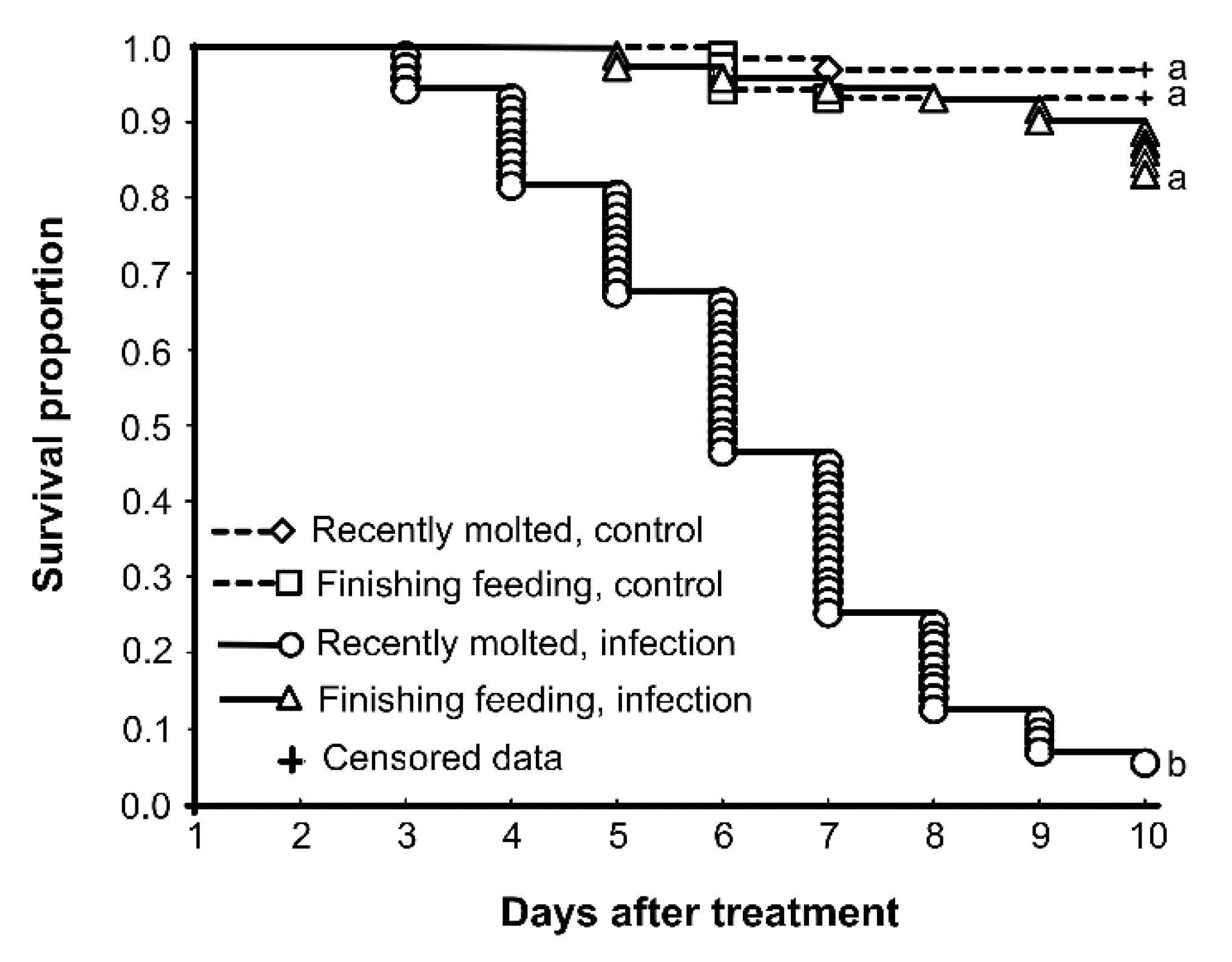
3.1. Susceptibility to M. robertsii

We repeated the survival assay in the present study, and the results were consistent with previous work [8]. The infection of recently molted larvae led to a significant decrease in survival, down to 5% over 10 days (Figure 1). Infection of finishing feeding larvae led to 85% survival over 10 days; this result did not significantly differ from controls, which showed 93–97% survival.

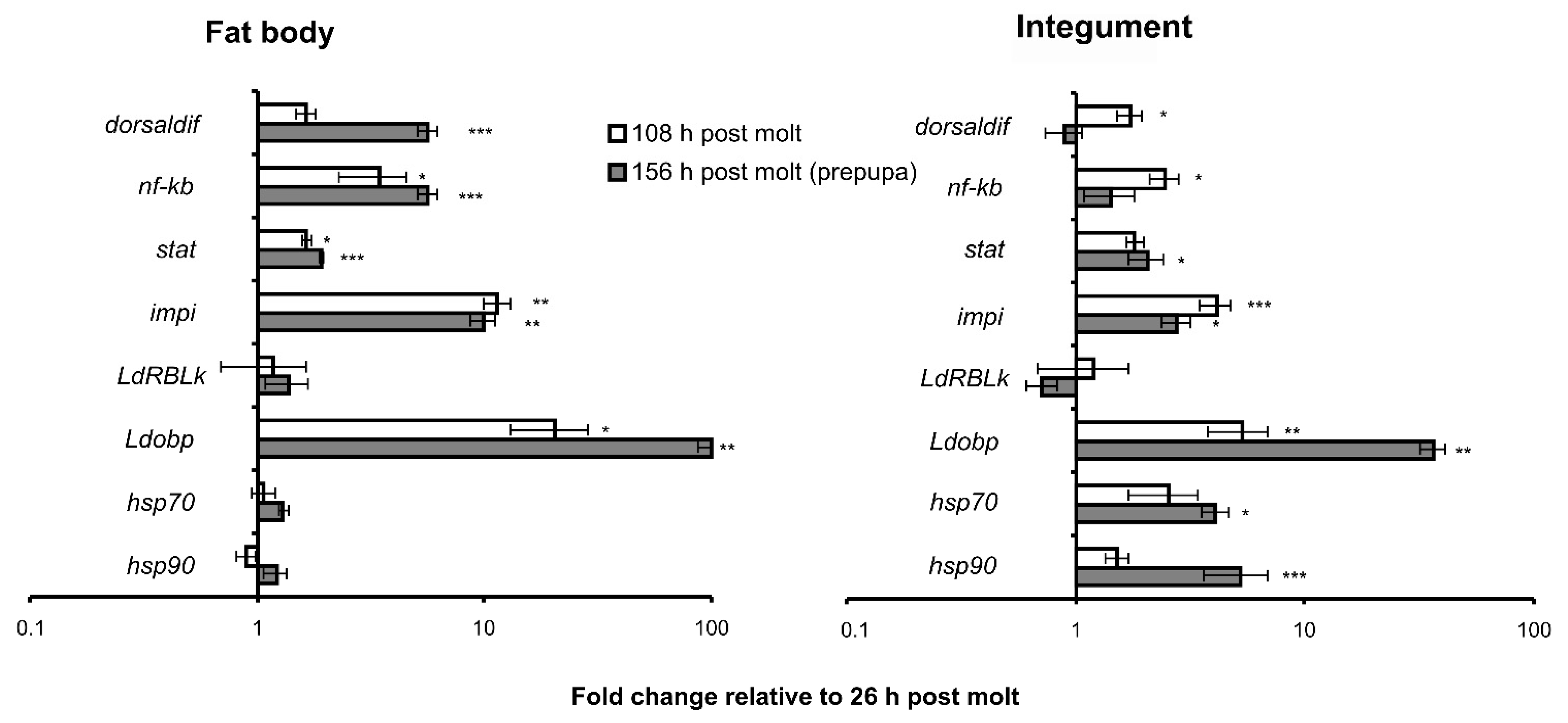
3.2. Changes in Gene Expression during Development

Transcription factors of Toll, IMD, and Jak–Stat immune signaling pathways (dorsaldif, nfkb, and stat) increased expression during the intermolt period (Figure 2). In particular, the upregulation of dorsaldif, nfkb, and stat in the fat body was time-dependent and reached 1.6–3.5-fold elevation in finishing feeding larvae and 2–6-fold elevation in prepupae compared to recently molted larvae. In the integument, significant upregulation of dorsaldif and nfkb was registered only in finishing feeding larvae (2–2.5-fold compared to recently molted larvae) but not in prepupae. Stat expression increased 1.8–2-fold in both finishing feeding larvae and prepupae compared to recently molted larvae, although significant differences were detected only in prepupae.
Expression of the impi gene also increased during larval development. The strongest upregulation was observed in the fat body, reaching 10–11-fold elevation in finishing feeding larvae and prepupae compared to recently molted larvae. In integument, this enhancement was 3–4-fold. LdRBLk gene expression did not significantly change as a function of larval stages in the fat body and integument. The Ldobp gene showed the strongest upregulation during the intermolt period: 21-fold and 100-fold in the fat body of finishing feeding larvae and prepupae, respectively, and 6-fold and 35-fold in the integument of finishing feeding larvae and prepupae compared to recently molted larvae, respectively. Hsp70 and hsp90 gene expression did not change during larval development in the fat body but significantly went up in the cuticle of the prepupa stage compared to recently molted larvae.

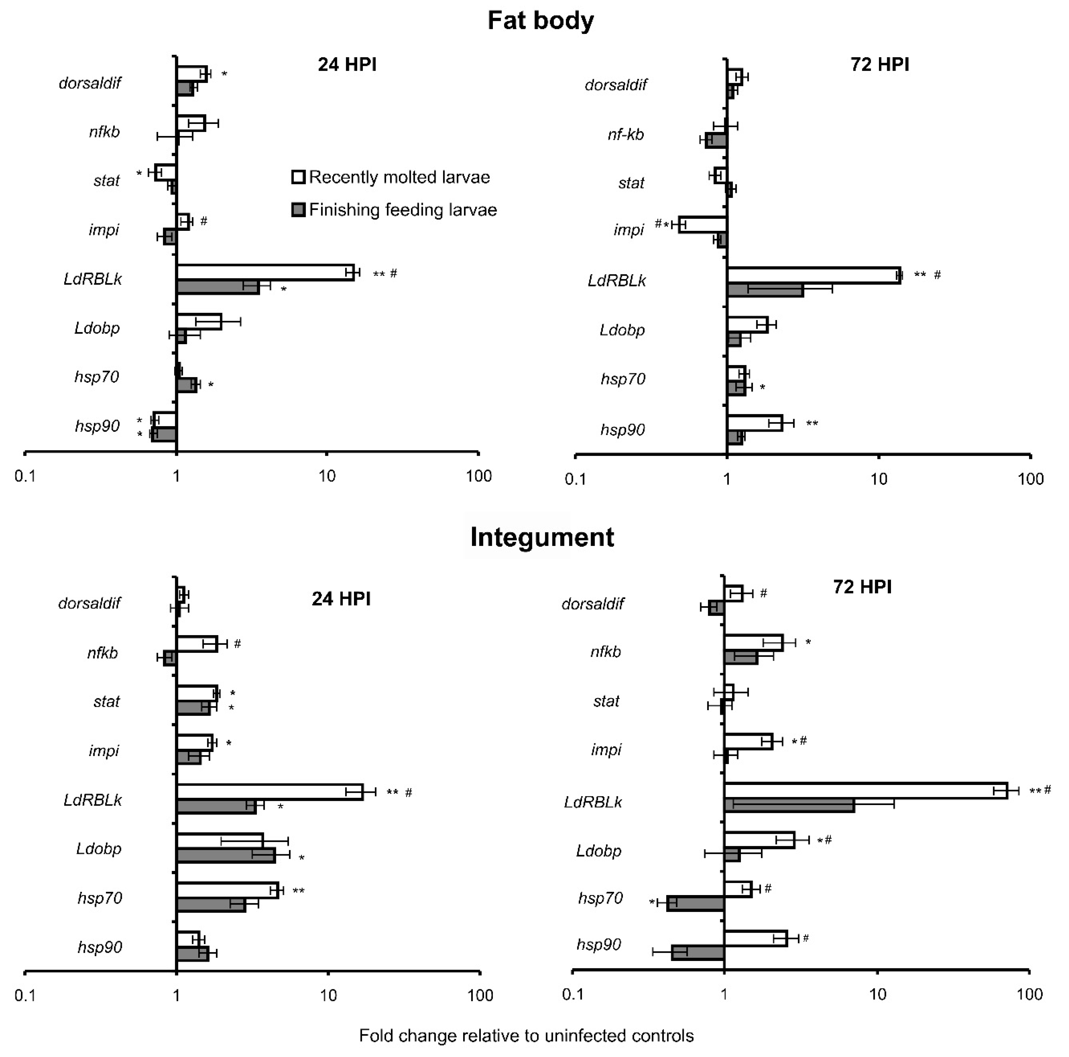
3.3. Alterations of Gene Expression in Response to M. robertsii Infection

Differential expression of the studied genes was registered after topical infection of CPB larvae with M. robertsii (Figure 3). More pronounced responses were detected in recently molted larvae compared to finishing feeding larvae and in the integument compared to the fat body.
Expression of genes of transcription factors dorsaldif, nfkb, and stat showed minor changes in response to M. robertsii infection; nonetheless, some of these alterations were significant (Figure 3). In particular, dorsaldif gene expression significantly increased in the fat body at the initial stage of mycosis (24 h) in recently molted larvae (1.6-fold relative to uninfected control). Nfkb gene expression was found to be significantly upregulated only in the integument of recently molted larvae at 72 h after infection (2.4-fold relative to control). Notably, nfkb was upregulated in the integuments of recently molted larvae compared to finishing feeding larvae only at 24 h after treatment, and the same effect was registered for dorsaldif at 72 h after treatment. The Stat gene tended to be slightly downregulated in the fat body at 24 h after infection initiation, with a significant decrease in recently molted larvae. By contrast, the stat gene was upregulated in the integument in response to infection (1.7–1.8-fold) in both recently molted and finishing feeding larvae at 24 h post-inoculation. At the letter stage of mycosis (72 h), there were no significant changes in stat expression in response to infection in the fat body and integument of both larval stages.
The impi gene was downregulated in the fat body at 72 h in response to infection in recently molted larvae but upregulated in the integument of the same larvae at both time points 24 and 72 h post-inoculation (1.7–2.1-fold relative to controls). There were no significant alterations of impi gene expression in finishing feeding larvae under the influence of the fungal infection in both tissues and at both time points.
The LdRBLk gene was dramatically upregulated in response to the infection in both tissues and more strongly in recently molted larvae. In particular, 14–15-fold upregulation in the fat body of recently molted larvae was observed at 24 and 72 h post-inoculation, whereas weaker (3.2–3.5-fold) upregulation was registered in finishing feeding larvae at these time points. In the integument, the LdRBLk gene was found to be upregulated 16-fold and 72-fold at 24 and 72 h, respectively, in recently molted larvae but only 3–7-fold at the same time points in finishing feeding larvae.
The Ldobp gene tended to be upregulated in response to the fungal infection. A significant increase in its expression was detected in the integument at 24 h after the inoculation in finishing feeding larvae (4.4-fold relative to control) and at 72 h after the inoculation in recently molted larvae (2.9-fold relative to control).
Hsp70 and hsp90 manifested complicated, not straightforward changes in expression, depending on larval stages, time after infection, tissue, and the genes. Hsp70 tended to be upregulated in the fat body in response to infection at both larval stages, but a significant increase was registered in finishing feeding larvae at 24 and 72 h (1.3–1.4-fold compared to control). In the integument, at 24 h after infection, the gene tended to be upregulated, and a significant increase was registered in recently molted larvae (4.6-fold relative to control). On the contrary, the gene was significantly downregulated at 72 h after infection in finishing feeding larvae (2.4-fold relative to control).
Hsp90 was significantly downregulated in the fat body at 24 h post-inoculation at both larval stages (1.4–1.5-fold relative to controls) but upregulated at 72 h after infection in recently molted larvae (2.3-fold relative to control). In the integument, hsp90 tended to be upregulated at 24 h post-inoculation at both larval stages, but the differences were not significant. At 72 h post-inoculation, hsp90 (just as hsp70) tended to be downregulated in response to infection in finishing feeding larvae, but the differences were also not significant.

4. Discussion

This study shows that during the intermolt period of CPB last-instar larvae, there is an increase in the expression of genes related to Toll, IMD, and Jak–Stat immune signaling pathways as well as to IMPIs, OBPs, and HSPs in the fat body and/or integument. These findings coincided with an increase in resistance to the fungal pathogen M. robertsii and are also consistent with previously obtained data on the enhancement of cellular immunity and cuticular defenses during this period [8]. In response to a fungal infection, most of the studied genes changed expression (up- or downregulation). Larvae that were more susceptible to the fungus (recently molted) showed more pronounced gene expression than less susceptible larvae (finishing feeding), apparently due to thin and weakly sclerotized integuments. In addition, changes in expression in response to the infection were overall more pronounced in the integument than in the fat body.
The expression of transcription factors of Toll and IMD pathways (dorsaldif and nfkb) increased during the intermolt period, especially in the fat body. The role of the Toll pathway in the defense against fungal infections is well known (reviewed by [25]), although it is not known which antimicrobial peptides are the effectors of this pathway in the CPB. Notably, the Toll pathway may also control hemocyte proliferation and encapsulation [26,27]. This observation is consistent with the increase in hemocyte density and in encapsulation activity during this intermolt period, as shown in our previous study [8]. Upregulation of transcription factors of the IMD pathway in response to fungal infections has been demonstrated in dipteran insects [11,28,29]. Notably, IMD transcription factor Rel2 is upregulated in antibiotic-treated insects in response to Beauveria and Isaria infections [11], probably indicating a specific response to a fungal infection but not to bacteria involved in the pathology. In our experiments, the upregulation of dorsaldif and nfkb in response to M. robertsii infection was rather weak (less than 2.4-fold relative to uninfected larvae) and was detected only in susceptible (recently molted) larvae. Nevertheless, we previously observed a significant 5–10-fold increase in the expression of these transcription factors in CPB larvae in response to topical infection with another fungus, B. bassiana (unpublished [30]). This fungus is the second most abundant species after the Metarhizium species in the soils of potato agrosystems [31]. Moreover, Beauveria is a leader in terms of the frequency of fungal infections in CPB populations [5,32]. We have also registered manifold stronger expression of the attacin gene (antimicrobial peptide of the IMD pathway) in the integument of CPB larvae in response to B. bassiana as compared to M. robertsii [14]. Probably, the level of dorsaldif and nfkb expression in CPB larvae depends on the fungus species and is more sensitive to the common pathogen B. bassiana.
A transcription factor of the Jak–Stat pathway (stat) showed an increase in expression in both tested tissues during the intermolt period in uninfected larvae as well as an increase in expression in the integument at the initial stages of M. robertsii infection (24 h post-inoculation). It has been shown that Jak–Stat cascade genes are expressed in response to fungal infections in lepidopterans and dipterans [10,11,28,33], and inhibition of the Jak–Stat pathway enhances the susceptibility of these insects to B. bassiana [10,33]. Accordingly, we can hypothesize that the overexpression of dorsaldif, nfkb, and stat during the intermolt period is associated with the formation of defenses against the fungal pathogens that are widespread in soil. It should be noted that during the last instar, the level of the 20-hydroxyecdysone titer increases, which enhances the activity of immuno-signaling cascades, and the production of antimicrobial peptides, as shown on Helicoverpa armigera [34]. Therefore, future studies may focus on the effect of 20-hydroxyecdysone on the induction of immunity in the Colorado potato beetle
The impi gene also increased expression at the end of the intermolt period in uninfected larvae. In response to the fungal infection, impi was upregulated mainly in the integument of susceptible (recently molted) larvae. It is known that IMPIs are produced as a reaction to fungal metalloproteinases, which contribute to the degradation of insect cuticle proteins [13]. It was shown on Galleria mellonella larvae that RNA interference-based silencing of impi leads to strong downregulation of its expression in the integument and less strong downregulation in the fat body, concurrently with an increase in susceptibility of the larvae to Metarhizium brunneum after topical infection [35]. In the present study, an increase in impi gene expression after the infection in recently molted larvae coincided with more severe damage and stronger melanization of their cuticle as compared to finishing feeding larvae [8]. The downregulation of impi in the fat body of the recently molted larvae is difficult to explain and may be related to the prioritization of immune responses between different tissues.
Expression of the gene encoding lectin LdRBLk did not change during the development of the insects under study but went up in response to the fungal infection in both tissues. The latter finding is in agreement with the results obtained earlier when the CPB was infected with M. robertsii and B. bassiana [14]. It is likely that this protein is inducible precisely in response to fungal infections but shows relatively stable basal expression in ontogeny. The strongest expression was observed here in susceptible (recently molted) larvae, especially in the integument. Thus, the expression of LdRBLk was symptomatic and corresponded to the level of lethality. It was previously reported that the structure of this protein allows it to have antifungal properties [14]. In particular, LdRBLk contains β-sheets that may interact with a fungal surface according to the carpet model [36], thereby resulting in destruction of the phospholipid bilayer of the membrane, followed by cell lysis.
We documented a significant increase in Ldobp gene expression during the intermolt period as well as its upregulation after the initiation of fungal infection. Wei et al. [16] registered upregulation of the obp gene mainly at 24–72 h post-inoculation by B. bassiana in the whole body of ants Solenopsis invicta. Those authors believe that OBPs are expressed mostly during the penetration of the cuticle by fungi. We also observed a significant increase in Ldobp expression only in the integument at 24 and 72 h post-inoculation. At this time point, Metarhizium usually does not yet circulate in the hemolymph of CPB larvae but is located in the integument and in encapsulated form under it [37]. It is noteworthy that in the larvae that are able to inactivate the fungal infection (finishing feeding), an increase in obp expression was observed at 24 h, whereas by 72 h, there was a decline to the control level. This result suggests that LdOBP can be considered a protein that exerts antifungal effects. This polypeptide contains the general odorant-binding protein domain (GOBP) [38]. Analysis of secondary structure of OBP from L. decemlineata by the self-optimized prediction method with alignment (SOPMA) revealed that it is a transmembrane polypeptide of 120 amino acid residues (aa) containing two transmembrane regions located within regions “aa 5–16” and “aa 54–56” (Figures S1 and S2). In these regions, the secondary structure of the protein folding is represented by a β-bridge, while the rest of the protein is folded into an α-helix interspersed by a random coil, consistent with previously described secondary structures of OBPs of insects [39]. This structure of the protein theoretically allows it to become integrated into the cell membrane of both insects and pathogens.
The expression of genes hsp70 and hsp90 increased at the end of larval development, but only in the integument. In response to the fungal infection, the genes also tended to be upregulated in the integument: in actively diseased (recently molted) larvae at 24 and 72 h post-inoculation and in resistant (finishing feeding) larvae only at 24 h post-inoculation. Previously, researchers also observed hsps upregulation as a reaction to fungal infections. For example, overexpression of hsp90 has been recorded in G. mellonella in response to B. bassiana infection [40] and Conidiobolus coronatus infections [19]. In CPB larvae, there are trends toward an increase in hsp90 expression in different tissues under the influence of M. robertsii and B. bassiana infections [14,41]. It is possible that induction of hsps expression in the integument during the intermolt period can enhance the defense against fungal infections because HSPs perform many general vitally important functions such as protein folding, unfolding, and localization, along with signaling and cell homeostasis [18]. Nonetheless, additional research is required to establish the role of HSPs in CPB ontogeny and immune responses.

5. Conclusions

This work uncovered changes in the expression of immune-system- and stress-related genes in CPB larvae during the intermolt period of last-instar larvae. Significant overexpression in the fat body and/or integument by the end of larval development was observed for transcription factors of immune signaling pathways (dorsaldif, nfkb, and stat) as well as for genes impi, Ldobp, hsp70, and hsp90 but not for the gene encoding β-lectin, LdRBLk. In addition, most of these genes underwent an alteration of expression after topical application of M. robertsii, mainly upregulation. At the same time, the changes in the integuments were more pronounced than in the fat body. We propose that the higher expression of immunity- and stress-related genes by the end of the intermolt period is an adaptation associated with the migration of larvae into soil, where there is a high risk of contact with entomopathogenic fungi. This notion is in agreement with the enhancement of cellular immunity and integumental defense in CPB larvae during this period [8]. Future research projects may focus on silencing of the analyzed genes, thereby providing an opportunity to enhance CPB susceptibility to native or introduced pathogens in potato agrosystems.

Supplementary Materials

The following are available online at https://www.mdpi.com/article/10.3390/insects13121168/s1, Figure S1: The predicted secondary structure of L. decemlineata OBP (LdOBP) by Self-Optimized Prediction Method with Alignment (SOPMA) method [42], Figure S2: The predicted transmembrane regions of L. decemlineata OBP (LdOBP) by the Dense Alignment Surface (DAS) method [43].

Author Contributions

Conceptualization, V.Y.K. and U.N.R.; methodology, U.N.R., V.Y.K. and V.V.G.; formal analysis, V.Y.K. and U.N.R.; investigation U.N.R., Y.A.N., O.N.Y. and V.Y.K.: resources, V.V.G., V.Y.K. and Y.A.N.; writing—original draft preparation, V.Y.K. and U.N.R.; writing—review and editing, V.Y.K., U.N.R., Y.A.N. and V.V.G.; funding acquisition, V.Y.K. and V.V.G.; visualization, V.Y.K. All authors have read and agreed to the published version of the manuscript.

Funding

This research was supported by Russian Science Foundation project no. 22-14-00309.

Institutional Review Board Statement

Not applicable.

Informed Consent Statement

Not applicable.

Data Availability Statement

The raw data of this article will be made available by the authors, without restrictions.

Acknowledgments

We are grateful to Vladimir Shilo for providing the equipment and for help with the collection of insects.

Conflicts of Interest

The authors declare no conflict of interest. The funders had no role in the design of the study; in the collection, analyses, or interpretation of the data; in the writing of the manuscript; or in the decision to publish the results.

References

  1. Critchlow, J.T.; Norris, A.; Tate, A.T. The legacy of larval infection on immunological dynamics over metamorphosis. Philos. Trans. R. Soc. B Biol. Sci. 2019, 374, 20190066. [Google Scholar] [CrossRef] [PubMed] [Green Version]
  2. Tate, A.T.; Graham, A.L. Dynamic Patterns of Parasitism and Immunity across Host Development Influence Optimal Strategies of Resource Allocation. Am. Nat. 2015, 186, 495–512. [Google Scholar] [CrossRef] [PubMed]
  3. Jaronski, S.T. Soil ecology of the entomopathogenic Ascomycetes: A critical examination of what we (think) we know. In Use of Entomopathogenic Fungi in Biological Pest Management; Ekesi, S., Maniania, N.K., Eds.; Research Signpost Inc.: Kerala, India, 2007; pp. 91–143. ISBN 978-81-308-0192-6. [Google Scholar]
  4. Scheepmaker, J.; Butt, T. Natural and released inoculum levels of entomopathogenic fungal biocontrol agents in soil in relation to risk assessment and in accordance with EU regulations. Biocontrol Sci. Technol. 2010, 20, 503–552. [Google Scholar] [CrossRef]
  5. Wraight, S.P.; Lacey, L.A.; Kabaluk, J.T.; Goettel, M.S. Potential for microbial biological control of coleopteran and hemipteran pests of potato. In Fruit, Vegetable and Cereal Science and Biotechnology 3 (Special Issue 1); Tennant, P., Benkeblia, N., Eds.; Global Science Book: Ikenobe, Japan, 2009; pp. 25–38. [Google Scholar]
  6. Weber, D.C.; Blackburn, M.B.; Jaronski, S.T. Biological and behavioral control of potato insect pests. In Insect Pests of Potato, 2nd ed.; Alyokhin, A., Rondon, S.I., Gao, Y., Eds.; Academic Press: Cambridge, MA, USA, 2022; pp. 231–276. [Google Scholar] [CrossRef]
  7. Alyokhin, A.; Kryukov, V. Ecology of a potato field. In Insect Pests of Potato, 2nd ed.; Alyokhin, A., Rondon, S.I., Gao, Y., Eds.; Academic Press: Cambridge, MA, USA, 2022; pp. 451–462. ISBN 9780128212370. [Google Scholar] [CrossRef]
  8. Tomilova, O.G.; Yaroslavtseva, O.N.; Ganina, M.D.; Tyurin, M.V.; Chernyak, E.I.; Senderskiy, I.V.; Noskov, Y.A.; Polenogova, O.V.; Akhanaev, Y.B.; Kryukov, V.Y.; et al. Changes in antifungal defence systems during the intermoult period in the Colorado potato beetle. J. Insect Physiol. 2019, 116, 106–117. [Google Scholar] [CrossRef] [PubMed]
  9. Butt, T.M.; Coates, C.J.; Dubovskiy, I.M.; Ratcliffe, N.A. Entomopathogenic fungi: New insights into host–pathogen interactions. In Advances in Genetics; Lovett, B., St. Leger, R.J., Eds.; Academic Press: Cambridge, MA, USA, 2016; Volume 94, pp. 307–364. ISBN 9780128046944. [Google Scholar]
  10. Geng, T.; Lv, D.-D.; Huang, Y.-X.; Hou, C.-X.; Qin, G.-X.; Guo, X.-J. JAK/STAT signaling pathway-mediated immune response in silkworm (Bombyx mori) challenged by Beauveria bassiana. Gene 2016, 595, 69–76. [Google Scholar] [CrossRef] [PubMed]
  11. Ramirez, J.L.; Muturi, E.J.; Barletta, A.B.; Rooney, A.P. The Aedes aegypti IMD pathway is a critical component of the mosquito antifungal immune response. Dev. Comp. Immunol. 2018, 95, 1–9. [Google Scholar] [CrossRef] [PubMed]
  12. Wei, G.; Lai, Y.; Wang, G.; Chen, H.; Li, F.; Wang, S. Insect pathogenic fungus interacts with the gut microbiota to accelerate mosquito mortality. Proc. Natl. Acad. Sci. USA 2017, 114, 5994–5999. [Google Scholar] [CrossRef] [PubMed] [Green Version]
  13. Vilcinskas, A. Coevolution between pathogen-derived proteinases and proteinase inhibitors of host insects. Virulence 2010, 1, 206–214. [Google Scholar] [CrossRef] [PubMed]
  14. Rotskaya, U.; Kryukov, V.; Kosman, E.; Tyurin, M.; Glupov, V. Identification of the Ricin-B-Lectin LdRBLk in the Colorado Potato Beetle and an Analysis of Its Expression in Response to Fungal Infections. J. Fungi 2021, 7, 364. [Google Scholar] [CrossRef]
  15. Scott, I.M.; Hatten, G.; Tuncer, Y.; Clarke, V.C.; Jurcic, K.; Yeung, K.K.-C. Proteomic Analyses Detect Higher Expression of C-Type Lectins in Imidacloprid-Resistant Colorado Potato Beetle Leptinotarsa decemlineata Say. Insects 2020, 12, 3. [Google Scholar] [CrossRef]
  16. Wei, Z.; Ortiz-Urquiza, A.; Keyhani, N.O. Altered Expression of Chemosensory and Odorant Binding Proteins in Response to Fungal Infection in the Red Imported Fire Ant, Solenopsis invicta. Front. Physiol. 2021, 12. [Google Scholar] [CrossRef] [PubMed]
  17. Kausar, S.; Abbas, M.N.; Yang, L.; Cui, H. Biotic and abiotic stress induces the expression of Hsp70/90 organizing protein gene in silkworm, Bombyx mori. Int. J. Biol. Macromol. 2020, 143, 610–618. [Google Scholar] [CrossRef] [PubMed]
  18. King, A.M.; MacRae, T.H. Insect Heat Shock Proteins During Stress and Diapause. Annu. Rev. Èntomol. 2015, 60, 59–75. [Google Scholar] [CrossRef] [PubMed]
  19. Wrońska, A.K.; Boguś, M.I. Heat shock proteins (HSP 90, 70, 60, and 27) in Galleria mellonella (Lepidoptera) hemolymph are affected by infection with Conidiobolus coronatus (Entomophthorales). PLoS ONE 2020, 15, e0228556. [Google Scholar] [CrossRef] [Green Version]
  20. Takov, D.; Ostoich, P.; Zubrik, M.; Pilarska, D. Coleopterans as model organisms in insect immunity: A review. North-West. J. Zool. 2022, 18, 104–111. [Google Scholar]
  21. Shi, X.-Q.; Guo, W.-C.; Wan, P.-J.; Zhou, L.-T.; Ren, X.-L.; Ahmat, T.; Fu, K.-Y.; Li, G.-Q. Validation of reference genes for expression analysis by quantitative real-time PCR in Leptinotarsa decemlineata (Say). BMC Res. Notes 2013, 6, 93. [Google Scholar] [CrossRef] [Green Version]
  22. National Library of Medicine. BLAST: Basic Local Alignment Search Tool. Available online: https://www.ncbi.nlm.nih.gov/tools/primer-blast/ (accessed on 13 December 2022).
  23. Integrated DNA Technologies. OligoAnalyzer Tool—Primer Analysis|IDT. Available online: https://eu.idtdna.com/pages/tools/oligoanalyzer (accessed on 13 December 2022).
  24. Hammer, Ø.; Harper, D.A.T.; Ryan, P.D. PAST: Paleontological statistics software package for education and data analysis. Palaeontol. Electron. 2001, 4, 1–9. [Google Scholar]
  25. Qu, S.; Wang, S. Interaction of entomopathogenic fungi with the host immune system. Dev. Comp. Immunol. 2018, 83, 96–103. [Google Scholar] [CrossRef]
  26. Hultmark, D. Drosophila immunity: Paths and patterns. Curr. Opin. Immunol. 2003, 15, 12–19. [Google Scholar] [CrossRef]
  27. Sorrentino, R.P.; Melk, J.P.; Govind, S. Genetic Analysis of Contributions of Dorsal Group and JAK-Stat92E Pathway Genes to Larval Hemocyte Concentration and the Egg Encapsulation Response in Drosophila. Genetics 2004, 166, 1343–1356. [Google Scholar] [CrossRef]
  28. Ramirez, J.L.; Dunlap, C.A.; Muturi, E.J.; Barletta, A.B.F.; Rooney, A.P. Entomopathogenic fungal infection leads to temporospatial modulation of the mosquito immune system. PLoS Negl. Trop. Dis. 2018, 12, e0006433. [Google Scholar] [CrossRef] [PubMed] [Green Version]
  29. Ramirez, J.L.; Schumacher, M.K.; Ower, G.; Palmquist, D.E.; Juliano, S.A. Impacts of fungal entomopathogens on survival and immune responses of Aedes albopictus and Culex pipiens mosquitoes in the context of native Wolbachia infections. PLoS Negl. Trop. Dis. 2021, 15, e0009984. [Google Scholar] [CrossRef] [PubMed]
  30. Kryukov, V.Y.; Kosman, E.S. Institute of Systematics and Ecology of Animals, SB RAS, Novosibirsk, Russia. Unpublished work. 2022. Available online: http://reznikova.net/wp-content/uploads/2017/02/Reznikova_Insight2016-1.pdf (accessed on 13 December 2022).
  31. Tyurin, M.; Kabilov, M.; Smirnova, N.; Tomilova, O.; Yaroslavtseva, O.; Alikina, T.; Glupov, V.; Kryukov, V. Can Potato Plants Be Colonized with the Fungi Metarhizium and Beauveria under Their Natural Load in Agrosystems? Microorganisms 2021, 9, 1373. [Google Scholar] [CrossRef] [PubMed]
  32. Sikura, A.I.; Sikura, L.V. Entomopathogenous fungi, bacteria, protozoa, viroses. In Colorado Potato Beetle, Leptinotarsa decemlineata Say Phylogeny, Morphology, Physiology, Ecology, Adaptation, Natural Enemies; Nauka Publishers: Moscow, Russia, 1981; pp. 299–313. (In Russian) [Google Scholar]
  33. Dong, Y.; Morton, J.C.; Ramirez, J.L.; Souza-Neto, J.A.; Dimopoulos, G. The entomopathogenic fungus Beauveria bassiana activate toll and JAK-STAT pathway-controlled effector genes and anti-dengue activity in Aedes aegypti. Insect Biochem. Mol. Biol. 2012, 42, 126–132. [Google Scholar] [CrossRef] [PubMed] [Green Version]
  34. Wang, J.-L.; Chen, L.; Tang, L.; Zhao, H.-B.; Liu, X.-S.; Wang, Y.-F. 20-hydroxyecdysone transcriptionally regulates humoral immunity in the fat body of Helicoverpa armigera. Insect Mol. Biol. 2014, 23, 842–856. [Google Scholar] [CrossRef]
  35. Grizanova, E.V.; Coates, C.J.; Butt, T.M.; Dubovskiy, I.M. RNAi-mediated suppression of insect metalloprotease inhibitor (IMPI) enhances Galleria mellonella susceptibility to fungal infection. Dev. Comp. Immunol. 2021, 122, 104126. [Google Scholar] [CrossRef]
  36. Zhang, D.; Lu, Y.; Chen, H.; Wu, C.; Zhang, H.; Chen, L.; Chen, X. Antifungal peptides produced by actinomycetes and their biological activities against plant diseases. J. Antibiot. 2020, 73, 265–282. [Google Scholar] [CrossRef]
  37. Tomilova, O.G.; Kryukov, V.Y.; Duisembekov, B.A.; Yaroslavtseva, O.N.; Tyurin, M.V.; Kryukova, N.A.; Skorokhod, V.; Dubovskiy, I.M.; Glupov, V.V. Immune-physiological aspects of synergy between avermectins and the entomopathogenic fungus Metarhizium robertsii in Colorado potato beetle larvae. J. Invertebr. Pathol. 2016, 140, 8–15. [Google Scholar] [CrossRef]
  38. EMBL’s European Bioinformatics Institute (EMBL-EBI). Pheromone/general odorant binding protein (IPR006170)—Inter Pro. Available online: https://www.ebi.ac.uk/interpro/entry/InterPro/IPR006170/ (accessed on 13 December 2022).
  39. Gaubert, A.; Amigues, B.; Spinelli, S.; Cambillau, C. Structure of odorant binding proteins and chemosensory proteins determined by X-ray crystallography. In Methodsin Enzymology; Pelosi, P., Knoll, W., Eds.; Academic Press: Cambridge, MA, USA, 2020; Volume 642, pp. 151–167. ISBN 9780128211571. [Google Scholar]
  40. Dubovskiy, I.; Whitten, M.; Kryukov, V.; Yaroslavtseva, O.N.; Grizanova, E.; Greig, C.; Mukherjee, K.; Vilcinskas, A.; Mitkovets, P.V.; Glupov, V.V.; et al. More than a colour change: Insect melanism, disease resistance and fecundity. Proc. R. Soc. B Boil. Sci. 2013, 280, 20130584. [Google Scholar] [CrossRef] [Green Version]
  41. Kryukov, V.Y.; Rotskaya, U.; Yaroslavtseva, O.; Polenogova, O.; Kryukova, N.; Akhanaev, Y.; Krivopalov, A.; Alikina, T.; Vorontsova, Y.L.; Slepneva, I.; et al. Fungus Metarhizium robertsii and neurotoxic insecticide affect gut immunity and microbiota in Colorado potato beetles. Sci. Rep. 2021, 11, 1299. [Google Scholar] [CrossRef]
  42. Geourjon, C.; Deléage, G. SOPMA: Significant improvement in protein secondary structure prediction by consensus prediction from multiple alignments. Comput. Appl. Biosci. 1995, 11, 681–684. [Google Scholar] [CrossRef] [PubMed]
  43. Cserzo, M.; Wallin, E.; Simon, I.; von Heijne, G.; Elofsson, A. Prediction of transmembrane alpha-helices in procaryotic membrane proteins: The Dense Alignment Surface method. Protein Eng. 1997, 10, 673–676. [Google Scholar] [CrossRef] [PubMed]
Figure 1. Survival of CPB larvae at different stages after topical infection with M. robertsii (5 × 10⁶ conidia/mL). Recently molted larvae were infected at 2 h post-molt in instar IV. Finishing feeding larvae were infected at 86 h post-molt in instar IV. Different letters show significant differences in survival dynamics (log rank test, χ2 > 110.3, df = 1, p < 0.0001).
Insects 13 01168 g001
Figure 2. Changes in expression of immunity- and stress-related genes during the intermolt period in CPB larvae (instar IV, no infection). Data are presented as fold changes relative to uninfected larvae at 26 h post-molt. Genes rp18, arf2, and arf19 were used as references. * p < 0.05, ** p < 0.01, and *** p < 0.001 relative to larvae at 26 h post-molt (Dunn’s test).
Insects 13 01168 g002
Figure 3. Alterations of expression of immunity- and stress-related genes in different stages of CPB larvae at 24 h post-inoculation (HPI) and 72 HPI with M. robertsii (5 × 10⁶ conidia/mL). Data are presented as fold changes relative to an uninfected control in each stage at each time point. Genes rp18, arf2, and arf19 were used as references. * p < 0.05, ** p < 0.01 relative to uninfected controls (Dunn’s test). # Significant differences in expression between infected recently molted larvae and infected finishing feeding larvae (Dunn’s test, p < 0.05).
Insects 13 01168 g003
Table 1. The list and description of genes and primer sequences used in the qPCR.
Gene NameGenBank Accession NumberGene SymbolPrimer Sequence (5′→3′) *Product Size (bp)PCR Efficiency (± SD)
60S ribosomal protein L18XM_023172940.1rp18For TAGAATCCTCAAAGCAGGTGGC
Rev CTGGACCAAAGTGTT CACTGC
1331.98 ± 0.02
ADP-ribosylation factor-like protein 4KC190027.1arf2For GTGCTCGCGAACCATGTGA
Rev AAACCTCCAATCCCTCGTGAAG
1401.94 ± 0.01
ADP-ribosylation factor-like protein 1XM_023169879.1arf19For CGGTGCTGGTAAAACGACAATATT
Rev TGACCTCCCAAATCCCAAACTT
1351.98 ± 0.02
DorsalDif-like proteinXM_023158121.1dorsaldifFor TGTGCGAAAAGGTGGCTAAAG
Rev ACTTGGGAGGGTTGGAAGTC
941.95 ± 0.05
NF-kappaB-like proteinXM_023174540.1nfkbFor AAGCAGCGGTTTGATTCGTTC
Rev AACTCGTCCAAGTTCTCCAGG
1201.96 ± 0.06
Signal transducer and activator of transcription proteinXM_023165198.1statFor AGGAGCAGAACACAGGGTAC
Rev TTTGCCTGGGAATTCTGTTGAC
1401.94± 0.03
Inducible metalloproteinase inhibitor proteinXM_023167894.1impiFor GCACCCTGTACAAGACCGT
Rev CCGTGCGTAACCAGGTATACA
1381.94 ± 0.02
Ricin-like β-lectin kGEEF01084863.1LdRLBLkFor GAACTGTTGATTGTCGTCACCATG
Rev TGGAAAGTTTGGGAGATGGAACT
1401.96 ± 0.04
Odorant binding proteinXM_023171367.1LdobpFor AGGTGAATCATGAAGTGCATTGC
Rev GTGGTACCTCTCTGCTTCCTC
961.93 ± 0.02
Heat shock protein 70KC544268.1hsp70For ACCCCGAAGAAGTCAGCTC
Rev TCCAGGCCGTAAGCGATG
2141.96 ± 0.04
Heat shock protein 90KC556802.1hsp90For GGGTGTAGTCGACTCTGAAGAC
Rev AGAGCTCCTCAAACAGTTCCAA
1251.89 ± 0.01
* Primer sequences for Rp18, Arf2, and Arf19 were taken from Shi et al. [21] and modified by Ulyana N. Rotskaya; the other primers were designed by Ulyana N. Rotskaya.
Publisher’s Note: MDPI stays neutral with regard to jurisdictional claims in published maps and institutional affiliations.

