Article

Bmapaf-1 is Involved in the Response against BmNPV Infection by the Mitochondrial Apoptosis Pathway

1
Jiangsu Key Laboratory of Sericultural Biology and Biotechnology, School of Biotechnology, Jiangsu University of Science and Technology, Zhenjiang 212018, China
2
Key Laboratory of Silkworm and Mulberry Genetic Improvement, Ministry of Agriculture and Rural Affairs, Sericultural Research Institute, Chinese Academy of Agricultural Science, Zhenjiang 212018, China
*
Authors to whom correspondence should be addressed.
These authors contributed equally to this work.
Insects 2020, 11(9), 647; https://doi.org/10.3390/insects11090647
Submission received: 4 August 2020 / Revised: 8 September 2020 / Accepted: 10 September 2020 / Published: 22 September 2020
(This article belongs to the Special Issue Biological Control and Insect Pathology)

Abstract

:

Simple Summary

Apaf-1 is involved in the apoptosis pathway and Bmapaf-1 showed a significant response to BmNPV infection in our previous transcriptome data. In this study, the underlying mechanism of Bmapaf-1 in response to BmNPV infection was studied. To preliminarily determine the relationship of Bmapaf-1 with BmNPV, the expression pattern of Bmapaf-1 was analyzed in different tissues of differentially resistant silkworm strains following virus infection. To further define the role of Bmapaf-1 in BmNPV infection, the alteration of BmNPV infection in BmN cells and the expression patterns of Bmcas-Nc and Bmcas-1 were analyzed following knockdown and overexpression of Bmapaf-1 using siRNA and the pIZT/V5-His-mCherry insect vector, respectively. Furthermore, to analyze whether Bmapaf-1 is involved in BmNPV infection by apoptosis, the inducer NSC348884 and inhibitor Z-DEVD-FMK were used.

Abstract

Discovery of the anti-BmNPV (Bombyx mori nuclearpolyhedrovirus) silkworm strain suggests that some kind of antiviral molecular mechanism does exist but is still unclear. Apoptosis, as an innate part of the immune system, plays an important role in the response against pathogen infections and may be involved in the anti-BmNPV infection. Several candidate genes involved in the mitochondrial apoptosis pathway were identified from our previous study. Bombyx mori apoptosis protease-activating factor-1 (Bmapaf-1) was one of them, but the antiviral mechanism is still unclear. In this study, sequences of BmApaf-1 were characterized. It was found to contain a unique transposase_1 functional domain and share high CARD and NB-ARC domains with other species. Relatively high expression levels of Bmapaf-1 were found at key moments of embryonic development, metamorphosis, and reproductive development. Further, the significant difference in expression of Bmapaf-1 in different tissues following virus infection indicated its close relationship with BmNPV, which was further validated by RNAi and overexpression in BmN cells. Briefly, infection of budded virus with enhanced green fluorescent protein (BV-EGFP) was significantly inhibited at 72 h after overexpression of Bmapaf-1, which was confirmed after knockdown of Bmapaf-1 with siRNA. Moreover, the downstream genes of Bmapaf-1, including Bmnedd2-like caspase (BmNc) and Bmcaspase-1 (Bmcas-1), were upregulated after overexpression of Bmapaf-1 in BmN cells, which was consistent with the RNAi results. Furthermore, the phenomenon of Bmapaf-1 in response to BmNPV infection was determined to be related to apoptosis using the apoptosis inducer NSC348884 and inhibitor Z-DEVD-FMK. Therefore, Bmapaf-1 is involved in the response against BmNPV infection by the mitochondrial apoptosis pathway. This result provides valuable data for clarifying the anti-BmNPV mechanism of silkworms and breeding of resistant silkworm strains.

1. Introduction

Sericulture has existed in China for more than 5000 years and is the main income for farmers who rear silkworms. BmNPV is a double-stranded DNA virus that causes serious economic losses ever year. Many silkworm strains have been found to have a high resistance to BmNPV infection [1,2], but the molecular mechanism of silkworm resistance to BmNPV is still unclear. With the fast development of biotechnology, many new technologies have been used to study the antiviral mechanism of silkworms, such as the RNA-seq transcriptome [3,4], isobaric tag for relative and absolute quantification (iTRAQ), and label-free proteomics [5,6]. Plenty of candidate genes and proteins involved in the response to BmNPV have been identified, but the functions of most of them are still unknown and require further validation in viral infection.
Apoptosis, also called programmed cell death (PCD), is a physiological process in pluricellular organisms. A significant characteristic of apoptosis is the removal of unwanted and potentially dangerous cells [7], which has been widely reported to play an important role in defense against viral infection [8]. A relatively substantial amount of evidence shows that mitochondria are one of the major organelles involved in signal transduction and activation of cell death [9]. The death of stimulated cells by apoptosis is triggered by proteins released from the mitochondrial intermembrane space, such as cytochrome c (cytc), and this released protein interacts with Apaf-1 and caspase-9 to form the apoptosome [10,11,12]. This pathway is known as the mitochondrial apoptosis pathway, which plays a central role in regulating mammalian cell apoptosis [13]. Once cytc is released into the cytosol, it binds with Apaf-1, and this permits the binding of deoxyadenosine triphosphate (dATP) or adenosine triphosphate (ATP), triggering its oligomerization to form the apoptosome [14,15]. After recruitment of multiple procaspase-9 molecules, autoactivation will start. Additionally, executioner caspase-3 can be activated by cleaved caspase-9, and apoptosis can proceed [16,17].
In our previous RNA-seq transcriptome analysis, among the midgut of different silkworm resistant strains following BmNPV infection, several candidate genes belonging to the mitochondrial apoptotic pathway were identified to be significantly differentially expressed after virus infection, including Bmapaf-1, Bmcytc, Bmcas-1, and BmNc [3]. It is little wonder that Drosophila contains the canonical apoptosome protein Apaf-1 (also known as CED4) and appears to use it. Apaf-1 is similar to that which has been described for mammalian cells and can activate downstream effector caspases by combining with caspase-9 and cytc [18,19]. It also has a close relationship with viral infection; the activation of Apaf-1 and caspase-9 in immortalized human hepatocytes was reported to inhibit core protein expression of the hepatitis C virus [20]. However, the role of BmApaf-1 in silkworms is still unknown. In this study, we explored the role of Bmapaf-1 in response to BmNPV infection in order to clarify the mechanism of the silkworm response to BmNPV.
To further study the antiviral mechanism of Bmapaf-1 in response to BmNPV, the expression of Bmapaf-1 was knocked down and overexpressed using siRNA and the pIZT-His-mCherry vector, respectively. The variation of BmNPV was recorded and determined using fluorescence microscopy and RT-qPCR, respectively. Moreover, the relationship of Bmapaf-1 and apoptosis was analyzed using the apoptosis inducer, NSC348884, and inhibitor, Z-DEVD-FMK.

2. Materials and Methods

2.1. Silkworm and BmNPV

The susceptible strain, YeB, and the resistant strain, YeA, and p50 (no relationship with YeA and YeB) were maintained in the Key Laboratory of Sericulture, School of Life Sciences, Jiangsu University of Science and Technology University, Zhenjiang, China. The first three instars silkworm larvae were fed with fresh mulberry leaves at 26 ± 1 °C, 75 ± 5% relative humidity, and a 12-h day/night cycle. The rearing temperature was reduced to 24 ± 1 °C for the last two instars, and the other conditions were the same with the first three instar larvae.
Budded virus of BmNPV containing enhanced green fluorescent protein tag (BV-EGFP) was generously donated by Professor Xu-dong Tang and was kept in our laboratory. The EGFP gene was inserted into the plasmid pFASTbac1 using the BamH I-Xho I site to generate a recombinant viral vector to express the EGFP protein under the drive of the polyhedron promoter. The titer of BV-EGFP (pfu/mL) was calculated using the method as described in our previous study [21]. The culture containing BV-EGFP (1 × 10⁸ pfu/mL) was used to infect BmN cells between different groups, and the control group was treated with equal volume culture.

2.2. Bioinformatics Analysis

The coding sequence (CDS) and the deduced amino acid sequences of BmApaf-1 (ID: NP_001186937.1) were aligned using the DNAMAN 8.0 software (LynnonBiosoft, San Ramon, CA, USA). The conserved motif of BmApaf-1 amino acid was predicted using the SMART server (http://smart.embl-heidelberg.de/). The analysis of homologous sequences of Apaf-1 in different species was conducted using the BLASTP tool (http://www.ncbi.nlm.nih.gov/). Amino acid sequence of BmApaf-1 and its homologs among different species were aligned using the MEGA-X software with the MUSCLE module. A neighbor-joining tree was generated using MEGA-X software with a bootstrap of 1000 replications and the LG+G DNA/Protein model.

2.3. Sample Preparation, RNA Extraction, and cDNA Synthesis

Each of the first day of fifth instar larva of YeA and YeB was inoculated with 2 μL of culture containing BV-EGFP (1.0 × 10⁸ pfu/mL) and then reared under standard conditions. The samples were prepared after BV-EGFP infection at 48 h. The midgut, hemolymph, fat body, and malpighian tubule of larva were dissected and washed with DEPC water, and 30 samples of each tissue were mixed together to minimize individual genetic differences. The whole bodies of 30 larvae at different developmental stages were mixed. Each experiment was repeated using three biological replicates. All samples were quick-frozen using liquid nitrogen, and then stored at −80 °C until use.
The total RNA of different silkworm tissues and BmN cells was isolated using TRIzol Reagent (Invitrogen, California, USA) following the manufacturer’s instructions. The concentrations and purities of RNA were quantified using the NanoDrop 2000 spectrophotometer (Thermo Fisher Scientific, Waltham, MA, USA). The RNA integrity was verified using a 1% agarose gel for electrophoresis. The first strand cDNA was synthesized with 1.0 μg RNA using the PrimeScript TM RT reagent Kit with gDNA Eraser (Perfect Real Time; TaKaRa, Kusatsu, Japan) following the manufacturer’s instructions. The quality of the synthetic cDNA was verified using the reference gene, B. mori glyceraldehyde-3-phosphate dehydrogenase (BmGAPDH).

2.4. RT-qPCR

The expression level of genes in this study was analyzed using RT-qPCR. All primer sequences with high amplification efficiency (≥95%) are listed in Table 1. RT-qPCR reactions were prepared with the NovoStart ®SYBR qPCR SuperMix Plus (Novoprotein, Shanghai, China) following the manufacturer’s instructions. Briefly, each 15 μL of reaction volume contained 2.0 μL of cDNA, 7.5 μL of SYBR SuperMix, 0.6 μL of each gene-specific primer (0.4 μM), and 4.3 μL of ddH2O. Reactions were carried out using the LightCycler® 96 System (Roche, Basel, Switzerland). The thermal cycling program included an initial denaturation at 95 °C for 5 min, and then 40 cycles under 95 °C for 5 s and 60 °C for 31 s. All measurements were repeated in triplicate. The 2−△△Ct method was adopted to calculate the relative expression level, based on the method described by Livak et al. (2001). BmGAPDH was used as an internal control [22]. Differences among the three repeats were analyzed using the SPSS Statistics 20 software (IBM, Endicott, NY, USA) with the one-way ANOVA method. A p-value less than 0.05 was considered as statistically significant among different groups.

2.5. Synthesis of siRNA

To knockdown the expression of Bmapaf-1 in BmN cells, two specific siRNAs targeting sequences located in the functional domain of Bmapaf-1 were selected and designed using the method in a previous study [23]. The target DNA sequence was inserted behind the T7 promoter, and the siRNA oligos were synthesized by SUNYA Biotechnology (Zhejiang, China; Table 2). The siRNA oligos were used to synthesize the template, which could transcript into siRNA using an In Vitro Transcription T7 Kit (for siRNA synthesis; TaKaRa, Japan) according to the manufacturer’s instructions. The concentration and purity of siRNAs were detected by the NanoDrop 2000 spectrophotometer (Thermo Fisher Scientific, Waltham, MA, USA). The integrity of siRNAs was determined by 3% agarose gel electrophoresis. The newly synthesized siRNA with good quality was stored at −80 °C until use.

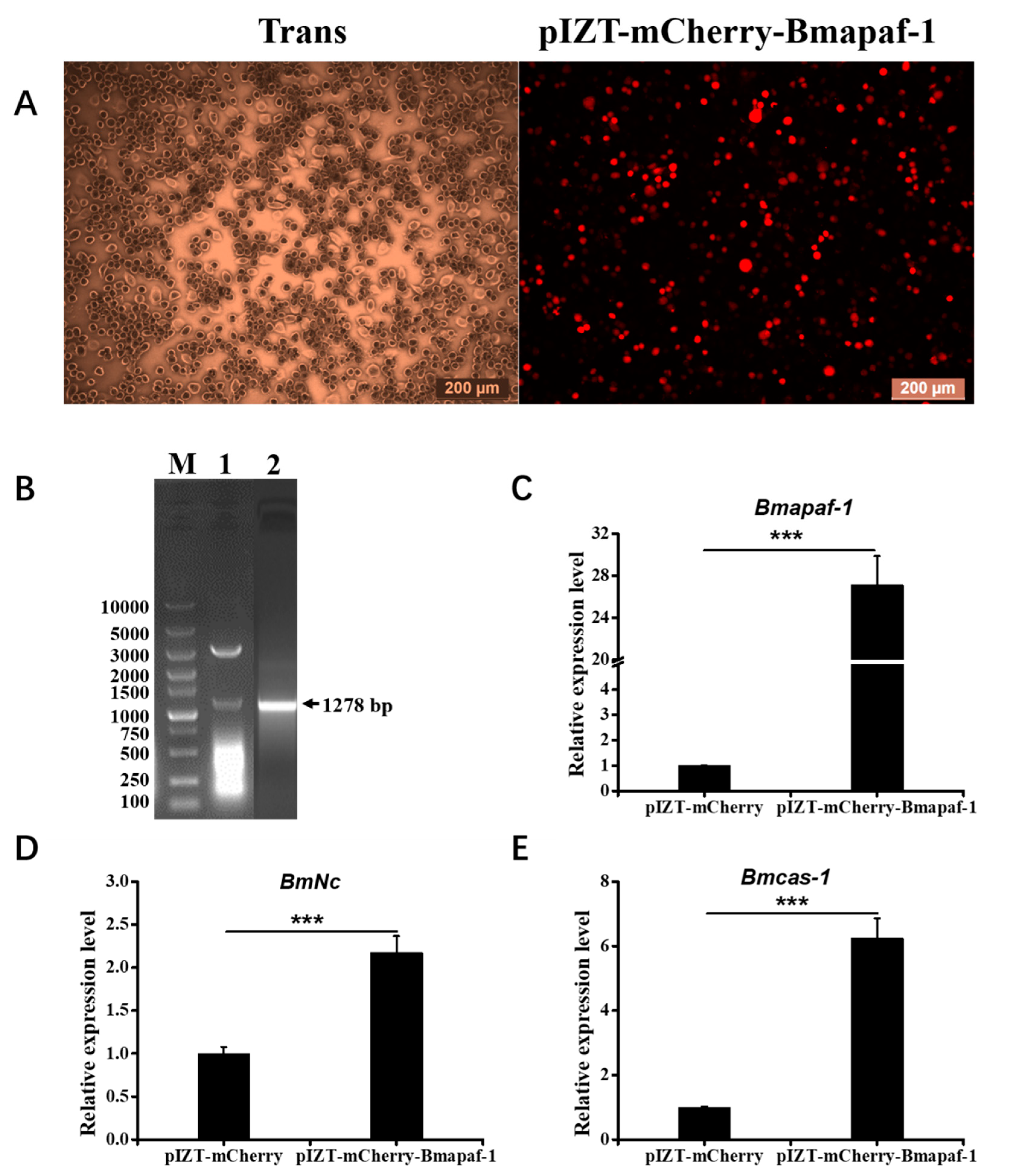
2.6. Construction of pIZT-mCherry-Bmapaf-1 Overexpression Vector

The functional domain of Bmapaf-1 was amplified from the cDNA of BmN cells with the Bmapaf-1KE primers (Table 1; the underline indicates Kpn I or EcoR I restriction sites). The purified PCR products were cloned into a pMD-19T vector for sequencing. The pMD-19T-Bmapaf-1 and the pIZT/V5-His-mCherry vector were digested with Kpn I and EcoR I (TaKaRa, Japan), and then ligated with T4 DNA ligase (TaKaRa, Kusatsu, Japan) at 16 °C overnight. Positive colonies were verified using PCR, and the recombinant expression vector pIZT/V5-His-mCherry-Bmapaf-1 was further verified by Kpn I and EcoR I digestion and sequenced by SUNYA Biotechnology (Zhejiang, China).

2.7. BmN Cell Culture, Transfection, and Fluorescence Signal Acquisition

The BmN cells derived from the silkworm ovary were cultured in TC-100 (AppliChem, Gatersleben, Germany) that contained 10% (v/v) fetal bovine serum (FBS; Thermo Fisher Scientific, Waltham, MA, USA) and 1% double-antibiotics (penicillin and streptomycin) at 28 °C [24]. The siRNA and overexpression vector of Bmapaf-1 were transfected into BmN cells using the Neofect TM DNA transfection reagent (NEOFECT, Beijing, China). Briefly, BmN cells were seeded into 30 mm culture flasks (approximately 1 × 10⁵ cells/well) before transfection. Then, 4.0 µg of siRNA or vector and 4.0 µL of transfection reagent were added successively into 200 µL of serum-free TC-100 to prepare the transfection solution, which was added into the culture medium to finish the transfection. The best efficiency was obtained at 24 h, and this time point was selected for all subsequent analysis.
The fluorescence signal in this study was captured at 24, 48, 72 h post-inoculation after knockdown or overexpression of Bmapaf-1 at 24 h using an inverted microscope DMi3000B camera (Leica, Solms, Germany), and the picture was processed with the Application Suite V4.6 software (Leica, Germany).

2.8. Inhibition and Induction of Apoptosis

Z-DEVD-FMK and NSC348884 reagents (Topscience; Beijing, China) were used for inhibition and induction of apoptosis, respectively. Both of these compounds were dissolved in DMSO to generate a 1 mM working solution. The final concentrations of 5 µM and 10 µM were selected for NSC348884 and Z-DEVD-FMK, respectively, based on the gradient concentration test on BmN cells. Inhibition and induction effects in this study were analyzed at 72 h after treatment with Z-DEVD-FMK and NSC348884.

3. Results

3.1. Characterization of the BmApaf-1 Sequence

The open reading fragment (ORF) of Bmapaf-1 (GenBank ID: NM_001200008.1) contained a complete 4302 bp, which encoded a 1433-amino acid protein. The theoretical isoelectric point (pI) and molecular weight (MW) were 7.04 and 16.30 kDa, respectively. BmApaf-1 protein contained three major functional domains: transposase_1, caspase recruitment domain (CARD), and a signaling motif NB-ARC (Figure S1).
BLASTP blast results showed that the amino acid sequence of BmApaf-1 was most similar to that of Bombyx mandarina (XP_028036457.1, 92.48% identity), followed by Manduca sexta (XP_030022507.1, 58.33%), Heliothis virescens (PCG76501.1, 52.45%), Trichoplusia ni (XP_026734098.1, 55.59%), Helicoverpa armigera (XP_021181657.1, 52.02%), and Papilio machaon (XP_014359375.1, 52.15%). A homologous alignment showed that a unique functional domain, transposase_1, was found in the BmApaf-1 amino acid sequence, compared to its homologs in other species, indicating BmApaf-1 might have an unknown and special role (Figure S1). Moreover, BmApaf-1 amino acid sequence shared high similarity in CARD and NB-ARC domains among different species; this indicated that BmApaf-1 might play an important role in the silkworm apoptosis pathway.
To determine the phylogenetic tree of Apaf-1 among different species, CDS sequences of BmApaf-1 and its homologs from other species were downloaded from NCBI. Accession numbers of BmApaf-1 homologs are listed in Table S1. A phylogenetic tree that contained BmApaf-1 and 15 other homologs was generated based on the DNA/protein model of JTT+G using MEGA X software (Figure S2). BmApaf-1 shared a low sequence similarity with its counterparts from Pieris rapae, Vanessa tameamea, and Bicyclus anynana, indicating that Apaf-1 might become divergent among Lepidoptera.

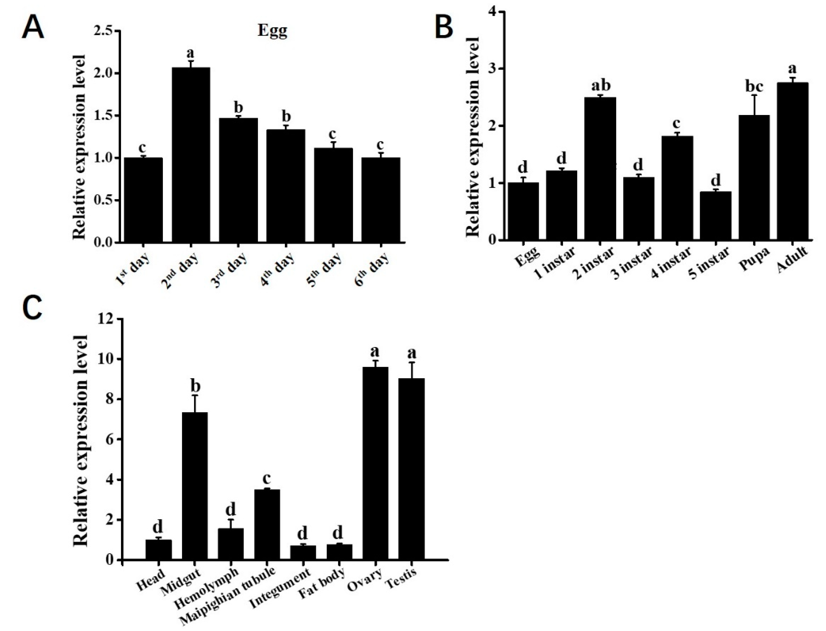
3.2. The Spatiotemporal Expression Pattern of Bmapaf-1

The silkworm p50 is a strain that is widely used in different laboratories, and its genome is available online. To preliminarily determine the biological function of Bmapaf-1, the relative expression level of Bmapaf-1 at different stages and different tissues of the p50 strain were detected by RT-qPCR. Among different development times of the egg, the highest expression level of Bmapaf-1 was found at the period when protuberance occurred (Figure 1A, 2nd day). Among the different development stages, the highest level was detected at the adult stage (Figure 1B), and the relatively high expression levels were observed in the testis and ovary (Figure 1C).

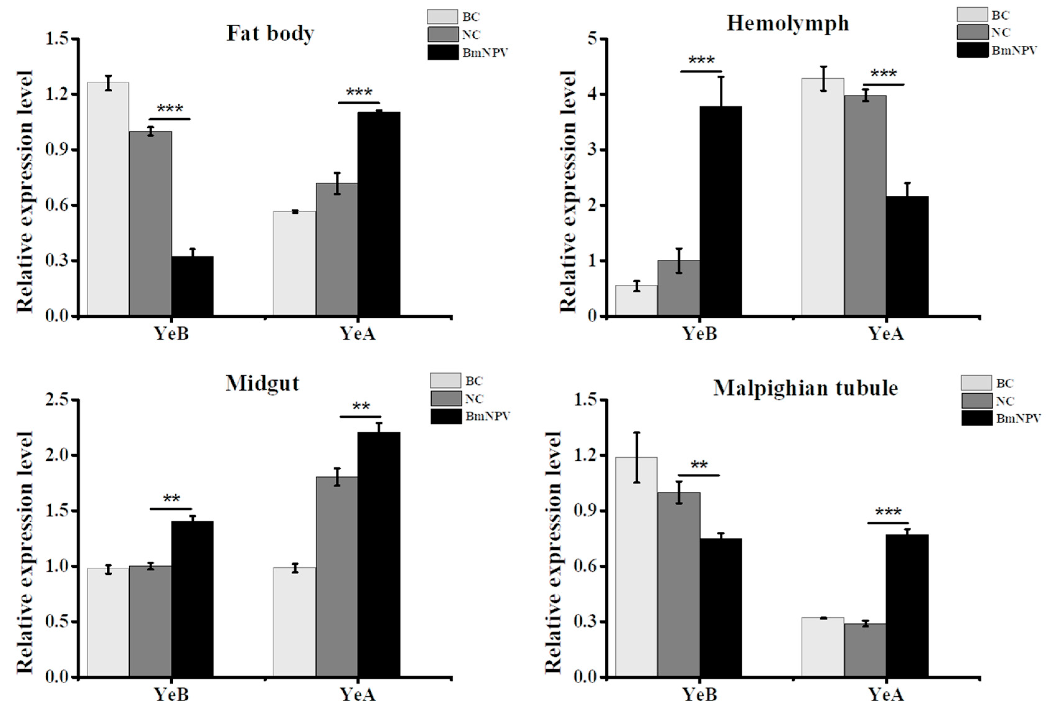
3.3. Bmapaf-1 Showed Significant Response to BmNPV Infection in Different Tissues

To preliminarily determine the relationship of Bmapaf-1 with BmNPV infection, the expression pattern of Bmapaf-1 was tested in different tissues of YeA (resistant strain) and YeB (susceptible strain) following BmNPV inoculation at 48 h, including the midgut, hemolymph, fat body, and malpighian tubule. The negative control is injected with BmN cell culture medium, and the blank control is given no injection. The resistant levels of YeA and YeB were tested in our previous report [2]. The medial lethal concentration (LC50) value of YeA was more than 109 OB/mL, but p50 was just about 105 OB/mL. Results showed that Bmapaf-1 had a reverse expression trend in the two different resistant strains, except in the midgut (Figure 2). Bmapaf-1 had a significantly higher expression level in selected tissues of YeA following virus infection, except in the hemolymph. Moreover, it had upregulated expression levels in the midgut and hemolymph of YeB following BmNPV infection and downregulated expression levels in the others (Figure 2). Generally, the significantly different expression levels of Bmapaf-1 in the two strains, following virus infection, indicated its vital role in response to BmNPV.

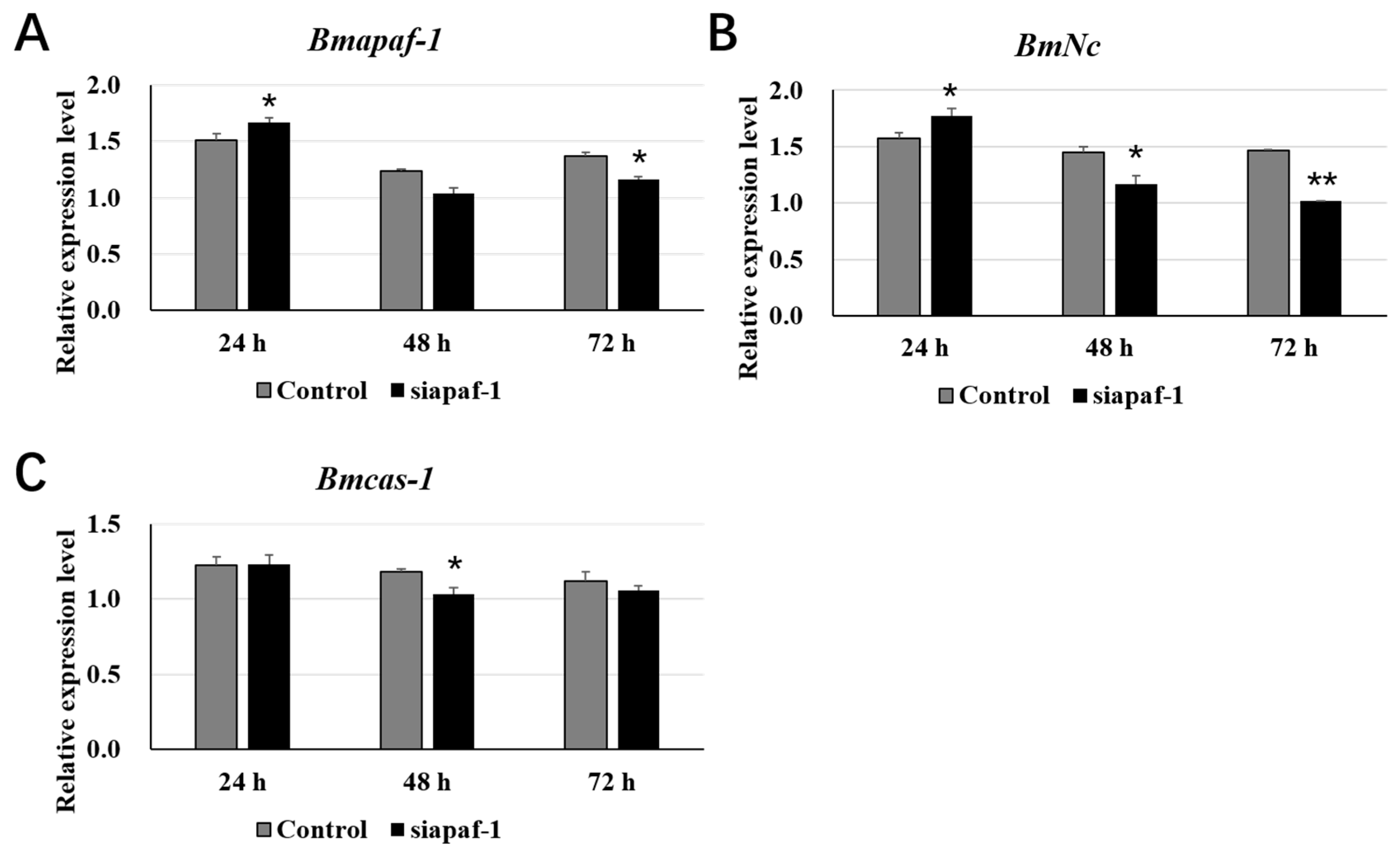
3.4. Selected Downstream Genes Were Downregulated after Knockdown of Bmapaf-1 in BmN Cells

To further study the role of Bmapaf-1 in response to BmNPV infection, two siRNAs targeting the functional domain of Bmapaf-1 were used to knockdown the expression of Bmapaf-1 in BmN cells. The preliminary experiment demonstrated that 4 μg of the siRNA targeting Bmapaf-1 (siapaf-1) was effective for interfering with the expression of Bmapaf-1 in BmN cells (data not shown). Expression levels of Bmapaf-1 were analyzed after transfecting with siapaf-1 at different times using RT-qPCR. The siRNA targeting red fluorescence protein (siRFP) was used as a negative control. Results showed that Bmapaf-1 was significantly downregulated after transfection with siapaf-1at 72 h, and this time point was selected for further analysis (Figure 3A).
In this study, two downstream genes of Bmapaf-1, i.e., BmNc and Bmcas-1, were selected by analyzing the mitochondrial apoptosis pathway of Drosophila. To get the relationship of Bmapaf-1 with BmNc and Bmcas-1, expression levels of the two genes were analyzed after transfection with siapaf-1 at different time points in BmN cells. BmNc showed a significant upregulation at 24 h post siapaf-1 transfection, and quickly downregulated at 48 and 72 h (Figure 3B). Bmcas-1 showed a significant downregulation at 48 h post siapaf-1 transfection (Figure 3C). These results indicated that Bmapaf-1 had a close relationship with the expression of BmNc and Bmcas-1.

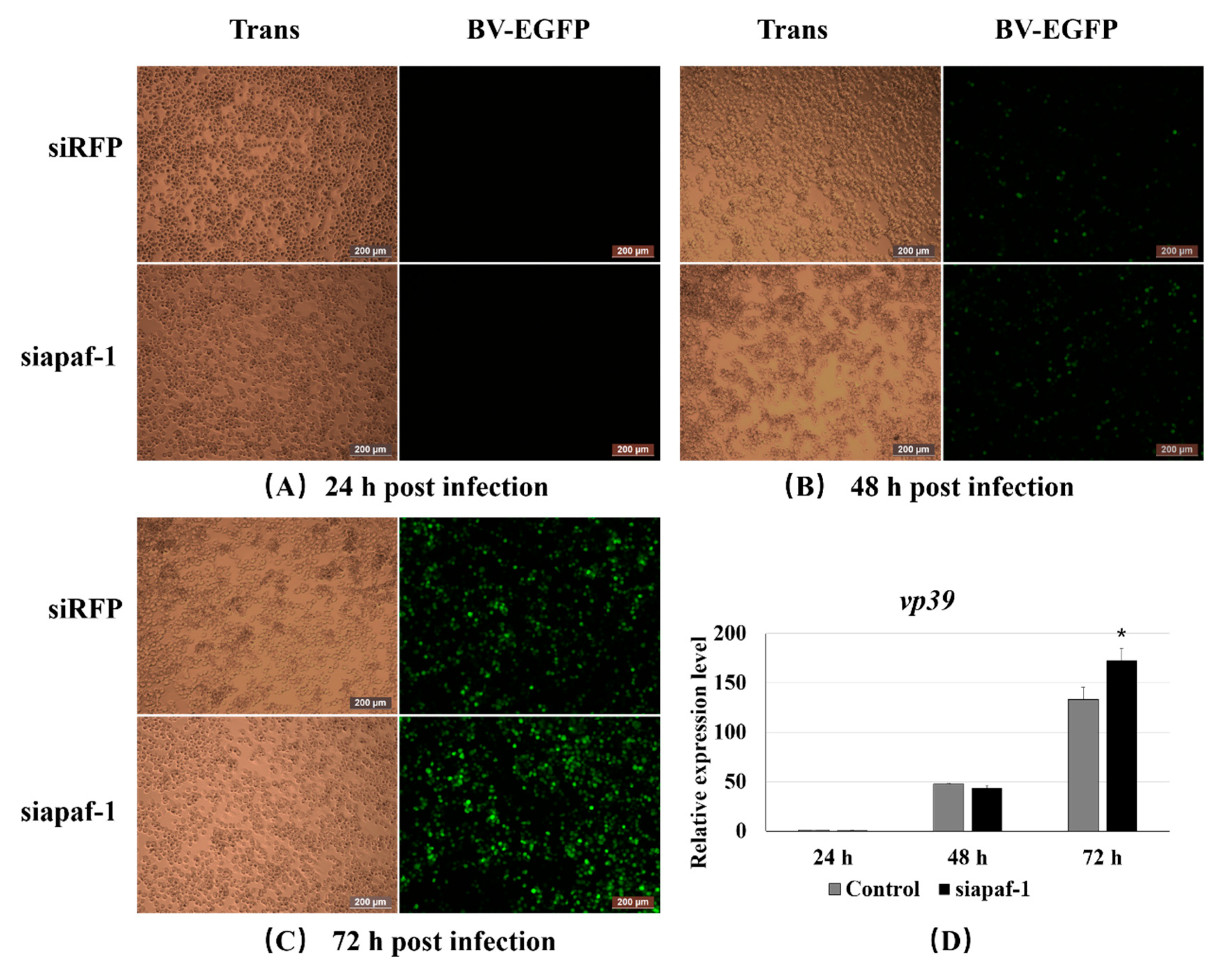
3.5. Knockdown of Bmapaf-1 Promoted BmNPV Infection in BmN Cells

To confirm the role of Bmapaf-1 during BmNPV infection, 20 μL of the culture medium containing BV-EGFP (1 × 10⁸ pfu/mL) was added into the BmN cells (30 mm dish) that had been transfected with siapaf-1 for 24 h. BmNPV infection signal was collected using fluorescence microscopy at 24, 48, and 72 h after inoculation with BV-EGFP and detected using RT-qPCR. The group transfected with siRFP was used as a negative control. The green fluorescence signal of the virus was significantly higher in the group treated with siapaf-1 at 72 h compared with the control (Figure 4C), but no difference was observed at 24 and 48 h (Figure 4A,B). To further validate the phenomenon, the capsid gene vp39 of BmNPV was used to determine viral replication in different groups using RT-qPCR. The expression level of vp39 was significantly higher in the siapaf-1 group than that in the control at 72 h (Figure 4D), but no significant difference was found at 24 and 48 h, which was consistent with the fluorescent images described above. These data indicated that Bmapaf-1 played an important role in response against BmNPV infection.

3.6. Overexpression of Bmapaf-1 Upregulated the Expression of Its Downstream Genes in BmN Cells

To overexpress Bmapaf-1 in BmN cells, a recombinant plasmid, pIZT-mCherry-Bmapaf-1, was constructed using double enzyme cutting (Figure 5B). The functional domain of Bmapaf-1 was inserted into the pIZT-mCherry vector between Kpn I and EcoR I. Subsequently, the recombinant plasmid was transfected into BmN cells using the Neofect TM DNA transfection reagent according to manufacturer’s instructions. The red fluorescence signal was observed using fluorescence microscope, and results indicated that pIZT-mCherry-Bmapaf-1 was transfected into BmN cells and successfully expressed (Figure 5A). It was also validated by the 30-fold upregulation of Bmapaf-1 in the transgenic BmN cell line using RT-qPCR, compared to the control group (Figure 5C).
To further validate the relationship of Bmapaf-1 with BmNc and Bmcas-1, expression levels of the two genes were determined between the transgenic BmN cell line and negative group using RT-qPCR. The expression level of BmNc in the transgenic cell line was more than two times higher than that in the control group (Figure 5D), and Bmcas-1 even reached a seven-fold enhancement in the transgenic cell line as compared with the negative group (Figure 5E). These results further validated the regulation relationship of Bmapaf-1 to BmNc and Bmcas-1.

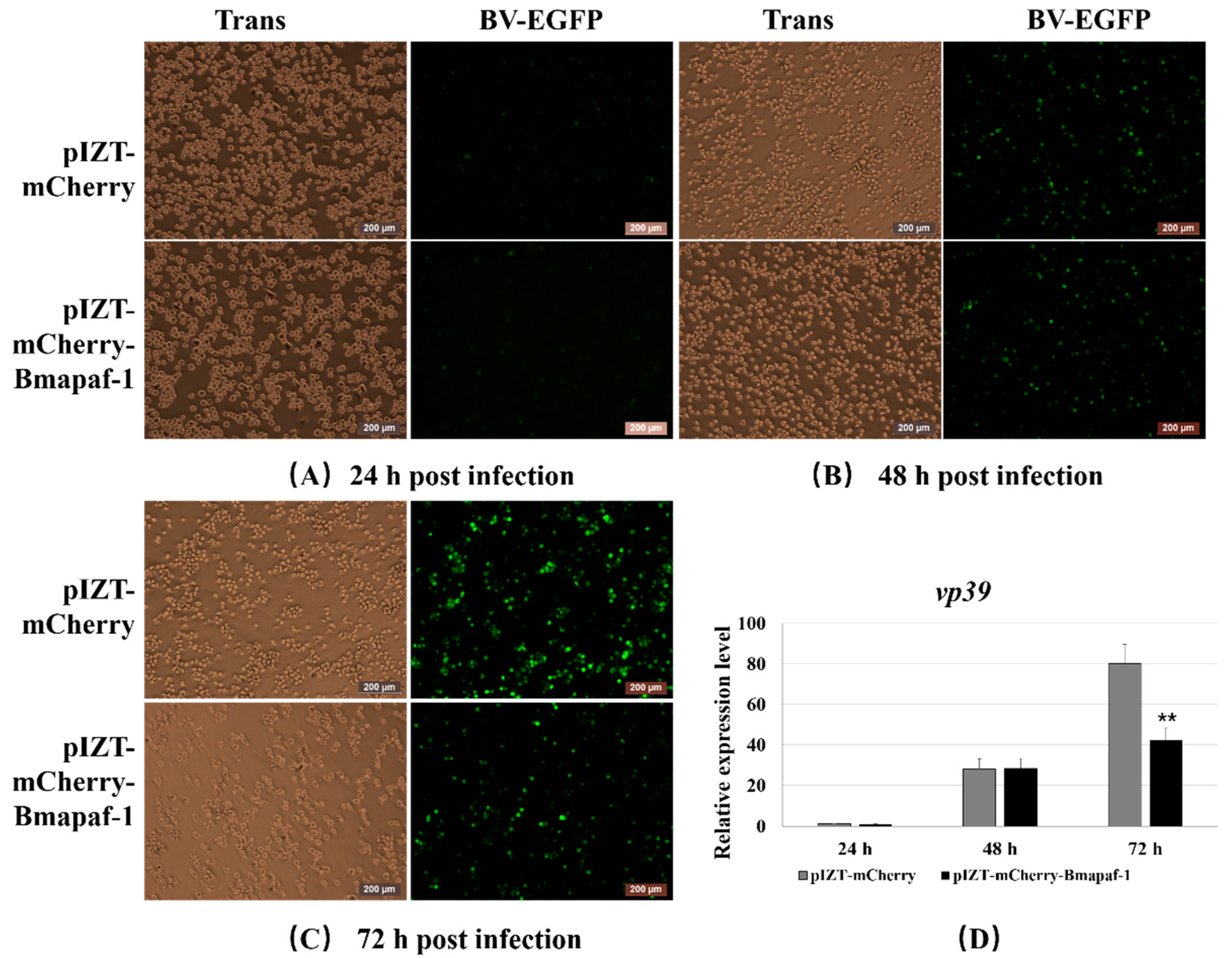
3.7. Overexpression of Bmapaf-1 Inhibited BmNPV Infection in BmN Cells

To further validate the role of Bmapaf-1 in response to BmNPV infection after RNAi, the variation of BmNPV infection was recorded in the transgenic cell line after inoculation with BV-EGFP using fluorescence microscopy at different time points. pIZT-mCherry was used as negative control. The green fluorescence signal of the virus was significantly lower in the transgenic cell line at 72 h post-inoculation compared with the negative control (Figure 6C), but no difference was observed at 24 and 48 h (Figure 6A,B). To further validate the result of Figure 6A–C, the capsid gene vp39 of BmNPV was analyzed to evaluate the viral replication in different groups using RT-qPCR. The expression level of vp39 was significantly downregulated in the transgenic cell line at 72 h compared to the control group (Figure 4D), but no significant difference was observed at 24 and 48 h, which was consistent with the fluorescent images described above. These data indicated that Bmapaf-1 played a vital role in anti-BmNPV infection.

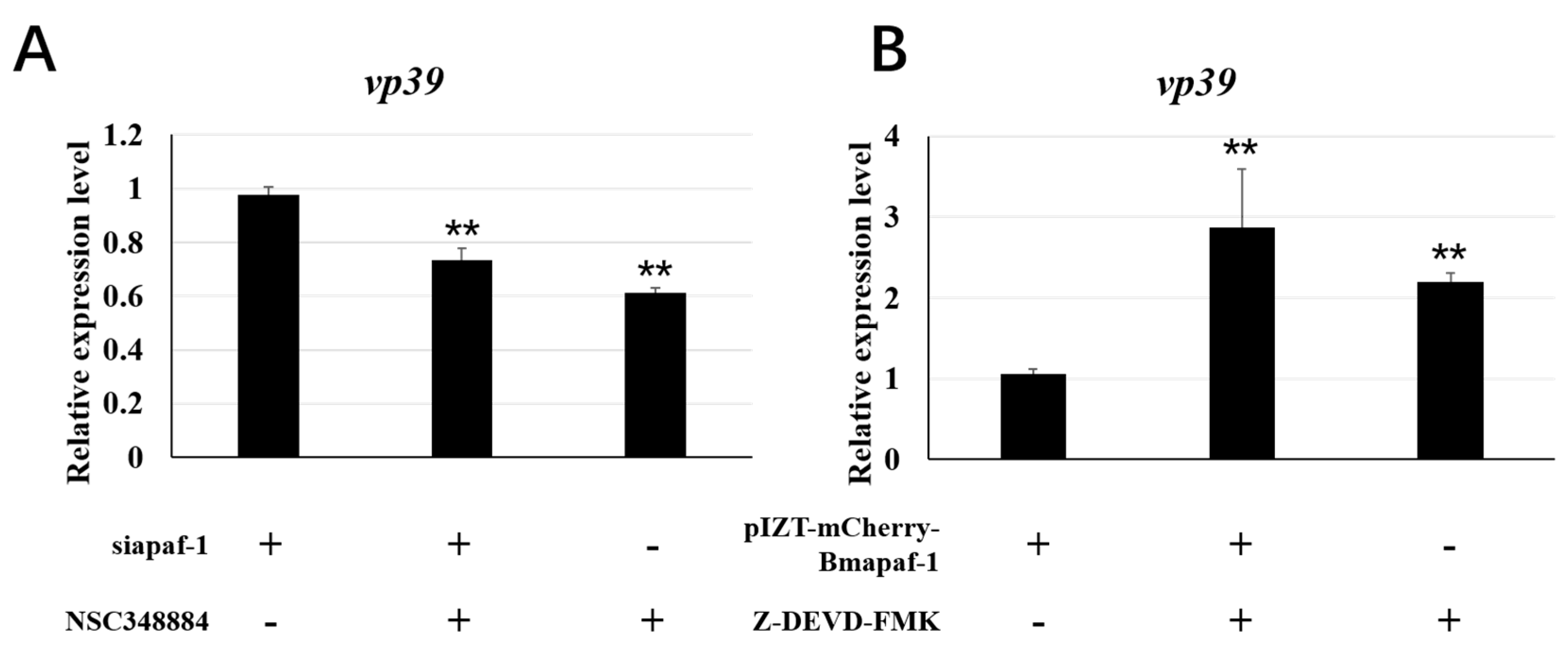
3.8. Apoptosis Regulated by Bmapaf-1 Involved in Response against BmNPV ISnfection
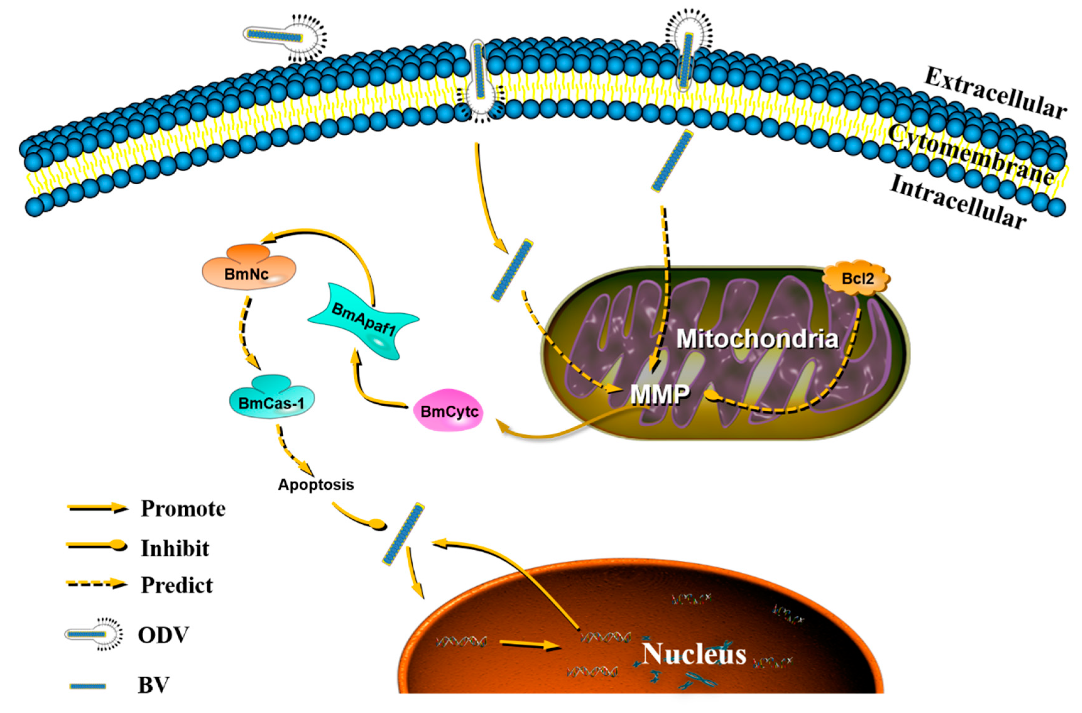
To analyze whether Bmapaf-1 was involved in BmNPV infection by regulating apoptosis, the variation of BmNPV replication was analyzed in the RNAi and overexpression group after treatment with the apoptosis inducer, NSC348884, and inhibitor, Z-DEVD-FMK, using RT-qPCR, respectively. BmNPV infection significantly increased after knockdown of Bmapaf-1 in BmN cells, as shown in Figure 4, but the infection of BV-EGFP was significantly inhibited in the groups treated with the inducer NSC348884 at 72 h after transfection with siapaf-1and siRFP (Figure 7A). Moreover, BmNPV infection was inhibited after overexpression of Bmapaf-1 in BmN cells, as shown in Figure 6, but the infection of BV-EGFP was significantly increased at 72 h after treatment with inhibitor Z-DEVD-FMK in the transgenic cell line and its negative control (Figure 7B). These results indicated that Bmapaf-1 was involved in the response against BmNPV infection by the apoptosis.

4. Discussions

BmNPV, one of the main silkworm pathogens, causes serious economic losses every year. However, the underlying molecular mechanism of silkworm in resistance to BmNPV infection remains unknown, even though many differentially resistant silkworm strains have been reported [2,25,26]. This suggests that there exists some kind of immune system in the silkworm organism, but this still needs further investigation. The antiviral mechanism of silkworms in response to the BmNPV infection is a complex process, and many candidate genes and proteins related to viral infection have been identified by high throughput techniques [3,6]. Clarification of these candidates’ functions will be useful to illustrate the mechanism. Bmapaf-1 was obtained from our previous transcriptome data of the midgut of two differentially resistant silkworm strains after feeding with BmNPV [3]. However, the role of Bmapaf-1 in the response against the BmNPV infection is still unknown.
Apoptosis is an effective immune pathway, leading to the self-destruction of the cell, playing a vital role in maintaining organism homeostasis. Furthermore, it also retains a highly evolutionarily conserved process in different species [27]. Diverse apoptotic stimuli, including viral infections, can trigger apoptosis through several apoptotic pathways, such as the mitochondrial apoptosis pathway [28]. The response mechanism of apoptosis might fit with other anti-viral mechanisms among different species based on the translational arrest [29]. Mitochondrial apoptosis has been reported to be involved in host defenses against environmental pressure by releasing Cytc into the cytoplasm to activate apoptosis, which also has been proven in silkworms in our previous report [25]. Moreover, BmCytc, released into the cytoplasm, could regulate its downstream genes to defend the BmNPV infection, including Bmapaf-1, BmNc, and Bmcas-1. To further confirm the apoptosis pathway, the function of Bmapaf-1 in response against BmNPV infection was determined in this study.
Bioinformatics analysis is a useful tool for predicting gene functional information; a relatively high conservation domain of BmApaf-1 in CARD and NB-ARC with selected other species indicated that it might play an important role in the silkworm apoptosis pathway (Figure S1). This was also found in the phylogenetic tree, with BmApaf-1 and 15 other homologs all belonging to Lepidoptera (Figure S2). Besides, a unique functional domain, transposase_1, in BmApaf-1 indicated that BmApaf-1 might have a special function, which still needs to be further analyzed (Figure S1). Furthermore, the relatively high expression level of Bmapaf-1 in the period of embryonic fast development indicated that it might be involved in embryonic development (Figure 1A). The significant high expression in pupa and adult revealed that it might be affected by ecdysone and was involved in the metamorphosis process (Figure 1B). Additionally, the significant high expression in the testis and ovary suggest potential roles in reproduction (Figure 1C).
In our previous transcriptome data, Bmapaf-1 was identified as a candidate antiviral gene for its differential expression after BmNPV infection, which was further validated by its significantly different expression in different tissues of two different resistant strains, YeA and YeB, after inoculation with BV-EGFP at 48 h (Figure 2). These results indicated that Bmapaf-1 had a close relationship with the virus infection. To analyze the role of Bmapaf-1 in the mitochondrial apoptosis pathway, its two downstream homologous genes in Drosophila were determined after knockdown and overexpression of Bmapaf-1 in BmN cells, that is, BmNc and Bmcas-1. The significant upregulation of BmNc and Bmcas-1 in the transgenic cell line showed that Bmapaf-1 could regulate the expression of the two genes (Figure 5), which was also validated after knockdown of Bmapaf-1 in BmN cells (Figure 3). Therefore, Bmapaf-1 is one of the upstream genes of BmNc and Bmcas-1 in the mitochondrial apoptosis pathway.
To further study the role of Bmapaf-1 in response to BmNPV infection, the variation of BmNPV was analyzed after knockdown and overexpression of Bmapaf-1 in BmN cells. Green fluorescence signals of BV-EGFP were significantly stronger at 72 h after knockdown of Bmapaf-1, compared to the control group (Figure 4A–C), but the effect was not observed at 24 h and 48 h, indicating knockdown of Bmapaf-1 could be beneficial for the virus infection. This phenomenon was also validated in the transgenic cell line (Figure 5A–C). Further, this phenomenon was also confirmed by the significantly higher expression level of vp39 in the RNAi group and the significantly lower expression level in transgenic group as compared with the control group (Figure 4D and Figure 6D). To further confirm whether this phenomenon was related to apoptosis, the apoptosis inducer NSC348884 and inhibitor Z-DEVD-FMK were used. The obvious downregulation of vp39 in the RNAi group and its negative control after treatment with the inducer NSC348884 at 72 h (Figure 7A) and the significant upregulation in the transgenic cell line and its negative control after treatment with the inhibitor Z-DEVD-FMK (Figure 7B) showed that Bmapaf-1 was involved in the antiviral infection by regulating the mitochondrial apoptosis pathway.
The significantly different resistance in silkworms in response against BmNPV infection indicates that there indeed exists some kind of immune response. Apoptosis, as one kind of innate immune system, plays a vital role in the response against the pathogen’s infection. Based on the results in this study and a previous one [25], as well as relevant reports, we speculated that mitochondria received some kind of signal as soon as budded viruses (BVs) entered into the host cell via clathrin-mediated endocytosis [30]. These signals can change the mitochondrial membrane potential, further changing permeability of the cell membrane [31], which is beneficial for the release of BmCytc into the cytoplasm [32]. Once BmCytc is released into the cytoplasm, it will combine with BmApaf-1 [25] to be an apoptotic complex that can promote the expression of downstream BmNc. After the activation of BmNc, its downstream protein BmCas-1 will be activated, and then regulate the process of apoptosis that can be used to respond to virus replication in BmN cells (Figure 8).

5. Conclusions

The significantly differential expression of Bmapaf-1 in different tissues of differential resistant strains following BmNPV infection verified the relationship between Bmapaf-1 and BmNPV. The anti-BmNPV function of Bmapaf-1 was confirmed by RNAi using the corresponding siRNA and overexpression using the pIZT/V5-His-mCherry insect vector harboring the Bmapaf-1 in vitro. Further results after treatment with apoptosis inducer, NSC348884, and inhibitor, Z-DEVD-FMK, showed Bmapaf-1 was involved in the response against BmNPV infection by the mitochondrial apoptosis pathway.

Supplementary Materials

The following are available online at https://www.mdpi.com/2075-4450/11/9/647/s1, Figure S1: Multiple sequence alignment of BmApaf-1 amino acids with its homologous proteins of other species. The deduced amino acid sequence of BmApaf-1 and its homologs in other species were retrieved from the NCBI database. Identical amino acids are highlighted in dark blue, and the positive amino acids are highlighted in pink and aqua. Figure S2: Phylogenetic analysis of BmApaf-1 using the neighbor-joining method. The tree was constructed with pairwise deletion of gaps in MEGA-X. The percentages on the branches indicate bootstrap values from 1000 replicates. The tree is drawn to scale, with branch lengths in the same units as those of the evolutionary distances used to infer the phylogenetic tree. The evolutionary distances were computed using the p-distance method. The analysis was included 16 amino acid sequences. All positions containing gaps and missing data were eliminated. Taxonomic names are shown on the right of the branches. Table S1. The list of accession numbers of proteins using in phylogenetic analysis.

Author Contributions

X.-y.W. conceived of and designed the experiments. X.-y.W., X.-y.D., Q.-y.C., K.-x.Z., and C.-x.Z. performed the experiments. X.-y.W. analyzed the data. X.-d.T., Y.-c.W., and M.-w.L. contributed reagents/materials/analysis tools. X.-y.W. wrote the paper. All authors have read and agreed to the published version of the manuscript.

Funding

This research received no external funding.

Acknowledgments

This work was supported by the National Natural Science Foundation of China, 31772523 and 31802137.

Conflicts of Interest

All authors declare no conflict of interest.

References

  1. Cheng, Y.; Wang, X.; Du, C.; Gao, J.; Xu, J. Expression Analysis of Several Antiviral Related Genes to BmNPV in Different Resistant Strains of Silkworm, Bombyx mori. J. Insect Sci. 2014, 14, 76. [Google Scholar] [CrossRef] [PubMed]
  2. Wang, X.Y.; Shao, Z.M.; Zhang, Y.J.; Vu, T.T.; Wu, Y.C.; Xu, J.P.; Deng, M.J. A (1)H NMR based study of hemolymph metabonomics in different resistant silkworms, Bombyx mori (Lepidotera), after BmNPV inoculation. J. Insect Physiol. 2019, 117, 103911. [Google Scholar] [CrossRef] [PubMed]
  3. Wang, X.Y.; Yu, H.Z.; Geng, L.; Xu, J.P.; Yu, D.; Zhang, S.Z.; Ma, Y.; Fei, D.Q. Comparative Transcriptome Analysis of Bombyx mori (Lepidoptera) Larval Midgut Response to BmNPV in Susceptible and Near-Isogenic Resistant Strains. PLoS ONE 2016, 11, e0155341. [Google Scholar] [CrossRef] [Green Version]
  4. Li, G.; Qian, H.; Luo, X.; Xu, P.; Yang, J.; Liu, M.; Xu, A. Transcriptomic analysis of resistant and susceptible Bombyx mori strains following BmNPV infection provides insights into the antiviral mechanisms. Int. J. Genom. 2016, 2016, 2086346. [Google Scholar]
  5. Yu, H.; Wang, X.; Xu, J.; Ma, Y.; Zhang, S.; Yu, D.; Fei, D.; Muhammad, A. iTRAQ-based quantitative proteomics analysis of molecular mechanisms associated with Bombyx mori (Lepidoptera) larval midgut response to BmNPV in susceptible and near-isogenic strains. J. Proteom. 2017, 165, 35–50. [Google Scholar] [CrossRef] [PubMed]
  6. Zhang, S.Z.; Wang, J.; Zhu, L.B.; Toufeeq, S.; Xu, X.; You, L.L.; Li, B.; Hu, P.; Xu, J.P. Quantitative label-free proteomic analysis reveals differentially expressed proteins in the digestive juice of resistant versus susceptible silkworm strains and their predicted impacts on BmNPV infection. J. Proteom. 2020, 210, 103527. [Google Scholar] [CrossRef]
  7. Smith, C.A.; Williams, G.T.; Kingston, R.; Jenkinson, E.J.; Owen, J.J.T. Apoptosis. Nature 1989, 338, 10. [Google Scholar] [CrossRef]
  8. Kvansakul, M. Viral Infection and Apoptosis. Viruses 2017, 9, 356. [Google Scholar] [CrossRef] [Green Version]
  9. Pradelli, L.A.; Bénéteau, M.; Ricci, J.-E. Mitochondrial control of caspase-dependent and -independent cell death. Cell. Mol. Life Sci. 2010, 67, 1589–1597. [Google Scholar] [CrossRef]
  10. Hakem, R.; Hakem, A.; Duncan, G.S.; Henderson, J.T.; Mak, T.W. Differential Requirement for Caspase 9 in Apoptotic Pathways In Vivo. Cell 1998, 94, 339–352. [Google Scholar] [CrossRef] [Green Version]
  11. Kuida, K.; Haydar, T.F.; Kuan, C.Y.; Gu, Y.; Flavell, R.A. Reduced Apoptosis and Cytochrome c–Mediated Caspase Activation in Mice Lacking Caspase 9. Cell 1998, 94, 325–337. [Google Scholar] [CrossRef] [Green Version]
  12. Li, K.; Li, Y.; Shelton, J.M.; Richardson, J.A.; Spencer, E.; Chen, Z.J.; Wang, X.; Williams, R.S. Cytochrome c Deficiency Causes Embryonic Lethality and Attenuates Stress-Induced Apoptosis. Cell 2000, 101, 389–399. [Google Scholar] [CrossRef] [Green Version]
  13. Clavier, A.; Rincheval-Arnold, A.; Colin, J.; Mignotte, B.; Guenal, I. Apoptosis in Drosophila: Which role for mitochondria? Apoptosis 2016, 21, 239–251. [Google Scholar] [CrossRef]
  14. Mohamad, N.; Gutierrez, A.; Nunez, M.; Cocca, C.; Martin, G.; Cricco, G.; Medina, V.; Rivera, E.; Bergoc, R. Mitochondrial apoptotic pathways. Biocell 2005, 29, 149–161. [Google Scholar] [CrossRef]
  15. Saleh, A. Negative regulation of the Apaf-1 apoptosome by Hsp70. Nat. Cell Biol. 2000, 2, 476–483. [Google Scholar] [CrossRef]
  16. Acehan, D.; Jiang, X.; Morgan, D.G.; Heuser, J.E.; Wang, X.; Akey, C.W. Three-Dimensional Structure of the Apoptosome: Implications for Assembly, Procaspase-9 Binding, and Activation. Mol. Cell 2002, 9, 423–432. [Google Scholar] [CrossRef]
  17. Adams, J.M.; Cory, S. Apoptosomes: Engines for caspase activation. Curr. Opin. Cell Biol. 2002, 14, 715. [Google Scholar] [CrossRef]
  18. Salvesen, G.S.; Abrams, J.M. Caspase activation-Stepping on the gas or releasing the brakes? Lessons from humans and flies. Oncogene 2004, 23, 2774–2784. [Google Scholar] [CrossRef] [Green Version]
  19. Wang, X. The expanding role of mitochondria in apoptosis. Genes Dev. 2001, 15, 2922–2933. [Google Scholar]
  20. Meyer, K.; Basu, A.; Saito, K.; Ray, R.B.; Ray, R. Inhibition of hepatitis C virus core protein expression in immortalized human hepatocytes induces cytochrome c-independent increase in Apaf-1 and caspase-9 activation for cell death. Virology 2005, 336, 198–207. [Google Scholar] [CrossRef] [Green Version]
  21. Wang, X.Y.; Shao, Z.M.; Chen, Q.Y.; Xu, J.P.; Sun, X.; Xu, Z.P.; Li, M.W.; Wu, Y.C. Knockdown of BmTCP-1beta Delays BmNPV Infection in vitro. Front. Microbiol. 2019, 10, 578. [Google Scholar] [CrossRef]
  22. Guo, H.; Jiang, L.; Xia, Q. Selection of reference genes for analysis of stress-responsive genes after challenge with viruses and temperature changes in the silkworm Bombyx mori. Mol. Genet. Genom. 2015, 291, 999–1004. [Google Scholar] [CrossRef]
  23. Yin, J.; Zhang, J.; Li, T.; Sun, X.; Li, M.-W. BmSd gene regulates the silkworm wing size by affecting the Hippo pathway. Insect Sci. 2019, 27, 655–664. [Google Scholar] [CrossRef]
  24. Ye, Y.; Liu, X.I.; Xiao, M.; Zhang, Z.I.; Li, M.A. A palmitoyltransferase Approximated gene Bm-app regulates wing development in Bombyx mori. Insect Sci. 2018, 27, 2–13. [Google Scholar]
  25. Wang, X.Y.; Wu, K.H.; Pang, H.L.; Xu, P.Z.; Li, M.W.; Zhang, G.Z. Study on the Role of Cytc in Response to BmNPV Infection in Silkworm, Bombyx mori (Lepidoptera). Int. J. Mol. Sci. 2019, 20, 4325. [Google Scholar] [CrossRef] [Green Version]
  26. Kang, L.; Shi, H.; Liu, X.; Zhang, C.; Yao, Q.; Wang, Y.; Chang, C.; Shi, J.; Cao, J.; Kong, J. Arginine kinase is highly expressed in a resistant strain of silkworm (Bombyx mori, Lepidoptera): Implication of its role in resistance to Bombyx mori nucleopolyhedrovirus. Comp. Biochem. Physiol. Part B Biochem. Mol. Biol. 2011, 158, 230–234. [Google Scholar] [CrossRef]
  27. Duprez, L.; Wirawan, E.; Berghe, T.V.; Vandenabeele, P. Major cell death pathways at a glance. Microbes Infect. 2009, 11, 1050–1062. [Google Scholar] [CrossRef]
  28. Wang, Q.; Ju, X.; Chen, L.; Chen, K. Caspase-1 from the silkworm, Bombyx mori, is involved in Bombyx mori nucleopolyhedrovirus infection. Z. Nat. C J. Biosci. 2016, 72, 147–153. [Google Scholar] [CrossRef]
  29. Nagamine, T.; Sako, Y. A Role for the Anti-Viral Host Defense Mechanism in the Phylogenetic Divergence in Baculovirus Evolution. PLoS ONE 2016, 11, e0156394. [Google Scholar] [CrossRef] [Green Version]
  30. Long, G.; Pan, X.Y.; Kormelink, R.; Vlak, J.M. Functional entry of baculovirus into insect and mammalian cells is dependent on clathrin-mediated endocytosis. J. Virol. 2006, 80, 8830–8833. [Google Scholar] [CrossRef] [Green Version]
  31. Sedlic, F.; Wakatsuki, T.; Pravdic, D.; Bosnjak, Z. Mitochondrial membrane potential regulates production of reactive oxygen species and opening of mitochondrial permeability transition pore. FASEB J. 2009, 23, 576. [Google Scholar]
  32. Pan, M.H.; Chen, M.; Huang, S.J.; Yu, Z.S.; Lu, C. Cloning and protein release of cytochrome c in the apoptosis cells of silkworm. Sci. Agric. Sin. 2009, 42, 2546–2551. [Google Scholar]
Figure 1. The spatiotemporal expression analysis of Bmapaf-1 using RT-qPCR. Relative expression levels of Bmapaf-1 among different egg development times (A); different developmental stages (B); and different tissues (C). 1st day, period of the longest embryo; 2nd day, period of protuberance occurred; 3rd day, prophase of protuberance rapid development; 4th day, period of shortening; 5th day, period of embryonic reversal; 6th day, head pigmentation period. BmGAPDH was used to normalize the data that were shown as the mean ± standard error, and the mean is from three independent repeats. The 2−△△Ct method was adopted to calculate the relative expression level. Differences among triple repeats were analyzed using the SPSS Statistics 20 software (IBM, USA) with the one-way ANOVA method. Different letters represented the significant difference (a, b, c; p < 0.05).
Insects 11 00647 g001
Figure 2. The analysis of Bmapaf-1 expression level in different tissues of different resistant strains following BmNPV infection using RT-qPCR. YeA was a resistant silkworm strain, and YeB was a susceptible silkworm strain. BC, blank control; NC, negative control. BmGAPDH was used to normalize the data that were shown as the mean ± standard error, and the mean was from three independent repeats. The 2−△△Ct method was adopted to calculate the relative expression level. Differences among triple repeats were analyzed using the SPSS Statistics 20 software (IBM, USA) with the one-way ANOVA method. Asterisks represented the significant difference, as follows: ** p < 0.01; *** p < 0.001.
Insects 11 00647 g002
Figure 3. Expression analysis of selected downstream genes after knockdown of Bmapaf-1 with siRNA at different time points using RT-qPCR. (A) Expression level analysis of Bmapaf-1 after transfection with siRNA at 24, 48, and 72 h. Analysis of BmNc (B) and Bmcas-1 (C) expression levels after knockdown of Bmapaf-1 at different time points. BmGAPDH was used to normalize the data that were showed as the mean ± standard error, and the mean was from three independent repeats. The 2−△△Ct method was adopted to calculate the relative expression level. Differences among triple repeats were analyzed using the SPSS Statistics 20 software (IBM, USA) with the one-way ANOVA method. Asterisks represent the significant difference, as follows: * p < 0.05; ** p < 0.01.
Insects 11 00647 g003
Figure 4. Analysis of BmNPV infection following knockdown of Bmapaf-1 at different times in BmN cells: 24 h (A); 48 h (B), and 72 h (C) after BV-EGFP infection. (D) The analysis of vp39 expression after knockdown of Bmapaf-1 at different times. Scale bar = 200 μm. Trans (white), optical transmission. EGFP (green), expressed following the replication of BV. BmGAPDH was used to normalize the data that were showed as the mean ± standard error, the mean was from three independent repeats. The 2−△△Ct method was adopted to calculate the relative expression level. Differences among triple repeats were analyzed using the SPSS Statistics 20 software (IBM, USA) with the one-way ANOVA method. Asterisks represent the significant difference, * p < 0.05.
Insects 11 00647 g004
Figure 5. Expression analysis of selected downstream genes after overexpression of Bmapaf-1 at different times in BmN cells. (A) Overexpression detection of Bmapaf-1 after transfection with pIZT-mCherry-Bmapaf-1 in BmN cells. Scale bar = 200 μm. Trans (white), optical transmission. mCherry (Red), fused expression with Bmapaf-1 protein. (B) The construction of pIZT-mCherry-Bmapaf-1: (1) validation of the recombinant vector using double enzyme digestion and (2) amplification of the functional domain of Bmapaf-1. (C) The expression level analysis of Bmapaf-1 after transfecting with recombinant vector using RT-qPCR. Expression level analysis of BmNc (D) and Bmcas-1 (E) after overexpression of Bmapaf-1. BmGAPDH was used to normalize the data that were shown as the mean ± standard error, and the mean was from three independent repeats. The 2−△△Ct method was adopted to calculate the relative expression level. Differences among triple repeats were analyzed using the SPSS Statistics 20 software (IBM, USA) with the one-way ANOVA method. Asterisks represent the significant difference, as follows: *** p < 0.001.
Insects 11 00647 g005
Figure 6. Analysis of BmNPV infection after overexpression of Bmapaf-1 at different times in BmN cells, 24 h (A), 48 h (B), and 72 h (C), after BV-EGFP infection. (D) The analysis of vp39 expression after overexpression of Bmapaf-1 at different times. Scale bar = 200 μm. Trans (white), optical transmission. EGFP (green), expressed following the replication of BV. BmGAPDH was used to normalize the data that were shown as the mean ± standard error, the mean was from three independent repeats. The 2−△△Ct method was adopted to calculate the relative expression level. Differences among triple repeats were analyzed using the SPSS Statistics 20 software (IBM, USA) with the one-way ANOVA method. Asterisks represent the significant difference, as follows: ** p < 0.01.
Insects 11 00647 g006
Figure 7. The analysis of BmNPV replication in the RNAi and overexpression group following regulation of apoptosis in BmN cells using RT-qPCR. (A) The replication of BmNPV in the RNAi group after treatment with inducer NSC348884. (B) The analysis of BmNPV replication in the transgenic BmN cell line following treatment with apoptosis inhibitor Z-DEVD-FMK. BmGAPDH was used to normalize the data that were shown as the mean ± standard error, the mean was from three independent repeats. The 2−△△Ct method was used to calculate the relative expression level. Differences among triple repeats were analyzed using the SPSS Statistics 20 software (IBM, USA) with the one-way ANOVA method. Asterisks represented the significant difference, as follows: ** p < 0.01.
Insects 11 00647 g007
Figure 8. The proposed function of BmApaf-1 in response against BmNPV infection by the mitochondrial apoptosis pathway. MMP, mitochondrial membrane potential.
Insects 11 00647 g008
Table 1. List of primers used in this study.
Genes NameForward Primers (5′-3′)Revers Primers (5′-3′)
Bmapaf-1TCACAACCCTCTAAAATCACACCAGCGACAGCCAGTAATGGGTGTATGAG
BmNcGAGGACGATGTGAGCAGGGATTTCAGCAGGAACGAAATGTAGC
Bmcas-1AACGGCAATGAAGACGAAGGGGTGCCCGTGCGAGATTTTA
BmGAPDHCCGCGTCCCTGTTGCTAATCTGCCTCCTTGACCTTTTGC
VP39CAACTTTTTGCGAAACGACTTGGCTACACCTCCACTTGCTT
Bmapaf-1 KEGGGGTACCAGGAAGCTGCTGCAGCACGGAATTCTATGTTTTCGACTTCGTTGAC
Table 2. Primers used to synthesize siRNA.
Primer NamesSequences (5′-3′)
Bmapaf-1-1 Olig-1GATCACTAATACGACTCACTATAGGGGCTAATCTGGTCATAGTTATT
Bmapaf-1-1 Olig-2AATAACTATGACCAGATTAGCCCCTATAGTGAGTCGTATTAGTGATC
Bmapaf-1-1 Olig-3AAGCTAATCTGGTCATAGTTACCCTATAGTGAGTCGTATTAGTGATC
Bmapaf-1-1 Olig-4GATCACTAATACGACTCACTATAGGGTAACTATGACCAGATTAGCTT
Bmapaf-1-2 Olig-1GATCACTAATACGACTCACTATAGGGGCTAATTATCACCCGCAAATT
Bmapaf-1-2 Olig-2AATTTGCGGGTGATAATTAGCCCCTATAGTGAGTCGTATTAGTGATC
Bmapaf-1-2 Olig-3AAGCTAATTATCACCCGCAAACCCTATAGTGAGTCGTATTAGTGATC
Bmapaf-1-2 Olig-4GATCACTAATACGACTCACTATAGGGTTTGCGGGTGATAATTAGCTT
RFP-Olig-1GATCACTAATACGACTCACTATAGGGGCACCCAGACCATGAGAATTT
RFP-Olig-2AAATTCTCATGGTCTGGGTGCCCCTATAGTGAGTCGTATTAGTGATC
RFP-Olig-3AAGCACCCAGACCATGAGAATCCCTATAGTGAGTCGTATTAGTGATC
RFP-Olig-4GATCACTAATACGACTCACTATAGGGATTCTCATGGTCTGGGTGCTT

