
Advancing Patient Care: How Artificial Intelligence Is Transforming Healthcare
 , and
, and
Abstract
:1. Introduction
2. Role of Artificial Intelligence in Healthcare
2.1. Disease Detection and Diagnosis and Medical Imaging
2.2. Treatment Planning and Personalized Medicine
2.3. Drug Discovery and Development
2.4. Predictive Analytics and Risk Assessment
3. Literature Review
3.1. Methodology
3.2. Results
3.2.1. AI in Cardiology
3.2.2. AI in Dermatology
3.2.3. AI in Gastroenterology
3.2.4. AI in Neurology and Neuroscience
3.2.5. AI in Ophthalmology
3.2.6. AI in Psychiatry
3.2.7. AI in Forensics and Toxicology
3.2.8. AI in Radiology
3.2.9. AI in Surgery
3.2.10. AI in Pathology
3.2.11. AI in Urology
3.2.12. AI in Obstetrics and Gynecology
AI in Obstetrics
AI in Gynecology
4. Discussion and Challenges
5. Conclusions
Author Contributions
Funding
Institutional Review Board Statement
Informed Consent Statement
Data Availability Statement
Conflicts of Interest
References
- Loh, E. Medicine and the rise of the robots: A qualitative review of recent advances of artificial intelligence in health. BMJ Lead. 2018, 2, 59–63. [Google Scholar] [CrossRef]
- Mistry, C.; Thakker, U.; Gupta, R.; Obaidat, M.S.; Tanwar, S.; Kumar, N.; Rodrigues, J.J.P.C. MedBlock: An AI-Enabled and Blockchain-Driven Medical Healthcare System for COVID-19. In Proceedings of the IEEE International Conference Communication, Montreal, QC, Canada, 14–23 June 2021; pp. 1–6. [Google Scholar]
- Turing, A.M. I. Computing machinery and intelligence. Mind 1950, 236, 433–460. [Google Scholar] [CrossRef]
- Salto-Tellez, M.; Maxwell, P.; Hamilton, P. Artificial intelligence-the third revolution in pathology. Histopathology 2019, 74, 372–376. [Google Scholar] [CrossRef] [PubMed] [Green Version]
- Kaplan, A.; Haenlein, M. Siri, Siri, in my hand: Who’s the fairest in the land? On the interpretations, illustrations, and implications of artificial intelligence. Bus. Horiz. 2019, 62, 15–25. [Google Scholar] [CrossRef]
- Bini, S.A. Artificial intelligence, machine learning, deep learning, and cognitive computing: What do these terms mean and how will they impact health care? J. Arthroplast. 2018, 33, 2358–2361. [Google Scholar] [CrossRef]
- Naylor, C.D. On the prospects for a (deep) learning health care system. JAMA 2018, 320, 1099–1100. [Google Scholar] [CrossRef]
- Alpaydin, E. Introduction to Machine Learning, 3rd ed.; The MIT Press: Cambridge, MA, USA, 2014; p. 3. [Google Scholar]
- Javaid, M.; Haleem, A.; Singh, R.P.; Suman, R.; Rab, S. Significance of machine learning in healthcare: Features, pillars and applications. Int. J. Intell. Netw. 2022, 3, 58–73. [Google Scholar] [CrossRef]
- LeCun, Y.; Bengio, Y.; Hinton, G. Deep learning. Nature 2015, 521, 436–444. [Google Scholar] [CrossRef]
- Liu, X.; Faes, L.; Kale, A.U.; Wagner, S.K.; Fu, D.J.; Bruynseels, A.; Mahendiran, T.; Moraes, G.; Shamdas, M.; Kern, C.; et al. A comparison of deep learning performance against health-care professionals in detecting diseases from medical imaging: A systematic review and meta-analysis. Lancet Digit. Health 2019, 1, e271–e297. [Google Scholar] [CrossRef]
- Liddy, E.D. Natural Language Processing. In Encyclopedia of Library and Information Science, 2nd ed.; Marcel Decker, Inc.: New York, NY, USA, 2001. [Google Scholar]
- Iroju, O.G.; Olaleke, J.O. A Systematic Review of Natural Language Processing in Healthcare. Int. J. Inf. Technol. Comput. Sci. 2015, 7, 44–50. [Google Scholar] [CrossRef] [Green Version]
- Bann, S.; Khan, M.; Hernandez, J.; Munz, Y.; Moorthy, K.; Datta, V.; Rockall, T.; Darzi, A. Robotics in Surgery. J. Am. Coll. Surg. 2003, 196, 784–795. [Google Scholar] [CrossRef]
- Hussain, A.; Malik, A.; Halim, M.U.; Ali, A.M. The use of robotics in surgery: A review. Int. J. Clin. Pract. 2014, 68, 1376–1382. [Google Scholar] [CrossRef]
- Jain, A.K.; Mao, J.; Mohiuddin, K.M. Artificial neural networks: A tutorial. Computer 1996, 29, 31–44. [Google Scholar] [CrossRef] [Green Version]
- Papik, K.; Molnár, B.; Schaefer, R.; Dombóvári, Z.; Tulassay, Z.; Féher, J. Application of neural networks in medicine—A review. Med. Sci. Monit. 1998, 4, 538–546. [Google Scholar]
- Abraham, T.H. Integrating mind and brain: Warren S. McCulloch, cerebral localization, and experimental epistemology. Endeav. 2003, 27, 32–36. [Google Scholar] [CrossRef]
- Itchhaporia, D.; Snow, P.B.; Almassy, R.J.; Oetgen, W.J. Artificial neural networks: Current status in cardiovascular medicine. J. Am. Coll. Cardiol. 1996, 28, 515–521. [Google Scholar] [CrossRef]
- Baxt, W.G. Application of artificial neural networks to clinical medicine. Lancet 1995, 346, 1135–1138. [Google Scholar] [CrossRef]
- Lisboa, P.J.; Taktak, A.F. The use of artificial neural networks in decision support in cancer: A systematic review. Neural Netw. 2006, 19, 408–415. [Google Scholar] [CrossRef]
- Chassagnon, G.; Vakalopolou, M.; Paragios, N.; Revel, M.-P. Deep learning: Definition and perspectives for thoracic imaging. Eur. Radiol. 2020, 30, 2021–2030. [Google Scholar] [CrossRef]
- Mirbabaie, M.; Stieglitz, S.; Frick, N.R.J. Artificial intelligence in disease diagnostics: A critical review and classification on the current state of research guiding future direction. Health Technol. 2021, 11, 693–731. [Google Scholar] [CrossRef]
- Ransohoff, D.F.; Feinstein, A.R. Problems of Spectrum and Bias in Evaluating the Efficacy of Diagnostic Tests. N. Engl. J. Med. 1978, 299, 926–930. [Google Scholar] [CrossRef] [PubMed]
- Lella, K.K.; Pja, A. A literature review on COVID-19 disease diagnosis from respiratory sound data. AIMS Bioeng. 2021, 8, 140–153. [Google Scholar] [CrossRef]
- Asch, F.M.; Abraham, T.; Jankowski, M.; Cleve, J.; Adams, M.; Romano, N.; Polivert, N.; Hong, H.; Lang, R. Accuracy and reproducibility of a novel artificial intelligence deep learning-based algorithm for automated calculation of ejection fraction in echocardiography. J. Am. Coll. Cardiol. 2019, 73 (Suppl. S1), 1447. [Google Scholar] [CrossRef]
- Retson, T.A.; Besser, A.H.; Sall, S.; Golden, D.; Hsiao, A. Machine Learning and Deep Neural Networks in Thoracic and Cardiovascular Imaging. J. Thorac. Imaging 2019, 34, 192–201. [Google Scholar] [CrossRef] [PubMed] [Green Version]
- Le, E.P.V.; Wang, Y.; Huang, Y.; Hickman, S.; Gilbert, F.J. Artificial intelligence in breast imaging. Clin. Radiol. 2019, 74, 357–366. [Google Scholar] [CrossRef]
- Evans, A.J.; Bauer, T.W.; Bui, M.M.; Cornish, T.C.; Duncan, H.; Glassy, E.F.; Hipp, J.; McGee, R.S.; Murphy, D.; Myers, C.; et al. US Food and Drug Administration approval of whole slide imaging for primary diagnosis: A key milestone is reached and new questions are raised. Arch. Pathol. Lab. Med. 2018, 142, 1383–1387. [Google Scholar] [CrossRef] [Green Version]
- Awwalu, J.; Garba, A.G.; Ghazvini, A.; Atuah, R. Artificial intelligence in personalized medicine application of AI algorithms in solving personalized medicine problems. Int. J. Comput. Theory Eng. 2015, 7, 439–443. [Google Scholar] [CrossRef] [Green Version]
- Blasiak, A.; Khong, J.; Kee, T. CURATE.AI: Optimizing Personalized Medicine with Artificial Intelligence. Slas Technol. 2020, 25, 95–105. [Google Scholar] [CrossRef]
- Paul, D.; Sanap, G.; Shenoy, S.; Kalyane, D.; Kalia, K.; Tekade, R.K. Artificial intelligence in drug discovery and development. Drug Discov. Today 2021, 26, 80–93. [Google Scholar] [CrossRef]
- Mak, K.K.; Pichika, M.R. Artificial intelligence in drug development: Present status and future prospects. Drug Discov. Today 2019, 24, 773–780. [Google Scholar] [CrossRef]
- Sellwood, M.A. Artificial intelligence in drug discovery. Fut. Sci. 2018, 10, 2025–2028. [Google Scholar] [CrossRef] [Green Version]
- Arnold, C. Inside the nascent industry of AI-designed drugs. Nat. Med. 2023, 29, 1292–1295. [Google Scholar] [CrossRef]
- Dias, R.; Torkamani, A. Artificial intelligence in clinical and genomic diagnostics. Genome Med. 2019, 11, 70. [Google Scholar] [CrossRef] [Green Version]
- Ramazzotti, D.; Lal, A.; Wang, B.; Batzoglou, S.; Sidow, A. Multi-omic tumor data reveal diversity of molecular mechanisms that correlate with survival. Nat. Commun. 2018, 9, 4453. [Google Scholar] [CrossRef] [Green Version]
- Attia, Z.I.; Kapa, S.; Lopez-Jimenez, F.; McKie, P.M.; Ladewig, D.J.; Satam, G.; Pellikka, P.A.; Enriquez-Sarano, M.; Noseworthy, P.A.; Munger, T.M.; et al. Screening for cardiac contractile dysfunction using an artificial intelligence–enabled electrocardiogram. Nat. Med. 2019, 25, 70–74. [Google Scholar] [CrossRef]
- Attia, Z.I.; Kapa, S.; Yao, X.; Lopez-Jimenez, F.; Mohan, T.L.; Pellikka, P.A.; Carter, R.E.; Shah, N.D.; Friedman, P.A.; Noseworthy, P.A. Prospective validation of a deep learning electrocardiogram algorithm for the detection of left ventricular systolic dysfunction. J. Cardiovasc. Electrophysiol. 2019, 30, 668–674. [Google Scholar] [CrossRef]
- Alsharqi, M.; Woodward, W.J.; Mumith, J.A.; Markham, D.C.; Upton, R.; Leeson, P. Artificial intelligence and echocardiography. Echo Res. Pract. 2018, 5, R115–R125. [Google Scholar] [CrossRef] [Green Version]
- Weng, S.F.; Reps, J.; Kai, J.; Garibaldi, J.M.; Qureshi, N. Can machine learning improve cardiovascular risk prediction using routine clinical data? PLoS ONE 2017, 12, e0174944. [Google Scholar] [CrossRef] [Green Version]
- Young, A.T.; Xiong, M.; Pfau, J.; Keiser, M.J.; Wei, M.L. Artificial Intelligence in Dermatology: A Primer. J. Investig. Dermatol. 2020, 140, 1504–1512. [Google Scholar] [CrossRef]
- Dick, V.; Sinz, C.; Mittlböck, M.; Kittler, H.; Tschandl, P. Accuracy of Computer-Aided Diagnosis of Melanoma: A Meta-analysis. JAMA Dermatol. 2019, 155, 1291–1299. [Google Scholar] [CrossRef]
- Esteva, A.; Kuprel, B.; Novoa, R.A.; Ko, J.; Swetter, S.M.; Blau, H.M.; Thrun, S. Dermatologist-level classification of skin cancer with deep neural networks. Nature 2017, 542, 115–118. [Google Scholar] [CrossRef] [PubMed]
- Kröner, P.T.; Engels, M.M.; Glicksberg, B.S.; Johnson, K.W.; Mzaik, O.; van Hooft, J.E.; Wallace, M.B.; El-Serag, H.B.; Krittanawong, C. Artificial intelligence in gastroenterology: A state-of-the-art review. World J. Gastroenterol. 2021, 27, 6794–6824. [Google Scholar] [CrossRef]
- Martin, D.R.; Hanson, J.A.; Gullapalli, R.R.; Schultz, F.A.; Sethi, A.; Clark, D.P. A Deep Learning Convolutional Neural Network Can Recognize Common Patterns of Injury in Gastric Pathology. Arch. Pathol. Lab. Med. 2020, 144, 370–378. [Google Scholar] [CrossRef] [PubMed] [Green Version]
- Pedersen, M.; Verspoor, K.; Jenkinson, M.; Law, M.; Abbott, D.F.; Jackson, G.D. Artificial intelligence for clinical decision support in neurology. Brain Commun. 2020, 2, fcaa096. [Google Scholar] [CrossRef]
- Hazlett, H.C.; Gu, H.; Munsell, B.C.; Kim, S.H.; Styner, M.; Wolff, J.J.; Elison, J.T.; Swanson, M.R.; Zhu, H.; Botteron, K.N.; et al. Early brain development in infants at high risk for autism spectrum disorder. Nature 2017, 542, 348–351. [Google Scholar] [CrossRef] [PubMed] [Green Version]
- Ienca, M.; Ignatiadis, K. Artificial Intelligence in Clinical Neuroscience: Methodological and Ethical Challenges. AJOB Neurosci. 2020, 11, 77–87. [Google Scholar] [CrossRef]
- Rathi, S.; Tsui, E.; Mehta, N.; Zahid, S.; Schuman, J.S. The Current State of Teleophthalmology in the United States. Ophthalmology 2017, 124, 1729–1734. [Google Scholar] [CrossRef]
- Gulshan, V.; Peng, L.; Coram, M.; Stumpe, M.C.; Wu, D.; Narayanaswamy, A.; Venugopalan, S.; Widner, K.; Madams, T.; Cuadros, J.; et al. Development and Validation of a Deep Learning Algorithm for Detection of Diabetic Retinopathy in Retinal Fundus Photographs. JAMA 2016, 316, 2402–2410. [Google Scholar] [CrossRef]
- Long, E.; Lin, H.; Liu, Z.; Wu, X.; Wang, L.; Jiang, J.; An, Y.; Lin, Z.; Li, X.; Chen, J.; et al. An artificial intelligence platform for the multihospital collaborative management of congenital cataracts. Nat. Biomed. Eng. 2017, 1, 0024. [Google Scholar] [CrossRef]
- Pham, K.T.; Nabizadeh, A.; Selek, S. Artificial Intelligence and Chatbots in Psychiatry. Psychiatr. Q. 2022, 93, 249–253. [Google Scholar] [CrossRef]
- Vieira, S.; Pinaya, W.H.L.; Mechelli, A. Using Deep Learning to Investigate the Neuroimaging Correlates of Psychiatric and Neurological Disorders: Methods and Applications. Neurosci. Biobehav. Rev. 2017, 74, 58–75. [Google Scholar] [CrossRef] [Green Version]
- Wankhade, T.D.; Ingale, S.W.; Mohite, P.M.; Bankar, N.J. Artificial Intelligence in Forensic Medicine and Toxicology: The Future of Forensic Medicine. Cureus 2022, 14, e28376. [Google Scholar] [CrossRef]
- Thurzo, A.; Kosnáčová, H.S.; Kurilová, V.; Kosmeľ, S.; Beňuš, R.; Moravanský, N.; Kováč, P.; Kuracinová, K.M.; Palkovič, M.; Varga, I. Use of Advanced Artificial Intelligence in Forensic Medicine, Forensic Anthropology and Clinical Anatomy. Healthcare 2021, 9, 1545. [Google Scholar] [CrossRef]
- Chary, M.A.; Manini, A.F.; Boyer, E.W.; Burns, M. The Role and Promise of Artificial Intelligence in Medical Toxicology. J. Med. Toxicol. 2020, 16, 458–464. [Google Scholar] [CrossRef]
- Hosny, A.; Parmar, C.; Quackenbush, J.; Schwartz, L.H.; Aerts, H.J.W.L. Artificial intelligence in radiology. Nat. Rev. Cancer 2018, 18, 500–510. [Google Scholar] [CrossRef]
- Chen, H.; Zheng, Y.; Park, J.H.; Heng, P.A.; Zhou, S.K. Iterative Multi-Domain Regularized Deep Learning for Anatomical Structure Detection and Segmentation from Ultrasound Images. In Proceedings of the International Conference on Medical Image Computing and Computer-Assisted Intervention, Athens, Greece, 17–21 October 2016; pp. 487–495. [Google Scholar] [CrossRef] [Green Version]
- Ghafoorian, M.; Karssemeijer, N.; Heskes, T.; van Uden, I.W.M.; Sanchez, C.I.; Litjens, G.; de Leeuw, F.E.; van Ginneken, B.; Marchiori, E.; Platel, B. Location Sensitive Deep Convolutional Neural Networks for Segmentation of White Matter Hyperintensities. Sci. Rep. 2017, 7, 5110. [Google Scholar] [CrossRef]
- Wang, H.; Zhou, Z.; Li, Y.; Chen, Z.; Lu, P.; Wang, W.; Liu, W.; Yu, L. Comparison of machine learning methods for classifying mediastinal lymph node metastasis of non-small cell lung cancer from 18F-FDG PET/CT images. EJNMMI Res. 2017, 7, 11. [Google Scholar] [CrossRef] [Green Version]
- Zhou, X.Y.; Guo, Y.; Shen, M.; Yang, G.Z. Application of artificial intelligence in surgery. Front. Med. 2020, 14, 417–430. [Google Scholar] [CrossRef]
- Hu, Y.; Zhang, L.; Li, W.; Yang, G.Z. Robotic Sewing and Knot Tying for Personalized Stent Graft Manufacturing. In Proceedings of the 2018 IEEE/RSJ International Conference on Intelligent Robots and Systems (IROS), Madrid, Spain, 1–5 October 2018; pp. 754–760. [Google Scholar] [CrossRef]
- Hu, Y.; Li, W.; Zhang, L.; Yang, G.Z. Designing, prototyping, and testing a flexible suturing robot for transanal endoscopic microsurgery. IEEE Robot. Autom. Lett. 2019, 4, 1669–1675. [Google Scholar] [CrossRef]
- Shademan, A.; Decker, R.S.; Opfermann, J.D.; Leonard, S.; Krieger, A.; Kim, P.C.W. Supervised autonomous robotic soft tissue surgery. Sci. Transl. Med. 2016, 8, 337ra64. [Google Scholar] [CrossRef]
- Cui, M.; Zhang, D.Y. Artificial intelligence and computational pathology. Lab. Investig. 2021, 101, 412–422. [Google Scholar] [CrossRef] [PubMed]
- Niazi, M.K.K.; Parwani, A.V.; Gurcan, M.N. Digital pathology and artificial intelligence. Lancet Oncol. 2019, 20, e253–e261. [Google Scholar] [CrossRef] [PubMed]
- Food and Drug Administration. IntelliSite Pathology Solution (PIPS, Philips Medical Systems). 2017. Available online: https://www.fda.gov/drugs/resources-information-approved-drugs/intellisite-pathology-solution-pips-philips-medical-systems (accessed on 8 June 2023).
- Food and Drug Administration. 510(k) Summary Aperio AT2 DX System, U.S. Department of Health and Human Services (ed) 2019. Available online: https://www.accessdata.fda.gov/cdrh_docs/pdf19/K190332.pdf (accessed on 8 June 2023).
- Araújo, T.; Aresta, G.; Castro, E.; Rouco, J.; Aguiar, P.; Eloy, C.; Polónia, A.; Campilho, A. Classification of breast cancer histology images using convolutional neural networks. PLoS ONE 2017, 12, e0177544. [Google Scholar] [CrossRef] [PubMed]
- Tumeh, P.C.; Hellmann, M.D.; Hamid, O.; Tsai, K.K.; Loo, K.L.; Gubens, M.A.; Rosenblum, M.; Harview, C.L.; Taube, J.M.; Handley, N.; et al. Liver Metastasis and Treatment Outcome with Anti-PD-1 Monoclonal Antibody in Patients with Melanoma and NSCLC. Cancer Immunol. Res. 2017, 5, 417–424. [Google Scholar] [CrossRef] [PubMed] [Green Version]
- Bera, K.; Schalper, K.A.; Rimm, D.L.; Velcheti, V.; Madabhushi, A. Artificial intelligence in digital pathology—New tools for diagnosis and precision oncology. Nat. Rev. Clin. Oncol. 2019, 16, 703–715. [Google Scholar] [CrossRef]
- Mezheyeuski, A.; Bergsland, C.H.; Backman, M.; Djureinovic, D.; Sjöblom, T.; Bruun, J.; Micke, P. Multispectral imaging for quantitative and compartment-specific immune infiltrates reveals distinct immune profiles that classify lung cancer patients. J. Pathol. 2018, 244, 421–431. [Google Scholar] [CrossRef]
- Balázs, A.; Rantalainen, M.; Hartman, J. Artificial intelligence as the next step towards precision pathology. J. Intern. Med. 2020, 288, 62–81. [Google Scholar] [CrossRef] [Green Version]
- Shaban, M.; Khurram, S.A.; Fraz, M.M.; Alsubaie, N.; Masood, I.; Mushtaq, S.; Hassan, M.; Loya, A.; Rajpoot, N.M. A Novel Digital Score for Abundance of Tumour Infiltrating Lymphocytes Predicts Disease Free Survival in Oral Squamous Cell Carcinoma. Sci. Rep. 2019, 9, 13341. [Google Scholar] [CrossRef] [Green Version]
- Hekler, A.; Utikal, J.S.; Enk, A.H.; Solass, W.; Schmitt, M.; Klode, J.; Schadendorf, D.; Sondermann, W.; Franklin, C.; Bestvater, F.; et al. Deep learning outperformed 11 pathologists in the classification of histopathological melanoma images. Eur. J. Cancer 2019, 118, 91–96. [Google Scholar] [CrossRef] [Green Version]
- Dong, F.; Irshad, H.; Oh, E.Y.; Lerwill, M.F.; Brachtel, E.F.; Jones, N.C.; Knoblauch, N.W.; Montaser-Kouhsari, L.; Johnson, N.B.; Rao, L.K.F.; et al. Computational pathology to discriminate benign from malignant intraductal proliferations of the breast. PLoS ONE 2014, 9, e114885. [Google Scholar] [CrossRef] [Green Version]
- Veta, M.; van Diest, P.J.; Willems, S.M.; Wang, H.; Madabhushi, A.; Cruz-Roa, A.; Gonzalez, F.; Larsen, A.B.; Vestergaard, J.S.; Dahl, A.B.; et al. Assessment of algorithms for mitosis detection in breast cancer histopathology images. Med. Image Anal. 2015, 20, 237–248. [Google Scholar] [CrossRef] [Green Version]
- Cireşan, D.C.; Giusti, A.; Gambardella, L.M.; Schmidhuber, J. Mitosis detection in breast cancer histology images with deep neural networks. Med. Image Comput. Comput.-Assist. Interv. 2013, 16 Pt 2, 411–418. [Google Scholar] [CrossRef] [Green Version]
- Couture, H.D.; Williams, L.A.; Geradts, J.; Nyante, S.J.; Butler, E.N.; Marron, J.S.; Perou, C.M.; Troester, M.A.; Niethammer, M. Image analysis with deep learning to predict breast cancer grade, ER status, histologic subtype, and intrinsic subtype. NPJ Breast Cancer 2018, 4, 30. [Google Scholar] [CrossRef] [Green Version]
- Sahiner, B.; Tozbikian, G.; Lozanski, G.; Gurcan, M.; Senaras, C. Creating synthetic digital slides using conditional generative adversarial networks: Application to Ki67 staining. Med. Imaging 2018 Digit. Pathol. 2018, 10581, 1058103. [Google Scholar] [CrossRef]
- Hossain, M.S.; Hanna, M.G.; Uraoka, N.; Nakamura, T.; Edelweiss, M.; Brogi, E.; Hameed, M.R.; Yamaguchi, M.; Ross, D.S.; Yagi, Y. Automatic quantification of HER2 gene amplification in invasive breast cancer from chromogenic in situ hybridization whole slide images. J. Med. Imaging 2019, 6, 047501. [Google Scholar] [CrossRef]
- Kott, O.; Linsley, D.; Amin, A.; Karagounis, A.; Jeffers, C.; Golijanin, D.; Gershman, B. Development of a deep learning algorithm for the histopathologic diagnosis and Gleason grading of prostate cancer biopsies: A pilot study. Eur. Urol. Focus 2021, 7, 347–351. [Google Scholar] [CrossRef]
- Baessler, B.; Nestler, T.; Pinto dos Santos, D.; Paffenholz, P.; Zeuch, V.; Pfister, D.; Heidenreich, A. Radiomics allows for detection of benign and malignant histopathology in patients with metastatic testicular germ cell tumors prior to post-chemotherapy retroperitoneal lymph node dissection. Eur. Radiol. 2020, 30, 2334–2345. [Google Scholar] [CrossRef]
- Idowu, I.O.; Fergus, P.; Hussain, A.; Dobbins, C.; Khalaf, M.; Eslava, R.V.C.; Keight, R. Artificial Intelligence for Detecting Preterm Uterine Activity in Gynecology and Obstetric Care. In Proceedings of the 2015 IEEE International Conference on Computer and Information Technology; Ubiquitous Computing and Communications; Dependable, Autonomic and Secure Computing; Pervasive Intelligence and Computing, Liverpool, UK, 26–28 October 2015; pp. 215–220. [Google Scholar] [CrossRef] [Green Version]
- Manna, C.; Nanni, L.; Lumini, A.; Pappalardo, S. Artificial intelligence techniques for embryo and oocyte classification. Reprod. Biomed. Online 2013, 26, 42–49. [Google Scholar] [CrossRef]
- Zhang, L.; Huang, J.; Liu, L. Improved deep learning network based in combination with cost-sensitive learning for early detection of ovarian cancer in color ultrasound detecting system. J. Med. Syst. 2019, 43, 251. [Google Scholar] [CrossRef]
- Hart, G.R.; Yan, V.; Huang, G.S.; Liang, Y.; Nartowt, B.J.; Muhammad, W.; Deng, J. Population-based screening for endometrial cancer: Human vs. machine intelligence. Front. Artif. Intell. 2020, 3, 539879. [Google Scholar] [CrossRef]
- Chen, J.; Remulla, D.; Nguyen, J.H.; Dua, A.; Liu, Y.; Dasgupta, P.; Hung, A.J. Current status of artificial intelligence applications in urology and their potential to influence clinical practice. BJU Int. 2019, 124, 567–577. [Google Scholar] [CrossRef] [PubMed]
- Sone, K.; Toyohara, Y.; Taguchi, A.; Miyamoto, Y.; Tanikawa, M.; Uchino-Mori, M.; Iriyama, T.; Tsuruga, T.; Osuga, Y. Application of artificial intelligence in gynecologic malignancies: A review. J. Obstet. Gynaecol. Res. 2021, 47, 2577–2585. [Google Scholar] [CrossRef] [PubMed]
- He, J.; Baxter, S.L.; Xu, J.; Xu, J.; Zhou, X.; Zhang, K. The practical implementation of artificial intelligence technologies in medicine. Nat. Med. 2019, 25, 30–36. [Google Scholar] [CrossRef] [PubMed]
- Jiang, F.; Jiang, Y.; Zhi, H.; Dong, Y.; Li, H.; Ma, S.; Wang, Y. Artificial intelligence in healthcare: Past, present and future. Stroke Vasc. Neurol. 2017, 2, 230–243. [Google Scholar] [CrossRef] [Green Version]
- Alabdulkareem, A. Artificial intelligence and dermatologists: Friends or foes? J. Dermatol. Dermatol. Surg. 2019, 23, 57–60. Available online: link.gale.com/apps/doc/A596299970/HRCA?u=anon~9a29d018&sid=googleScholar&xid=e1fd2a5d (accessed on 15 July 2023). [CrossRef]
- Mittelstadt, B.D.; Allo, P.; Taddeo, M.; Wachter, S.; Floridi, L. The ethics of algorithms: Mapping the debate. Big Data Soc. 2016, 3, 2053951716679679. [Google Scholar] [CrossRef] [Green Version]
- Cabitza, F.; Rasoini, R.; Gensini, G.F. Unintended Consequences of Machine Learning in Medicine. JAMA 2017, 318, 517–518. [Google Scholar] [CrossRef]


| Term | Definition |
|---|---|
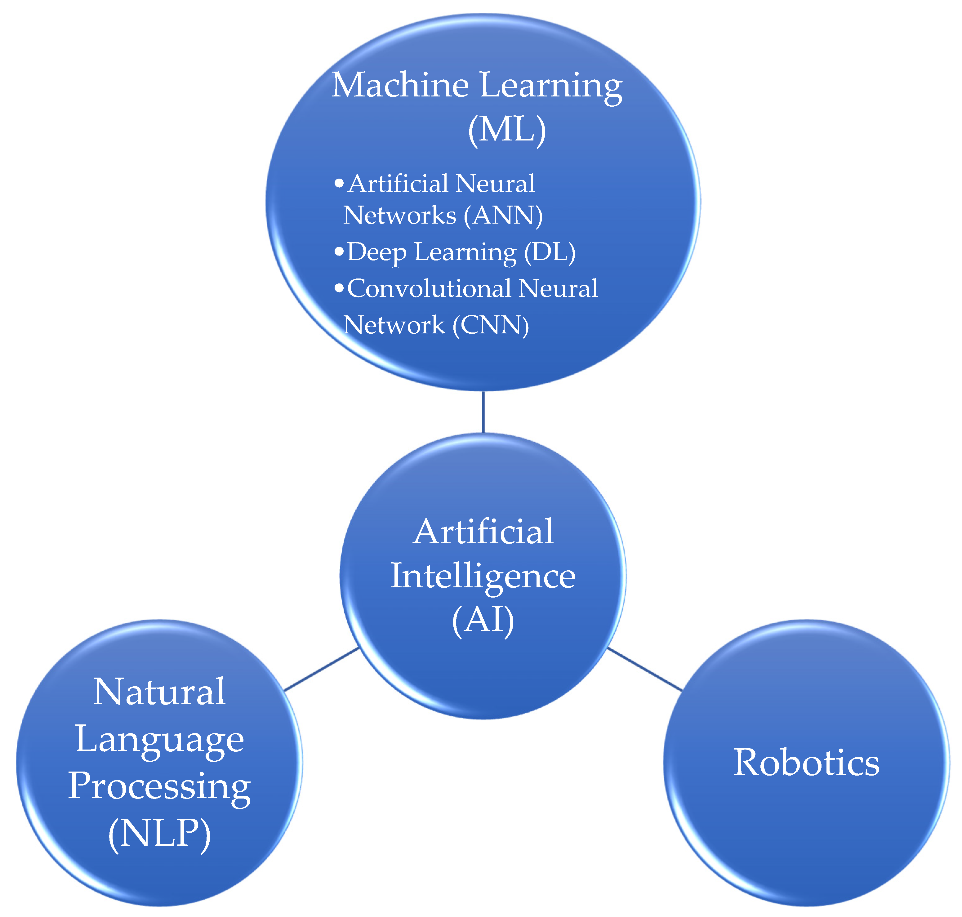
| Artificial Intelligence (AI) | The first definition was been given in 1950 by Alan Turing, the founding father of AI, as the science and engineering of making intelligent machines, especially intelligent computer programs [3]. According to Salto-Tellez M. et al. [4], AI represents a range of advanced machine technologies that can derive meaning and understanding from extensive data inputs, in ways that mimic human capabilities. In the present context of medical practice, a specific definition may be a system’s ability to correctly interpret external data, to learn from such data, and to use those learnings to achieve specific goals and tasks through flexible adaptation [5]. |
| Machine Learning (ML) | ML, a subset of artificial intelligence, exhibits the experiential “learning” associated with human intelligence, while also having the capacity to learn and improve its analyses through the use of computational algorithms [6,7]. Alpaydin E. [8] defined machine learning as the field of programming computers to optimize a performance criterion using example data or past experience. ML-based tools are used in the healthcare system to provide various treatment alternatives and individualized treatments and improve the overall efficiency of hospitals and healthcare systems while lowering the cost of care [9]. |
| Deep Learning (DL) | Deep Learning, a subset of Machine Learning, refers to a deep neural network, which is a specific configuration where neurons are organized in multiple successive layers that can independently learn representations of data and progressively extract complex features, performing tasks such as computer vision and natural language processing (NLP) [10]. In experimental settings across multiple medical specialties, DL performs equivalently to healthcare professionals for detecting diseases from medical imaging [11]. |
| Natural Language Processing (NLP) | Natural Language Processing is a theoretically-motivated range of computational techniques for analyzing and representing naturally-occurring texts at one or more levels of linguistic analysis for the purpose of achieving human-like language processing for a range of tasks or applications [12]. NLP techniques have been used to structure information in healthcare systems by extracting relevant information from narrative texts so as to provide data for decision-making [13]. |
| Robotics | The robot has been defined as “a reprogrammable multifunctional manipulator designed to move material, parts, tools, or specialized devices through variable programmed motions for the performance of a variety of tasks” by the Robot Institute of America [14]. The term “robotics” refers to the study and use of robots. Robotic assistance has been shown to improve the safety and performance of intracorporeal suturing, which is heavily required in urological and gynecological procedures [15]. |
| Artificial Neural Network (ANN) | An Artificial Neural Network, a subset of Machine Learning, is a computational model inspired by the biological neural networks in the human brain. These systems are mainly used for pattern identification and processing and are able to progressively improve their performance based on analytic results from previous tasks [16,17,18]. Many areas have been integrating the use of ANNs to facilitate the diagnosis, prognosis, and treatment of many diseases [19,20,21]. |
| Convolutional Neural Network (CNN) | A Convolutional Neural Network is a Deep Learning algorithm specifically designed for image and video processing, primarily used in medical image analysis and diagnostics. CNNs have demonstrated superior performance as compared with classical machine learning algorithms and in some cases achieved comparable or better performance than clinical experts [22]. |
| Medical Specialty | Year of Study | Author | Application |
|---|---|---|---|
| Cardiology | 2019 | Attia Z.I. [38] | Screening for cardiac contractile dysfunction |
| 2019 | Attia Z.I. [39] | Detection of left ventricular systolic dysfunction | |
| 2018 | Alsharqi M. [40] | Echocardiography analysis | |
| 2017 | Weng S.F. [41] | Cardiovascular risk prediction | |
| Dermatology | 2020 | Young A.T. [42] | Diagnosis of skin lesions |
| 2019 | Dick V. [43] | Diagnosis of melanoma | |
| 2017 | Esteva A. [44] | Classification of skin cancer | |
| Gastroenterology | 2021 | Kröner P.T. [45] | Detection of various lesions |
| 2020 | Martin D.R. [46] | Detecting current Helicobacter pylori infection | |
| Neurology and Neuroscience | 2020 | Pedersen M. [47] | Diagnosis of neurological diseases |
| 2017 | Hazlett H.C. [48] | Diagnosis of autism | |
| 2020 | Ienca M. [49] | Diagnosis of Alzheimer’s disease | |
| Ophthalmology | 2017 | Rathi S. [50] | Teleophthalmology for retinopathy and glaucoma |
| 2016 | Gulshan V. [51] | Detection of diabetic retinopathy | |
| 2017 | Long E. [52] | Diagnosis of congenital cataracts | |
| Psychiatry | 2022 | Pham K.T. [53] | Classification of psychiatric disorders |
| 2017 | Vieira S. [54] | Classification of schizophrenia patients | |
| 2018 | Loh E. [1] | Prediction of suicide attempts | |
| Forensics and Toxicology | 2022 | Wankhade T.D. [55] | Detection of various samples |
| 2021 | Thurzo A. [56] | Identification of a cadaver | |
| 2020 | Chary M.A. [57] | Identification of drug use patterns | |
| Radiology | 2018 | Hosny A. [58] | Recognition of complex radiographic patterns |
| 2016 | Chen H. [59] | Detection in ultrasonography | |
| 2017 | Ghafoorian M. [60] | Segmentation in magnetic resonance imaging (MRI) | |
| 2017 | Wang H. [61] | Classification of mediastinal lymph node metastasis | |
| Surgery | 2020 | Zhou X.Y. [62] | Advances in surgery |
| 2018 | Hu Y. [63] | Robotic sewing and knot tying | |
| 2019 | Hu Y. [64] | Suturing robot for transanal endoscopic microsurgery | |
| 2016 | Shademan A. [65] | Robotic soft tissue surgery | |
| Pathology | 2021 | Cui M. [66] | Digitizing histopathology |
| 2019 | Niazi M.K.K. [67] | Whole-slide imaging | |
| 2017 | FDA [68] | IntelliSite Pathology Solution | |
| 2019 | FDA [69] | Summary Aperio AT2 DX system | |
| 2017 | Araújo T. [70] | Classification of breast cancer | |
| 2017 | Tumeh P.C. [71] | Identification of the immune cell populations | |
| 2019 | Bera K. [72] | Quantitative evaluation of histological and morphological patterns | |
| 2018 | Mezheyeuski A. [73] | Classification of lung cancer patients | |
| 2020 | Balázs A. [74] | Detection of metastasis and micrometastasis | |
| 2019 | Shaban M. [75] | Prediction of disease-free survival in oral squamous cell carcinoma | |
| 2019 | Hekler A. [76] | Classification of histopathological melanoma images | |
| 2014 | Dong F. [77] | Distinction between benign and malignant intraductal proliferations of the breast | |
| 2015 | Veta M. [78] | Mitosis detection in breast cancer | |
| 2013 | Cireşan D.C. [79] | Mitosis detection in breast cancer | |
| 2018 | Couture H.D. [80] | Prediction of breast cancer grade | |
| 2018 | Sahiner B. [81] | Application to Ki67 staining | |
| 2019 | Hossain M.S. [82] | Automatic quantification of HER2 gene amplification | |
| Urology | 2021 | Kott O. [83] | Diagnosis of prostate cancer and Gleason grading |
| 2020 | Baessler B. [84] | Detection of metastatic testicular germ cell tumors | |
| Obstetrics and Gynecology | 2015 | Idowu I. [85] | Detection of true labor and diagnosis of premature labor |
| 2013 | Manna C. [86] | Identification of most viable oocytes and embryos | |
| 2019 | Zhang L. [87] | Diagnosis of ovarian tumor | |
| 2020 | Hart G. [88] | Early detection of endometrial cancer |
| Examples of AI Systems Applications in Pathology |
|---|
| 1. Differentiate between benign and malignant tumors |
| 2. Grading of dysplasia and in situ lesions [70] |
| 3. Metastasis and micrometastasis detection [74] |
| 4. Relationships between different immune cell populations [70,71] |
| 5. IHC/ISH scoring of multiple biomarkers and topography of the immune response [72] |
| 6. Mitosis detection [78,79] |
Disclaimer/Publisher’s Note: The statements, opinions and data contained in all publications are solely those of the individual author(s) and contributor(s) and not of MDPI and/or the editor(s). MDPI and/or the editor(s) disclaim responsibility for any injury to people or property resulting from any ideas, methods, instructions or products referred to in the content. |
© 2023 by the authors. Licensee MDPI, Basel, Switzerland. This article is an open access article distributed under the terms and conditions of the Creative Commons Attribution (CC BY) license (https://creativecommons.org/licenses/by/4.0/).
Share and Cite
Poalelungi, D.G.; Musat, C.L.; Fulga, A.; Neagu, M.; Neagu, A.I.; Piraianu, A.I.; Fulga, I. Advancing Patient Care: How Artificial Intelligence Is Transforming Healthcare. J. Pers. Med. 2023, 13, 1214. https://doi.org/10.3390/jpm13081214
Poalelungi DG, Musat CL, Fulga A, Neagu M, Neagu AI, Piraianu AI, Fulga I. Advancing Patient Care: How Artificial Intelligence Is Transforming Healthcare. Journal of Personalized Medicine. 2023; 13(8):1214. https://doi.org/10.3390/jpm13081214
Chicago/Turabian StylePoalelungi, Diana Gina, Carmina Liana Musat, Ana Fulga, Marius Neagu, Anca Iulia Neagu, Alin Ionut Piraianu, and Iuliu Fulga. 2023. "Advancing Patient Care: How Artificial Intelligence Is Transforming Healthcare" Journal of Personalized Medicine 13, no. 8: 1214. https://doi.org/10.3390/jpm13081214