
Advancing Osteoporosis Evaluation Procedures: Detailed Computational Analysis of Regional Structural Vulnerabilities in Osteoporotic Bone

Abstract
:1. Introduction
2. Materials and Methods
2.1. Anatomical Data
2.2. Ethics Approval
2.3. 3D Model Generation
2.4. Automated Osteological Sampling
2.5. Analysis
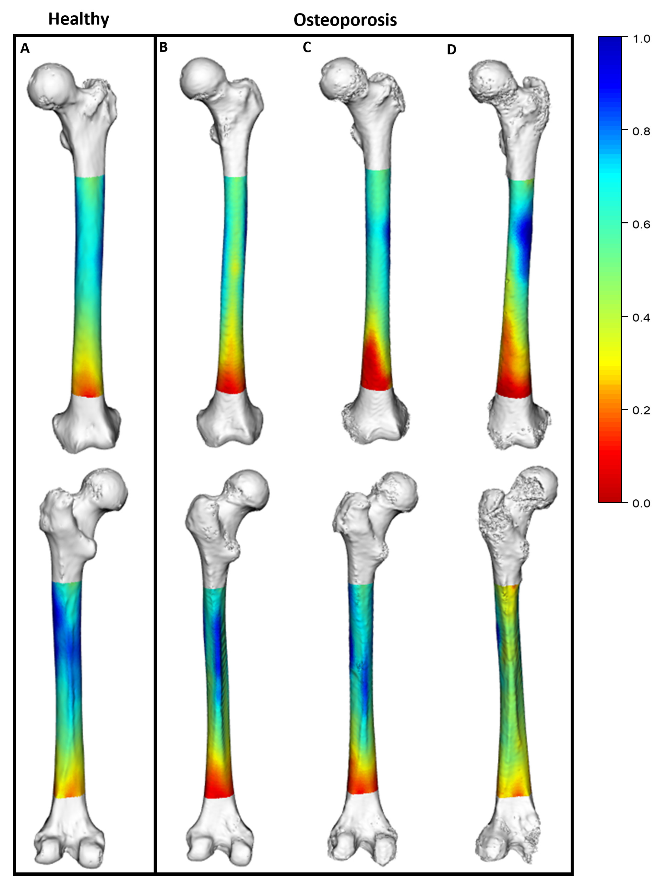
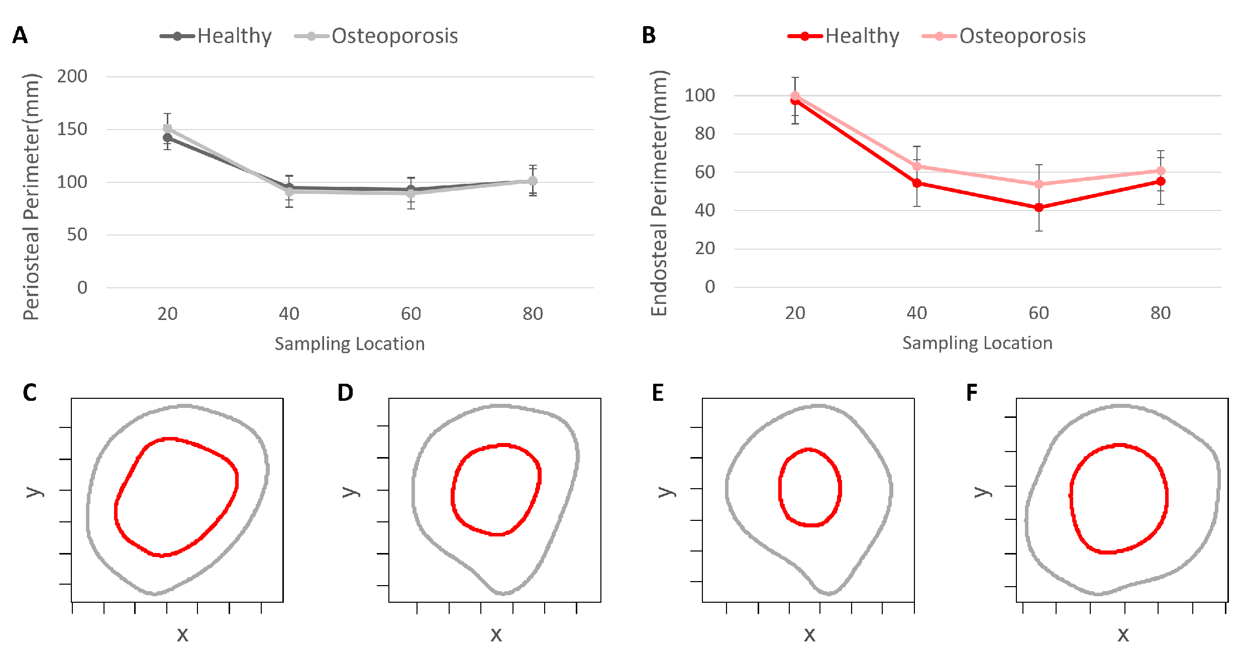
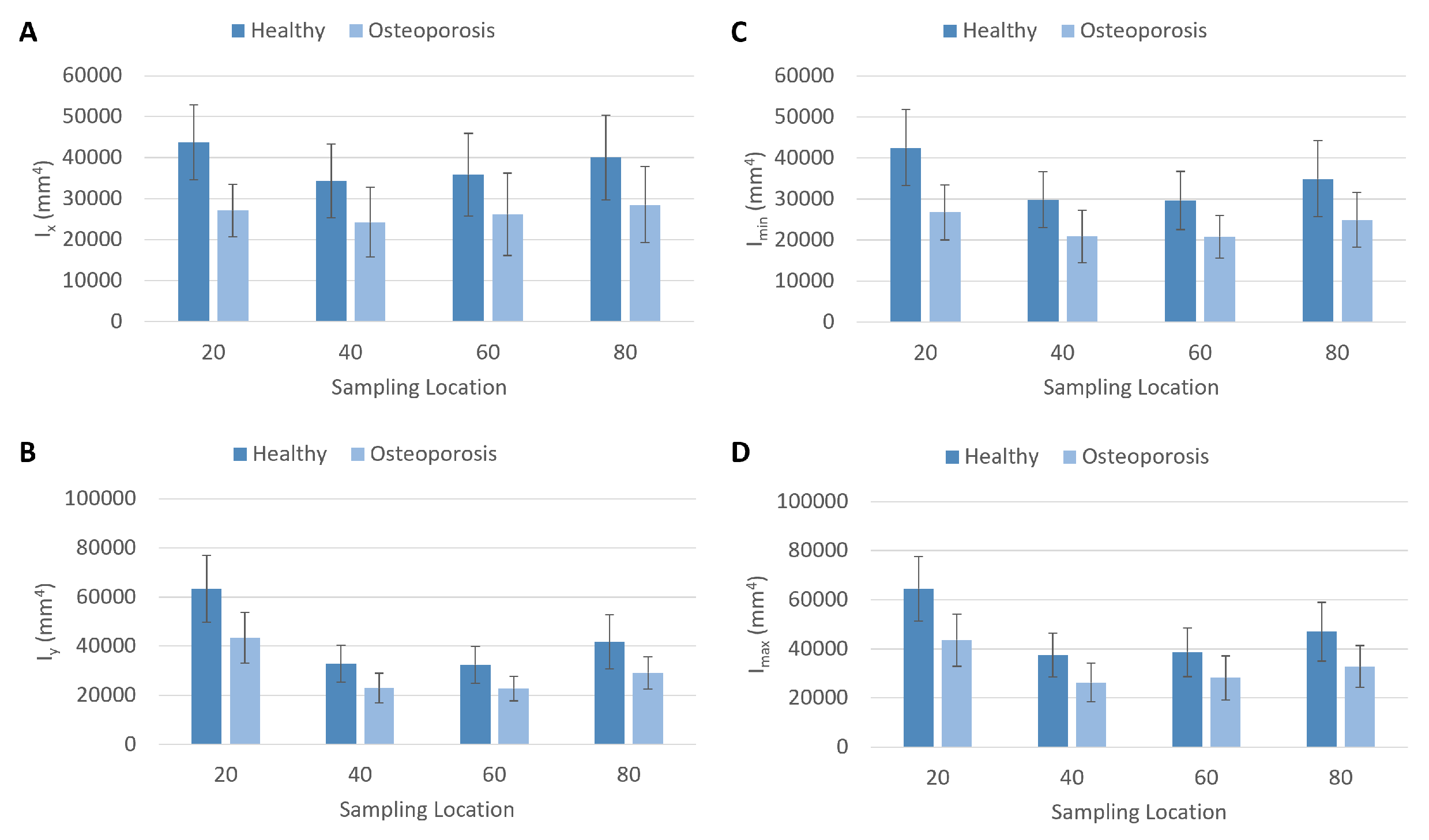
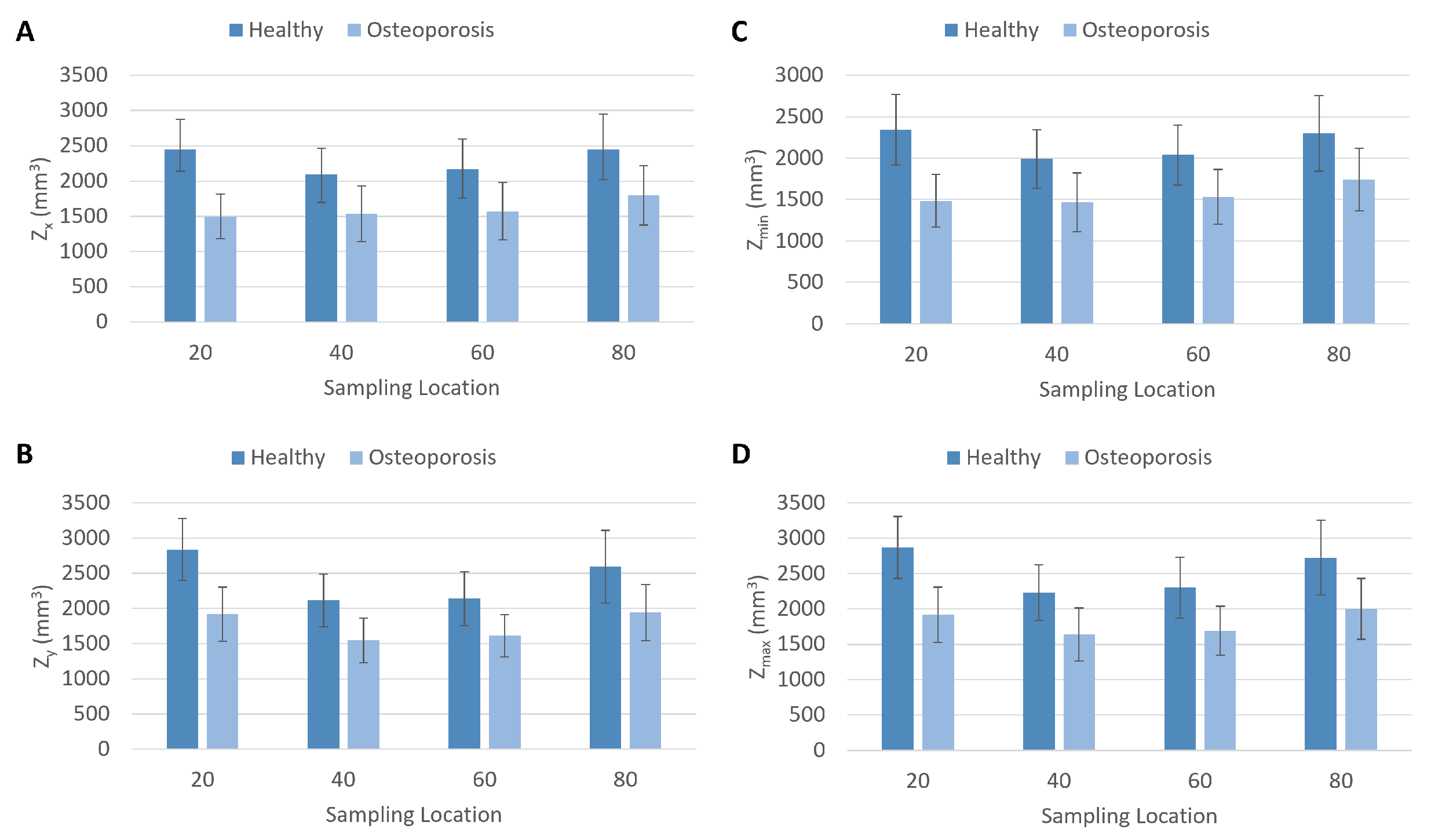
3. Results
4. Discussion
5. Conclusions
Supplementary Materials
Author Contributions
Funding
Institutional Review Board Statement
Informed Consent Statement
Data Availability Statement
Acknowledgments
Conflicts of Interest
Abbreviations
| CT | Computed tomography |
| UB | Universtity at Buffalo |
| CTSI | Clinical and Translational Science Institute |
| KI | Kittler–Illingworth |
| Area moment of inertia around the x axis | |
| Area moment of inertia around the y axis | |
| Minimum area moment of inertia | |
| Maximum area moment of inertia | |
| Section modulus about the x axis | |
| Section modulus about the y axis | |
| Minimum section modulus | |
| Maximum section modulus | |
| Theta | |
| Polar section modulus | |
| J | Polar moment of inertia |
| CA | Cortical area |
| BL | Biomechanical length |
References
- Caliri, A.; De Filippis, L.; Bagnato, G.; Bagnato, G. Osteoporotic fractures: Mortality and quality of life. Panminerva Med. 2007, 49, 21. [Google Scholar] [PubMed]
- Glinkowski, W.; Narloch, J.; Krasuski, K.; Śliwczyński, A. The increase of osteoporotic hip fractures and associated one-year mortality in Poland: 2008–2015. J. Clin. Med. 2019, 8, 1487. [Google Scholar] [CrossRef] [PubMed]
- Bergh, C.; Möller, M.; Ekelund, J.; Brisby, H. 30-day and 1-year mortality after skeletal fractures: A register study of 295,713 fractures at different locations. Acta Orthop. 2021, 92, 739–745. [Google Scholar] [CrossRef] [PubMed]
- Carpenter, R.D.; Beaupré, G.S.; Lang, T.F.; Orwoll, E.S.; Carter, D.R. New QCT analysis approach shows the importance of fall orientation on femoral neck strength. J. Bone Miner. Res. 2005, 20, 1533–1542. [Google Scholar] [CrossRef]
- Mayhew, P.M.; Thomas, C.D.; Clement, J.G.; Loveridge, N.; Beck, T.J.; Bonfield, W.; Burgoyne, C.J.; Reeve, J. Relation between age, femoral neck cortical stability, and hip fracture risk. Lancet 2005, 366, 129–135. [Google Scholar] [CrossRef]
- Lotz, J.; Cheal, E.; Hayes, W. Stress distributions within the proximal femur during gait and falls: Implications for osteoporotic fracture. Osteoporos. Int. 1995, 5, 252–261. [Google Scholar] [CrossRef]
- Sollmann, N.; Löffler, M.T.; Kronthaler, S.; Böhm, C.; Dieckmeyer, M.; Ruschke, S.; Kirschke, J.S.; Carballido-Gamio, J.; Karampinos, D.C.; Krug, R.; et al. MRI-based quantitative osteoporosis imaging at the spine and femur. J. Magn. Reson. Imaging 2021, 54, 12–35. [Google Scholar] [CrossRef]
- Bergh, C.; Möller, M.; Ekelund, J.; Brisby, H. Mortality after sustaining skeletal fractures in relation to age. J. Clin. Med. 2022, 11, 2313. [Google Scholar] [CrossRef]
- Canton, G.; Giraldi, G.; Dussi, M.; Ratti, C.; Murena, L. Osteoporotic distal femur fractures in the elderly: Peculiarities and treatment strategies. Acta Biomed. 2019, 90, 25. [Google Scholar]
- Larsen, P.; Ceccotti, A.A.; Elsoe, R. High mortality following distal femur fractures: A cohort study including three hundred and two distal femur fractures. Int. Orthop. 2020, 44, 173–177. [Google Scholar] [CrossRef]
- Kammerlander, C.; Riedmüller, P.; Gosch, M.; Zegg, M.; Kammerlander-Knauer, U.; Schmid, R.; Roth, T. Functional outcome and mortality in geriatric distal femoral fractures. Injury 2012, 43, 1096–1101. [Google Scholar] [CrossRef]
- Kim, J.; Kang, S.B.; Nam, K.; Rhee, S.H.; Won, J.W.; Han, H.S. Retrograde intramedullary nailing for distal femur fracture with osteoporosis. Clin. Orthop. Surg. 2012, 4, 307–312. [Google Scholar] [CrossRef]
- Hollensteiner, M.; Sandriesser, S.; Bliven, E.; von Rüden, C.; Augat, P. Biomechanics of osteoporotic fracture fixation. Curr. Osteoporos. Rep. 2019, 17, 363–374. [Google Scholar] [CrossRef]
- Poole, K.E.; Treece, G.M.; Mayhew, P.M.; Vaculík, J.; Dungl, P.; Horák, M.; Štěpán, J.J.; Gee, A.H. Cortical thickness mapping to identify focal osteoporosis in patients with hip fracture. PLoS ONE 2012, 7, e38466. [Google Scholar] [CrossRef]
- Wainwright, S.A.; Marshall, L.M.; Ensrud, K.E.; Cauley, J.A.; Black, D.M.; Hillier, T.A.; Hochberg, M.C.; Vogt, M.T.; Orwoll, E.S.; Study of Osteoporotic Fractures Research Group. Hip fracture in women without osteoporosis. J. Clin. Endocrinol. Metab. 2005, 90, 2787–2793. [Google Scholar] [CrossRef]
- Treece, G.M.; Poole, K.E.; Gee, A.H. Imaging the femoral cortex: Thickness, density and mass from clinical CT. Med. Image Anal. 2012, 16, 952–965. [Google Scholar] [CrossRef]
- De Bakker, P.M.; Manske, S.L.; Ebacher, V.; Oxland, T.R.; Cripton, P.A.; Guy, P. During sideways falls proximal femur fractures initiate in the superolateral cortex: Evidence from high-speed video of simulated fractures. J. Biomech. 2009, 42, 1917–1925. [Google Scholar] [CrossRef]
- Poole, K.E.; Skingle, L.; Gee, A.H.; Turmezei, T.D.; Johannesdottir, F.; Blesic, K.; Rose, C.; Vindlacheruvu, M.; Donell, S.; Vaculik, J.; et al. Focal osteoporosis defects play a key role in hip fracture. Bone 2017, 94, 124–134. [Google Scholar] [CrossRef]
- Poole, K.E.; Treece, G.M.; Gee, A.H.; Brown, J.P.; McClung, M.R.; Wang, A.; Libanati, C. Response to: Comment on:“Denosumab rapidly increases cortical bone in key locations of the femur: A 3D bone mapping study in women with osteoporosis”. J. Bone Miner. Res. 2015, 30, 1939–1940. [Google Scholar] [CrossRef]
- Endo, B.; Takahashi, H. Various methods for measuring the geometrical properties of the long bone cross section with respect to mechanics. J. Anthropol. Soc. Jpn. 1982, 90, 1–16. [Google Scholar] [CrossRef]
- Profico, A.; Bondioli, L.; Raia, P.; O’Higgins, P.; Marchi, D. morphomap: An R package for long bone landmarking, cortical thickness, and cross-sectional geometry mapping. Am. J. Phys. Anthropol. 2021, 174, 129–139. [Google Scholar] [CrossRef] [PubMed]
- Doube, M.; Kłosowski, M.M.; Arganda-Carreras, I.; Cordelières, F.P.; Dougherty, R.P.; Jackson, J.S.; Schmid, B.; Hutchinson, J.R.; Shefelbine, S.J. BoneJ: Free and extensible bone image analysis in ImageJ. Bone 2010, 47, 1076–1079. [Google Scholar] [CrossRef] [PubMed]
- Morimoto, N.; De León, M.S.P.; Zollikofer, C.P. Exploring femoral diaphyseal shape variation in wild and captive chimpanzees by means of morphometric mapping: A test of Wolff’s law. Anat. Rec. 2011, 294, 589–609. [Google Scholar] [CrossRef] [PubMed]
- Puymerail, L. The functionally-related signatures characterizing the endostructural organisation of the femoral shaft in modern humans and chimpanzee. C. R. Palevol 2013, 12, 223–231. [Google Scholar] [CrossRef]
- Kivell, T.L.; Davenport, R.; Hublin, J.J.; Thackeray, J.F.; Skinner, M.M. Trabecular architecture and joint loading of the proximal humerus in extant hominoids, Ateles, and Australopithecus africanus. Am. J. Phys. Anthropol. 2018, 167, 348–365. [Google Scholar] [CrossRef]
- Wallace, J.M.; Rajachar, R.M.; Allen, M.R.; Bloomfield, S.A.; Robey, P.G.; Young, M.F.; Kohn, D.H. Exercise-induced changes in the cortical bone of growing mice are bone-and gender-specific. Bone 2007, 40, 1120–1127. [Google Scholar] [CrossRef]
- Carlson, K.J.; Judex, S. Increased non-linear locomotion alters diaphyseal bone shape. J. Exp. Biol. 2007, 210, 3117–3125. [Google Scholar] [CrossRef]
- Shaw, C.N.; Stock, J.T. Intensity, repetitiveness, and directionality of habitual adolescent mobility patterns influence the tibial diaphysis morphology of athletes. Am. J. Phys. Anthropol. 2009, 140, 149–159. [Google Scholar] [CrossRef]
- Niinimäki, S.; Narra, N.; Härkönen, L.; Abe, S.; Nikander, R.; Hyttinen, J.; Knüsel, C.; Sievänen, H. The relationship between loading history and proximal femoral diaphysis cross-sectional geometry. Am. J. Hum. 2017, 29, e22965. [Google Scholar] [CrossRef]
- Jepsen, K.J.; Bigelow, E.M.; Schlecht, S.H. Women build long bones with less cortical mass relative to body size and bone size compared with men. Clin. Orthop. Relat. Res. 2015, 473, 2530–2539. [Google Scholar] [CrossRef]
- Wark, J.D. Osteoporosis: A global perspective. Bull. World Health Organ. 1999, 77, 424. [Google Scholar]
- Siris, E.; Boonen, S.; Mitchell, P.; Bilezikian, J.; Silverman, S. What’s in a name? What constitutes the clinical diagnosis of osteoporosis? Osteoporos. Int. 2012, 23, 2093–2097. [Google Scholar] [CrossRef]
- Siris, E.; Adler, R.; Bilezikian, J.; Bolognese, M.; Dawson-Hughes, B.; Favus, M.; Harris, S.; Jan de Beur, S.; Khosla, S.; Lane, N.; et al. The clinical diagnosis of osteoporosis: A position statement from the National Bone Health Alliance Working Group. Osteoporos. Int. 2014, 25, 1439–1443. [Google Scholar] [CrossRef]
- Johnell, O.; Kanis, J.A.; Oden, A.; Johansson, H.; De Laet, C.; Delmas, P.; Eisman, J.A.; Fujiwara, S.; Kroger, H.; Mellstrom, D.; et al. Predictive value of BMD for hip and other fractures. J. Bone Miner. Res. 2005, 20, 1185–1194. [Google Scholar] [CrossRef]
- Lepor, H. Prostatic Diseases; W.B. Saunders Company: Philadelphia, PA, USA, 2000; p. 966. [Google Scholar]
- Wysocki, M.A.; Doyle, S. The impact of CT-data segmentation variation on the morphology of osteological structure. Proc. SPIE Med. Imag. 2021, 11595. [Google Scholar] [CrossRef]
- Kittler, J.; Illingworth, J. Minimum error thresholding. Pattern Recognit. 1986, 19, 41–47. [Google Scholar] [CrossRef]
- Wysocki, M.A.; Doyle, S. Enhancing biomedical data validity with standardized segmentation finite element analysis. Sci. Rep. 2022, 12, 1–9. [Google Scholar] [CrossRef]
- Fedorov, A.; Beichel, R.; Kalpathy-Cramer, J.; Finet, J.; Fillion-Robin, J.C.; Pujol, S.; Bauer, C.; Jennings, D.; Fennessy, F.; Sonka, M.; et al. 3D Slicer as an image computing platform for the Quantitative Imaging Network. Magn. Reson. Imaging 2012, 30, 1323–1341. [Google Scholar] [CrossRef]
- Cignoni, P.; Callieri, M.; Corsini, M.; Dellepiane, M.; Ganovelli, F.; Ranzuglia, G. Meshlab: An open-source mesh processing tool. In Proceedings of the Eurographics Italian Chapter Conference, Salerno, Italy, 2–4 July 2008; Volume 2008, pp. 129–136. [Google Scholar]
- Wysocki, M.A.; Doyle, S. Optimization of decimation protocols for advancing the validity of 3D model data. Proc. SPIE Med. Imag. 2022, 12031. [Google Scholar] [CrossRef]
- Autodesk, Inc. Meshmixer. Version 3.5.474. 2017. Available online: www.meshmixer.com (accessed on 1 July 2021).
- Trinkaus, E.; Churchill, S.E.; Ruff, C.B.; Vandermeersch, B. Long bone shaft robusticity and body proportions of the Saint-Césaire 1 Châtelperronian Neanderthal. J. Archaeol. Sci. 1999, 26, 753–773. [Google Scholar] [CrossRef]
- White, T.D.; Black, M.T.; Folkens, P.A. Human Osteology; Academic Press: Cambridge, MA, USA, 2011. [Google Scholar]
- Ruff, C.B. Long bone articular and diaphyseal structure in Old World monkeys and apes. I: Locomotor effects. Am. J. Phys. Anthropol. 2002, 119, 305–342. [Google Scholar] [CrossRef] [PubMed]
- Marchi, D. The cross-sectional geometry of the hand and foot bones of the Hominoidea and its relationship to locomotor behavior. J. Hum. Evol. 2005, 49, 743–761. [Google Scholar] [CrossRef] [PubMed]
- Marchi, D. Relative strength of the tibia and fibula and locomotor behavior in hominoids. J. Hum. Evol. 2007, 53, 647–655. [Google Scholar] [CrossRef] [PubMed]
- Rodríguez Casal, A.; Pateiro López, B. Generalizing the convex hull of a sample: The R package alphahull. J. Stat. Softw. 2010, 34, 1–28. [Google Scholar]
- Schlager, S.; Schlager, M.S. Package ‘Rvcg’; R Package. 2014. Available online: https://cran.microsoft.com/snapshot/2014-12-24/web/packages/Rvcg/Rvcg.pdf (accessed on 6 February 2023).
- Schlager, S. Morpho and Rvcg–Shape Analysis in R: R-Packages for Geometric Morphometrics, Shape Analysis and Surface Manipulations. In Statistical Shape and Deformation Analysis; Academic press: Cambridge, MA, USA, 2017; pp. 217–256. [Google Scholar]
- Keitt, T.H. Coherent ecological dynamics induced by large-scale disturbance. Nature 2008, 454, 331–334. [Google Scholar] [CrossRef]
- Hijmans, R.J.; Van Etten, J.; Cheng, J.; Mattiuzzi, M.; Sumner, M.; Greenberg, J.A.; Lamigueiro, O.P.; Bevan, A.; Racine, E.B.; Shortridge, A.; et al. Package ‘Raster’. R Package. 2015, p. 734. Available online: https://cran.r-project.org/web/packages/raster/raster.pdf (accessed on 6 February 2023).
- Team, R.C. R: A Language and Environment for Statistical Computing; R Foundation for Statistical Computing: Vienna, Austria, 2020. [Google Scholar]
- Murdoch, D.; Adler, D.; Nenadic, O. Package ‘rgl’. R Package. 2023. Available online: https://cran.r-project.org/web/packages/rgl/rgl.pdf (accessed on 6 February 2023).
- Gunz, P.; Mitteroecker, P.; Bookstein, F.L. Semilandmarks in three dimensions. In Modern Morphometrics in Physical Anthropology; Kluwer Academic/Plenum Pbulishers: New York, NY, USA, 2005; pp. 73–98. [Google Scholar]
- Frelat, M.A.; Katina, S.; Weber, G.W.; Bookstein, F.L. A novel geometric morphometric approach to the study of long bone shape variation. Am. J. Phys. Anthropol. 2012, 149, 628–638. [Google Scholar] [CrossRef]
- Morimoto, N.; Nakatsukasa, M.; de León, M.S.P.; Zollikofer, C.P. Femoral ontogeny in humans and great apes and its implications for their last common ancestor. Sci. Rep. 2018, 8, 1–11. [Google Scholar] [CrossRef]
- Garn, S.M.; Poznanski, A.K.; Nagy, J.M. Bone measurement in the differential diagnosis of osteopenia and osteoporosis. Radiology 1971, 100, 509–518. [Google Scholar] [CrossRef]
- Ruff, C.; Holt, B.; Trinkaus, E. Who’s afraid of the big bad Wolff?:“Wolff’s law” and bone functional adaptation. Am. J. Phys. Anthropol. 2006, 129, 484–498. [Google Scholar] [CrossRef]
- Ruff, C.B.; Trinkaus, E.; Walker, A.; Larsen, C.S. Postcranial robusticity in Homo. I: Temporal trends and mechanical interpretation. Am. J. Phys. Anthropol. 1993, 91, 21–53. [Google Scholar] [CrossRef]
- Stock, J.; Pfeiffer, S. Linking structural variability in long bone diaphyses to habitual behaviors: Foragers from the southern African Later Stone Age and the Andaman Islands. Am. J. Phys. Anthropol. 2001, 115, 337–348. [Google Scholar] [CrossRef]
- Trinkaus, E.; Ruff, C.B. Femoral and tibial diaphyseal cross-sectional geometry in Pleistocene Homo. PaleoAnthropology 2012, 2012, 13–62. [Google Scholar]
- Lacoste Jeanson, A.; Santos, F.; Dupej, J.; Veleminska, J.; Bruzek, J. Sex-specific functional adaptation of the femoral diaphysis to body composition. Am. J. Hum. Biol. 2018, 30, e23123. [Google Scholar] [CrossRef]
- Lacoste Jeanson, A.; Santos, F.; Villa, C.; Banner, J.; Bruzek, J. Architecture of the femoral and tibial diaphyses in relation to body mass and composition: Research from whole-body CT scans of adult humans. Am. J. Phys. Anthropol. 2018, 167, 813–826. [Google Scholar] [CrossRef]
- Ruff, C.B. New approaches to structural evolution of limb bones in primates. Folia Primatol. 1989, 53, 142–159. [Google Scholar] [CrossRef]
- Ruff, C.B.; Hayes, W.C. Cross-sectional geometry of Pecos Pueblo femora and tibiae—A biomechanical investigation: I. Method and general patterns of variation. Am. J. Phys. Anthropol. 1983, 60, 359–381. [Google Scholar] [CrossRef]
- Sládek, V.; Berner, M.; Sailer, R. Mobility in central European late Eneolithic and early bronze age: Femoral cross-sectional geometry. Am. J. Phys. Anthropol. 2006, 130, 320–332. [Google Scholar] [CrossRef]
- Lieberman, D.E.; Polk, J.D.; Demes, B. Predicting long bone loading from cross-sectional geometry. Am. J. Phys. Anthropol. 2004, 123, 156–171. [Google Scholar] [CrossRef]
- Koso, R.E.; Terhoeve, C.; Steen, R.G.; Zura, R. Healing, nonunion, and re-operation after internal fixation of diaphyseal and distal femoral fractures: A systematic review and meta-analysis. Int. Orthop. 2018, 42, 2675–2683. [Google Scholar] [CrossRef]


Disclaimer/Publisher’s Note: The statements, opinions and data contained in all publications are solely those of the individual author(s) and contributor(s) and not of MDPI and/or the editor(s). MDPI and/or the editor(s) disclaim responsibility for any injury to people or property resulting from any ideas, methods, instructions or products referred to in the content. |
© 2023 by the authors. Licensee MDPI, Basel, Switzerland. This article is an open access article distributed under the terms and conditions of the Creative Commons Attribution (CC BY) license (https://creativecommons.org/licenses/by/4.0/).
Share and Cite
Wysocki, M.A.; Doyle, S.T. Advancing Osteoporosis Evaluation Procedures: Detailed Computational Analysis of Regional Structural Vulnerabilities in Osteoporotic Bone. J. Pers. Med. 2023, 13, 321. https://doi.org/10.3390/jpm13020321
Wysocki MA, Doyle ST. Advancing Osteoporosis Evaluation Procedures: Detailed Computational Analysis of Regional Structural Vulnerabilities in Osteoporotic Bone. Journal of Personalized Medicine. 2023; 13(2):321. https://doi.org/10.3390/jpm13020321
Chicago/Turabian StyleWysocki, Matthew A., and Scott T. Doyle. 2023. "Advancing Osteoporosis Evaluation Procedures: Detailed Computational Analysis of Regional Structural Vulnerabilities in Osteoporotic Bone" Journal of Personalized Medicine 13, no. 2: 321. https://doi.org/10.3390/jpm13020321