Article

Adequacy of Disease Control by Supraomohyoid Neck Dissection in cT1/T2 Tongue Cancer

1
Department of Otorhinolaryngology and Head and Neck Surgery, Chang Gung Memorial Hospital, Linkou Branch, Yaoyuan 33302, Taiwan
2
Unit of Otorhinolaryngology-Head and Neck Surgery, IRCCS Ospedale Policlinico San Martino, 16132 Genova, Italy
3
Department of Surgical Sciences and Integrated Diagnostics (DISC), University of Genova, 16132 Genova, Italy
4
Department of Plastic Surgery, Chang Gung Memorial, Linkou Branch, Taoyuan 333, Taiwan
5
Department of Otolaryngology, Head and Neck Surgery, Chang Gung Memorial, Keelung Branch, Keelung 20401, Taiwan
6
School of Medicine, Chang Gung University, Taoyuan 333, Taiwan
7
Department of Medical Oncology, Internal Medicine, Chang Gung Memorial, Linkou Branch, Taoyuan 333, Taiwan
8
Department of Radiation Oncology, Chang Gung Memorial, Linkou Branch, Taoyuan 333, Taiwan
9
Graduate Institute of Clinical Medical Sciences, Chang Gun Medical College, Chang Gung University, Taoyuan 33302, Taiwan
*
Author to whom correspondence should be addressed.
These authors contributed equally to this work.
J. Pers. Med. 2022, 12(9), 1535; https://doi.org/10.3390/jpm12091535
Submission received: 28 August 2022 / Revised: 13 September 2022 / Accepted: 14 September 2022 / Published: 19 September 2022
(This article belongs to the Section Clinical Medicine, Cell, and Organism Physiology)

Abstract

:
Background: Patients affected by oral tongue squamous cell carcinoma (OTSCC) underwent a supraomohyoid neck dissection (SOHND) or modified radical neck dissection (mRND) according to the clinical nodal status (cN0 vs. cN+). We investigate whether the type of neck dissection affects survival with the presence of extranodal extension (ENE) and multiple nodal metastases (MNM). Methods: We conducted a retrospective study enrolling surgically treated patients affected by cT1/T2 OTSCC and MNM or ENE. The outcomes assessed were: overall survival (OS), disease-free survival (DFS), and neck-control- and metastases-free survival (NC-MFS). Survival curves were plotted by the Kaplan–Meier method and the log-rank test. Furthermore, we conducted a multivariable analysis with the Cox regression model. Results: We included a total of 565 patients (36% cT1, 64% cT2). Of these, 501 patients underwent a SOHND, and 64 underwent an mRND. A total of 184 patients presented rpN+, with 28.7% of these in the SOHND group and 62.5% of these in the mRND group. We identified no significant differences in OS, DFS, and NC-MFS in the whole pN+ cohort, in the MNM, and the ENE subgroups. In the multivariable analysis, the type of ND did not affect OS and DFS. Conclusions: Treating cT1-2 N0/+ tongue cancer with SOHND is oncologically safe. ENE and MNM patients do not benefit from an mRND.

1. Introduction

Oral cavity squamous cell carcinoma (OSCC) contributes substantially to the global cancer burden, with relevant cancer-specific mortality in the region of 31% in most major referral centers [1,2,3,4]. More than fifty-two thousand cases are diagnosed annually in the United States [5]. The tongue is one of the most common subsites involved, found in up to half of all cases [6], with a propensity for early and extensive local invasion and regional lymphatic spread, leading to significant disease- and treatment-related morbidity. Regional neck metastasis represents an ominous prognostic factor in OSCC. In oral tongue squamous cell carcinoma (OTSCC), the presence of just one metastatic lymph node (LN) commits patients to an advanced-stage disease category [7]. It has been shown to determine up to a 50% decrease in overall survival (OS) [8,9,10,11,12]. Moreover, OTSCC has a higher propensity for lymphovascular invasion (LVI), perineural invasion (PNI), and neck dissemination than other OSCC subsites, one of the most significant prognostic factors and varies from 6% to 85% [13,14,15,16,17]. Thus, historically modified radical neck dissection (mRND) has been part of the treatment of OTSCC. However, the cosmetic and functional defects associated with mRND prompted the search for less morbid alternatives [18,19,20].
Detailed examination of lymphatic drainage pathways showed that levels I–II were at the most significant risk of metastasis in OSCC, with levels IV and V playing minor roles. Consequently, to overcome these issues and minimize morbidities in the past decades, elective selective neck dissection (SND) was described to clear the nodal levels with a higher chance of occult metastases [21]. Afterward, several studies demonstrated that in OTSCC, the most commonly nodal levels at risk of metastasis are levels I, II, and III [4,13,22]; therefore, supraomohyoid neck dissection (SOHND) resulted in achieving comparable oncological outcomes to mRND [23,24]. The impact of selective neck dissection in patients with clinically evident nodal disease (cN+) is more controversial. Recent studies [25,26] reported reasonable regional control with selective neck dissection in selected patients with cervical nodal metastases, mainly when adjuvant treatment was applied. Nevertheless, several questions need to be answered: which subpopulation of patients would benefit more from a less extensive dissection? How often are skip metastases, defined as nodal metastases that bypass levels I, II, or both, localized directly to levels III, IV, or V [27], found in early-stage OTSCC? This study aimed to investigate whether, in a group of clinically early-stage OTSCC with the presence of detrimental risk factors, such as extranodal extension (ENE) and multiple nodal metastases, an mRND might determine a survival advantage.

2. Materials and Methods

2.1. Patients Selection

We conducted a retrospective analysis of all patients affected by clinically early-stage (cT1-T2) cancer of the mobile tongue treated at our institute between March 1997 and May 2017. The institutional review board approved the study at Chang Gung Medical Foundation, Taiwan (IRB No. 202101807B0). The inclusion criteria were: age over 18 years old; a biopsy-proven squamous cell carcinoma (SCC) of the mobile tongue; at least one year of follow-up, or an earlier date of recurrence or death; clinical stage (cT1-T2) according to AJCC 8th edition [7]; treated with curative intent with upfront surgery including neck dissection as part of the treatment. In addition, we excluded patients with a previous history of head and neck cancer; treatment with neoadjuvant therapy; advanced or very advanced local stage; unavailable follow-up information; prior chemotherapy for any cancer and/or radiotherapy in the head and neck area; synchronous head and neck squamous cell carcinoma; presence of distant metastases.

2.2. Diagnostic Work-Up, Treatment Policy, and Follow-Up

All patients received a complete tumor survey, including a detailed medical history, comprehensive physical examination, blood tests, computed tomography (CT) or magnetic resonance imaging (MRI) of the head and neck, chest radiographs, abdominal sonography, bone scan, or positron emission tomography (PET). Tumor excision was performed according to the lesion’s extent with a macroscopic surgical margin of at least 1 cm. Free flap reconstruction was performed when needed. Patients without clinically suspicious lymph node metastases (cN0) underwent supraomohyoid neck dissection (SOHND). A modified radical neck dissection (mRND) was performed in the presence of clinically evident nodal disease (cN+). Adjuvant treatment was discussed by the multidisciplinary head and neck tumor board, including radiotherapy or chemoradiotherapy [28,29]. Clinical data recorded included gender, age, smoking, and treatment details. Pathological details recorded were surgical margin status, diameter, adverse pathological features, pN category, and nodal metastases. All tumors were re-staged according to the 8th edition of the UICC-AJCC Staging System [7]. Follow-up included clinical examination every two months during the first and second year, every three months in the third year, every four months in the fourth year, and every six months during the fifth year. No routine radiological follow-up was performed unless patients had symptoms or suspicious clinical finding(s) of recurrence for which CT/PET was carried out.

2.3. Survival Analysis
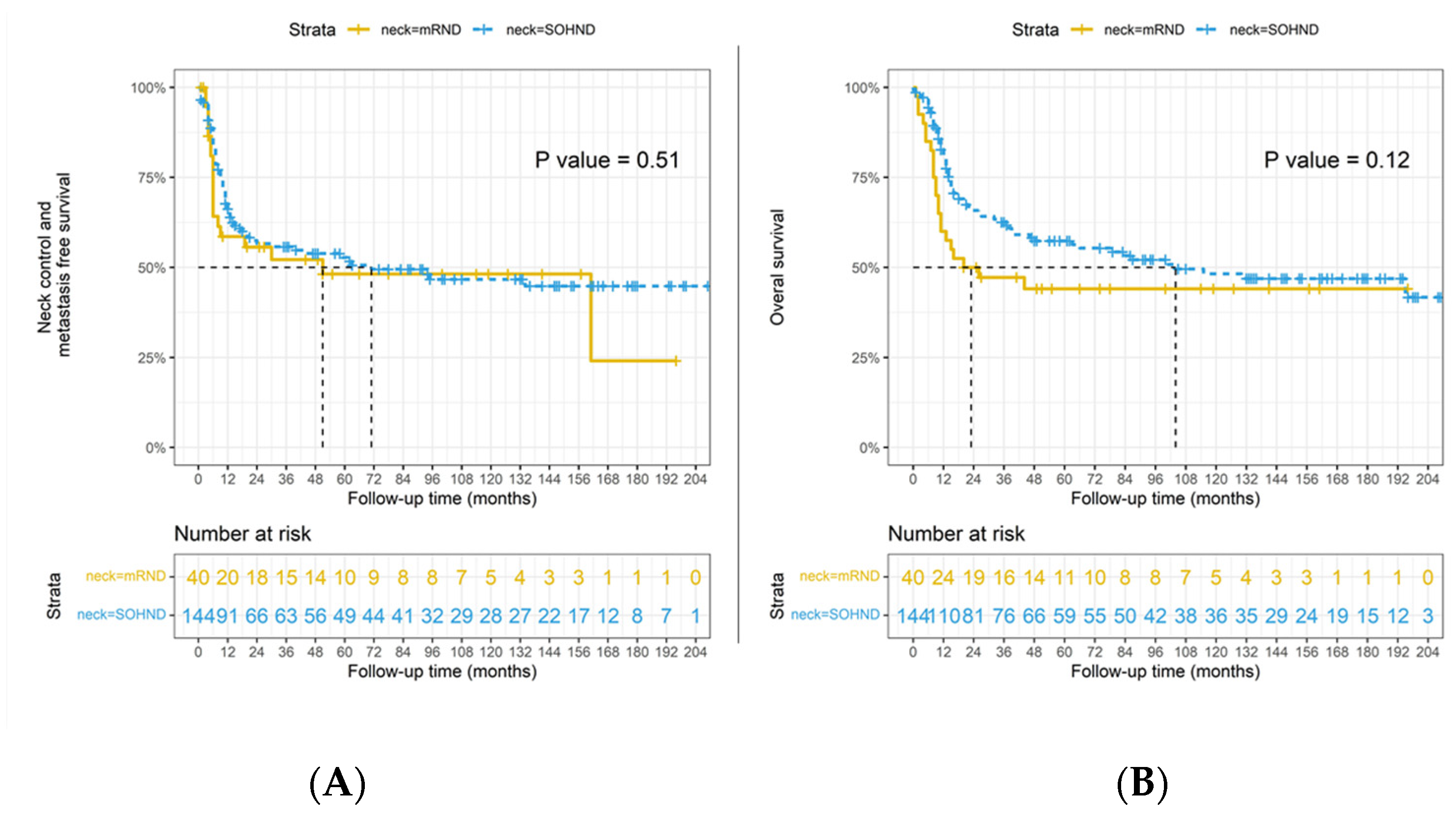
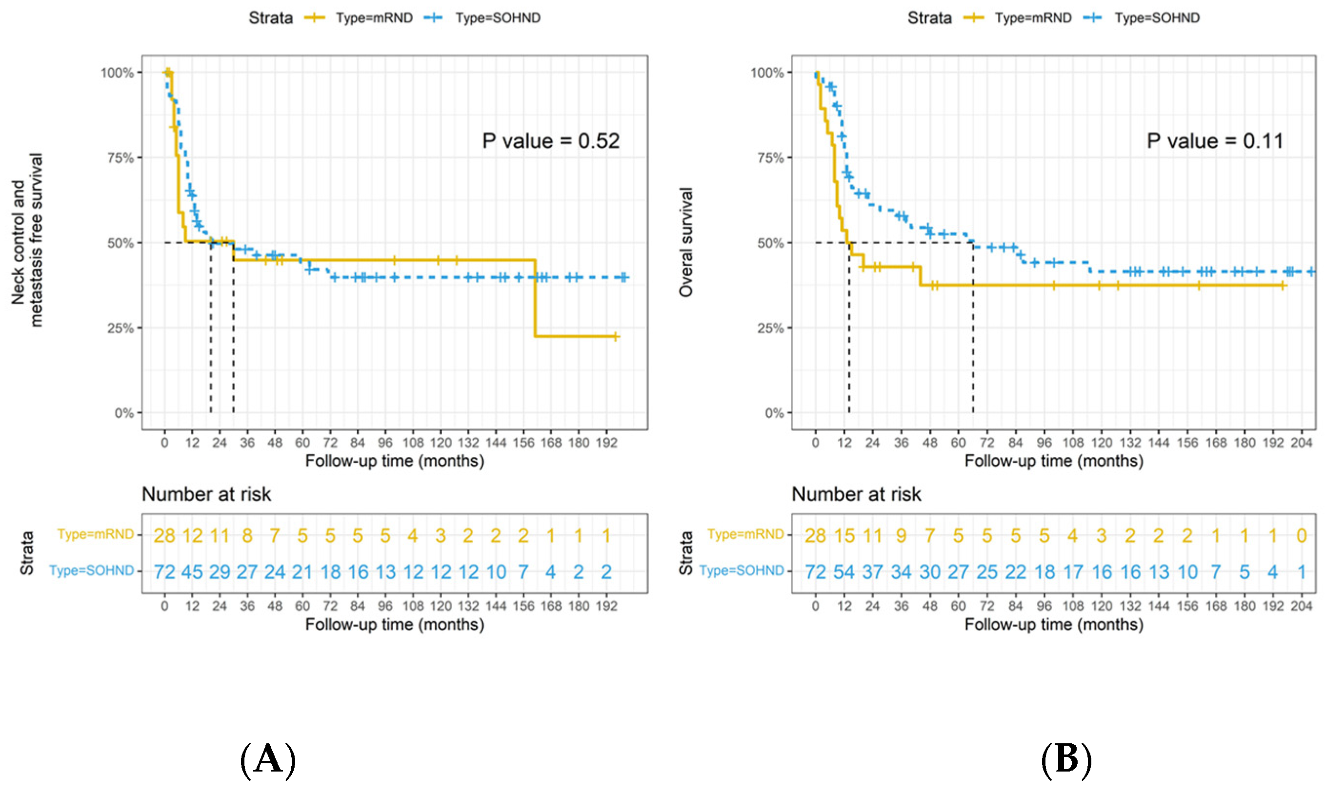
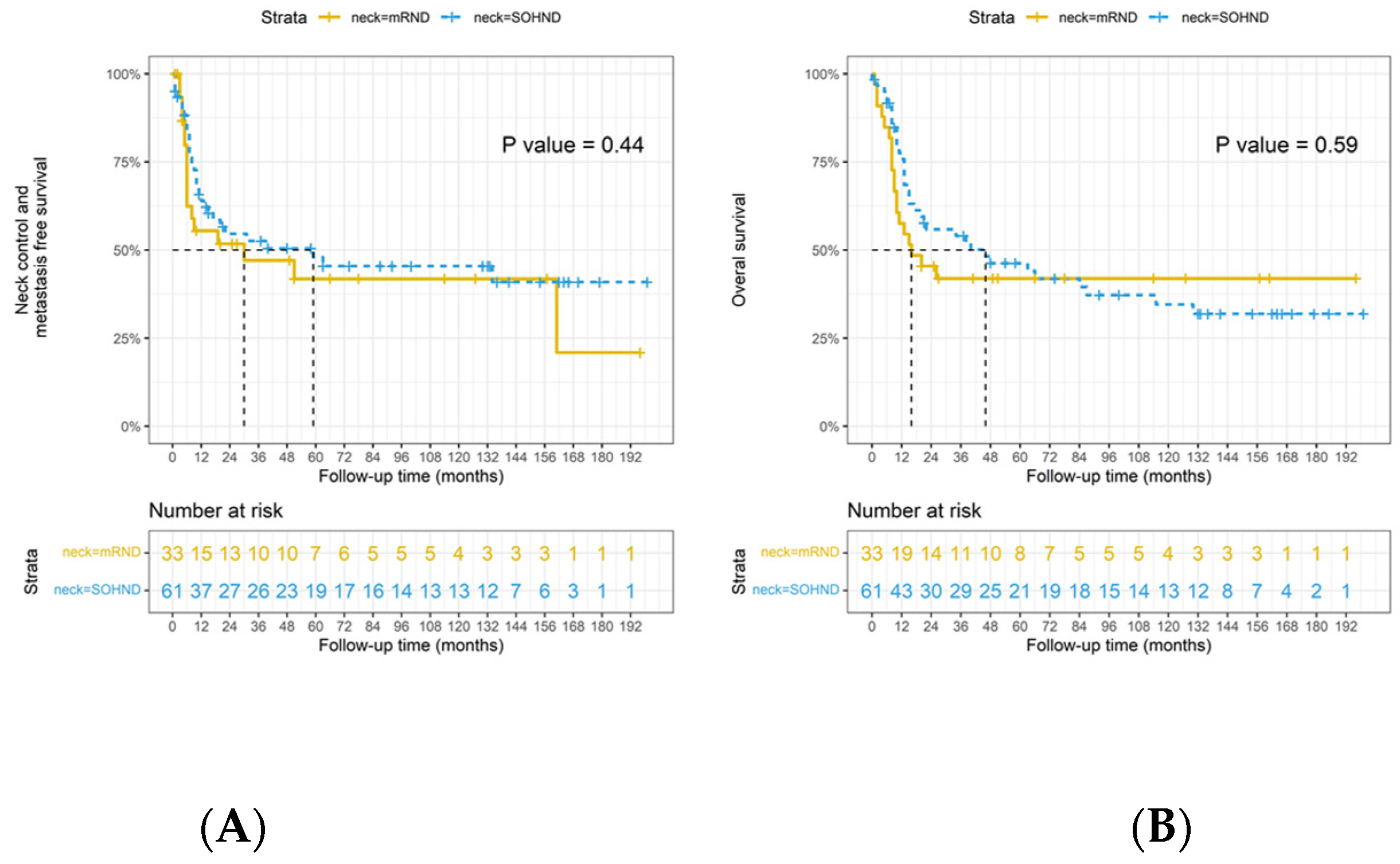
The x2 test evaluated differences and relationships between categorical parameters. Survival analysis was performed considering as outcomes the overall survival (OS), defined as the time between the date of treatment and the date of death; disease-free survival (DFS), defined as the time between the date of treatment and recurrence; and neck control and metastases-free survival (NC-MFS), as defined as a regional or distant relapse of the disease. We evaluated the impact of the different types of neck dissections on the aforementioned oncological outcomes. We first assessed the whole cohort of patients; then, we further analyzed the subgroups of patients affected by multiple nodal disease and extranodal extension (ENE) because these are the subcategory believed to benefit from more extended neck treatment. Survival curves were plotted by the Kaplan–Meier method, and the log-rank test compared the differences between curves. To reduce the effect of confounder values in OS and DFS, with conducted a multivariable analysis with the Cox regression model. Statistical analyses were performed by SPSS 18.0 software (SPSS Inc., Chicago, IL, USA), and p < 0.05 was considered statistically significant. The survival curves were constructed with packages of “survival” and “survminer” in R language version 4.1.0 (R Foundation for Statistical Computing, Vienna, Austria) [30,31,32,33].

3. Results

3.1. Patients, Disease, and Treatment Characteristics

We collected data from 583 patients affected by cT1/T2 OTSCC treated at our institution between March 1997 and May 2017. After a careful assessment, we excluded 18 patients due to missing data, identifying a total number of 565 patients amenable to the study. The clinical characteristics of the patients are presented in Supplementary Table S1. The mean age of the study group was 49.73 years old; it was mainly composed of males (85.8%). Among this cohort, 256 patients (45.3%) were initially classified as cT1 and 309 (54.7%) as cT2. After the pathology report’s results and evaluation of the depth of invasion, 175 were staged as pT1 (31%), 199 (35.3%) as pT2, and 191 (33.8%) as pT3, according to the AJCC 8th edition staging system. After preoperative staging, 501 patients were considered negative for regional metastasis (cN0) and therefore underwent an elective SOHND, whereas 64 patients showed a suspicious pathological lymph nodes disease (cN+); consequently, according to the department’s policy, an mRND was performed. The pathologic characteristics of the cohort are listed in Table 1. The occult nodal metastases (cN0/pN+) rate was 28.7% in the SOHND group. In the mRND group, nodal metastases were confirmed in 62.5% of cases, with 184 patients affected by regional disease. Regarding the site of pathologically confirmed metastasis (Supplementary Table S2), level I was the most involved (58.1%), followed by level II (55.9%) and level III (26.6%); level IV and V were affected in a small percentage of cases: 9.3% and 1.5%, respectively. No skip metastases were detected. In the SOHND group, 71.3% of the patients were pN0, 11% pN1, 5.6% pN2, and 12.2% were affected by pN3b disease. In the mRND group, the composition was: pN0 37.5%, pN1 6.2%, pN2 4.7% and pN3b 51.6%. Patients that received mRND had a higher rate of pN3b with a statistically significant difference compared to the SOHND cohort (p < 0.001). Results are summarized in Table 2.
We further analyzed the subgroups of patients with multiple nodal metastases (Table 3) and ENE disease. These are the subcategory believed to benefit from more extensive neck treatment. In addition, we identified a different ENE distribution in patients who underwent mRND, with a higher rate of extracapsular spread than the SOHND group, 89.3% and 61.1%, respectively (p < 0.001).

3.2. Survival Analysis by Type on Neck Dissection

Considering the OS as the endpoint, in the pathologic positive lymph nodes group, the 3 years OS rate was 63% for the SOHND and 47% for the mRND; we did not observe a statistically significant difference between the two (p = 0.119) (Figure 1). Moreover, no difference was revealed in regional relapse and distant failure rate (p = 0.509) (Figure 1). The NC-MFS curves did not show survival advantages in the mRND group compared to the SOHND group.
In the cohort of patients affected by multiple nodal diseases, the 3 years OS rate was 58% in the SOHND group and 43% in the mRND group (Figure 2). There was no significant difference (p = 0.105) in the overall death rate between the two treatments. Furthermore, no statistically significant different trend (p = 0.523) was observed in DFS and NC-MFS. (Figure 2). Lastly, regarding the subgroup of patients with ENE’s presence, the 3 years OS was 54% in patients subjected to SOHND and 42% in patients who underwent mRND. No statistically significant differences were identified affecting any of the outcomes assessed: OS (p = 0.592), DFS (p = 0.444), and NC-MFS (Figure 3).
The associations of clinicopathologic factors, including age, pathologic T stage, pathologic N stage, the type of ND, histologic grade, and PNI status with DFS and OS, are summarized in Table 4. At the multivariable analysis, the risk factors that significantly affected the DFS were: a positive lymph node (pN+ENE−), (HR: 1.603; 95% C.I.: 1.085–2.369, p = 0.018), and the presence of PNI (HR 1.398, 95% C.I.: 1.023–1.910, p = 0.035), whereas an age ≥ 50 had a protective effect (p = 0.043). Furthermore, the combination of pN+ with ENE increased the risk of recurrence b twofold (HR: 2.034, 95% C.I.: 1.381–2.995; p ≤ 0.001).
Analyzing the OS, the variables determining a negative impact on outcome were an increase in pT stage: pT2 (HR: 2.245, 95% C.I.: 1.310–3.848, p = 0.003) and pT3 (HR: 2.910, 95% C.I.:1.691–5.007, p < 0.001), respectively; furthermore, the presence of pN+ ENE(−) and (pN(+) ENE(+), HR: 3.756; 95% C.I.: 2.510–5.769; p < 0.001) were confirmed to represent a detrimental prognosticator likewise in OS. On the other hand, the covariable “type of neck dissection” showed no significant effect on any of the survival outcomes studied.

4. Discussion

The indications and the extent of neck dissection in early-stage tongue cancer have historically been a matter of debate for different reasons, arguing the risk–benefit evaluation between the probability of neck metastases, complications, and the prognostic influence of delayed diagnosis of metastasis during follow-up [23]. Unfortunately, retrospective studies on cN0 cT1/T2 OTSCC did not help solve this problem; moreover, in cN+, the rationale between the extent of the dissection and improvement of survival is not well defined yet [34]. With this study, we report the clinical outcomes of cT1/T2 OTSCC following a retrospective pathology review of specimens according to AJCC 8th edition guidelines [7]; evaluate the impact of SOHND versus mRND in pN0 and pN+; and, in the case of ENE, we evaluate the percentage of skip metastasis.
Decades after Crile first described the radical neck dissection, shoulder and neck pain following the procedure was viewed as a minor and acceptable side effect [35]. Even the initial description of the disability after radical neck dissection in 1951 [36] characterized postoperative discomfort as “variable and seldom incapacitating”; they noted that most patients “continue without much difficulty to their living as before surgery” [36]. However, a systematic review of the literature reported that the prevalence of shoulder pain and dysfunction after mRND was markedly higher compared with SND (range, 9–25%). Moreover, mRND was associated with a reduced health-related quality of life [37]. Therefore, surgeons nowadays are motivated to modify the classic radical neck dissection to preserve function and improve life’s quality [20] while achieving equivalent oncologic effectiveness [38,39]. Thus, over the past 50 years, there have been sequential developments in modified, functional, and selective neck dissections [40]. Moreover, surgeons kept paying more attention to mini-invasive approaches, hidden-scar incisions, and postoperative rehabilitation [35,41].
Concerning oral tongue cancer, regional lymph node metastasis incidence varies from 6% to 85% [11,13,15]. Since then, the need for lymph node clearance in cT1-2N0 tongue cancers has been a matter of much debate. Finally, in a randomized controlled trial from 2015, D’Cruz et al. demonstrated that END in patients with early-stage oral carcinoma improves overall and disease-free survival compared to observation [42]. They reported a rate of 30% of occult nodal metastasis (false negative cN0/pN+), in line with our result of 28.7% and ones from previous literature [22], reaching 35% in cases of floor-of-mouth involvement [43]. The following year, a metanalysis confirmed that END significantly reduces the regional failure rate and improves DSS in patients with cT1-2N0 oral cancer [44].
An up-to-date meta-analysis, including 19 studies, reported that the occult nodal metastasis rate ranges from 10.2% to 44.6% (mean 24.4%) [16]. The authors, analyzing T1 and T2 separately, found that the occult metastasis rate was not significantly different from the studies with more T2 cases than T1, which means that the nodal spread does not depend only on the T stage; thus, elective neck treatment might be needed even in minor cases.
A milestone study carried out in India [42] and those that followed [16,44] demonstrated that all cT1/T2 OTSCC need to receive elective neck dissection since 25–30% is an unacceptable rate of subclinical neck metastasis to avoid neck clearance, despite T stage. However, we decided to enroll only patients affected by early clinical stage (cT1–T2), given that, in similar studies, an advanced stage demonstrated an influence on the DSS and OS, regardless of the type of neck dissection [45].
Nevertheless, the presence of N+ is one of the more significant prognosticators [11]. Thus, elective mRND has been part of treating patients with cN+ squamous cell carcinoma of the oral tongue [46,47]; ideally, it should be avoided in patients with clinically false-positive and true-negative lymph nodes. We noted that 37.5% of patients that received MRND for cN+ disease turned to be pN0 (false positive), meaning that more than one-third of the cohort were overtreated and, instead, could have undergone SOHND obtaining an equal cure rate (the covariable “type of neck dissection” was demonstrated to not significantly affect any of the survival outcomes assessed). Similarly, Iype et al. reported no significant difference in regional recurrence between pN0 and pN+ oral cancer patients treated with SOHND; there was no difference even in regional recurrence outside the surgical field (level IV–V), confirming the rarity of lower neck occult disease. In line with our findings, Kowalski demonstrated 0% of level V involvement/recurrence in N1-2a oral cancer treated with I–III therapeutic neck dissection [24].
Additionally, from another perspective, a few studies have demonstrated an exceedingly low-level V metastasis rate in patients who underwent mRND for pN+ oral cancer, ranging from 0% to 6% in the case of multiple I–IV nodal metastases [48,49]. On the contrary, we encountered only 1.5% level V metastasis following MRND in cN+ patients. Therefore, even in the context of multiple regional metastases, the spread along the nodal echelons has been demonstrated to be constant and predictable.
The evidence from the abundant literature might guide us to safely propose SOHND in cT1-2 N+ oral squamous cell carcinoma [24,35,43,48,49,50,51]. Our current study pointed it out specifically for tongue cancer; moreover, the literature further corroborates our findings that, in the subset of patients affected by T1/2 N1/2, loco-regional control seems to depend on the proper use of adjuvant treatment [28,29] rather than the extension of the neck dissection [24,43,52,53]. Multiple nodal metastases and ENE mainly affect OS, increasing distant failure, not loco-regional control.
Regarding treatment choice in the presence of ENE, our investigation introduces another valuable piece of information: ENE needs to be distinguished from N3b. Our survival analysis in ENE cases has not indicated any benefits in performing mRND compared to SOHND. Recent studies supported these findings and demonstrated how the pN3b category was more predictive of poor prognosis than ENE alone [2,12]. Interestingly, single nodal metastasis with ENE was not significantly associated with a poor prognosis. Once again, a high number of positive nodes [53,54] showed a prognostic weight overwhelming the presence of ENE and other survival indicators. These patients are a minority; ENE is often observed in necks with multiple metastases [2]. We demonstrated that, in cT1-2 N0/+ tongue cancer, SOHND is oncologically safe, even in the presence of ENE.
The strength of our work is that we focused on a large and homogeneous cohort of cT1/T2 OTSCC patients treated in a single institution with uniform treatment guidelines, comparing two different treatment strategies. However, the retrospective nature of this study represents its principal limit. We hope that further research will clarify whether a particular subset of patients with multiple lower neck nodal metastasis might benefit or not benefit from mRND, followed by adjuvant treatment. More interesting pathological factors could be studied in such a setting, such as the lymph-node size, the worst pattern of invasion, and the micro- vs. macro-ENE, to justify the need for adjuvant treatment.

5. Conclusions

Treating cT1-2 N0/+ tongue cancer with SOHND is oncologically safe and justified by the close-to-zero rate of skip metastasis. Furthermore, ENE and multiple nodes do not benefit from a more extensive neck dissection since it might affect the distant control primarily.

Supplementary Materials

The following supporting information can be downloaded at: https://www.mdpi.com/article/10.3390/jpm12091535/s1; Table S1: The clinical characteristics of the patients, Table S2: Lymph node level involved in patients who received modified radical neck dissection (mRND).

Author Contributions

Conceptualization, A.I. and S.-F.H.; methodology, C.-T.L. and S.-F.H.; software S.-F.H.; validation, C.-T.L., C.-K.T. and C.-J.K.; formal analysis, S.-F.H.; investigation, A.-C.C. and C.-K.Y.; resources, C.-T.L. and S.-F.H.; data curation, C.-J.K., H.-M.W. and T.-C.J.C.; writing—original draft preparation, A.I. and F.M.; writing—review and editing, A.I., F.M. and S.-F.H.; visualization, F.M.; supervision, C.-T.L., C.-K.T., C.-J.K. and S.-F.H.; project administration, S.-F.H.; funding acquisition, S.-F.H. All authors have read and agreed to the published version of the manuscript.

Funding

This study was supported by grants (CMRPG3L0861, CMRPG3J0593, CMRPG3M1101, and CMRPB53) from Chang Gung Memorial Hospital and a grant (MOST 110-2314-B-182-045-MY3) from the Ministry of Science and Technology, Executive Yuan, Taiwan, ROC.

Institutional Review Board Statement

The study was conducted in accordance with the Declaration of Helsinki and approved by Institution Review Board at Chang Gung Medical Foundation, Taiwan (IRB No. 202101807B0).

Informed Consent Statement

Informed consent was obtained from all subjects involved in the study.

Data Availability Statement

The data was available by request to the correspondence author.

Acknowledgments

The authors thank all members of the Cancer Center, Chang Gung Memorial Hospital, Linkou, Taiwan, for their invaluable help.

Conflicts of Interest

The authors declare no conflict of interest.

References

  1. Xu, B.; Salama, A.M.; Valero, C.; Yuan, A.; Khimraj, A.; Saliba, M.; Zanoni, D.K.; Ganly, I.; Patel, S.G.; Katabi, N.; et al. The prognostic role of histologic grade, worst pattern of invasion, and tumor budding in early oral tongue squamous cell carcinoma: A comparative study. Virchows Arch. Int. J. Pathol. 2021, 47, 597–606. [Google Scholar] [CrossRef]
  2. Tsai, T.Y.; Iandelli, A.; Marchi, F.; Huang, Y.; Tai, S.F.; Hung, S.Y.; Kao, H.K.; Chang, K.P. The Prognostic Value of Lymph Node Burden in Oral Cavity Cancer: Systematic Review and Meta-Analysis. Laryngoscope 2021, 132, 88–95. [Google Scholar] [CrossRef]
  3. Zanoni, D.; Montero, P.; Migliacci, J.; Shah, J.; Ganly, I.; Patel, S.G. Survival outcomes after treatment of cancer of the oral cavity (1985–2015). Oral Oncol. 2020, 90, 115–121. [Google Scholar] [CrossRef] [PubMed]
  4. Balasubramanian, D.; Thankappan, K.; Battoo, A.J.; Rajapurkar, M.; Kuriakose, M.A.; Iyer, S. Isolated skip nodal metastasis is rare in T1 and T2 oral tongue squamous cell carcinoma. Otolaryngol. Head Neck Surg. Off. J. Am. Acad. Otolaryngol. Head Neck Surg. 2012, 147, 275–277. [Google Scholar] [CrossRef] [PubMed]
  5. Henley, S.J.; Ward, E.M.; Scott, S.; Anderson, R.N.; Firth, A.U.; Thomas, C.C.; Islami, F.; Weir, H.K.; Lewis, D.R.; Sherman, R.L.; et al. Annual Report to the Nation on the Status of Cancer, Part I: National Cancer Statistics. Cancer 2020, 126, 2225–2249. [Google Scholar] [CrossRef] [PubMed]
  6. Marur, S.; Forastiere, A.A. Head and Neck Squamous Cell Carcinoma: Update on Epidemiology, Diagnosis, and Treatment. Mayo Clin. Proc. 2016, 91, 386–396. [Google Scholar] [CrossRef]
  7. American Joint Committee on Cancer. AJCC Cancer Staging Manual, 8th ed.; Springer: New York, NY, USA, 2017. [Google Scholar]
  8. Lee, H.; Roh, J.L.; Cho, K.J.; Choi, S.H.; Nam, S.Y.; Kim, S.Y. Number of positive lymph nodes better predicts survival for oral cavity cancer. J. Surg. Oncol. 2019, 119, 675–682. [Google Scholar] [CrossRef]
  9. Ebrahimi, A.; Gupta, R.; Luk, P.; Low, T.H.H.; McDowell, L.; Magarey, M.J.; Smith, P.N.; Perriman, D.M.; Schulte, K.M.; Veness, M.; et al. Number of nodal metastases and the American Joint Committee on cancer staging of head and neck cutaneous squamous cell carcinoma: A multicenter study. Oral Oncol. 2020, 111, 104855. [Google Scholar] [CrossRef] [PubMed]
  10. Subramaniam, N.; Balasubramanian, D.; Kumar, N.; Murthy, S.; Vijayan, S.N.; Nambiar, A.; Vidhyadharan, S.; Thankappan, K.; Iyer, S. Lymph node staging systems in oral squamous cell carcinoma: A comparative analysis. Oral Oncol. 2019, 97, 92–98. [Google Scholar] [CrossRef]
  11. Balasubramanian, D.; Vijayan, S.; Nambiar, A.; Mattavelli, D. Predictive nomograms for oral tongue squamous cell carcinoma applying the American Joint Committee on Cancer/Union Internationale Contre le Cancer 8th edition staging system. Head Neck 2021, 43, 1043–1055. [Google Scholar] [CrossRef]
  12. Mattavelli, D.; Ferrari, M.; Taboni, S.; Morello, R.; Paderno, A.; Rampinelli, V.; Del Bon, F.; Lombardi, D.; Grammatica, A.; Bossi, P.; et al. The 8th TNM classification for oral squamous cell carcinoma: What is gained, what is lost, and what is missing. Oral Oncol. 2020, 111, 104937. [Google Scholar] [CrossRef] [PubMed]
  13. Shah, J.P.; Strong, E.; Spiro, R.H.; Vikram, B. Surgical grand rounds. Neck dissection: Current status and future possibilities. Clin. Bull. 1981, 11, 25–33. [Google Scholar] [PubMed]
  14. Satgunaseelan, L.; Allanson, B.M.; Asher, R.; Reddy, R.; Low, H.T.; Veness, M.; Iyer, N.G.; Smee, R.I.; Palme, C.E.; Gupta, R.; et al. The incidence of squamous cell carcinoma of the oral tongue is rising in young non-smoking women: An international multi-institutional analysis. Oral Oncol. 2020, 110, 104875. [Google Scholar] [CrossRef]
  15. Lindberg, R. Distribution of cervical lymph node metastases from squamous cell carcinoma of the upper respiratory and digestive tracts. Cancer 1972, 29, 1446–1449. [Google Scholar] [CrossRef]
  16. Choi, K.Y.; Park, S.C.; Kim, J.H.; Lee, D.J. The occult nodal metastasis rate of early tongue cancer (T1-T2): A protocol for a systematic review and meta-analysis. Medicine 2021, 100, e24327. [Google Scholar] [CrossRef]
  17. Chatterjee, D.; Bansal, V.; Malik, V.; Bhagat, R.; Punia, R.S.; Handa, U. Tumor Budding and Worse Pattern of Invasion Can Predict Nodal Metastasis in Oral Cancers and Associated with Poor Survival in Early-Stage Tumors. Ear Nose Throat J. 2019, 98, E112–E119. [Google Scholar] [CrossRef]
  18. Ahlberg, A.; Nikolaidis, P.; Engström, T.; Gunnarsson, K.; Johansson, H.; Sharp, L.; Laurell, G. Morbidity of supraomohyoidal and modified radical neck dissection combined with radiotherapy for head and neck cancer: A prospective longitudinal study. Head Neck 2012, 34, 66–72. [Google Scholar] [CrossRef]
  19. Popescu, B.; Berteşteanu, S.V.G.; Grigore, R.; Scăunaşu, R.; Popescu, C.R. Functional implications of radical neck dissection and the impact on the quality of life for patients with head and neck neoplasia. J. Med. Life 2012, 5, 410–413. [Google Scholar]
  20. Terrell, J.E.; Welsh, D.E.; Bradford, C.R.; Chepeha, D.B.; Esclamado, R.M.; Hogikyan, N.D.; Wolf, G.T. Pain, quality of life, and spinal accessory nerve status after neck dissection. Laryngoscope 2000, 110, 620–626. [Google Scholar] [CrossRef]
  21. Huang, S.F.; Kang, C.J.; Lin, C.Y.; Fan, K.H.; Yen, T.C.; Wang, H.M.; Chen, I.H.; Liao, C.T.; Cheng, A.J.; Chang, J.T.C. Neck treatment of patients with early stage oral tongue cancer: Comparison between observation, supraomohyoid dissection, and extended dissection. Cancer 2008, 112, 1066–1075. [Google Scholar] [CrossRef]
  22. Flach, G.B.; Tenhagen, M.; de Bree, R.; Brakenhoff, R.H.; van der Waal, I.; Bloemena, E.; Kuik, D.J.; Castelijns, J.A.; Leemans, C.R. Outcome of patients with early stage oral cancer managed by an observation strategy towards the N0 neck using ultrasound guided fine needle aspiration cytology: No survival difference as compared to elective neck dissection. Oral Oncol. 2013, 49, 157–164. [Google Scholar] [CrossRef] [PubMed]
  23. Kowalski, L.P.; Sanabria, A. Elective neck dissection in oral carcinoma: A critical review of the evidence. Acta Otorhinolaryngol. Ital. 2007, 27, 113–117. [Google Scholar] [PubMed]
  24. Kowalski, L.P.; Carvalho, A.L. Feasibility of supraomohyoid neck dissection in N1 and N2a oral cancer patients. Head Neck 2002, 24, 921–924. [Google Scholar] [CrossRef] [PubMed]
  25. Andersen, P.E.; Warren, F.; Spiro, J.; Burningham, A.; Wong, R.; Wax, M.K.; Shah, J.P.; Cohen, J.I. Results of selective neck dissection in management of the node-positive neck. Arch. Otolaryngol. Head Neck Surg. 2002, 128, 1180–1184. [Google Scholar] [CrossRef]
  26. Patel, R.S.; Clark, J.R.; Gao, K.; O’Brien, C.J. Effectiveness of selective neck dissection in the treatment of the clinically positive neck. Head Neck 2008, 30, 1231–1236. [Google Scholar] [CrossRef]
  27. Byers, R.M.; Weber, R.S.; Andrews, T.; McGill, D.; Kare, R.; Wolf, P. Frequency and therapeutic implications of “skip metastases” in the neck from squamous carcinoma of the oral tongue. Head Neck 1997, 19, 14–19. [Google Scholar] [CrossRef]
  28. Lin, C.Y.; Fan, K.H.; Lee, L.Y.; Hsueh, C.; Yang, L.Y.; Ng, S.H.; Wang, H.M.; Hsieh, C.H.; Lin, C.H.; Tsao, C.K.; et al. Precision Adjuvant Therapy Based on Detailed Pathologic Risk Factors for Resected Oral Cavity Squamous Cell Carcinoma: Long-Term Outcome Comparison of CGMH and NCCN Guidelines. Int. J. Radiat. Oncol. Biol. Phys. 2020, 106, 916–925. [Google Scholar] [CrossRef]
  29. Pfister, D.G.; Spencer, S.; Adelstein, D.; Adkins, D.; Anzai, Y.; Brizel, D.M.; Bruce, J.Y.; Busse, P.M.; Caudell, J.J.; Cmelak, A.J.; et al. Head and Neck Cancers, Version 2.2020, NCCN Clinical Practice Guidelines in Oncology. J. Natl. Compr. Cancer Netw. 2020, 18, 873–898. [Google Scholar] [CrossRef]
  30. R Core Team. The R Project for Statistical Computing. 2021. Available online: https://www.R-project.org/ (accessed on 27 August 2022).
  31. Therneau, T.M. A Package for Survival Analysis in R. R Package Version 3.4-0. 2022. Available online: https://CRAN.R-project.org/package=survival (accessed on 27 August 2022).
  32. Therneau, T.M.; Grambsch, P.M. Modeling Survival Data: Extending the Cox Model, 1st ed.; Springer: New York, NY, USA, 2000. [Google Scholar] [CrossRef]
  33. Kassambra, A.; Kosinski, M.; Biecek, P.; Fabian, S. Drawing Survival Curves Using “ggplot2”. R Package Version 0.4.9. 2021. Available online: https://CRAN.R-project.org/package=survminer (accessed on 27 August 2022).
  34. de Bree, R.; van den Brekel, M.W.M. Elective neck dissection versus observation in the clinically node negative neck in early oral cancer: Do we have the answer yet? Oral Oncol. 2015, 51, 963–965. [Google Scholar] [CrossRef]
  35. Ferlito, A.; Rinaldo, A.; Silver, C.E.; Shah, J.P.; Suárez, C.; Medina, J.E.; Kowalski, L.P.; Johnson, J.T.; Strome, M.; Rodrigo, J.P.; et al. Neck dissection: Then and now. Auris Nasus Larynx 2006, 33, 365–374. [Google Scholar] [CrossRef]
  36. Ewing, M.; Martin, H. Disability following radical neck dissection; an assessment based on the postoperative evaluation of 100 patients. Cancer 1952, 5, 873–883. [Google Scholar] [CrossRef]
  37. Gane, E.M.; Michaleff, Z.A.; Cottrell, M.A.; McPhail, S.M.; Hatton, A.L.; Panizza, B.J.; O’Leary, S.P. Prevalence, incidence, and risk factors for shoulder and neck dysfunction after neck dissection: A systematic review. Eur. J. Surg. Oncol. 2017, 43, 1199–1218. [Google Scholar] [CrossRef] [PubMed]
  38. Ferlito, A.; Rinaldo, A. Osvaldo Suárez: Often-forgotten father of functional neck dissection (in the non-Spanish-speaking literature). Laryngoscope 2004, 114, 1177–1178. [Google Scholar] [CrossRef] [PubMed]
  39. Gavilán, C.; Gavilán, J. Five-year results of functional neck dissection for cancer of the larynx. Arch. Otolaryngol. Head Neck Surg. 1989, 115, 1193–1196. [Google Scholar] [CrossRef] [PubMed]
  40. Bocca, E.; Pignataro, O.; Oldini, C.; Cappa, C. Functional neck dissection: An evaluation and review of 843 cases. Laryngoscope 1984, 94, 942–945. [Google Scholar] [CrossRef]
  41. Nibu, K.I.; Ebihara, Y.; Ebihara, M.; Kawabata, K.; Onitsuka, T.; Fujii, T.; Saikawa, M. Quality of life after neck dissection: A multicenter longitudinal study by the Japanese Clinical Study Group on Standardization of Treatment for Lymph Node Metastasis of Head and Neck Cancer. Int. J. Clin. Oncol. 2010, 15, 33–38. [Google Scholar] [CrossRef]
  42. D’Cruz, A.K.; Vaish, R.; Kapre, N.; Dandekar, M.; Gupta, S.; Hawaldar, R.; Agarwal, J.P.; Pantvaidya, G.; Chaukar, D.; Deshmukh, A.; et al. Elective versus Therapeutic Neck Dissection in Node-Negative Oral Cancer. N. Engl. J. Med. 2015, 373, 521–529. [Google Scholar] [CrossRef]
  43. Iype, E.M.; Sebastian, P.; Mathew, A.; Balagopal, P.G.; Varghese, B.T.; Thomas, S. The role of selective neck dissection (I-III) in the treatment of node negative (N0) neck in oral cancer. Oral Oncol. 2008, 44, 1134–1138. [Google Scholar] [CrossRef]
  44. Abu-Ghanem, S.; Yehuda, M.; Carmel, N.N.; Leshno, M.; Abergel, A.; Gutfeld, O.; Fliss, D.M. Elective Neck Dissection vs Observation in Early-Stage Squamous Cell Carcinoma of the Oral Tongue With No Clinically Apparent Lymph Node Metastasis in the Neck: A Systematic Review and Meta-analysis. JAMA Otolaryngol. Head Neck Surg. 2016, 142, 857–865. [Google Scholar] [CrossRef]
  45. Kolli, V.R.; Datta, R.V.; Orner, J.B.; Hicks, W.L.; Loree, T.R. The role of supraomohyoid neck dissection in patients with positive nodes. Arch. Otolaryngol. Head Neck Surg. 2000, 126, 413–416. [Google Scholar] [CrossRef] [Green Version]
  46. Byers, R.M. Modified neck dissection. A study of 967 cases from 1970 to 1980. Am. J. Surg. 1985, 150, 414–421. [Google Scholar] [CrossRef]
  47. Byers, R.M.; Wolf, P.F.; Ballantyne, A.J. Rationale for elective modified neck dissection. Head Neck Surg. 1988, 10, 160–167. [Google Scholar] [CrossRef] [PubMed]
  48. Lim, Y.C.; Koo, B.S.; Lee, J.S.; Choi, E.C. Level V lymph node dissection in oral and oropharyngeal carcinoma patients with clinically node-positive neck: Is it absolutely necessary? Laryngoscope 2006, 116, 1232–1235. [Google Scholar] [CrossRef] [PubMed]
  49. Jayasuriya, N.S.S.; Mannapperuma, N.T.; Siriwardana, S.; Attygalla, A.M.; DeSilva, S.; Jinadasa, H.; Ekanayaka, R.; Dias, D.K.; Wadusinghearachi, S.; Perera, I. Incidence of metastasis to level V lymph nodes in clinically positive necks among Sri Lankan patients with oral squamous cell carcinoma. Br. J. Oral Maxillofac. Surg. 2021, 59, 771–775. [Google Scholar] [CrossRef] [PubMed]
  50. Battoo, A.J.; Hedne, N.; Ahmad, S.Z.; Thankappan, K.; Iyer, S.; Kuriakose, M.A. Selective neck dissection is effective in N1/N2 nodal stage oral cavity squamous cell carcinoma. J. Oral Maxillofac. Surg. Off. J. Am. Assoc. Oral Maxillofac. Surg. 2013, 71, 636–643. [Google Scholar] [CrossRef]
  51. Subramaniam, N.; Balasubramanian, D.; Murthy, S.; Thankappan, K.; Iyer, S. European Journal of Surgical Oncology Predictors of loco-regional control in stage I/II oral squamous cell carcinoma classi fi ed by AJCC 8th edition. Eur. J. Surg. Oncol. 2019, 45, 2126–2130. [Google Scholar] [CrossRef]
  52. Larson, A.R.; Kemmer, J.; Formeister, E.; El-Sayed, I.; Ha, P.; George, J.; Ryan, W.; Chan, E.; Heaton, C. Beyond Depth of Invasion: Adverse Pathologic Tumor Features in Early Oral Tongue Squamous Cell Carcinoma. Laryngoscope 2020, 130, 1715–1720. [Google Scholar] [CrossRef]
  53. Ho, A.S.; Kim, S.; Tighiouart, M.; Gudino, C.; Mita, A.; Scher, K.S.; Laury, A.; Prasad, R.; Shiao, S.L.; Van Eyk, J.E.; et al. Metastatic lymph node burden and survival in oral cavity cancer. J. Clin. Oncol. 2017, 35, 3601–3609. [Google Scholar] [CrossRef]
  54. Choi, Y.; Bin-Manie, M.; Roh, J.L.; Cho, K.J.; Lee, Y.S.; Choi, S.H.; Nam, S.Y.; Kim, S.Y. Metastatic lymph node burden predictive of survival in patients undergoing primary surgery for laryngeal and hypopharyngeal cancer. J. Cancer Res. Clin. Oncol. 2019, 145, 2565–2572. [Google Scholar] [CrossRef]
Figure 1. (A) Disease-free survival differences in pN+ patients by types of neck dissection (n = 184, p = 0.51); (B) overall survival differences in pN+ patients by types of neck dissection (p = 0.12). Dotted line indicated the survival time that half of the patients had no events.
Jpm 12 01535 g001
Figure 2. (A) Disease-free survival differences in multiple nodal disease by types of neck dissection (n =100, p = 0.52); (B) overall survival differences in multiple nodal disease by types of neck dissection (p = 0.11). Neck-control-distant metastases-free survival in multiple nodal disease by types of neck dissection. Dotted line indicated the survival time that half of the patients had no events.
Jpm 12 01535 g002
Figure 3. (A) Disease-free survival differences in extranodal extension+ (ENE+) by types of neck dissection (n = 94, p = 0.44); (B) overall survival differences in extranodal extension+ (ENE+) by types of neck dissection (p = 0.59). Neck-control-distant metastases-free survival in extranodal extension+ (ENE+) by types of neck dissection. Dotted line indicated the survival time that half of the patients had no events.
Jpm 12 01535 g003
Table 1. Patient’s clinical data and pathological characteristics.
CharacteristicsTotal(%)pN0(%)pN+(%)p
cT stage T125645.321556.4 4122.3<0.001
T2309 54.7 15143.610777.7
cN stageN038167.4-----
N18414.9-----
N2b9015.9-----
N2c101.8-----
pT stageT117531.015841.179.2<0.001
T219935.213936.56032.6
T319133.88422.010758.2
pN stageN038167.4-----
N15910.4-----
N2b305.3-----
N2c10.2-----
N3b9416.6-----
GradingWell17731.314137.03619.6<0.001
Moderate33158.621556.411663.0
Poor5710.1256.63217.4
PNINo38868.729076.19853.3<0.001
Yes17731.39123.98646.7
VINo-------
Yes-------
LINo54195.837799.016489.1<0.001
yes244.241.02010.9
PNI: perineural invasion; VI: vascular invasion; LI: lymphatic invasion.
Table 2. Relationship between pathologic nodal status and type of ND in different AJCC editions.
AJCC 7th Ed.
pN Status
SOHND (%)mRND (%)AJCC 8th Ed.
pN Status
SOHND (%)mRND (%)p
pN0381 (67.4)357 (71.3)24 (37.5)381 (67.4)357 (71.3)24 (37.5)<0.001
pN184 (14.9)72 (14.4)12 (18.8)59 (10.4)55 (11.0)4 (6.2)
pN2a 25 (4.4)17 (3.4)8 (12.5)
pN2b90 (15.9)66 (13.2)24 (37.5)30 (5.3)27 (5.4)3 (4.7)
pN2c10 (1.8)6 (1.2)4 (6.3)1 (0.2)1 (0.2)0 (0.0)
pN3b 69 (12.2)44 (8.8)25 (36.2)
Total 501 (100.0)64 (100.0) 501 (100.0)64 (100.0)
SOHND: supraomohyoid neck dissection; mRND: modified radical neck dissection.
Table 3. Relationship between extranodal extension (ENE) and type of neck dissection in pathologic multiple nodal disease.
AllSOHND (%)mRND (%)p
ENE−3128 (38.90)3 (10.70)<0.001
ENE+6944 (61.10)25 (89.30)
Total1007228
SOHND: supraomohyoid neck dissection; mRND: modified radical neck dissection; ENE: extranodal extension.
Table 4. Multivariate Cox regression analysis for factors influencing disease-free survival and overall survival.
DFS OS
VariablesHR (95% C.I.)pHR (95% C.I.)pHR (95% C.I.)pHR (95% C.I.)p
Age
  <50 years oldRef0.037Ref Ref
  ≥50 years old0.735 (0.550–0.982) 0.737 (0.548–0.991)0.0430.984 (0.715–1.354)0.921
Primary tumor status
  pT1Ref Ref-Ref Ref-
  pT21.489 (1.018–2.179)0.0401.144 (0.766–1.707)0.5112.862 (1.696–4.829)<0.0012.245 (1.310–3.848)0.003
  pT32.261 (1.569–3.258)<0.0011.417 (0.933–2.151)0.1025.032 (3.052–8.298)<0.0012.910 (1.691–5.007)<0.001
Lymph node metastasis
  pN (−) ENE(−)Ref Ref-Ref Ref-
  pN (+) ENE(−)2.045(1.420–2.945)<0.0011.603 (1.085–2.369)0.0182.632(1.732–4.000)<0.0012.001 (1.294–3.094)<0.001
  pN (+) ENE(+)2.743(1.947–3.865)<0.0012.034 (1.381–2.995)<0.0015.491(3.828–7.876)<0.0013.756 (2.510–5.622)<0.001
Type of neck dissection
  SOHND Ref Ref-Ref Ref-
  mRND1.624(1.094–2.411)0.0161.188 (0.783–1.802)0.4182.471 (1.673–3.649)<0.0011.348 (0.887–2.048)0.162
Tumor cell differentiation
  Well/Moderate Ref-Ref
  Poor1.683 (1.113–2.545)0.0141.275 (0.824–1.973)0.2751.212 (0.722–2.034)0.468
PNI
  NoRef<0.001Ref-Ref Ref
  Yes1.816(1.363–2.419) 1.398 (1.023–1.910)0.0351.644 (1.190–2.271)0.0031.006 (0.714–1.417)0.972
SOHND: supraomohyoid neck dissection; mRND: modified radical neck dissection; ENE: extranodal extension; DFS: disease-free survival; OS: overall survival.
Publisher’s Note: MDPI stays neutral with regard to jurisdictional claims in published maps and institutional affiliations.

