
Dealing with Corticosteroid and High-Dose Cyclosporine Therapy in a Pyoderma Gangrenosum Patient Contracting a COVID-19 Infection

 , ,
, ,
Abstract
:1. Introduction
2. Case Report
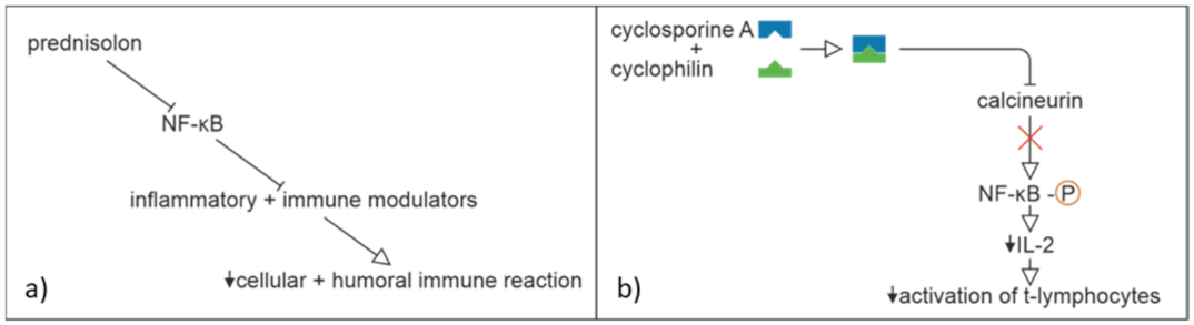
3. Discussion
Author Contributions
Funding
Institutional Review Board Statement
Informed Consent Statement
Data Availability Statement
Acknowledgments
Conflicts of Interest
References
- Yen, C.-C.; Lin, S.-Y.; Chen, S.-C.; Chiu, Y.-W.; Chang, J.-M.; Hwang, S.-J. COVID-19 vaccines in patients with maintenance hemodialysis. J. Pers. Med. 2021, 11, 789. [Google Scholar] [CrossRef] [PubMed]
- Marzano, A.; Borghi, A.; Meroni, P.L.; Cugno, M. Pyoderma gangrenosum and its syndromic forms: Evidence for a link with autoinflammation. Br. J. Dermatol. 2016, 175, 882–891. [Google Scholar] [CrossRef] [PubMed] [Green Version]
- Johnson, K.M.; Belfer, J.J.; Peterson, G.R.; Boelkins, M.R.; Dumkow, L.E. Managing COVID-19 in renal transplant recipients: A review of recent literature and case supporting corticosteroid-sparing immunosuppression. Pharmacother. J. Hum. Pharmacol. Drug Ther. 2020, 40, 517–524. [Google Scholar] [CrossRef] [PubMed]
- Khurana, A.; Sethia, K. Using cyclosporine in the COVID era: An emergent need for caution. J. Am. Acad. Dermatol. 2020, 83, e315–e316. [Google Scholar] [CrossRef]
- World Health Organization (WHO). Corticosteroids for COVID-19. In Living Guid; WHO: Geneva, Switzerland, 2020; pp. 1–25. [Google Scholar]
- Sun, Y.; Zhou, J.; Ye, K. White blood cells and severe COVID-19: A mendelian randomization study WBC COVID-19 SNPs associated with WBC SNPs associated with COVID-19. J. Pers. Med. 2021, 11, 195. [Google Scholar] [CrossRef]
- Zhou, F.; Yu, T.; Du, R.; Fan, G.; Liu, Y.; Liu, Z.; Xiang, J.; Wang, Y.; Song, B.; Gu, X.; et al. Clinical course and risk factors for mortality of adult inpatients with COVID-19 in Wuhan, China: A retrospective cohort study. Lancet 2020, 395, 1054–1062. [Google Scholar] [CrossRef]
- Deutsche Dermatologische Gesellschaft, B. Deutsche Dermatologische Gesellschaft B. S1-Leitlinie Pyoderma Gangrenosum. 2020. AWMF. Available online: https://www.awmf.org/uploads/tx_szleitlinien/013-091l_S1_Pyoderma-gangrenosum_2020-10_1.pdf (accessed on 28 November 2021).
- Monari, P.; Moro, R.; Motolese, A.; Misciali, C.; Baraldi, C.; Fanti, P.A.; Caccavale, S.; Puviani, M.; Olezzi, D.; Zampieri, P.; et al. Epidemiology of pyoderma gangrenosum: Results from an Italian prospective multicentre study. Int. Wound J. 2018, 15, 875–879. [Google Scholar] [CrossRef]
- Powell, F.C.; Su, W.D.; Perry, H.O. Pyoderma gangrenosum: Classification and management. J. Am. Acad. Dermatol. 1996, 34, 392–395. [Google Scholar] [CrossRef]
- Abenavoli, L.; Dastoli, S.; Bennardo, L.; Boccuto, L.; Passante, M.; Silvestri, M.; Proietti, I.; Potenza, C.; Luzza, F.; Nisticò, S.P. The skin in celiac disease patients: The other side of the coin. Medicina 2019, 55, 1–17. [Google Scholar] [CrossRef] [Green Version]
- Maverakis, E.; Ma, C.; Shinkai, K.; Fiorentino, D.; Callen, J.P.; Wollina, U.; Marzano, A.V.; Wallach, D.; Kim, K.; Cheng, M.Y.; et al. Diagnostic criteria of ulcerative pyoderma gangrenosum: A Delphi consensus of international experts. JAMA Dermatol. 2018, 154, 461–466. [Google Scholar] [CrossRef]
- Jockenhöfer, F.; Wollina, U.; Salva, K.; Benson, S.; Dissemond, J. The PARACELSUS score: A novel diagnostic tool for pyoderma gangrenosum. Br. J. Dermatol. 2018, 180, 615–620. [Google Scholar] [CrossRef] [PubMed]
- Marzano, A.V.; Cugno, M.; Trevisan, V.; Fanoni, D.; Venegoni, L.; Berti, E.; Crosti, C. Role of inflammatory cells, cytokines and matrix metalloproteinases in neutrophil-mediated skin diseases. Clin. Exp. Immunol. 2010, 162, 100–107. [Google Scholar] [CrossRef] [PubMed]
- Braswell, S.F.; Kostopoulos, T.C.; Ortega-Loayza, A.G. Pathophysiology of pyoderma gangrenosum (PG): An updated review. J. Am. Acad. Dermatol. 2015, 73, 691–698. [Google Scholar] [CrossRef] [PubMed]
- Skopis, M.; Bag-Ozbek, A. Pyoderma gangrenosum: A review of updates in diagnosis, pathophysiology and management. J 2021, 4, 367–375. [Google Scholar] [CrossRef]
- Wang, E.A.; Steel, A.; Luxardi, G.; Mitra, A.; Patel, F.; Cheng, M.Y.; Wilken, R.; Kao, J.; De Ga, K.; Sultani, H.; et al. Classic ulcerative pyoderma gangrenosum is a T Cell-Mediated disease targeting follicular adnexal structures: A hypothesis based on molecular and clinicopathologic studies. Front. Immunol. 2018, 8, 1980. [Google Scholar] [CrossRef]
- Wise, C.A.; Gillum, J.D.; Seidman, C.E.; Lindor, N.M.; Veile, R.; Bashiardes, S.; Lovett, M. Mutations in CD2BP1 disrupt binding to PTP PEST and are responsible for PAPA syndrome, an autoinflammatory disorder. Hum. Mol. Genet. 2002, 11, 961–969. [Google Scholar] [CrossRef]
- Merad, M.; Martin, J.C. Pathological inflammation in patients with COVID-19: A key role for monocytes and macrophages. Nat. Rev. Immunol. 2020, 20, 355–362. [Google Scholar] [CrossRef]
- Sinha, P.; Matthay, M.A.; Calfee, C.S. Is a “Cytokine Storm” relevant to COVID-19? JAMA Intern. Med. 2020, 180, 1152–1154. [Google Scholar] [CrossRef]
- Kox, M.; Waalders, N.J.B.; Kooistra, E.J.; Gerretsen, J.; Pickkers, P. Cytokine levels in critically Ill patients with COVID-19 and other conditions. JAMA 2020, 324, 1565–1567. [Google Scholar] [CrossRef]
- Mahil, S.K.; Dand, N.; Mason, K.J.; Yiu, Z.Z.; Tsakok, T.; Meynell, F.; Coker, B.; McAteer, H.; Moorhead, L.; Mackenzie, T.; et al. Factors associated with adverse COVID-19 outcomes in patients with psoriasis—Insights from a global registry–based study. J. Allergy Clin. Immunol. 2021, 147, 60–71. [Google Scholar] [CrossRef]
- Beecker, J.; Cooper, C.; Gisondi, P.; Gooderham, M.; Hong, C.H.; Kirchhof, M.G.; Lynde, C.W.; Maari, C.; Poulin, Y.; Puig, L. Position statement for a pragmatic approach to immunotherapeutics in patients with in flammatory skin diseases during the coronavirus disease 2019 pandemic and beyond. J. Eur. Acad. Dermatol. Venereol. 2021, 35, 797–806. [Google Scholar] [CrossRef] [PubMed]
- Wang, C.; Rademaker, M.; Baker, C.; Foley, P. COVID-19 and the use of immunomodulatory and biologic agents for severe cutaneous disease: An Australian/New Zealand consensus statement. Australas. J. Dermatol. 2020, 61, 210–216. [Google Scholar] [CrossRef] [PubMed] [Green Version]
- Guisado-Vasco, P.; Valderas-Ortega, S.; Carralón-González, M.M.; Roda-Santacruz, A.; González-Cortijo, L.; Sotres-Fernández, G.; Martí-Ballesteros, E.M.; Luque-Pinilla, J.M.; Almagro-Casado, E.; La Coma-Lanuza, F.J.; et al. Clinical characteristics and outcomes among hospitalized adults with severe COVID-19 admitted to a tertiary medical center and receiving antiviral, antimalarials, glucocorticoids, or immunomodulation with tocilizumab or cyclosporine: A retrospective observational study (COQUIMA cohort). EClinicalMedicine 2020, 28, 100591. [Google Scholar] [PubMed]
- de Wilde, A.H.; Zevenhoven-dobbe, J.C.; van der Meer, Y.; Thiel, V.; Narayanan, K.; Makino, S.; Snijder, E.J.; van Hemert, M.J. Communication cyclosporin A inhibits the replication of diverse coronaviruses. J. Gen. Virol. 2011, 92, 2542–2548. [Google Scholar] [CrossRef]
- Rochwerg, B.; Siemieniuk, R.A.; Agoritsas, T.; Lamontagne, F.; Askie, L.; Lytvyn, L.; Agarwal, A.; Leo, Y.-S.; Macdonald, H.; Zeng, L.; et al. A living WHO guideline on drugs for covid-19. BMJ 2020, 370, 1–14. [Google Scholar]
- Poulsen, N.N.; von Brunn, A.; Hornum, M.; Blomberg Jensen, M. Cyclosporine and COVID-19: Risk or favorable? Am. J. Transpl. 2020, 20, 2975–2982. [Google Scholar] [CrossRef]
- de Almeida, S.M.V.; Soares, J.C.S.; dos Santos, K.L.; Alves, J.E.F.; Ribeiro, A.G.; Jacob, Í.T.T.; da Silva Ferreira, C.J.; Santos, J.C.D.; de Oliveira, J.F.; de Lima, M.D.C.A. COVID-19 therapy: What weapons do we bring into battle? Bioorg. Med. Chem. 2020, 28, 115757. [Google Scholar] [CrossRef]
- Khurana, A.; Saxena, S. Immunosuppressive agents for dermatological indications in the ongoing COVID-19 pandemic: Rationalizing use and clinical applicability. Dermatol. Ther. 2020, 33, e13639. [Google Scholar] [CrossRef]
- Kim, J.H.; Perfect, J.R. Infection and cyclosporine. Rev. Infect. Dis. 1989, 11, 677–690. [Google Scholar] [CrossRef]
- Rudnicka, L.; Glowacka, P.; Goldust, M.; Sikora, M.; Sar-Pomian, M.; Rakowska, A.; Samochockiet, Z.; Olszewska, M. Cyclosporine therapy during the COVID-19 pandemic. J. Am. Acad. Dermatol. 2020, 83, e151–e152. [Google Scholar] [CrossRef]
- Chatham, W.W.; Kimberly, R.P. Treatment of lupus with corticosteroids. SAGE J. 2001, 10, 140–147. [Google Scholar] [CrossRef] [PubMed]
- Tsokos, G.C. Principles of Molecular Rheumatology; Humana Press: Totowa, NJ, USA, 2000; pp. 439–449. [Google Scholar]
- Cour, M.; Ovize, M.; Argaud, L. Cyclosporine A: A valid candidate to treat COVID-19 patients with acute respiratory failure? Crit. Care 2020, 24, 1–3. [Google Scholar] [CrossRef] [PubMed]
- Fang, R.; Wang, J.; Jiang, X.-Y.; Wang, S.-H.; Cheng, H.; Zhou, Q. Case report: A novel mutation in NFKB1 associated with pyoderma gangrenosum. Front. Genet. 2021, 10, 673453. [Google Scholar] [CrossRef] [PubMed]
- Kumar, G.; Patel, D.; Hererra, M.; Jefferies, D.; Sakhuja, A.; Cpa, M.M.; Dalton, D.; Nanchal, R.; Guddati, A.K. Do high-dose corticosteroids improve outcomes in hospitalized COVID-19 patients? J. Med. Virol. 2022, 1, 372–379. [Google Scholar] [CrossRef]
- McPHIE, M.L.; Kirchhof, M.G. Pyoderma gangrenosum treated with secukinumab: A case report. SAGE Open Med. Case Rep. 2020, 1, 2050313X20940430. [Google Scholar] [CrossRef]
- Vallerand, I.A.; Hardin, J. Ustekinumab for the treatment of recalcitrant pyoderma gangrenosum: A case report. SAGE Open Med. Case Rep. 2019, 1, 2050313X19845206. [Google Scholar] [CrossRef]
- McKenzie, F.; Arthur, M.; Ortega-Loayza, A.G. Pyoderma Gangrenosum: What Do We Know Now? Curr. Dermatol. Rep. 2018, 7, 147–157. [Google Scholar] [CrossRef]
- Kolios, A.; Maul, J.-T.; Meier, B.; Kerl, K.; Traidl-Hoffmann, C.; Hertl, M.; Zillikens, D.; Röcken, M.; Ring, J.; Facchiano, A.; et al. Canakinumab in adults with steroid-refractory pyoderma gangrenosum. Br. J. Dermatol. 2015, 173, 1216–1223. [Google Scholar] [CrossRef] [Green Version]
- Brenner, M.; Ruzicka, T.; Plewig, G.; Thomas, P.; Herzer, P. Targeted treatment of pyoderma gangrenosum in PAPA (pyogenic arthritis, pyoderma gangrenosum and acne) syndrome with the recombinant human interleukin-1 receptor antagonist anakinra. Br. J. Dermatol. 2009, 161, 1199–1201. [Google Scholar] [CrossRef]
- Kochar, B.; Herfarth, N.; Mamie, C.; Navarini, A.A.; Scharl, M.; Herfarth, H.H. Tofacitinib for the treatment of pyoderma gangrenosum. Clin. Gastroenterol. Hepatol. 2019, 17, 991–993. [Google Scholar] [CrossRef] [Green Version]
- Nasifoglu, S.; Heinrich, B.; Welzel, J. Successful therapy for pyoderma gangrenosum with a Janus kinase 2 inhibitor. Br. J. Dermatol. 2018, 179, 504–505. [Google Scholar] [CrossRef] [PubMed]


| Major Criteria | Minor Criteria |
|---|---|
| Rapidly progressing course of disease (P) | Prompt alleviation of symptoms by immunosuppressants (P *) |
| Reddish-violaceous wound border (P) | Characteristically irregular shape of the ulcer (P) |
| Exclusion of relevant differential diagnoses (P) | Extreme pain (P) |
| Neutrophilic infiltrate found in the ulcer edge (D) | Lesion at site of trauma (P) |
| Exclusion of infection (D) | |
| Pathergy (D) | |
| History of inflammatory bowel disease or inflammatory arthitis (D) | |
| History of papule, pustule, or vesicle ulcerating within 4 days of appearing (D) | |
| Peripheral erythema, undermining border, and tenderness at ulceration site; (D) | |
| multiple ulcerations, at least 1 on an anterior lower leg (D) | |
| Cribriform or “wrinkled paper” scar(s) at healed ulcer sites (D) | |
| Decreased ulcer size within 1 month of initiating immunosuppressive medication(s) (D) |
| Laboratory Parameter | Value | Reference |
|---|---|---|
| Hemoglobin | 9.8 g/dL | 13.3–17.7 g/dL |
| Hematocrite | 30% | 40–52% |
| Red Blood Cells | 3.67 × 10⁶/mm3 | 4.40–5.90 × 10⁶/mm3 |
| Blood Platelets (in Ethylenediaminetetraacetic Acid (EDTA)) | 544 million/μL | 3.9–10.6 million/μL |
| Reticulocytes | 2.05% | 0.50–2.00% |
| Neutrophiles | 70.7% | 40.0–70.0% |
| Lymphocytes | 14.3% | 20.0–45.0% |
| Eosinohiles | 0.7% | <4.0% |
| Basophiles | 0.4% | <2.0% |
| Monocytes | 13.9% | <12.0% |
| Creatinin | 0.55 mg/dL | 0.80–1.30 mg/dL |
| Uric Acid | 4.5 mg/dL | 2.6–7.2 mg/dL |
| Sodium | 133 mmol/L | 135–145 mmol/L |
| Potassium | 4.6 mmol/L | 3.5–5.1 mmol/L |
| Chloride | 99 mmol/L | 97–107 mmol/L |
| CRP (c-reactive proteine ultrasensitive) | 198.3 mg/L | <8.0 mg/L |
| Protein total | 60.3 g/L | 58.0–83.0 g/L |
| AST (spartate aminotransferase) | 31 U/L | <42 U/L |
| GPT (alanine transaminase) | 33 U/L | <40 U/L |
| LDH (lactaatdehydrogenase) | 125 U/L | <250 U/L |
| Alkaline Phosphatases | 57 U/L | 30–120 U/L |
| Gamma-GT (glutamyl transferase) | 34 U/L | <50 U/L |
| Bilirubin Total | 0.4 mg/dL | 0.2–1.2 mg/dL |
| Bilirubin Direct | 0.1 mg/dL | <0.3 mg/dL |
Publisher’s Note: MDPI stays neutral with regard to jurisdictional claims in published maps and institutional affiliations. |
© 2022 by the authors. Licensee MDPI, Basel, Switzerland. This article is an open access article distributed under the terms and conditions of the Creative Commons Attribution (CC BY) license (https://creativecommons.org/licenses/by/4.0/).
Share and Cite
May, M.R.; Rübben, A.; Lennertz, A.; Vanstreels, L.; Leijs, M. Dealing with Corticosteroid and High-Dose Cyclosporine Therapy in a Pyoderma Gangrenosum Patient Contracting a COVID-19 Infection. J. Pers. Med. 2022, 12, 173. https://doi.org/10.3390/jpm12020173
May MR, Rübben A, Lennertz A, Vanstreels L, Leijs M. Dealing with Corticosteroid and High-Dose Cyclosporine Therapy in a Pyoderma Gangrenosum Patient Contracting a COVID-19 Infection. Journal of Personalized Medicine. 2022; 12(2):173. https://doi.org/10.3390/jpm12020173
Chicago/Turabian StyleMay, Marcella Ricardis, Albert Rübben, Andrea Lennertz, Luk Vanstreels, and Marike Leijs. 2022. "Dealing with Corticosteroid and High-Dose Cyclosporine Therapy in a Pyoderma Gangrenosum Patient Contracting a COVID-19 Infection" Journal of Personalized Medicine 12, no. 2: 173. https://doi.org/10.3390/jpm12020173