1. Introduction
Cleft lip and/or palate (CLP) is one of the most common malformations, with a majority of isolated types, observed in ca. 1.7 per 1000 live births. The etiology of cleft remains unclear, although the combination of genetic and environmental factors is indisputable. In addition to the genetic factors, epidemiology considers environmental factors, the highlighting of drugs, alcohol and smoking. As well as the folic acid intake of the mother. It has been observed that the frequency of cases varies according to each race in a given country. In most cases, the deformity is unilateral, but a bilateral version is also possible [
1,
2,
3].
CLP is a congenital deformity, and the malformation occurs in the womb. Normally, palatal shelves draw nearer and fuse in the 5th to 12th week while in utero. The disturbance in mesenchymal and endodermal cell proliferation in embryogenesis guides to disturbances in development that may manifest as clefts [
4]. The division of clefts includes cleft palate and cleft lip with or without cleft palate [
5]. This could be also divided into cleft lip (CL), cleft lip and palate (CLP) and cleft palate (CPO). Cleft palate may influence soft tissues formation and can be also limited to the uvula [
4]. Isolated cleft lip affects ca. 15% of all cleft cases, whereas isolated cleft palate affects 40% of the patients [
6].
The aim of this narrative review is to focus on the medical assistance that patients will need to be provided throughout their lifetime. It is also important to state, how important the cooperation between many specialists is and how difficult and multifactorial the treatment of these individuals. Patients are affected by a large number of impaired functions, such as problems with breathing, swallowing, chewing, sucking, speaking and hearing [
7]. Among the necessary procedures, there are surgeries, orthodontic treatment, laryngologist and speech therapy [
8,
9]. The oral cavity is a main target for treatment, supporting facial tissues and influencing a patients’ life. The support of genetics and psychology is crucial for patients with clefts nowadays [
10]. Many procedures performed by many specialists are needed in cleft patients; therefore, a leader that confers with other specialists would be a perfect solution for cleft patients. The patient requires constant control and referring to other specialist; therefore, the leader should see the patient most often and refer to others when necessary.
The most important questions that should be stated in this research are what the steps for the treatment of patients with clefts are and whether or not there should be a person that coordinates that treatment.
2. Inclusion and Exclusion Criteria
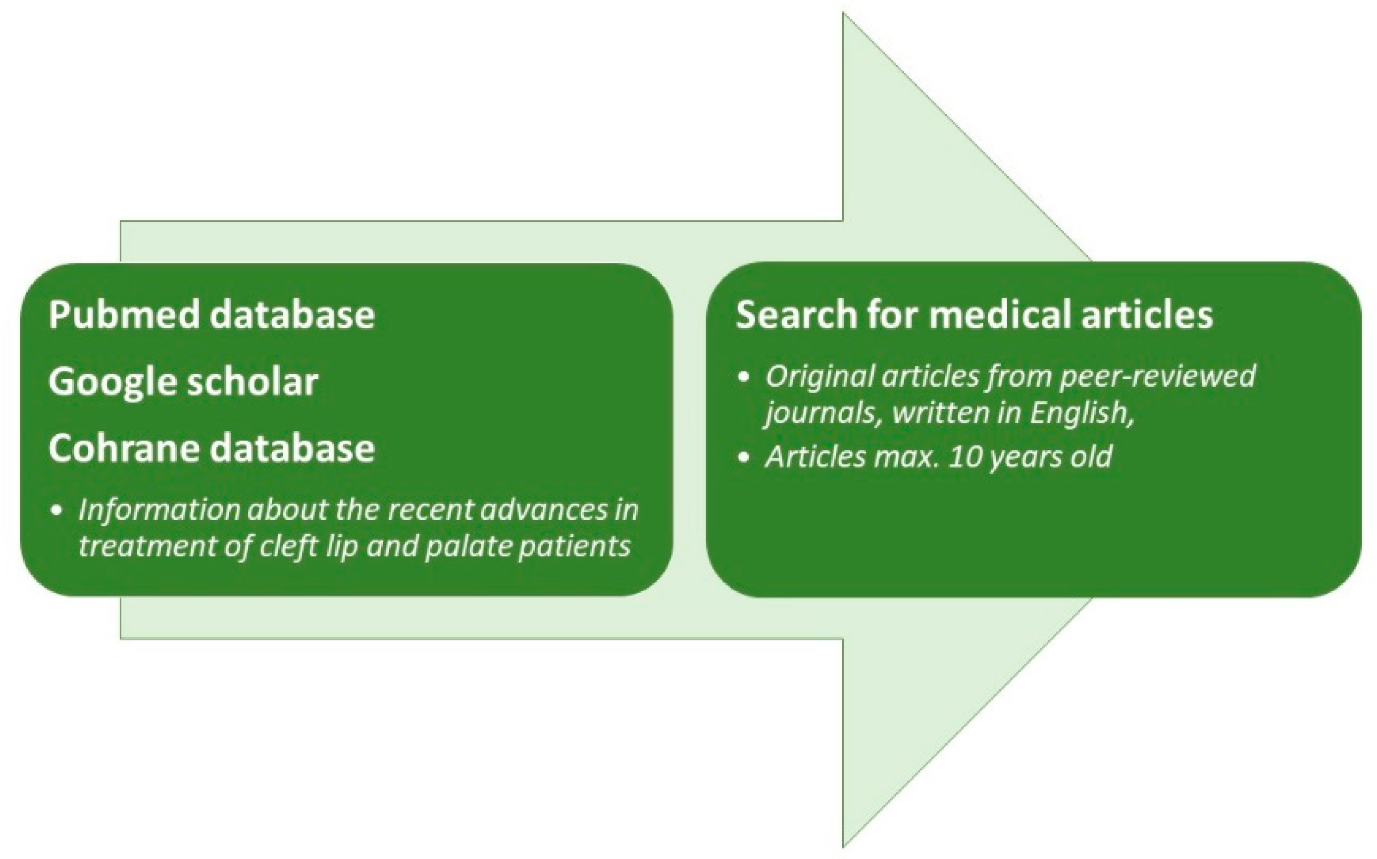
Three databases (PubMed, Google Scholar and Cochrane) were searched to obtain the most recent information on the topic of cleft lip and palate treatment. The search criteria included the words cleft lip and/or palate with a combination of the words: multidisciplinary treatment, multidisciplinary approach, surgical treatment, dental treatment, orthodontic treatment and medical care. The articles that were taken into account were peer-reviewed original and review papers published in English. The exclusion criteria assumes that the paper should not be older than 10 years (if possible), and the scientific background should be clear.
Figure 1 presents the basic inclusion criteria to conduct that study. The case reports were incorporated into the study only if the novel technique of treatment was introduced in that paper. The aspect of the papers should be practical and have clinical significance. The authors tried to summarize all types of treatment that are performed in patients that require multidisciplinary approach.
Two of the authors (A.P.-S. and M.M.) are workers of a program that establishes burden of care in patients with cleft lip and palate for many years. Both of them are specialists in orthodontics. The aim of this program is to bind doctors of many specialties into the treatment of children with congenital facial deformities, among them clefts. The third author works in diagnostic laboratory and has experience with genetics and in vitro procedures.
Two authors (A.P.-S. and I.D.-I.) searched for the articles. Only the articles that both authors accepted were taken into account when writing this review. The third author (M.M.) accepted the final quantity and quality of the chosen articles and was responsible for the final approval of this paper.
3. Newest Treatments of Cleft Lip and/or Palate
3.1. Early, Pre-Surgical Treatment
The presurgical procedure should involve presurgical nasoalveolar molding (PNAM also known as Figueroa’s NAM technique). It was firstly introduced by Grayson in 2004 [
11]. This reduces cleft stigma, especially around the nasal and lip area. The observation shows reduction of columnella width and improvement of columnella height. It also shows reduction of bi-ala width and increases bi-ala height [
12,
13]. The pre-surgical preparation may also require a NAM-plate, which is an acrylic orthodontic device to stimulate the maxillary growth and change the growth pattern of the patient with cleft. The novel plates are prepared as a 3D print. NAM plate is helpful with rotation of the premaxilla and reducing the amount of cleft fissure [
14]. In addition to those results, the change in the maxillary width in the canine and molar region is also observed [
15]. It should be applied as fast as possible due to the possible problems with neonate adaptation. After the preparation, the plate should be worn daily with only a short time for cleaning the plate twice a day [
13]. Presurgical preparation also changes the rotation of the incisal bone and therefore the alveolar bone shape becomes less triangular, more natural and the cleft gap spontaneously reduces [
12]. The long-term follow-up shows though, that only the shape of the dental arch remains stable in time, other malformations still coexist [
16]. The possible ways of mobilizing the soft tissues are lip massage and lip taping in order to make skin more elastic and give it more possibilities to close over the alveolar bone [
17,
18].
3.2. Lip and/orPalate Closure
The preliminary surgical procedures for patients with clefts are cheilo- and palatoplasty, as well as alveolar bone grafting. In most countries, lip and palatal procedures are separated, with different timings 3–6 months for lip and 6–18 months for palatal closure [
19,
20]. A C-flap technique was recently presented by Jung et al. [
18]. This procedure creates a longer lip during the surgery and therefore, the malformation of philtrum and Cupid’s bow peak is lower than with the traditional method. The choice of the method of surgical reparation depends on the surgeon’s experience and the type of the cleft. Most surgeons incorporated Fischer’s technique for the lip repair with modified Millard rotation-advancement flap [
21,
22,
23]. Among many surgical procedures to close the palate, nonradical intravelar veloplasty is one of the methods that might be used to restore it [
24].
After the surgery of the lip is performed, the postoperative care should include scar management, massage and observation of the wound potential dehiscence and silicone gel as well as steroid administration [
19]. The novel methods of scar management require a laser to reduce the scar tissue by its softening and flattening [
25].
3.3. Bone Grafting
Most of the patients with cleft of the alveolar bone need bone grafting to restore the shape of the bone for the future teeth movement and prosthetic restoration. The timing of the bone graft is under debate—several techniques of that are used. The most common donor site is iliac crest (most preferable, due to up to 50 mL possible cancellous bone gain) or calvarial bone. The autogenic bone is the best material that could be used in any kind of graft. The bone should be grafted gently, as crushing of the bone increases its resorption and in consequence reduces its volume and quantity [
19,
26,
27,
28]. The bone grafting is usually performed at the mixed dentition stage and besides the restoration of collapsed nasal bone, it becomes a base for the ala nasi. The reconstruction of longitude of the alveolar bone makes it possible to restore the teeth loss [
7]. Now, the bone grafting should be based on the 3D planning, which enables it to produce actual and fit-to-size bone graft [
29]. Additionally, 3D scans might be used for printing scaffolds for bone grafting formed from the bio-glass [
8].
4. Genetics Changes Underlying the Cleft Lip and/or Palate
The genetic factors in the hereditary of clefts play a crucial role. Among the most common genes in non-syndromic clefts are named: CDH1 (locus 16q22.1), COL2A1 (12q13.11), CRISPLD2 (16q24.1), FOXE1 (9q22.33), GRHL3 (1p36.11), IRF6 (1q32.2), JAG2 (14q32.33), MSX1 (4p16.2), PAX7 (1p36.13), ROCK1 (18q11.1), SUMO1 (2q33.1), TBX22 (Xq21.1), TCOF1 (5q32-q33.1) and TGFA (2p13.3) [
4,
24]. The genetic sequence could be important for the patient in terms of future reproductive plans. With that knowledge one could plan pregnancy more consciously and the prenatal diagnosis might be crucial in those kids. Knowing that PAX9 and MSX1 genes may predispose to hypodontia, the prenatal diagnostic is also possible in that area [
30,
31,
32]. Further studies regarding genetics of clefts and accompanying dismorphic features could give the doctors wider knowledge on how to treat patients and how to help them in the future. Knowing the genetics of the individuals could help doctors to predict the accompanying problems of the patient and find the best option for their care. If genetic engineering comes into account in the nearest future, the human species could probably exclude the genetic mutation leading to clefts for future individuals. The scientific world discusses the aspects of genetic engineering, but the authors want to be careful with those kinds of statements, especially for the near future.
5. Medical Teams Facing the Cleft Lip and/or Palate Treatment and Diagnostics
5.1. Pediatrician
The pediatric care of the child with cleft starts at the first diagnostics process. A newborn has difficulties sucking through a regular nipple, due to the lip seal that does not exist. The gap in the lip and in the palate does not let the patient eat normally. Additionally, the gap in the roof of the mouth makes it impossible to produce the build-up of pressure in the mouth. Most of the babies require a special nipple that should be adjusted before leaving the neonatal hospital [
6]. The pediatrician could also be a leader of the team, coordinate the other specialist’s work timing, as well as be a support in psychosocial problems, especially when communication problems appear [
33].
5.2. Logopedics and Phoniatrics
The cleft patients have affected muscle phonation. This dysfunction applies to muscle levator velirud palatini. The most common observation is retardation of consonant sounds (p, b, t, d k, g). The velopharyngeal dysfunction (VPD) is corelated with hypernasality, problems with compensatory articulation and air emission. Nasal resonance and difficulties in articulation are also very common [
6,
34]. Due to palatal shortage, the nasal speech is present twice more often in cleft toddlers than in healthy individuals [
35]. Although more than 60% of the patients with clefts present understandable speech, more than 70% need speech therapy [
36].
It should also be considered by the speech therapist and consulted with the laryngologist, whether the nasal emission is obligatory or compensatory, or is it caused by misarticulation. The last phenomenon is observed when pharyngeal sound is substituted by and oral sound. The speech therapist should be able to recognize, whether this condition is a result of abnormal velopharyngeal structure [
37]. To obtain longer palatal length, Furlow’s palatoplasty is the method of choice [
38]. Primary as well as secondary palatoplasty give the same results [
39].
5.3. Laryngologist
Laryngological problems usually refer to infections of the middle ear and may result in hearing loss. The biomechanism of otitis media in this situation bases on dysfunction of muscle levator veli palatini, which opens the Eustachian tube [
6]. The middle ear effusion may be present at birth and therefore the patient may fail the hearing test [
40]. Acoustic rhinometry may be a method of choice when considering the nasal patency, as within the years patients require many surgeries. Acoustic rhinometry helps measure the laryngological problems with the nose and the potential need of the surgical reconstruction [
41]. A large number of rhinoplasties are performed on juveniles, the rib grafts are the most common donor sites to restore the nasal shape, length and width. The second most common procedure is nasal osteotomy [
42].
5.4. Dentists
In patients with cleft lip or palate, dental issues may refer to several from below presented the changes in number of teeth, their shape and time of eruption. The incidence of the neonatal teeth is considered to be common [
43]. Microdontic lateral maxillary incisors (or missing ones) are also a very common issue. Another very frequent dental problem is ectopic teeth eruption, specifically referring to the canine that erupts at the palatal side. Delayed tooth eruption and enamel hypoplasia [
44] are causes of dental lamina injuries [
6,
45]. Another dental anomaly is the presence of supernumerary teeth, which affects almost one fourth of the cleft patients and is probably caused by division of dental lamina during prenatal time [
46]. The delayed tooth development refers mainly to the cleft side [
47]. Dental anomalies refer mostly to the cleft side [
48].
Before the reconstructive operation, the patient should be consulted with the pediatric dentist in terms of diet and oral cavity hygiene. After the surgical procedure, topical fluoride and sealants applications should be considered [
45]. The fluoride should be administered at least twice a year [
40].
It is also a very common issue that the oral hygiene in cleft individuals is improper [
45]. Additionally with demarcated or diffused opacity and enamel hypoplasia this may lead to higher predisposition to dental caries [
49]. It had been observed that teeth hypoplasia is strongly related to the palatal defect, as well as number and type of performed surgeries [
50].
The patients with clefts are more susceptible to caries; therefore, the early preventive advise for caries prevention should be performed. If necessary, the restorative care and endodontic treatment should be performed [
45,
51]. Children are at a higher risk of developing early childhood caries [
40]. Although the recessions are not present, it has been proven that the keratinized gingiva is thinner around the cleft area [
52]. The checkups should be common, especially at the stage of active orthodontic treatment with any fixed device, as the hygiene would be more difficult to deal with at this stage [
40]. The pediatric dentist could also be another “link” between all members of the multidisciplinary team and a potential leader in treatment planning [
45].
5.4.1. Orthodontic Procedures
The most common orthodontic problems refer to malocclusions, which often needs orthognathic surgery in the future. The most frequent ones are crossbites and partial open bites, especially at the surgery side. Among the angle classification, the most frequent are Class I malocclusions, but in difference when compared to the population of healthy individuals, more frequent are Class III malocclusions with dominance of pseudomesioclussion connected to maxillary hypoplasia, whereas, in healthy individuals, Class II malocclusions are three to eight times more frequent. There are also theses that hypoplasia is related to the whole midface complex [
53,
54,
55]. It is also popular to use GOSLON-Yardick scale for defining dentoalveolar malocclusions among patients with clefts. It is a five-category scale that helps divide malocclusions and plan patient’s treatment needs. The GOSLON index is presented in
Table 1. GOSLON 3 and higher are observed in ca. 60–70% of patients [
56].
The treatment of patients with clefts is a long procedure—most of the patients start their treatment during early childhood, according to Roguzińska at al. [
57], at the mean age of 4.83 years and lasts for more than 9 years for unilateral clefts and 4.53 years and over 10 years, respectively, for bilateral clefts. Orthodontic procedures require removable and fixed appliances [
57]. Removable appliances are used to gain space for the teeth or keep the space if there was premature loss; therefore, transversal screws are used in those kind of appliances [
45].
The novel techniques in orthodontic diagnostics are based on dental scans, which is especially helpful in patients with clefts as the 3D image is more accurate and precise and therefore the individual appliances produced for the patients are expected to fit patients’ dental arch better [
58,
59].
5.4.2. Prosthodontics
Prosthodontist should be a part of the cleft team. The result of smile attractiveness and naturality depends mainly on the quality of prosthodontic approach. In the past, the majority of dental restorations concentrated around veneers and dental crowns. The issue of prosthodontist is to improve the visual aspect, especially of the damaged and malformed teeth. Dental bridges and removable prostheses were replacing the missing teeth [
60,
61]. Right now, the most desirable reconstruction of the missing teeth is dental implant. The high survival rate is observed among the dental implants placed in the alveolar bone grafts, although long-term esthetic follow-ups would be desired [
62,
63]. The pink esthetics due to thinner gingival margin and lower bone levels is worse in individuals with clefts, although the peri-implant parameters remain similar to the control group. The best results were obtained when the bone grafts were performed 3 months before the implant placement [
64].
5.4.3. Plastic and Orthognathic Surgery
Plastic and orthognathic surgery pertains to adulthood of the individual. It should include lip revision to correct labial shape and outcome, secondary palatoplasty (correction of velopharyngeal dysfunction), correction of maxillary hypoplasia, rhinoplasty and velopharyngeal incompetence surgery [
19]. Due to the characteristic nasal speech of the patients with cleft, their improvement in velopharyngeal function is required to correct them into the non-nasal speech [
65,
66].
Maxillary hypoplasia is one of the most visible stigmata of the cleft. Oral distractors or orthognathic surgery should cause less visibility in the cleft malformations [
19]. Osteotomy (most frequently Le Fort I) is performed to improve the patient’s life quality and facial appearance. It should be combined with bone grafting to avoid velopharyngeal function (VPF) disturbances, but still, the patients should be informed before the Le Fort I osteotomy that they may require more surgeries to improve their speech [
67]. Most of the patients would be willing to have a rhinoplasty simultaneously when the Le Fort procedure is performed [
9]. In patients with clefts, the usual revision rhinoplasty requires cartilage grafts [
68].
6. Other Treatment Needs
The facial deformation, congenital pain and need for of repair procedures may influence a patient’s life [
69,
70]. The will to look better and nicer is the psychosocial problem of individuals with cleft. The need for acceptance pushes these individuals to put posts on the internet with positively stigmatized words such as beauty [
71]. Therefore, the need for psychologist might be crucial. It has been shown, though, that improvement of smile and occlusion with orthodontic treatment act the same when compared to healthy individuals; there is no difference in life quality among those two groups [
72].
Patients with clefts present masticatory muscle function overload, which may require orthodontic and prosthetic treatment as well as physiotherapy [
73,
74,
75]. The ideal treatment planning to restore the proper bite, intercuspidation and function would require using facebows and articulators in order to not overload the temporomandibular joint and plan the equal pressure on all points on the teeth [
66].
7. Discussion
Patients with clefts require multidisciplinary treatment throughout all of their lives. Knowing the overall problems of orofacial deformities in patients with clefts may result in the expansion of the need for multidisciplinary treatment. The most common malocclusions present in cleft patients are crossbites and open bites, especially on the cleft side. The problem of malocclusions lies in the underdevelopment of hypoplastic maxilla [
53]. Although the patient requires several reconstruction surgeries (lip reconstruction, palatal closure and bone grafts) and plastic surgeries to improve the overall result, the result of the treatment lies in orthodontic-orthognatic preparation in most of the cases. Lacking the palatal suture and presence of the scar on the palate rotates the palatal bone mesially, especially in the frontal region. The patient requires maxillary segmentalization to improve the stability of maxillary complex after the surgical, orthognathic procedures in adulthood [
53,
76]. It is also important to establish vertical and sagittal relation of the jaws, as it gives a higher knowledge on the 3-dimensional problem of malocclusion [
77].
In addition to reconstruction and surgical procedures, patients with clefts require many specialists on their way to the “normality”. Speech therapists, laryngologists and phoniatrics take care of the development of speech and loss of nasality typical for cleft patients [
34,
35,
36,
37,
38,
39,
40,
41,
42]. Due to several dental anomalies that are present in the patients, those patients require additional multidisciplinary dental care, including a child dentist, orthodontist and finally, in most of the cases, prosthodontist to achieve symmetrical and the most natural effect that is possible [
78,
79].
The team that takes care of the patient with cleft should cooperate with each other very strongly. In the authors opinion, creating the programs of orthodontic care burden, as one in our country, is a great idea to create a web of specialists is extremely beneficial. Creating the schemes of the treatment would keep the patient and the parents well-informed and makes it easier to cooperate between the specialists of all the medical specialties. It would also be easier to incorporate the novel treatment methods to those patients in a cohesive and cooperative team. It is also easier to refer the patient to the specialist of other disciplines in that case. In the authors opinion, it is mandatory to have a leader in this kind of team, although it would be disputable who it should be. The main goal of the leader would be creating a kind of web and bind other specialists when the care is applied to the patient. The cleft patients require specialists that cooperate, but also have high experience in this topic. The “evidence-based” medicine shows that the greater the experience of the doctor, results in the best possible outcomes. This also applies to that topic.
8. Conclusions
To sum up, clefts are very common birth defects that require multidisciplinary approach. Clefts require many surgeries—reconstructive and, later, plastic ones—to restore the continuity of tissues and impaired functions. The first surgeries take place in the neonatal period and the whole treatment is long-term, requires many specialists and lasts until adulthood. Due to the most frequent and systematic checkups (at least twice a year), the leader of the team, which refers patient to the other specialist, in the authors mind, could be a pediatrician, dentist or orthodontist.
9. Limitations
The first limitation of this study is incorporating only three databases into the search criteria. The authors decided to do that because of the nature of this research, knowing that neither a systematic review nor meta-analysis could be carried out on this topic. The authors are aware that this topic could be widened, and each section of this review could be treated as a separate paper.
The experience of the authors of this study, based on the dental and orthodontic knowledge, mainly due to the fact that it is the main area of expertise of the authors. Working with the cleft patients, we made some observations regarding the schemes of treatment in general and tried to present it.
Author Contributions
Conceptualization, A.P.-S.; methodology, A.P.-S., I.D.-I.; validation, A.P.-S., I.D.-I., formal analysis, A.P.-S.; investigation, A.P.-S., M.M.; resources, A.P.-S., I.D.-I.; data curation, A.P.-S., I.D.-I.; writing—original draft preparation, A.P.-S.; writing—review and editing, M.M.; visualization, I.D.-I.; supervision, M.M.; funding acquisition, M.M. All authors have read and agreed to the published version of the manuscript.
Funding
This research received no external funding.
Institutional Review Board Statement
Not applicable.
Informed Consent Statement
Not applicable.
Conflicts of Interest
The authors declare no conflict of interest.
References
- Mossey, P.A.; Little, J.; Munger, R.G.; Dixon, M.J.; Shaw, W.C. Cleft lip and palate. Lancet 2009, 374, 1773–1785. [Google Scholar] [CrossRef] [PubMed]
- Supplement, D.; Candotto, V.; Oberti, L.; Gabrione, F.; Greco, G.; Rossi, D.; Romano, M.; Mummolo, S. Current concepts on cleft lip and palate etiology. J. Biol. Regul. Homeost. Agents 2019, 33, 145–151. [Google Scholar]
- Cuozzo, F.D.M.; Espinosa, M.M.; Da Silva, K.T.S.; De Barros, Y.B.A.M.; Bandeca, M.C.; Aranha, A.M.F.; Borges, A.H.; Volpato, L.E.R. Cleft lip and palate in a Brazilian subpopulation. J. Int. Oral Health 2013, 5, 15–20. [Google Scholar] [PubMed]
- Martinelli, M.; Palmieri, A.; Carinci, F.; Scapoli, L. Non-syndromic Cleft Palate: An Overview on Human Genetic and Environmental Risk Factors. Front. Cell Dev. Biol. 2020, 8, 592271. [Google Scholar] [CrossRef] [PubMed]
- Crockett, D.J.; Goudy, S.L. Cleft Lip and Palate. Facial Plast. Surg. Clin. N. Am. 2014, 22, 573–586. [Google Scholar] [CrossRef]
- Vyas, T.; Gupta, P.; Kumar, S.; Gupta, R.; Gupta, T.; Singh, H.P. Cleft of lip and palate: A review. J. Fam. Med. Prim. Care 2020, 9, 2621–2625. [Google Scholar] [CrossRef]
- Rychlik, D.; Wojcicki, P.; Koźlik, M. Osteoplasty of the alveolar cleft defect. Adv. Clin. Exp. Med. 2012, 21, 255–262. [Google Scholar]
- Brézulier, D.; Chaigneau, L.; Jeanne, S.; Lebullenger, R. The Challenge of 3D Bioprinting of Composite Natural Polymers PLA/Bioglass: Trends and Benefits in Cleft Palate Surgery. Biomedicines 2021, 9, 1553. [Google Scholar] [CrossRef]
- Raffaini, M.; Cocconi, R.; Spinelli, G.; Agostini, T. Simultaneous Rhinoseptoplasty and Orthognathic Surgery: Outcome Analysis of 250 Consecutive Patients Using a Modified Le Fort I Osteotomy. Aesthetic Plast. Surg. 2018, 42, 1090–1100. [Google Scholar] [CrossRef]
- Al-Namankany, A.; Alhubaishi, A. Effects of cleft lip and palate on children’s psychological health: A systematic review. J. Taibah Univ. Med. Sci. 2018, 13, 311–318. [Google Scholar] [CrossRef]
- Grayson, B.H.; Maull, D. Nasoalveolar molding for infants born with clefts of the lip, alveolus, and palate. Clin. Plast. Surg. 2004, 31, 149–158. [Google Scholar] [CrossRef] [PubMed] [Green Version]
- Bhutiani, N.; Tripathi, T.; Verma, M.; Bhandari, P.S.; Rai, P. Assessment of Treatment Outcome of Presurgical Nasoalveolar Molding in Patients with Cleft Lip and Palate and Its Postsurgical Stability. Cleft Palate Craniofac. J. 2020, 57, 700–706. [Google Scholar] [CrossRef] [PubMed]
- Koya, S.; Shetty, S.; Husain, A.; Khader, M. Presurgical Nasoalveolar Molding Therapy Using Figueroa’s NAM Technique in Unilateral Cleft Lip and Palate Patients: A Preliminary Study. J. Clin. Pediatr. Dent. 2016, 40, 410–416. [Google Scholar] [CrossRef]
- De Oliveira, N.V.; Tou, G.A.A.; Silva, R.S.; Rezende, S.E.; Pretti, H.; Macari, S. The First-Year Follow-Up of a Cleft Lip and Palate Patient Treated with Nasoalveolar Molding (NAM). Braz. Dent. J. 2020, 31, 190–196. [Google Scholar] [CrossRef] [PubMed]
- Ruíz-Escolano, M.G.; Martínez-Plaza, A.; Fernández-Valadés, R.; Cortés-Sánchez, R.; Muñoz-Miguelsanz, M.A.; Velasco-Ortega, E.; Perez-Ureña, M.B.; Matar-Satuf, K.; España-López, A.J. Nasoalveolar Molding Therapy for the Treatment of Unilateral Cleft Lip and Palate Improves Nasal Symmetry and Maxillary Alveolar Dimensions. J. Craniofac. Surg. 2016, 27, 1978–1982. [Google Scholar] [CrossRef] [PubMed]
- Papadopoulos, M.A.; Koumpridou, E.N.; Vakalis, M.L.; Papageorgiou, S.N. Effectiveness of pre-surgical infant orthopedic treatment for cleft lip and palate patients: A systematic review and meta-analysis. Orthod. Craniofac. Res. 2012, 15, 207–236. [Google Scholar] [CrossRef]
- Worley, M.L.; Patel, K.G.; Kilpatrick, L.A. Cleft Lip and Palate. Clin. Perinatol. 2018, 45, 661–678. [Google Scholar] [CrossRef]
- Jung, S.; Chung, K.H.; Chang, S.-Y.; Ohrmman, D.; Lim, E.; Lo, L.-J. A new technique for perioral muscle reconstruction and lip lengthening in complete unilateral cleft lip. J. Plast. Reconstr. Aesthetic Surg. 2020, 73, 749–757. [Google Scholar] [CrossRef] [PubMed]
- Raghavan, U.; Rao, D.; Ullas, G.; Vijayadev, V. Postoperative Management of Cleft Lip and Palate Surgery. Facial Plast. Surg. 2018, 34, 605–611. [Google Scholar] [CrossRef] [Green Version]
- Kobus, K.; Kobus-Zaleśna, K. Timing of cleft lip and palate repair. Dev. Period Med. 2014, 18, 79–83. [Google Scholar]
- Chong, D.K.; Swanson, J.W. The Essential Anatomical Subunit Approximation Unilateral Cleft Lip Repair. Plast. Reconstr. Surg. 2016, 138, 91e–94e. [Google Scholar] [CrossRef] [PubMed]
- Mittermiller, P.A.; Martin, S.; Johns, D.N.; Perrault, D.; Jablonka, E.M.; Khosla, R.K. Improvements in Cleft Lip Aesthetics with the Fisher Repair Compared to the Mohler Repair. Plast. Reconstr. Surg. Glob. Open 2020, 8, e2919. [Google Scholar] [CrossRef] [PubMed]
- Patel, T.A.; Patel, K.G. Comparison of the Fisher Anatomical Subunit and Modified Millard Rotation-Advancement Cleft Lip Repairs. Plast. Reconstr. Surg. 2019, 144, 238e–245e. [Google Scholar] [CrossRef] [PubMed]
- Rossell-Perry, P.; Romero-Narvaez, C.; Olivencia-Flores, C.; Marca-Ticona, R.; Anaya, M.H.; Cordova, J.P.; Luque-Tipula, M. Effect of Nonradical Intravelar Veloplasty in Patients with Unilateral Cleft Lip and Palate: A Comparative Study and Systematic Review. J. Craniofac. Surg. 2021, 32, 1999–2004. [Google Scholar] [CrossRef]
- Peng, L.; Tang, S.; Li, Q. Intense pulsed light and laser treatment regimen improves scar evolution after cleft lip repair surgery. J. Cosmet. Dermatol. 2018, 17, 752–755. [Google Scholar] [CrossRef] [PubMed]
- Pan, W.; Wu, C.; Yang, Z.; Duan, Z.; Su, Z.; Wang, P.; Zheng, Q.; Li, C. Secondary Alveolar Bone Grafting and Iliac Cancellous Bone Harvesting for Patients with Alveolar Cleft. J. Craniofac. Surg. 2016, 27, 883–891. [Google Scholar] [CrossRef] [PubMed]
- Siegenthaler, M.; Bettelini, L.; Brudnicki, A.; Rachwalski, M.; Fudalej, P.S. Early versus late alveolar bone grafting in unilateral cleft lip and palate: Dental arch relationships in pre-adolescent patients. J. Cranio Maxillofacial Surg. 2018, 46, 2052–2057. [Google Scholar] [CrossRef] [PubMed]
- Rychlik, D.; Wójcicki, P. Bone Graft Healing in Alveolar Osteoplasty in Patients with Unilateral Lip, Alveolar Process, and Palate Clefts. J. Craniofac. Surg. 2012, 23, 118–123. [Google Scholar] [CrossRef]
- Dowgierd, K.; Pokrowiecki, R.; Borowiec, M.; Sokolowska, Z.; Dowgierd, M.; Wos, J.; Kozakiewicz, M.; Krakowczyk, Ł. Protocol and Evaluation of 3D-Planned Microsurgical and Dental Implant Reconstruction of Maxillary Cleft Critical Size Defects in Adolescents and Young Adults. J. Clin. Med. 2021, 10, 2267. [Google Scholar] [CrossRef]
- Paradowska-Stolarz, A. MSX1 gene in the etiology orofacial deformities. Postepy Hig. Med. Dosw. 2015, 69, 1499–1504. [Google Scholar]
- Kirac, D.; Eraydin, F.; Avcilar, T.; Ulucan, K.; Ozdemir, F.; Guney, A.I.; Kaspar, E.; Keshi, E.; Isbir, T. Effects of PAX9 and MSX1 gene variants to hypodontia, tooth size and the type of congenitally missing teeth. Cell. Mol. Biol. 2016, 62, 78–84. [Google Scholar] [CrossRef] [PubMed] [Green Version]
- Nasroen, S.; Maskoen, A.; Soedjana, H.; Hilmanto, D.; Gani, B.A. IRF6 rs2235371 as a risk factor for non-syndromic cleft palate only among the Deutero-Malay race in Indonesia and its effect on the IRF6 mRNA expression level. Dent. Med. Probl. 2022, 59, 59–65. [Google Scholar] [CrossRef] [PubMed]
- Kummer, A.W. A Pediatrician’s Guide to Communication Disorders Secondary to Cleft Lip/Palate. Pediatr. Clin. N. Am. 2018, 65, 31–46. [Google Scholar] [CrossRef] [PubMed]
- Larangeira, F.R.; Dutka, J.; Whitaker, M.E.; de Souza, O.M.V.; Lauris, J.; da Silva, M.J.F.; Pegoraro-Krook, M.I. Speech nasality and nasometry in cleft lip and palate. Braz. J. Otorhinolaryngol. 2016, 82, 326–333. [Google Scholar] [CrossRef]
- Hardin-Jones, M.A.; Chapman, K.L. The Implications of Nasal Substitutions in the Early Phonology of Toddlers with Repaired Cleft Palate. Cleft Palate Craniofac. J. 2018, 55, 1258–1266. [Google Scholar] [CrossRef]
- Bruneel, L.; Bettens, K.; Van Lierde, K. The relationship between health-related quality of life and speech in patients with cleft palate. Int. J. Pediatr. Otorhinolaryngol. 2019, 120, 112–117. [Google Scholar] [CrossRef]
- Oren, L.; Kummer, A.; Boyce, S. Understanding Nasal Emission During Speech Production: A Review of Types, Terminology, and Causality. Cleft Palate Craniofac. J. 2020, 57, 123–126. [Google Scholar] [CrossRef]
- Chang, B.L.; Yu, J.W.; Nikonova, E.; Low, D.W.; Taylor, J.A.; Jackson, O. Palatal Measurements Pre- and Post-Furlow Cleft Palate Repair: Analysis of Palatal Lengthening and Comparison Within Cleft Types. Cleft Palate Craniofac. J. 2019, 56, 601–609. [Google Scholar] [CrossRef]
- Rudnicki, P.A.; Tsang, C.; Vecchiotti, M.A.; Scott, A.R. Palatal Motion After Primary and Secondary Furlow Palatoplasty. JAMA Otolaryngol. Neck Surg. 2017, 143, 111–115. [Google Scholar] [CrossRef]
- Lewis, C.W.; Jacob, L.S.; Lehmann, C.U.; Krol, D.; Gereige, R.; Karp, J.; Fisher-Owens, S.; Braun, P.; Segura, A.; Section on Oral Health. The Primary Care Pediatrician and the Care of Children with Cleft Lip and/or Cleft Palate. Pediatrics 2017, 139, e20170628. [Google Scholar] [CrossRef] [Green Version]
- Banari, A.S.; Datana, S.; Agarwal, S.S.; Bhandari, S.K. Evaluation of Nasal Patency Among Patients with Unilateral Cleft Lip and Palate: Cleft Versus Non-Cleft Side. Cleft Palate Craniofac. J. 2021, 58, 340–346. [Google Scholar] [CrossRef] [PubMed]
- Chouairi, F.; Torabi, S.; Gabrick, K.S.; Persing, J.A.; Alperovich, M. Secondary Cleft Rhinoplasty in 1720 Patients: Are National Practices Consistent with Guidelines? Cleft Palate Craniofac. J. 2020, 57, 438–443. [Google Scholar] [CrossRef] [PubMed]
- Yilmaz, R.B.N.; Cakan, D.G.; Mesgarzadeh, N. Prevalence and management of natal/neonatal teeth in cleft lip and palate patients. Eur. J. Dent. 2016, 10, 54–58. [Google Scholar] [CrossRef] [PubMed] [Green Version]
- Mazur, M.; Westland, S.; Guerra, F.; Corridore, D.; Vichi, M.; Maruotti, A.; Nardi, G.M.; Ottolenghi, L. Objective and subjective aesthetic performance of icon® treatment for enamel hypomineralization lesions in young adolescents: A retrospective single center study. J. Dent. 2018, 68, 104–108. [Google Scholar] [CrossRef] [PubMed]
- Luzzi, V.; Zumbo, G.; Guaragna, M.; Di Carlo, G.; Ierardo, G.; Sfasciotti, G.L.; Bossù, M.; Vozza, I.; Polimeni, A. The Role of the Pediatric Dentist in the Multidisciplinary Management of the Cleft Lip Palate Patient. Int. J. Environ. Res. Public Health 2021, 18, 9487. [Google Scholar] [CrossRef] [PubMed]
- Menezes, C.; De Arruda, J.-A.; Silva, L.-V.; Monteiro, J.-L.; Caribé, P.; Álvares, P.; Almeida, M.-C.; Coelli, J.-C.; Goldemberg, F.; Silveira, M.; et al. Nonsyndromic cleft lip and/or palate: A multicenter study of the dental anomalies involved. J. Clin. Exp. Dent. 2018, 10, e746–e750. [Google Scholar] [CrossRef]
- Park, H.-M.; Han, D.-H.; Baek, S.-H. Comparison of tooth development stage of the maxillary anterior teeth before and after secondary alveolar bone graft: Unilateral cleft lip and alveolus vs unilateral cleft lip and palate. Angle Orthod. 2014, 84, 989–994. [Google Scholar] [CrossRef]
- Celikoglu, M.; Buyuk, S.; Sekerci, A.; Cantekin, K.; Candirli, C. Maxillary Dental Anomalies in Patients with Cleft Lip and Palate: A Cone Beam Computed Tomography Study. J. Clin. Pediatr. Dent. 2015, 39, 183–186. [Google Scholar] [CrossRef]
- Shen, C.-A.; Guo, R.; Li, W. Enamel defects in permanent teeth of patients with cleft lip and palate: A cross-sectional study. J. Int. Med Res. 2019, 47, 2084–2096. [Google Scholar] [CrossRef] [Green Version]
- Korolenkova, M.V.; Starikova, N.V.; Udalova, N.V. The role of external aetiological factors in dental anomalies in non-syndromic cleft lip and palate patients. Eur. Arch. Paediatr. Dent. 2019, 20, 105–111. [Google Scholar] [CrossRef]
- Askar, H.; Krois, J.; Rohrer, C.; Mertens, S.; Elhennawy, K.; Ottolenghi, L.; Mazur, M.; Paris, S.; Schwendicke, F. Detecting white spot lesions on dental photography using deep learning: A pilot study. J. Dent. 2021, 107, 103615. [Google Scholar] [CrossRef] [PubMed]
- Wyrębek, B.; Cudziło, D.; Plakwicz, P. Evaluation of periodontal tissues in growing patients with bilateral cleft lip and palate. A pilot study. Dev. Period Med. 2017, 21, 154–161. [Google Scholar] [CrossRef] [PubMed]
- Paradowska-Stolarz, A.; Kawala, B. Occlusal Disorders among Patients with Total Clefts of Lip, Alveolar Bone, and Palate. BioMed. Res. Int. 2014, 2014, 583416. [Google Scholar] [CrossRef] [PubMed] [Green Version]
- Kozanecka, A.; Sarul, M.; Kawala, B.; Antoszewska-Smith, J. Objectification of Orthodontic Treatment Needs: Does the Classification of Malocclusions or a History of Orthodontic Treatment Matter? Adv. Clin. Exp. Med. 2016, 25, 1303–1312. [Google Scholar] [CrossRef]
- Sneha, P.; Ravi, P.; Arunachalam, D.; Raja, V.B.K.K. Zygomatic Hypoplasia in Patients with Non-syndromic Cleft Lip and Palate: A Case–Control Study. J. Maxillofac. Oral Surg. 2018, 17, 425–431. [Google Scholar] [CrossRef]
- Mars, M.; Plint, D.A.; Houston, W.J.; Bergland, O.; Semb, G. The Goslon Yardstick: A new system of assessing dental arch relationships in children with unilateral clefts of the lip and palate. Cleft Palate J. 1987, 24, 314–322. [Google Scholar]
- Roguzińska, S.; Pelc, A.; Mikulewicz, M. Orthodontic-care burden for patients with unilateral and bilateral cleft lip and palate. Dent. Med. Probl. 2020, 57, 411–416. [Google Scholar] [CrossRef]
- Sarul, M.; Lewandowska, B.; Kawala, B.; Kozanecka, A.; Antoszewska-Smith, J. Objectively measured patient cooperation during early orthodontic treatment: Does psychology have an impact? Adv. Clin. Exp. Med. 2017, 26, 1245–1251. [Google Scholar] [CrossRef] [Green Version]
- Kustrzycka, D.; Marschang, T.; Mikulewicz, M.; Grzebieluch, W. Comparison of the Accuracy of 3D Images Obtained fromDifferent Types of Scanners: A Systematic Review. J. Health Eng. 2020, 2020, 8854204. [Google Scholar] [CrossRef]
- Reisberg, D.J. Dental and Prosthodontic Care for Patients with Cleft or Craniofacial Conditions. Cleft Palate Craniofac. J. 2000, 37, 534–537. [Google Scholar] [CrossRef]
- Calik, K.; Chmiel, G.; Więckiewicz, W. Rehabilitacja protetyczna pacjenta z całkowitym rozszczepem podniebienia pierwotnego i wtórnego-opis przypadku = Prosthodontic rehabilitation of a patient with a complete cleft of the primary and secondary palate-case report. E-Dentico Dwumiesięcznik Stomatol. Prakt. 2012, 66–71. [Google Scholar]
- Loboda, M.; Mituś-Kenig, M.; Marcinkowska-Mituś, A.; Piątkowski, G.; Pawłowska, E. Prosthetic rehabilitation of patients with unilateral complete cleft of the primary and secondary palate. Dev. Period Med. 2014, 18, 123–128. [Google Scholar] [PubMed]
- Sales, P.; Oliveira-Neto, O.; Torres, T.; de Lima, F. Effectiveness of dental implants placed in bone graft area of cleft Patients. Int. J. Oral Maxillofac. Surg. 2019, 48, 1109–1114. [Google Scholar] [CrossRef]
- Alberga, J.M.; Stellingsma, K.; Meijer, H.J.A.; Oostenbrink, H.A.; Vissink, A.; Raghoebar, G.M. Dental implant placement in alveolar cleft patients: A retrospective comparative study on clinical and aesthetic outcomes. Int. J. Oral Maxillofac. Surg. 2020, 49, 952–959. [Google Scholar] [CrossRef] [PubMed]
- Li, L.; Strum, D.; Larson, S.; Preciado, D. Quality of life outcomes following velopharyngeal insufficiency surgery. Int. J. Pediatr. Otorhinolaryngol. 2019, 127, 109643. [Google Scholar] [CrossRef] [PubMed]
- Ruda, J.M.; Krakovitz, P.; Rose, A.S. A Review of the Evaluation and Management of Velopharyngeal Insufficiency in Children. Otolaryngol. Clin. N. Am. 2012, 45, 653–669. [Google Scholar] [CrossRef]
- Harjunpää, R.; Alaluusua, S.; Leikola, J.; Heliövaara, A. Le Fort I osteotomy in cleft patients: Maxillary advancement and velopharyngeal function. J. Cranio Maxillofac. Surg. 2019, 47, 1868–1874. [Google Scholar] [CrossRef]
- Lin, S.; Hsiao, Y.-C.; Chang, C.-S.; Chen, P.K.-T.; Chen, J.-P. Diced Cartilage Graft for Revision Rhinoplasty in a 64-year-old Cleft Patient: A Case Report. Plast. Reconstr. Surg. Glob. Open 2016, 4, e813. [Google Scholar] [CrossRef]
- Hartmann, A.; Welte-Jzyk, C.; Al-Nawas, B.; Daubländer, M. Neurophysiological changes and chronic pain in cleft patients. J. Cranio-Maxillofacial Surg. 2019, 47, 1875–1880. [Google Scholar] [CrossRef]
- Kaczorowska, N.; Mikulewicz, M. Impact of Cleft Palate Anastomosis in Cleft Lip and Palate Patients with Coexisting Cleft Lip Anastomosis Scar Based on Cephalometric Measurements. Appl. Sci. 2022, 12, 1104. [Google Scholar] [CrossRef]
- Sycinska-Dziarnowska, M.; Stepien, P.; Janiszewska-Olszowska, J.; Grocholewicz, K.; Jedlinski, M.; Grassi, R.; Mazur, M. Analysis of Instagram® Posts Referring to Cleft Lip. Int. J. Environ. Res. Public Health 2020, 17, 7404. [Google Scholar] [CrossRef] [PubMed]
- Francisco, I.; Caramelo, F.; Fernandes, M.H.; Vale, F. A Comparative Study of Oral Health-Related Quality of Life among Cleft Lip and Palate Patients and Their Families during Orthodontic Treatment. Int. J. Environ. Res. Public Health 2021, 18, 12826. [Google Scholar] [CrossRef] [PubMed]
- Krezel, J.D.; Friel, T.; Waia, S.; Clark, P.; Taylor, P.D. Prosthetic Rehabilitation of a Repaired Cleft Palate with Use of a Two-Part Hinged Magnet Retained Removable Prosthesis. J. Prosthodont. 2021, 30, 454–457. [Google Scholar] [CrossRef]
- Szyszka-Sommerfeld, L.; Budzyńska, A.; Lipski, M.; Kulesza, S.; Woźniak, K. Assessment of Masticatory Muscle Function in Patients with Bilateral Complete Cleft Lip and Palate and Posterior Crossbite by means of Electromyography. J. Health Eng. 2020, 2020, 8828006. [Google Scholar] [CrossRef]
- Wieckiewicz, M.; Shiau, Y.-Y.; Boening, K. Pain of Temporomandibular Disorders: From Etiology to Management. Pain Res. Manag. 2018, 2018, 4517042. [Google Scholar] [CrossRef]
- Fariña, R.; Diaz, A.; Pantoja, R.; Bidart, C. Treatment of Maxillary Hypoplasia in Cleft Lip and Palate: Segmental Distraction Osteogenesis with Hyrax Device. J. Craniofac. Surg. 2018, 29, 411–414. [Google Scholar] [CrossRef]
- Alam, M.K.; Alfawzan, A.A. Dental Characteristics of Different Types of Cleft and Non-cleft Individuals. Front. Cell Dev. Biol. 2020, 8, 789. [Google Scholar] [CrossRef]
- Marzouk, T.; Alves, I.L.; Wong, C.L.; DeLucia, L.; McKinney, C.M.; Pendleton, C.; Howe, B.J.; Marazita, M.L.; Peter, T.K.; Kopycka-Kedzierawski, D.T.; et al. Association between Dental Anomalies and Orofacial Clefts: A Meta-analysis. JDR Clin. Transl. Res. 2021, 6, 368–381. [Google Scholar] [CrossRef]
- Runte, C.; Dirksen, D. Symmetry and Aesthetics in Dentistry. Symmetry 2021, 13, 1741. [Google Scholar] [CrossRef]
| Publisher’s Note: MDPI stays neutral with regard to jurisdictional claims in published maps and institutional affiliations. |
© 2022 by the authors. Licensee MDPI, Basel, Switzerland. This article is an open access article distributed under the terms and conditions of the Creative Commons Attribution (CC BY) license (https://creativecommons.org/licenses/by/4.0/).


