
Adipose-Derived Stem Cells for Facial Rejuvenation


Abstract
:1. Introduction
2. Harvesting Autologous-Derived Agents
3. The Powerful Action of the ADSCs in Facial Rejuvenation
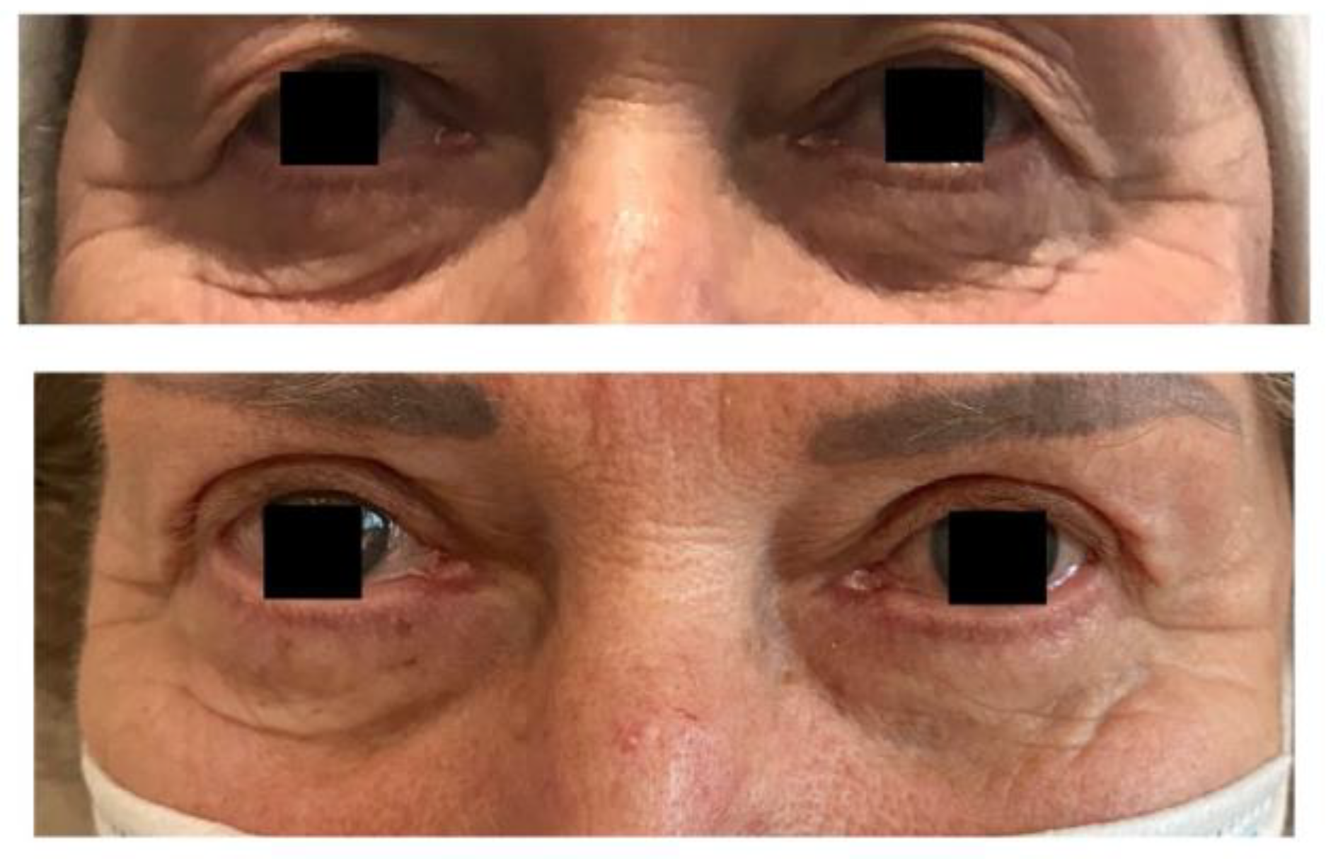
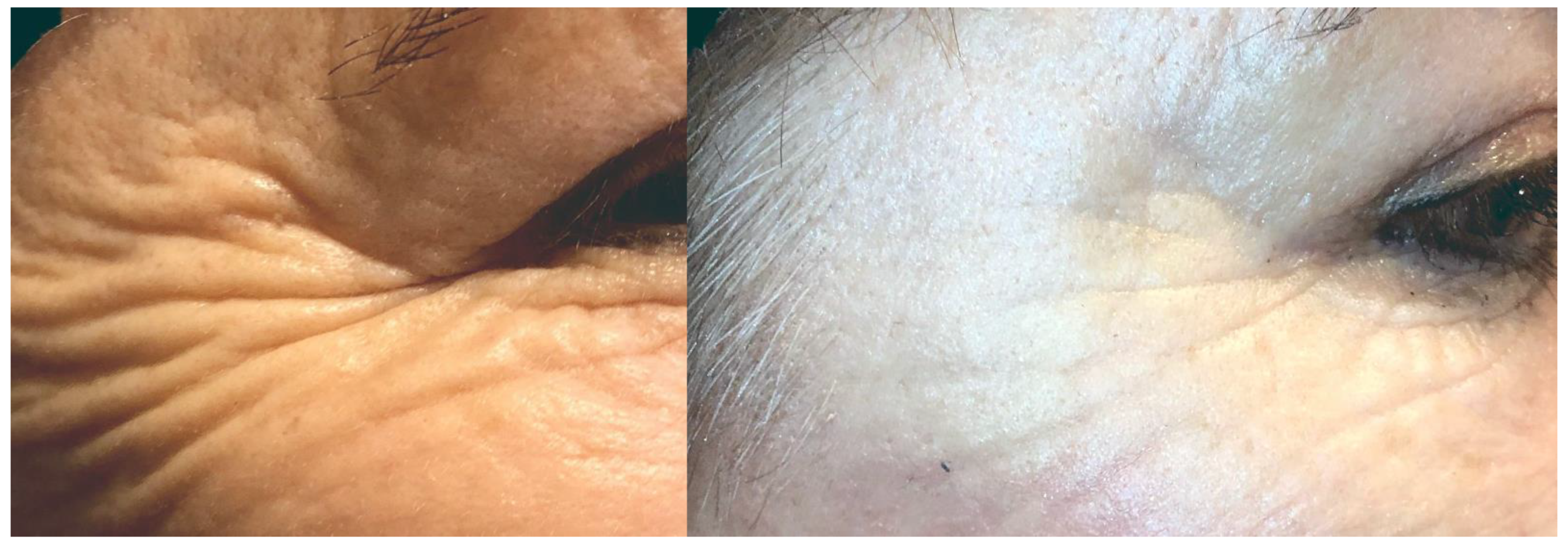
4. Clinical Indications for SVF
5. Complications
Future Trends
6. Conclusions
7. Key Points
Author Contributions
Funding
Informed Consent Statement
Conflicts of Interest
References
- Daar, A.S.; Greenwood, H.L. A proposed definition of regenerative medicine. J. Tissue Eng. Regen. Med. 2007, 1, 179–184. [Google Scholar] [CrossRef] [PubMed]
- Avantaggiato, A.; Palmieri, A.; Carinci, F.; Pasin, M.; Bertuzzi, G. Biostimulation and biorevitalization: Effects on human skin fibroblasts. Ann. Oral Maxillofac. Surg. 2013, 1, 11. [Google Scholar] [CrossRef] [Green Version]
- Gentile, P.; Garcovich, S. Systematic Review: Adipose-Derived Mesenchymal Stem Cells, Platelet-Rich Plasma and Biomaterials as New Regenerative Strategies in Chronic Skin Wounds and Soft Tissue Defects. Int. J. Mol. Sci. 2021, 22, 1538. [Google Scholar] [CrossRef] [PubMed]
- Graziani, F.; Ivanovski, S.; Cei, S.; Ducci, F.; Tonetti, M.; Gabriele, M. The in vitro effect of different PRP concentrations on osteoblasts and fibroblasts. Clin. Oral Implant. Res. 2006, 17, 212–219. [Google Scholar] [CrossRef] [Green Version]
- Anitua, E.; Sánchez, M.; Zalduendo, M.M.; de la Fuente, M.; Prado, R.; Orive, G.; Andía, I. Fibroblastic response to treatment with different preparations rich in growth factors. Cell Prolif. 2009, 42, 162–170. [Google Scholar] [CrossRef]
- Maisel-Campbell, A.L.; Ismail, A.; Reynolds, K.A.; Poon, E.; Serrano, L.; Grushchak, S.; Farid, C.; West, D.P.; Alam, M. A systematic review of the safety and effectiveness of platelet-rich plasma (PRP) for skin aging. Arch. Dermatol. Res. 2019, 312, 301–315. [Google Scholar] [CrossRef]
- Alves, R.; Grimalt, R. A Review of Platelet-Rich Plasma: History, Biology, Mechanism of Action, and Classification. Ski. Appendage Disord. 2018, 4, 18–24. [Google Scholar] [CrossRef] [PubMed]
- Stevens, H.P. ACA-Technik: “stromal vascular fraction”, “platelet-rich plasma” und Mikrofett zur körpereigenen Regeneration und Hautverjüngung. J. Ästhetische Chir. 2019, 12, 77–83. [Google Scholar] [CrossRef] [Green Version]
- Elghblawi, E. Platelet-rich plasma, the ultimate secret for youthful skin elixir and hair growth triggering. J. Cosmet. Dermatol. 2017, 17, 423–430. [Google Scholar] [CrossRef]
- Keller, G.; Snodgrass, R. Life span of multipotential hematopoietic stem cells in vivo. J. Exp. Med. 1990, 171, 1407–1418. [Google Scholar] [CrossRef] [Green Version]
- Bacakova, L.; Zarubova, J.; Travnickova, M.; Musilkova, J.; Pajorova, J.; Slepicka, P.; Kasalkova, N.S.; Svorcik, V.; Kolska, Z.; Motarjemi, H.; et al. Stem cells: Their source, potency and use in regenerative therapies with focus on adipose-derived stem cells—A review. Biotechnol. Adv. 2018, 36, 1111–1126. [Google Scholar] [CrossRef] [PubMed]
- Padoin, A.V.; Braga-Silva, J.; Martins, P.; Rezende, K.; Rezende, A.R.D.R.; Grechi, B.; Gehlen, D.; Machado, D.C. Sources of Processed Lipoaspirate Cells: Influence of Donor Site on Cell Concentration. Plast. Reconstr. Surg. 2008, 122, 614–618. [Google Scholar] [CrossRef]
- Tsekouras, A.; Mantas, D.; Tsilimigras, D.I.; Moris, D.; Kontos, M.; Zografos, G.C. Comparison of the viability and yield of adipose-derived stem cells (ASCs) from different donor areas. In Vivo 2017, 31, 1229–1234. [Google Scholar]
- Di Taranto, G.; Cicione, C.; Visconti, G.; Isgrò, M.A.; Barba, M.; Di Stasio, E.; Stigliano, E.; Bernardini, C.; Michetti, F.; Salgarello, M.; et al. Qualitative and quantitative differences of adipose-derived stromal cells from superficial and deep subcutaneous lipoaspirates: A matter of fat. Cytotherapy 2015, 17, 1076–1089. [Google Scholar] [CrossRef]
- Coleman, S.R. Structural Fat Grafting. Aesthetic Surg. J. 1998, 18, 386–388. [Google Scholar] [CrossRef] [Green Version]
- Travnickova, M.; Pajorova, J.; Zarubova, J.; Krocilova, N.; Molitor, M.; Bacakova, L. The Influence of Negative Pressure and of the Harvesting Site on the Characteristics of Human Adipose Tissue-Derived Stromal Cells from Lipoaspirates. Stem Cells Int. 2020, 2020, 1–13. [Google Scholar] [CrossRef] [PubMed]
- Centurión, P.; Gamarra, R.; Caballero, G.; Kaufmann, P.; Delgado, P. Optimizing harvesting for facial lipografting with a new photochemical stimulation concept: One STEP technique™. Eur. J. Plast. Surg. 2020, 43, 733–742. [Google Scholar] [CrossRef]
- Mojallal, A.; Auxenfans, C.; Lequeux, C.; Braye, F.; Damour, O. Influence of negative pressure when harvesting adipose tissue on cell yield of the stromal–vascular fraction. Bio-Med. Mater. Eng. 2008, 18, 193–197. [Google Scholar] [CrossRef]
- Cheriyan, T.; Kao, H.K.; Qiao, X.; Guo, L. Low Harvest Pressure Enhances Autologous Fat Graft Viability. Plast. Reconstr. Surg. 2014, 133, 1365–1368. [Google Scholar] [CrossRef]
- Duscher, D.; Atashroo, D.; Maan, Z.; Luan, A.; Brett, E.A.; Barrera, J.; Khong, S.M.; Zielins, E.R.; Whittam, A.J.; Hu, M.S.; et al. Ultrasound-Assisted Liposuction Does Not Compromise the Regenerative Potential of Adipose-Derived Stem Cells. STEM CELLS Transl. Med. 2016, 5, 248–257. [Google Scholar] [CrossRef]
- Chung, M.T.; Zimmermann, A.S.; Paik, K.J.; Morrison, S.D.; Hyun, J.S.; Lo, D.D.; McArdle, A.; Montoro, D.T.; Walmsley, G.G.; Senarath-Yapa, K.; et al. Isolation of Human Adipose-Derived Stromal Cells Using Laser-Assisted Liposuction and Their Therapeutic Potential in Regenerative Medicine. Stem Cells Transl. Med. 2013, 2, 808–817. [Google Scholar] [CrossRef]
- Fontes, T.; Brandão, I.; Negrão, R.; Martins, M.J.; Monteiro, R. Autologous fat grafting: Harvesting techniques. Ann. Med. Surg. 2018, 36, 212–218. [Google Scholar] [CrossRef]
- Rigotti, G.; Marchi, A.; Galie, M.; Baroni, G.; Benati, D.; Krampera, M.; Pasini, A.; Sbarbati, A. Clinical treatment of radiotherapy tissue damage by lipoaspirate transplant: A healing process mediated by adipose-derived adult stem cells. Plast. Reconstr. Surg. 2007, 119, 1409–1422. [Google Scholar] [CrossRef]
- Ferraro, G.A.; De Francesco, F.; Tirino, V.; Cataldo, C.; Rossano, F.; Nicoletti, G.; D’Andrea, F. Effects of a New Centrifugation Method on Adipose Cell Viability for Autologous Fat Grafting. Aesthetic Plast. Surg. 2010, 35, 341–348. [Google Scholar] [CrossRef] [PubMed]
- Asilian, A.; Siadat, A.H.; Iraji, R. Comparison of fat maintenance in the face with centrifuge versus filtered and washed fat. J. Res. Med Sci. 2014, 19, 556–561. [Google Scholar]
- Surowiecka, A.; Piekarski, M.; Pototschnig, H. Stromal vascular fraction and emulsified fat as regenerative tools in rejuvenation of the lower eyelid area. Dermatol. Ther. 2021, 34, e14937. [Google Scholar] [CrossRef]
- Mestak, O.; Sukop, A.; Hsueh, Y.-S.; Molitor, M.; Mestak, J.; Matejovska, J.; Zarubova, L. Centrifugation versus PureGraft for fatgrafting to the breast after breast-conserving therapy. World J. Surg. Oncol. 2014, 12, 178. [Google Scholar] [CrossRef] [PubMed] [Green Version]
- Alharbi, Z.; Opländer, C.; Almakadi, S.; Fritz, A.; Vogt, M.; Pallua, N. Conventional vs. micro-fat harvesting: How fat harvesting technique affects tissue-engineering approaches using adipose tissue-derived stem/stromal cells. J. Plast. Reconstr. Aesthetic Surg. 2013, 66, 1271–1278. [Google Scholar] [CrossRef]
- Tonnard, P.; Verpaele, A.; Peeters, G.; Hamdi, M.; Cornelissen, M.; Declercq, H. Nanofat grafting: Basic research and clinical applications. Plast. Reconstr. Surg. 2013, 132, 1017–1026. [Google Scholar] [CrossRef]
- Azzam, A.; Kholosy, H.; Abouarab, M. The efficacy of autologous Nanofat Injections in the treatment of infraorbital dark colouration. Egyp. J. Plast. Reconstr. Surg. 2019, 43, 445–452. [Google Scholar]
- Oh, D.S.; Kim, D.H.; Roh, T.S. Correction of Dark Coloration of the Lower Eyelid Skin with Nanofat Grafting. Arch. Aesthetic Plast. Surg. 2014, 20, 92–96. [Google Scholar] [CrossRef]
- Xue, E.Y.; Narvaez, L.; Chu, C.K.; Hanson, S.E. Fat Processing Techniques. Semin. Plast. Surg. 2020, 34, 011–016. [Google Scholar] [CrossRef]
- Shukla, L.; Morrison, W.A.; Shayan, R. Adipose-derived stem cells in radiotheraphy injury: A new frontier. Front. Surg. 2015, 2, 1–12. [Google Scholar] [CrossRef] [Green Version]
- De Ugarte, D.A.; Alfonso, Z.; Zuk, P.A.; Elbarbary, A.; Zhu, M.; Ashjian, P. Differential expression of stem cell mobilization-associated molecules on multi-lineage cells from adipose tissue and bone marrow. Immunol. Lett. 2003, 89, 267–270. [Google Scholar] [CrossRef]
- Strużyna, J.; Pojda, Z. Zastosowania komórek macierzystych z tkanki tłuszczowej w medycynie regeneracyjnej. Chir. Plast. i Oparzenia/Plast. Surg. Burn. 2015, 3, 151–157. [Google Scholar] [CrossRef]
- Suh, A.; Pham, A.; Cress, M.J.; Pincelli, T.; TerKonda, S.P.; Bruce, A.J.; Zubair, A.C.; Wolfram, J.; Shapiro, S.A. Adipose-derived cellular and cell-derived regenerative therapies in dermatology and aesthetic rejuvenation. Ageing Res. Rev. 2019, 54, 100933. [Google Scholar] [CrossRef] [PubMed]
- Gimble, J.M.; Katz, A.J.; Bunnell, B. Adipose-Derived Stem Cells for Regenerative Medicine. Circ. Res. 2007, 100, 1249–1260. [Google Scholar] [CrossRef] [PubMed]
- Xiong, S.; Yi, C.; Pu, L.L. An Overview of Principles and New Techniques for Facial Fat Grafting. Clin. Plast. Surg. 2020, 47, 7–17. [Google Scholar] [CrossRef] [PubMed]
- Zhang, S.; Dong, Z.; Peng, Z.; Lu, F. Anti-Aging Effect of Adipose-Derived Stem Cells in a Mouse Model of Skin Aging Induced by D-Galactose. PLoS ONE 2014, 9, e97573. [Google Scholar] [CrossRef] [Green Version]
- Esteves, C.L.; Donadeu, F.X. Pericytes and their potential in regenerative medicine across species. Cytom. Part A 2018, 93, 50–59. [Google Scholar] [CrossRef] [Green Version]
- Pond, C.M. Adipose tissue, the anatomists’ Cinderella, goes to the ball at last, and meets some influential partners. Postgrad. Med J. 2000, 76, 671–673. [Google Scholar] [CrossRef] [Green Version]
- Kane, H.; Lynch, L. Innate Immune Control of Adipose Tissue Homeostasis. Trends Immunol. 2019, 40, 857–872. [Google Scholar] [CrossRef] [PubMed]
- Jiang, S.; Quan, Y.; Wang, J.; Cai, J.; Lu, F. Fat Grafting for Facial Rejuvenation Using Stromal Vascular Fraction Gel Injection. Clin. Plast. Surg. 2020, 47, 73–79. [Google Scholar] [CrossRef] [PubMed]
- Chen, A.; Zhang, L.; Chen, P.; Zhang, C.; Tang, S.; Chen, X. Comparison of the Efficacy and Safety of Cell-Assisted Lipotransfer and Platelet-Rich Plasma Assisted Lipotransfer: What Should We Expect from a Systematic Review with Meta-Analysis? Cell Transplant. 2021, 30, 0963689721989607. [Google Scholar] [CrossRef] [PubMed]
- Park, B.-S.; Jang, K.A.; Sung, J.-H.; Park, J.-S.; Kwon, Y.H.; Kim, K.J.; Kim, W.-S. Adipose-Derived Stem Cells and Their Secretory Factors as a Promising Therapy for Skin Aging. Dermatol. Surg. 2008, 34, 1323–1326. [Google Scholar] [CrossRef] [PubMed]
- Kim, E.H.; Kim, Y.C.; Lee, E.-S.; Kang, H.Y. The vascular characteristics of melasma. J. Dermatol. Sci. 2007, 46, 111–116. [Google Scholar] [CrossRef] [PubMed]
- Gaur, M.; Dobke, M.; Lunyak, V.V. Mesenchymal Stem Cells from Adipose Tissue in Clinical Applications for Dermatological Indications and Skin Aging. Int. J. Mol. Sci. 2017, 18, 208. [Google Scholar] [CrossRef] [PubMed] [Green Version]
- Chen, S.; He, Z.; Xu, J. Application of adipose-derived stem cells in photoaging: Basic science and literature review. Stem Cell Res. Ther. 2020, 11, 1–15. [Google Scholar] [CrossRef]
- Mazini, L.; Rochette, L.; Admou, B.; Amal, S.; Malka, G. Hopes and Limits of Adipose-Derived Stem Cells (ADSCs) and Mesenchymal Stem Cells (MSCs) in Wound Healing. Int. J. Mol. Sci. 2020, 21, 1306. [Google Scholar] [CrossRef] [PubMed] [Green Version]
- McKesey, J.; Tovar-Garza, A.; Pandya, A.G. Melasma Treatment: An Evidence-Based Review. Am. J. Clin. Dermatol. 2020, 21, 173–225. [Google Scholar] [CrossRef] [PubMed]
- Menkes, S.; Luca, M.; Soldati, G.; Polla, L. Subcutaneous Injections of Nanofat Adipose-derived Stem Cell Grafting in Facial Rejuvenation. Plast. Reconstr. Surg.-Glob. Open 2020, 8, e2550. [Google Scholar] [CrossRef] [PubMed]
- Yoshimura, K. Cell-Assisted Lipotransfer and Therapeutic Use of Adipose Stem Cells Thereafter. Aesthetic Plast. Surg. 2020, 44, 1266–1267. [Google Scholar] [CrossRef]
- Yoshimura, K.; Sato, K.; Aoi, N.; Kurita, M.; Hirohi, T.; Harii, K. Cell-assisted lipotransfer (CAL) for cosmetic breast augmentation-supportive use of adipose-derived stem/stromal cells. Aesthet. Plast. Surg. 2008, 32, 48–55. [Google Scholar] [CrossRef] [PubMed] [Green Version]
- Modarressi, A. Platlet Rich Plasma (PRP) Improves Fat Grafting Outcomes. World J. Plast. Surg. 2013, 2, 6–13. [Google Scholar]
- Wei, H.; Gu, S.-X.; Liang, Y.-D.; Liang, Z.-J.; Chen, H.; Zhu, M.-G.; Xu, F.-T.; He, N.; Wei, X.-J.; Li, H.-M. Nanofat-derived stem cells with platelet-rich fibrin improve facial contour remodeling and skin rejuvenation after autologous structural fat transplantation. Oncotarget 2017, 8, 68542–68556. [Google Scholar] [CrossRef] [Green Version]
- Eto, H.; Kato, H.; Suga, H.; Aoi, N.; Doi, K.; Kuno, S.; Yoshimura, K. The Fate of Adipocytes after Nonvascularized Fat Grafting. Plast. Reconstr. Surg. 2012, 129, 1081–1092. [Google Scholar] [CrossRef] [Green Version]
- Schendel, S.A. Enriched Autologous Facial Fat Grafts in Aesthetic Surgery: 3D Volumetric Results. Aesthetic Surg. J. 2015, 35, 913–919. [Google Scholar] [CrossRef] [Green Version]
- Yin, Y.; Li, J.; Li, Q.; Zhang, A.; Jin, P. Autologous fat graft assisted by stromal vascular fraction improves facial skin quality: A randomized controlled trial. J. Plast. Reconstr. Aesthetic Surg. 2020, 73, 1166–1173. [Google Scholar] [CrossRef] [PubMed] [Green Version]
- Almadori, A.; Griffin, M.; Ryan, C.M.; Hunt, D.F.; Hansen, E.; Kumar, R.; Abraham, D.J.; Denton, C.P.; Butler, P.E.M. Stem cell enriched lipotransfer reverses the effects of fibrosis in systemic sclerosis. PLoS ONE 2019, 14, e0218068. [Google Scholar] [CrossRef]
- Sterodimas, A.; Nicaretta, B.; Boriani, F. Composite Face Lifting. Ann. Plast. Surg. 2020, 85, e20–e23. [Google Scholar] [CrossRef]
- Amirkhani, M.A.; Shoae-Hassani, A.; Soleimani, M.; Hejazi, S.; Ghalichi, L.; Nilforoushzadeh, M.A. Rejuvenation of facial skin and improvement in the dermal architecture by transplantation of autologous stromal vascular fraction: A clinical study. BioImpacts 2016, 6, 149–154. [Google Scholar] [CrossRef]
- Bernardini, F.P.; Gennai, A.; Izzo, L.; Zambelli, A.; Repaci, E.; Baldelli, I.; Orcioni, G.F.; Hartstein, M.E.; Santi, P.L.; Quarto, R. Superficial Enhanced Fluid Fat Injection (SEFFI) to Correct Volume Defects and Skin Aging of the Face and Periocular Region. Aesthetic Surg. J. 2015, 35, 504–515. [Google Scholar] [CrossRef] [Green Version]
- Surowiecka, A. The step-up approach in rejuvenation of the midface: A combination of minimally invasive procedures. Funct. Med. Med. Aesthethics 2021. [Google Scholar]
- Stevens, H.P.; Donners, S.; de Bruijn, J. Introducing Platelet-Rich Stroma: Platelet-Rich Plasma (PRP) and Stromal Vascular Fraction (SVF) Combined for the Treatment of Androgenetic Alopecia. Aesthet. Surg. J. 2018, 38, 811–822. [Google Scholar] [CrossRef] [PubMed]
- Yao, Y.; Cai, J.; Zhang, P.; Liao, Y.; Yuan, Y.; Dong, Z.; Lu, F. Adipose Stromal Vascular Fraction Gel Grafting: A New Method for Tissue Volumization and Rejuvenation. Dermatol. Surg. 2018, 44, 1278–1286. [Google Scholar] [CrossRef]
- Chunlan, L.; Guanchu, L.; Linwang, T.; Jiao, Z. Application of Adipose Stem Cell Glue in Facial and Breast Plastic. Clin. Med. Res. 2020, 9, 132. [Google Scholar] [CrossRef]
- Tonnard, P.; Verpaele, A.; Carvas, M. Fat Grafting for Facial Rejuvenation with Nanofat Grafts. Clin Plast Surg. 2020, 47, 53–62. [Google Scholar] [CrossRef]
- Yang, Y.-H.K.; Ogando, C.R.; See, C.W.; Chang, T.-Y.; Barabino, G.A. Changes in phenotype and differentiation potential of human mesenchymal stem cells aging in vitro. Stem Cell Res. Ther. 2018, 9, 1–14. [Google Scholar] [CrossRef] [PubMed] [Green Version]
- Piccolo, N.S.; Piccolo, M.S.; Piccolo, M.T.S. Fat Grafting for Treatment of Burns, Burn Scars, and Other Difficult Wounds. Clin. Plast. Surg. 2015, 42, 263–283. [Google Scholar] [CrossRef]
- Condé-Green, A.; Marano, A.A.; Lee, E.S.; Reisler, T.; Price, L.A.; Milner, S.M.; Granick, M. Fat Grafting and Adipose-Derived Regenerative Cells in Burn Wound Healing and Scarring. Plast. Reconstr. Surg. 2016, 137, 302–312. [Google Scholar] [CrossRef] [PubMed]
- Zhou, B.-R.; Xu, Y.; Guo, S.-L.; Wang, Y.; Zhu, F.; Permatasari, F.; Wu, D.; Yin, Z.-Q.; Luo, D. The Effect of Conditioned Media of Adipose-Derived Stem Cells on Wound Healing after Ablative Fractional Carbon Dioxide Laser Resurfacing. BioMed Res. Int. 2013, 2013, 1–9. [Google Scholar] [CrossRef] [Green Version]
- Lee, Y.I.; Kim, S.; Kim, J.; Kim, J.; Chung, K.B.; Lee, J.H. Randomized controlled study for the anti-aging effect of human adipocyte-derived mesenchymal stem cell media combined with niacinamide after laser therapy. J. Cosmet. Dermatol. 2021, 20, 1774–1781. [Google Scholar] [CrossRef]
- Verpaele, A.; Tonnard, P.; Jeganathan, C.; Ramaut, L. Nanofat Needling. Plast. Reconstr. Surg. 2019, 143, 1062–1065. [Google Scholar] [CrossRef] [PubMed]
- Evans, G.R.D.; Widgerow, A.D. Stem cells and tissue engineering in plastic surgery: An update. Plast. Aesthetic Res. 2020, 2020. [Google Scholar] [CrossRef]
- Fafián-Labora, J.A.; Morente-López, M.; Arufe, M.C. Effect of aging on behaviour of mesenchymal stem cells. World J. Stem Cells 2019, 11, 337–346. [Google Scholar] [CrossRef] [PubMed]
- Yu, P.; Yuan, R.; Yang, X.; Qi, Z. Adipose tissue, aging, and metabolism. Curr. Opin. Endocr. Metab. Res. 2019, 5, 11–20. [Google Scholar] [CrossRef]
- Cárdenas-Camarena, L.; Gerardo, L.-P.A.; Durán, H.; Bayter-Marin, J.E. Strategies for Reducing Fatal Complications in Liposuction. Plast. Reconstr. Surg.-Glob. Open 2017, 5, e1539. [Google Scholar] [CrossRef] [PubMed] [Green Version]
- Karina, K.; Rosliana, I.; Rosadi, I.; Schwartz, R.; Sobariah, S.; Afini, I.; Widyastuti, T.; Remelia, M.; Wahyuningsih, K.A.; Pawitan, J.A. Safety of Technique and Procedure of Stromal Vascular Fraction Therapy: From Liposuction to Cell Administration. Sci. 2020, 2020, 1–11. [Google Scholar] [CrossRef] [PubMed]
- Cansancao, A.L.; Condé-Green, A.; David, J.A.; Cansancao, B.; Vidigal, R.A. Use of Tranexamic Acid to Reduce Blood Loss in Liposuction. Plast. Reconstr. Surg. 2018, 141, 1132–1135. [Google Scholar] [CrossRef] [PubMed]
- Wolf, D.A.; Beeson, W.; Rachel, J.D.; Keller, G.S.; Hanke, C.W.; Waibel, J.; Leavitt, M.; Sacopulos, M. Mesothelial Stem Cells and Stromal Vascular Fraction for Skin Rejuvenation. Facial Plast. Surg. Clin. North Am. 2018, 26, 513–532. [Google Scholar] [CrossRef] [PubMed] [Green Version]
- Yang, Z.; Jin, S.; He, Y.; Zhang, X.; Han, X.; Li, F. Comparison of Microfat, Nanofat and Extracellular Matrix/Stromal Vascular Fraction Gel for Skin Rejuvenation: Basic Research and Clinical Applications. Aesthetic Surg. J. 2021, sjab033. [Google Scholar] [CrossRef]
- Wang, J.V.; Schoenberg, E.; Saedi, N.; Ibrahim, O. Platelet-rich Plasma, Collagen Peptides, and Stem Cells for Cutaneous Rejuvenation. J Clin Aesthet Dermatol 2020, 13, 44–49. [Google Scholar] [PubMed]
- Rubio, D.; Garcia-Castro, J.; Martín, M.C.; De La Fuente, R.; Cigudosa, J.C.; Lloyd, A.C.; Bernad, A. Spontaneous Human Adult Stem Cell Transformation. Cancer Res. 2005, 65, 3035–3039. [Google Scholar] [CrossRef] [PubMed] [Green Version]
- Goto, H.; Shimono, Y.; Funakoshi, Y.; Imamura, Y.; Toyoda, M.; Kiyota, N.; Minami, H. Adipose-derived stem cells enhance human breast cancer growth and cancer stem cell-like properties through adipsin. Oncogene 2019, 38, 767–779. [Google Scholar] [CrossRef]
- Klinger, M.; Losurdo, A.; Lisa, A.V.E.; Morenghi, E.; Vinci, V.; Corsi, F.; Albasini, S.; Leonardi, M.C.; Jereczek-Fossa, B.A.; Veronesi, P.; et al. Safety of autologous fat grafting in breast cancer: A multicenter Italian study among 17 senonetwork breast units autologous fat grafting safety: A multicenter Italian retrospective study. Breast Cancer Res. Treat. 2021, 1–9. [Google Scholar] [CrossRef] [PubMed]
- Calabrese, C.; Kothari, A.; Badylak, S.; Di Taranto, G.; Marcasciano, M.; Sordi, S.; Barellini, L.; Torto, F.L.; Tarallo, M.; Gaggelli, I.; et al. Oncological safety of stromal vascular fraction enriched fat grafting in two-stage breast reconstruction after nipple sparing mastectomy: Long-term results of a prospective study. Eur. Rev. Med Pharmacol. Sci. 2018, 22, 4768–4777. [Google Scholar]
- Ohashi, M. Fat Grafting for Facial Rejuvenation with Cryopreserved Fat Grafts. Clin. Plast. Surg. 2019, 47, 63–71. [Google Scholar] [CrossRef] [PubMed]
- Kim, S.; Edelson, R.L.; Sumpio, B.; Kwei, S.; Narayan, D. A Unique Case of Allogeneic Fat Grafting Between Brothers. Plast. Reconstr. Surg.-Glob. Open 2016, 4, e1032. [Google Scholar] [CrossRef] [PubMed]


| Study | Study Type | Patients and Methods | Outcomes | Conclusion |
|---|---|---|---|---|
| Adipose-Derived Mesenchymal Stem Cells, Platelet-Rich Plasma and Biomaterials as New Regenerative Strategies in Chronic Skin Wounds and Soft Tissue Defects [3] | Review | 72 articles met the criteria | 84% of studies showed effective outcomes of autologous therapies | PRP, ADSC are safe and can be used for the therapy skin defects and wounds. |
| The in vitro effect of different PRP concentrations on osteoblasts and fibroblasts [4] | Experimental | Peripheral blood was collected from three healthy volunteers, human oral fibroblasts and osteoblasts were cultured with activated and non-activated PRP as various concentrations | PRP stimulates fibroblasts and osteoblasts to proliferate. The maximum effect was obtained at platelet concentration of 2.5×. | Platelet concentration in PRP may affect the final results. Study showed that platelet concentration of 2.5× is most optimal. |
| Fibroblastic response to treatment with different preparations rich in growth factors [5] | Experimental | Sixteen fibroblast cultures obtained from three different anatomical sites (skin, synovium and tendon) of 16 donors | Maximum proliferation rate of fibroblasts was obtained with PRP with twofold or fourfold platelet concentration. PRR stimulated HA synthesis (p < 0.05). | Platelet concentration in PRP may affect the final results. |
| A systematic review of the safety and effectiveness of platelet-rich plasma (PRP) for skin aging [6] | Review | 24 studies, 480 patients after PRP | High satisfaction was noted among patients even though only 50% of skin improvement was observed. | PRP injections are safe and beneficial in skin aging. |
| Sources of processed lipoaspirate cells: influence of donor site on cell concentration [12] | A prospective cross-sectional study | 25 females who underwent liposuction in four or more zones. | The lower abdomen and the inner thigh may have higher processed lipoaspirate cell concentrations. | Different body areas contain various number of active cells, which might influence the final outcome of lipotransfer. |
| Comparison of the viability and yield of adipose-derived stem cells (ASCs) from different donor areas [13] | A prospective study | 40 females who underwent liposuction. Lipoaspirates and SVF were processed. | Inner and outer thigh showed a significantly higher number of ADSC compared to abdominal, waist, and inner knee samples (p < 0.05). | Different body areas contain various number of active cells, which might influence the final outcome of lipotransfer. |
| Qualitative and quantitative differences of adipose-derived stromal cells from superficial and deep subcutaneous lipoaspirates: a matter of fat [14] | A prospective study | 16 females who underwent liposuction for elective breast augmentation. Three cadavers to collect full-thickness skin and abdominal wall specimens | Superficial adipose tissue contained a higher stromal tissue compound, along with a higher proportion of CD105-positive cells, compared with deep adipose tissue. | Layers of fat tissue contain various number of active cells, including stem cells which might influence the final outcome of lipotransfer |
| The Influence of Negative Pressure and of the Harvesting Site on the Characteristics of Human Adipose Tissue-Derived Stromal Cells from Lipoaspirates [16] | A comparative study | 15 healthy volunteers, who underwent tumescent liposuction. ADSC were isolated and cultured. | Higher initial cell yields from the outer thigh region than from the abdomen region. Negative pressure did not influence the cell yields from the outer thigh region, whereas the yields from the abdomen region were higher under high negative pressure than under low negative pressure. | For in vitro culturing and for use in tissue engineering, negative pressure while harvesting lipoaspirates does not influence the outcomes. |
| Optimizing harvesting for facial lipografting with a new photochemical stimulation concept: One STEP technique™ [17] | A prospective study. | 245 patients who underwent facial lipofilling | The novel technique of STEP™ showed good results in facial lipofilling. | Long-term results of skin improvement were seen starting from 2 month. |
| Influence of negative pressure when harvesting adipose tissue on cell yield of the stromal vascular fraction [18] | A comparative study | 3 patients, 6 different harvesting techniques. Cell yielding was performed. | Negative pressure is a factor influencing the number of SVF cells harvested | Harvesting techniques may influence the number of stem cells in lipoaspirate. |
| Low harvest pressure enhances autologous fat graft viability [19] | A comparative study | 3 patients who underwent lipoaspiration at two pressures: high −760 mmHg and low −250mmHg. Cell counting. | The cell count was 47% higher when tissue was aspirated with low pressure. | Harvesting techniques may influence the number of stem cells in lipoaspirate. |
| Ultrasound-assisted liposuction does not compromise the regenerative potential of adipose-derived stem cells [20] | A prospective study | 3 females who underwent ultrasound-assisted liposuction (UAL), cell culturing (CD34+/CD31−/CD45−) and mice injecting | Cutaneous regeneration and neovascularization were significantly enhanced in mice treated with ADSC harvested with UAL | UAL is a successful method of obtaining fully functional ADSC for regenerative medicine purposes. |
| Isolation of human adipose-derived stromal cells using laser-assisted liposuction and their therapeutic potential in regenerative medicine [21] | A prospective study | 12 females who underwent laser-assisted liposuction (Nd-YAG), cell culturing (CD34+/CD31−/CD45−) and mice injecting | Laser-assisted liposuction appears to negatively impact the biology of ASCs | Cell harvest using suction-assisted liposuction is preferable. |
| Clinical treatment of radiotherapy tissue damage by lipoaspirate transplant: a healing process mediated by adipose-derived adult stem cells [23] | A prospective study | 20 patients with radiation damage, 31 months follow-up. | Transplanted tissue stimulated neoangiogenesis and tissue regeneration. | SVF is a safe and useful method to treat radiation damages. |
| Effects of a new centrifugation method on adipose cell viability for autologous fat grafting [24] | A prospective study | 10 patients underwent Coleman technique; fat aspired from 10 patients was centrifuged at 1300 rpm for 5 min and from 10 patients at 3000 rpm for 3 min | 1300 rpm resulted in better density of adipose tissue, with good cell viability and increased ability to preserve a significant number of progenitor cells | Forces of centrifugation may impact the final results of lipofilling. |
| Comparison of fat maintenance in the face with centrifuge versus filtered and washed fat [25] | A prospective single-blind analysis | 32 healthy patients undergoing nasolabial fold fat transplantation | No significant difference in the survival of grafted fat between the fat-processing with centrifuge at 3400 rpm for 1-min and fat washed in the sieve | Centrifugation does not decrease the survival of cells in fat graft. |
| Stromal vascular fraction and emulsified fat as regenerative tools in rejuvenation of the lower eyelid area [26] | An observative study | 16 patients underwent tumescent liposuction and injection of SVF and emulsified fat into the lower eyelid area. | Clinical outcomes were rated as exceptional, very improved, or improved in all patients, with an average GAIS score of 1.6. No serious adverse events occurred. | The study’s results suggest that SVF and emulsified fat are safe and effective tools for skin rejuvenation and correction of volume deficiencies in the lower eyelid area. |
| Centrifugation versus PureGraft for fat grafting to the breast after breast-conserving therapy [27] | A prospective study | 30 patients who received fat grafts into breasts either after centrifugation or by washing. 30 months follow-up. BREAST-Q analysis. | No significant difference in BREAST-Q between two groups. | Macrofat grafting is of an unpredictable duration. |
| Conventional vs. micro-fat harvesting: how fat harvesting technique affects tissue-engineering approaches using adipose tissue-derived stem/stromal cells [28] | A prospective study | 10 patients, one side harvested with a conventional fat harvesting by the Coleman cannula (3 mm, one-hole blunt tip) and the micro-fat-harvesting technique by the st’RIM cannula (2 mm, multi-perforated hole blunt tip) on contralateral area. | Viability and migration of isolated ADSC obtained from micro-harvested lipoaspirates were significantly higher. | The different sizes and surface of fatty tissue obtained by using different cannula sizes influence the effects. |
| Nanofat grafting: basic research and clinical applications [29] | A prospective study | 67 patients underwent facial lipofilling with a 27-gauge needle. | Adipose-derived stem cells were richly present in the nanofat sample | Nanofat is a source of ADSC. |
| The efficacy of autologous Nanofat Injections in the treatment of infraorbital dark colouration [30] | A prospective study | 10 female patients, emulsified fat tissue injection into the lower eyelid for dark circles. | Significant improvement or improvement was seen in 70% of cases. | Emulsified fat is a source of regenerative cells. |
| Correction of Dark Coloration of the Lower Eyelid Skin with Nanofat Grafting [31] | A prospective study | 19 patients underwent simultaneous transconjunctival lower eyelid blepharoplasty with nanofat injection into lower eyelid | The procedure was safe, and all patients had improvement. | SVF can be simultaneously injected with lower eyelid blepharoplasty. |
| Study | Study Type | Patients and Methods | Outcomes |
|---|---|---|---|
| Adipose-derived cellular and cell-derived regenerative therapies in dermatology and aesthetic rejuvenation [36] | Review | Review summarizes the use of adipose-derived products in hair growth, scar improvement, skin ischemia-reperfusion recovery, and facial rejuvenation | Cellular and cell-derived products are safe and effective in skin rejuvenation |
| Anti-Aging Effect of Adipose-Derived Stem Cells in a Mouse Model of Skin Aging Induced by D-Galactose [39] | Experimental | Six-week-old nude mice were subcutaneously injected with D-gal daily for 8 weeks | Transplanted ADSC were detectable for 14 days. ADSC inhibited advanced glycation, increased the SOD level and decreased the malondialdehyde level. |
| Pericytes and their potential in regenerative medicine across species [40] | Review | 92 articles on pericytes. The regenerative potential of human pericytes (CD146+/CD45−/CD34−) was evaluated | Human pericytes have a regenerative potential, stimulate neoangiogenesis. |
| Comparison of the Efficacy and Safety of Cell-Assisted Lipotransfer and Platelet-Rich Plasma Assisted Lipotransfer: What Should We Expect from a Systematic Review with Meta-Analysis? [44] | Review | Evaluation of the efficacy and safety of CAL and PRP, 36 studies, 1697 patients | CAL and PRP-assisted lipotransfer significantly improved the fat survival rate (CAL vs. non-CAL: 71% vs. 48%, p < 0.0001; PRP vs. non-PRP: 70% vs. 40%, p < 0.0001; CAL vs. PRP: 71% vs. 70%, p = 0.7175 |
| Adipose-derived stem cells and their secretory factors as a promising therapy for skin aging. [45] | A prospective study | 3 micropigs, ADSCs injected intradermally, twice in a 14-day interval | ADSCs and their secretory factors can be used in cosmetic dermatology and anti-aging medicine. |
| The vascular characteristics of melasma [46] | A prospective study | 50 Korean women with melasma, Immunohistochemistry to determine the expression of factor VIIIa-related antigen and VEGF in melasma. | The expression of VEGF was significantly increased in melasma |
| Mesenchymal Stem Cells from Adipose Tissue in Clinical Applications for Dermatological Indications and Skin Aging [47] | Review | 248 articles regarding skin aging | ADSC have a beneficial impact on skin aging, however further studies are necessary to establish the optimal, long-lasting and, importantly, safe strategies for ADSCs. |
| Application of adipose-derived stem cells in photoaging: basic science and literature review [48] | Review | 178 articles regarding photoaging and ADSC | ADSCs are potential to address photoaging problem and might treat skin cancer. |
| Hopes and Limits of Adipose-Derived Stem Cells (ADSCs) and Mesenchymal Stem Cells (MSCs) in Wound Healing [49] | Review | 159 articles regarding ADSC in wound healing | ADSC can be used as a therapeutic strategy in wound healing and skin aging. |
| Innate Immune Control of Adipose Tissue Homeostasis [42] | Review | 123 articles | Adipose immune cells play a crucial role in maintaining local homeostasis and contributes to the regulation of systemic metabolism |
| Study | Study Type | Patients and Methods | Outcomes |
|---|---|---|---|
| Subcutaneous Injections of Nanofat Adipose-derived Stem Cell Grafting in Facial Rejuvenation [51] | Prospective study | 50 patients for non-surgical facial rejuvenation were enrolled. They underwent subcutaneous nanofat injections. All patients confirmed an improvement in skin quality and a lifting effect. | Nanofat is a safe and efficient method in dermal rejuvenation. |
| Cell-assisted lipotransfer (CAL) for cosmetic breast augmentation-supportive use of adipose-derived stem/stromal cells [53]. | Prospective study | 70 patients (60 breast augmentation), rest face rejuvenation. The total volume of harvested fat was 1118, while CAL volume to the left breast was 268 mL and 277 to the right. | Postoperative atrophy of injected fat was minimal. Cyst formation or microcalcification was detected in four patients. Almost all the patients were satisfied with augmentation. |
| Platelet Rich Plasma (PRP) Improves Fat Grafting Outcomes [54] | An original study | A description of a method of PRP and microfat (Coleman technique) combination and usage for facial rejuvenation. | Addition of PRP to fat grafts offers a better fat grafting survival, a less bruising and inflammation reaction, and easier application of fat grafts due to liquefaction effect of PRP. |
| Nanofat-derived stem cells with platelet-rich fibrin improve facial contour remodelling and skin rejuvenation after autologous structural fat transplantation [55] | A comparative study | 62 patients with soft tissue depression or signs of aging who underwent combined nanofat, PRF, and autologous fat structural transplantation had been compared to 77 control group patients who underwent traditional autologous fat transplantation. Flow cytometry after one of the following rabbit anti-human primary antibodies was added: CD29-PE, CD44-PE, CD49d-PE, CD54-PE, CD90-PE, or CD105-PE. Incubation with CD34-PE, CD45-PE, CD106-PE. Microfat, nanofat, and PRF were mixed. Follow-ups occurred 7 days, 3 months, 6 months, and 12 months after operation | Transplants that combine newly isolated nanofat, which has a rich stromal vascular fraction (SVF), with PRF and autologous structural fat granules may therefore be a safe, highly effective, and long-lasting method for remodelling facial contours and rejuvenating the skin. |
| The fate of adipocytes after non-vascularized fat grafting: evidence of early death and replacement of adipocytes [56] | Experimental | Cultured human adipocytes and cellular components of fat tissue | Adipocytes are viable for 3 days, and those located within 300 μm of the tissue edge survived. |
| Enriched autologous facial fat grafts in aesthetic surgery: 3D volumetric results [57] | A prospective study | 12 females, 50 cc of autologous lipoaspirated fat, SVF injection (mean 18,4 cc). Evaluation of the results with 3dMD photogrammetric system, average follow up 12.6 months | Graft survival was dependent from amount of ADSC in the fat graft. |
| Autologous fat graft assisted by stromal vascular fraction improves facial skin quality: A randomized controlled trial [58] | A randomized control trial | CAL in 25 study group and fat only in 25 control group. The SVF cells were counted, tested in terms of viability, and characterized. The volumes of whole faces were determined by using a 3D scanner. Time of observation 12 months | CAL improves the outcomes and guarantees better and longer results. |
| Stem cell-enriched lipotransfer reverses the effects of fibrosis in systemic sclerosis [59] | Open cohort study | 62 patients with scleroderma, injected with CAL, result evaluation with Mouth Handicap in Systemic Sclerosis Scale-MHISS, Cell viability, DNA content, protein secretion of known fibrotic mediators including growth factor- β1 (TGF β-1) and connective tissue growth factor (CTGF) using ELISA analysis | Fibrosis associated genes were down regulated: Matrix metalloproteinase-8 (MMMP-8), Platelet derived growth factor-β (PDGF-β) and Integrin Subunit Beta 6 (ITG-β6). CAL significantly improved the effects of oro-facial fibrosis. |
| Fat Grafting for Facial Rejuvenation Using Stromal Vascular Fraction Gel Injection [43] | Observative study | 32 patients underwent transconjunctival eye bag removal with SVF-gel injection and 42 patients only received SVF-gel injection to correct tear trough deformity or infraorbital hollow. | High satisfaction was noted among patients treated with SVF-gel injection for periorbital rejuvenation with fairly low complication rates. |
| Composite Face Lifting: The Combination of Stromal Enriched Lipograft with Face Minilift and Upper and Lower Blepharoplasty: A Review of 210 Cases [60] | Prospective study | 210 patients, evaluation after 6 months. Combination of minilift and upper and lower blepharoplasty with CAL. The amount of CAL transplanted varied from 22 to 56 mL per side (mean, 41) | Improvement of skin laxity, and skin quality that was synergic to surgery results. A safe method, no severe adverse events were reported. |
| Rejuvenation of facial skin and improvement in the dermal architecture by transplantation of autologous stromal vascular fraction: a clinical study [61] | Clinical study | 16 patients, VF was harvested from 100 mL of harvested fat tissue, injected into NLF | Improvement in skin elasticity and density, improvement in thickness, as well as neovascularization were observed. No adverse events were reported. |
| Superficial Enhanced Fluid Fat Injection (SEFFI) to Correct Volume Defects and Skin Aging of the Face and Periocular Region [62] | Clinical study | 98 patients with aesthetic procedures simultaneously with superficial liquid fat. minimal-incisions vertical endoscopic lift (n = 51), primary blepharoplasty (n = 35), neck lift (n = 23), and revisional blepharoplasty (n = 12), which were performed in a typical way. In second step during same procedure, the harvested and processed fat was injected in superficial layers with a 23G sharp needle | Volume restoration and improvement skin quality was observed. 3 minor side effects (cysts) reported. |
| The step-up approach in rejuvenation of the midface: a combination of minimally invasive procedures [63] | Clinical study | 35 patients, 5 with simultaneous CAL and PDO barbed threads. | Simultaneous volumization, skin rejuvenation and tissue elevation were obtained. Prolonged edema up to 3 weeks was reported in 2 cases. |
| Introducing Platelet-Rich Stroma: Platelet-Rich Plasma (PRP) and Stromal Vascular Fraction (SVF) Combined for the Treatment of Androgenetic Alopecia [64] | Clinical study | 10 male patients suffering from AGA at stage II to III, have been treated with a single injection of autologous PRS (ACPSVF: combination of PRP and SVF) | Hair density was significantly increased after 6 weeks and 12 weeks postinjection (p = 0.013 and p < 0.001). In hair-to-hair matching analyses, new hair grew from active follicles. Furthermore, nonfunctioning hair follicles filled with hyperkeratotic plugs, up to today assumed incapable of forming new hair, proved to grow new hair. No side effects were noted after treatment. |
| Adipose Stromal Vascular Fraction Gel Grafting: A New Method for Tissue Volumization and Rejuvenation [65] | Retrospective single center study | 127 patients after SVF gel and 78 after conventional lipostransfer. SVF-gel were harvested and examined histologically | Mild side effects (swelling) in 10% of cases. 77% patients were very satisfied or satisfied with the results. |
| Application of Adipose Stem Cell Glue in Facial and Breast Plastic [66] | Retrospective study | 60 patients who underwent facial and breast fat transplantation. Follow-up 8 months. | The satisfaction of patients after CAL was higher than after macrofat. |
| Nanofat grafting: basic research and clinical applications [67] | Clinical study | Nanofat grafting was performed in 67 cases to correct superficial rhytides, scars, and dark lower eyelids. In the research study, three fat samples (macrofat, microfat, nanofat) were analyzed. | No viable adipocytes were observed in the nanofat sample. Adipose-derived stem cells were still richly present in the nanofat sample. Cell cultures showed an equal proliferation and differentiation capacity of the stem cells from the three samples. Clinical applications showed remarkable improvements in skin quality 6 months postoperatively. No infections, fat cysts, granulomas, or other unwanted side effects were observed. |
| Changes in phenotype and differentiation potential of human mesenchymal stem cells aging in vitro [68] | Experimental | Human bone marrow-derived MSCs were passaged in vitro and cultivated, the gene expression profile and adipogenic and osteogenic was evaluated. | In vitro aging MSCs gradually lost the typical fibroblast-like shape, CD146 expression decreased. Stem cells undergo senescent changes. |
| Fat grafting for treatment of burns, burn scars, and other difficult wounds [69] | Observation study | 240 patients with burns and chronic wound were treated with lipofilling with SVF. | Potential complications of the procedure are infection, edema, vessel injury, ulcerations. When used into scars it acts antifibrotic. |
| Fat Grafting and Adipose-Derived Regenerative Cells in Burn Wound Healing and Scarring: A Systematic Review of the Literature [70] | Systematic review | 6 murine, 12 human studies | Lack of big, randomized studies, subjective improvement in scar appearance. |
| The effect of conditioned media of adipose-derived stem cells on wound healing after ablative fractional carbon dioxide laser resurfacing [71] | Prospective study | 19 patients, CO2 laser, ADSC- CM applied topically over the wounds after CO2 resurfacing. These were cultured, human ADSC harvested from two liposuctions. | Application of ADSC improved healing. |
| Randomized controlled study for the anti-aging effect of human adipocyte-derived mesenchymal stem cell media combined with niacinamide after laser therapy [72] | Prospective study | 25 patients, CO2 laser followed by topical application of DSC on a niacinamide vehicle. | Improvement in healing and wrinkles. |
| Nanofat Needling: A Novel Method for Uniform Delivery of Adipose-Derived Stromal Vascular Fraction into the Skin [73] | Clinical study | Topical application of nanofat with microneedling. | Improvement in skin quality and patient satisfaction was observed. |
Publisher’s Note: MDPI stays neutral with regard to jurisdictional claims in published maps and institutional affiliations. |
© 2022 by the authors. Licensee MDPI, Basel, Switzerland. This article is an open access article distributed under the terms and conditions of the Creative Commons Attribution (CC BY) license (https://creativecommons.org/licenses/by/4.0/).
Share and Cite
Surowiecka, A.; Strużyna, J. Adipose-Derived Stem Cells for Facial Rejuvenation. J. Pers. Med. 2022, 12, 117. https://doi.org/10.3390/jpm12010117
Surowiecka A, Strużyna J. Adipose-Derived Stem Cells for Facial Rejuvenation. Journal of Personalized Medicine. 2022; 12(1):117. https://doi.org/10.3390/jpm12010117
Chicago/Turabian StyleSurowiecka, Agnieszka, and Jerzy Strużyna. 2022. "Adipose-Derived Stem Cells for Facial Rejuvenation" Journal of Personalized Medicine 12, no. 1: 117. https://doi.org/10.3390/jpm12010117