
Review on the Role of Salivary Biomarkers in the Diagnosis of Mild Traumatic Brain Injury and Post-Concussion Syndrome
 , and
, and
Abstract
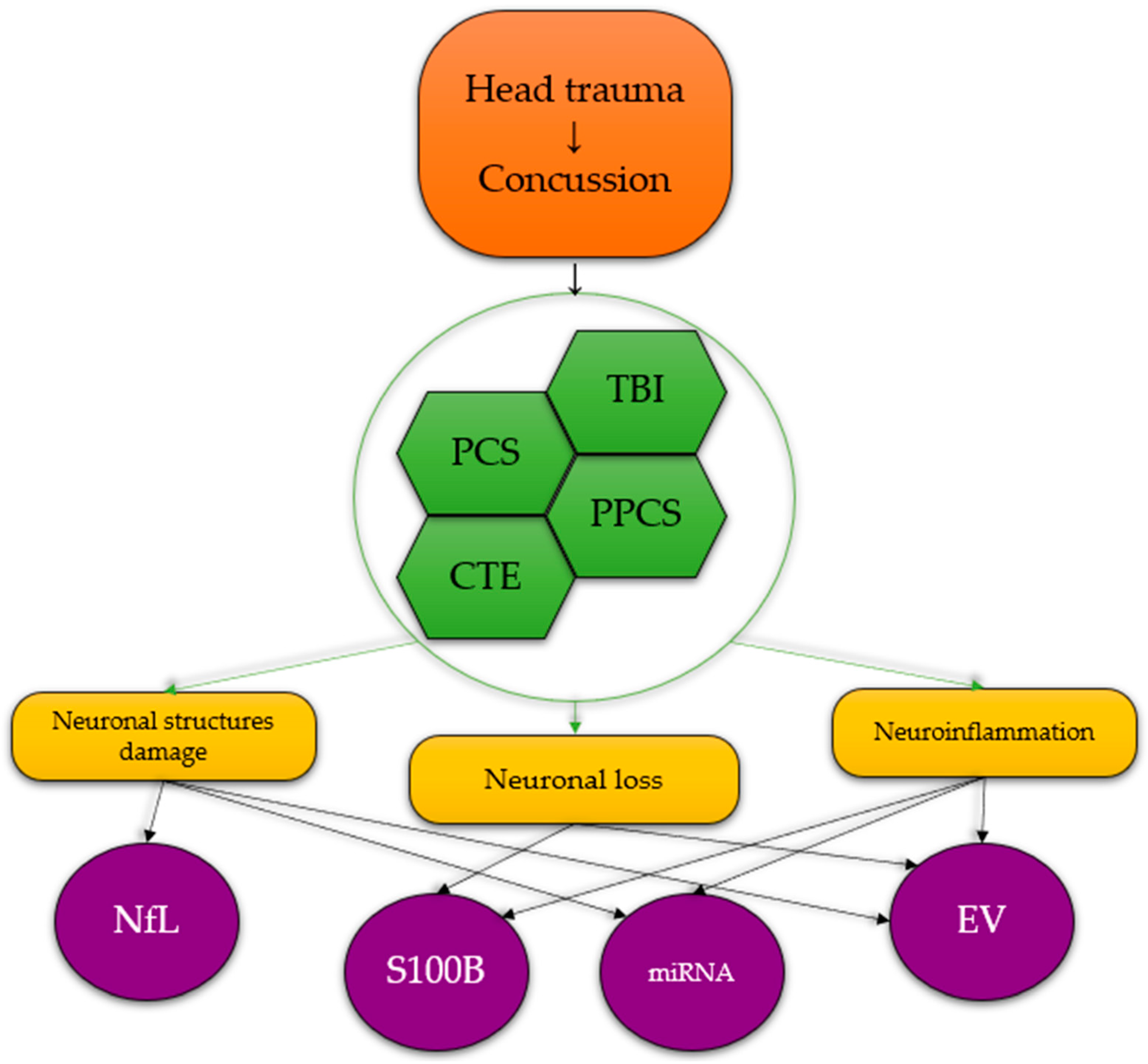
:1. Introduction
2. Post-Concussion Syndrome—Epidemiology and Diagnosis Criteria
3. Salivary Biomarkers
3.1. S100B
3.2. Neurofilament Light Chain (NfL)
3.3. MicroRNAs
3.4. Extracellular Vesicles
4. Conclusions
Funding
Conflicts of Interest
References
- Mavroudis, I.; Kazis, D.; Chowdhury, R.; Petridis, F.; Costa, V.; Balmus, I.-M.; Ciobica, A.; Luca, A.-C.; Radu, I.; Dobrin, R.P.; et al. Post-Concussion Syndrome and Chronic Traumatic Encephalopathy: Narrative Review on the Neuropathology, Neuroimaging and Fluid Biomarkers. Diagnostics 2022, 12, 740. [Google Scholar] [CrossRef] [PubMed]
- Ling, H.; Hardy, J.; Zetterberg, H. Neurological consequences of traumatic brain injuries in sports. Mol. Cell. Neurosci. 2015, 66, 114–122. [Google Scholar] [CrossRef] [PubMed]
- DeKosky, S.T.; Blennow, K.; Ikonomovic, M.D.; Gandy, S. Acute and chronic traumatic encephalopathies: Pathogenesis and biomarkers. Nat. Rev. Neurol. 2013, 9, 192–200. [Google Scholar] [CrossRef] [PubMed] [Green Version]
- Cantu, R. Concussion Classification: Ongoing Controversy. In Sebastianelli, W.J.; Slobounov, S.M., Ed.; Foundations of Sport-Related Brain Injuries; Springer: Berlin/Heidelberg, Germany, 2006; pp. 87–110. [Google Scholar]
- Saatman, K.E.; Duhaime, A.-C.; Bullock, R.; Maas, A.I.; Valadka, A.; Manley, G.T. Classification of Traumatic Brain Injury for Targeted Therapies. J. Neurotrauma 2008, 25, 719–738. [Google Scholar] [CrossRef] [Green Version]
- Teasdale, G.; Jennett, B. Assessment of coma and impaired consciousness. A practical scale. Lancet Lond. Engl. 1974, 2, 81–84. [Google Scholar] [CrossRef]
- Nakase-Richardson, R.; Sherer, M.; Seel, R.T.; Hart, T.; Hanks, R.; Arango-Lasprilla, J.C.; Yablon, S.A.; Sander, A.M.; Barnett, S.D.; Walker, W.C.; et al. Utility of post-traumatic amnesia in predicting 1-year productivity following traumatic brain injury: Comparison of the Russell and Mississippi PTA classification intervals. J. Neurol. Neurosurg. Psychiatry 2011, 82, 494–499. [Google Scholar] [CrossRef]
- Greenwald, B.D.; Ambrose, A.F.; Armstrong, G.P. Mild Brain Injury. Rehabil. Res. Pract. 2012, 2012, 469475. [Google Scholar] [CrossRef]
- Malec, J.F.; Brown, A.W.; Leibson, C.L.; Flaada, J.T.; Mandrekar, J.N.; Diehl, N.N.; Perkins, P.K. The Mayo Classification System for Traumatic Brain Injury Severity. J. Neurotrauma 2007, 24, 1417–1424. [Google Scholar] [CrossRef]
- Langer, L.K.; Alavinia, S.M.; Lawrence, D.W.; Munce, S.E.P.; Kam, A.; Tam, A.; Ruttan, L.; Comper, P.; Bayley, M.T. Prediction of risk of prolonged post-concussion symptoms: Derivation and validation of the TRICORDRR (Toronto Rehabilitation Institute Concussion Outcome Determination and Rehab Recommendations) score. PLoS Med. 2021, 18, e1003652. [Google Scholar] [CrossRef]
- Boake, C.; McCauley, S.R.; Levin, H.S.; Pedroza, C.; Contant, C.F.; Song, J.X.; Brown, S.A.; Goodman, H.; Brundage, S.I.; Diaz-Marchan, P.J. Diagnostic Criteria for Postconcussional Syndrome After Mild to Moderate Traumatic Brain Injury. J. Neuropsychiatry 2005, 17, 350–356. [Google Scholar] [CrossRef]
- McCauley, S.R.; Wilde, E.A.; Miller, E.R.; Robertson, C.S.; McCarthy, J.J.; Levin, H.S. Comparison of ICD-10 and DSM-IV Criteria for Postconcussion Syndrome/Disorder Stephen R. Rev. Iberoam. Neuropsicol. 2018, 1, 63–81. [Google Scholar]
- Permenter, C.M.; Fernández-de Thomas, R.J.; Sherman, A. Postconcussive Syndrome. StatPearls; StatPearls Publishing: Treasure Island, FL, USA, 2022. [Google Scholar]
- Clark, C.N.; Edwards, M.J.; Ong, B.E.; Goodliffe, L.; Ahmad, H.; Dilley, M.D.; Betteridge, S.; Griffin, C.; Jenkins, P.O. Reframing postconcussional syndrome as an interface disorder of neurology, psychiatry and psychology. Brain 2022, 145, 1906–1915. [Google Scholar] [CrossRef]
- Yoshizawa, J.M.; Schafer, C.A.; Schafer, J.J.; Farrell, J.J.; Paster, B.J.; Wong, D.T.W. Salivary Biomarkers: Toward Future Clinical and Diagnostic Utilities. Clin. Microbiol. Rev. 2013, 26, 781–791. [Google Scholar] [CrossRef] [Green Version]
- Kaufman, E.; Lamster, I.B. The Diagnostic Applications of Saliva—A Review. Crit. Rev. Oral Biol. Med. 2002, 13, 197–212. [Google Scholar] [CrossRef] [Green Version]
- Dhima, M.; Salinas, T.J.; Wermers, R.A.; Weaver, A.L.; Koka, S. Preference changes of adult outpatients for giving saliva, urine and blood for clinical testing after actual sample collection. J. Prosthodont. Res. 2013, 57, 51–56. [Google Scholar] [CrossRef]
- E Abraham, J.; Maranian, M.J.; Spiteri, I.; Russell, R.; Ingle, S.; Luccarini, C.; Earl, H.M.; Pharoah, P.P.; Dunning, A.M.; Caldas, C. Saliva samples are a viable alternative to blood samples as a source of DNA for high throughput genotyping. BMC Med. Genom. 2012, 5, 19. [Google Scholar] [CrossRef] [Green Version]
- Melguizo-Rodríguez, L.; Costela-Ruiz, V.; Manzano-Moreno, F.; Ruiz, C.; Illescas-Montes, R. Salivary Biomarkers and Their Application in the Diagnosis and Monitoring of the Most Common Oral Pathologies. Int. J. Mol. Sci. 2020, 21, 5173. [Google Scholar] [CrossRef]
- Salazar-Ruales, C.; Arguello, J.-V.; López-Cortés, A.; Cabrera-Andrade, A.; García-Cárdenas, J.M.; Guevara-Ramírez, P.; Peralta, P.; Leone, P.E.; Paz-Y-Miño, C. Salivary MicroRNAs for Early Detection of Head and Neck Squamous Cell Carcinoma: A Case-Control Study in the High Altitude Mestizo Ecuadorian Population. BioMed Res. Int. 2018, 2018, 9792730. [Google Scholar] [CrossRef] [Green Version]
- Dadas, A.; Washington, J.; Diaz-Arrastia, R.; Janigro, D. Biomarkers in traumatic brain injury (TBI): A review. Neuropsychiatr. Dis. Treat. 2018, 14, 2989–3000. [Google Scholar] [CrossRef] [Green Version]
- Hier, D.B.; Obafemi-Ajayi, T.; Thimgan, M.S.; Olbricht, G.R.; Azizi, S.; Allen, B.; Hadi, B.A.; Wunsch, D.C. Blood biomarkers for mild traumatic brain injury: A selective review of unresolved issues. Biomark. Res. 2021, 9, 70. [Google Scholar] [CrossRef]
- Zhang, J.; Puvenna, V.; Janigro, D. Biomarkers of Traumatic Brain Injury and Their Relationship to Pathology. In Translational Research in Traumatic Brain Injury; Laskowitz, D., Grant, G., Eds.; CRC Press/Taylor and Francis Group: Boca Raton, FL, USA, 2016; Chapter 12. [Google Scholar]
- Dadas, A.; Janigro, D. The role and diagnostic significance of cellular barriers after concussive head trauma. Concussion 2018, 3, CNC53. [Google Scholar] [CrossRef] [Green Version]
- Murcko, R.; Marchi, N.; Bailey, D.; Janigro, D. Diagnostic biomarker kinetics: How brain-derived biomarkers distribute through the human body, and how this affects their diagnostic significance: The case of S100B. Fluids Barriers CNS 2022, 19, 32. [Google Scholar] [CrossRef] [PubMed]
- Donato, R. Intracellular and extracellular roles of S100 proteins. Microsc. Res. Tech. 2003, 60, 540–551. [Google Scholar] [CrossRef]
- Chong, Z.; Changyaleket, B.; Xu, H.; Dull, R.; Schwartz, D. Identifying S100B as a Biomarker and a Therapeutic Target For Brain Injury and Multiple Diseases. Curr. Med. Chem. 2016, 23, 1571–1596. [Google Scholar] [CrossRef] [PubMed]
- Sedaghat, F.; Notopoulos, A. S100 protein family and its application in clinical practice. Hippokratia 2008, 12, 198–204. [Google Scholar] [PubMed]
- Michetti, F.; D'Ambrosi, N.; Toesca, A.; Puglisi, M.A.; Serrano, A.; Marchese, E.; Corvino, V.; Geloso, M.C. The S100B story: From biomarker to active factor in neural injury. J. Neurochem. 2019, 148, 168–187. [Google Scholar] [CrossRef] [Green Version]
- Tsoporis, J.N.; Mohammadzadeh, F.; Parker, T.G. Intracellular and Extracellular Effects of S100B in the Cardiovascular Response to Disease. Cardiovasc. Psychiatry Neurol. 2010, 2010, 206073. [Google Scholar] [CrossRef] [Green Version]
- Thelin, E.P.; Nelson, D.W.; Bellander, B.-M. A review of the clinical utility of serum S100B protein levels in the assessment of traumatic brain injury (Wien). Acta Neurochir. 2017, 159, 209–225. [Google Scholar] [CrossRef] [Green Version]
- Traxdorf, M.; Wendler, O.; Tziridis, K.; Bauer, J.; Scherl, C. S100B in serum and saliva: A valid invasive or non-invasive biomarker in obstructive sleep apnea? Eur. Rev. Med. Pharmacol. Sci. 2016, 20, 4766–4774. [Google Scholar]
- Janigro, D.; Kawata, K.; Silverman, E.; Marchi, N.; Diaz-Arrastia, R. Is Salivary S100B a Biomarker of Traumatic Brain Injury? A Pilot Study. Front. Neurol. 2020, 11, 528. [Google Scholar] [CrossRef]
- Yeung, C.; Bhatia, R.; Bhattarai, B.; Sinha, M. Role of Salivary Biomarkers in Predicting Significant Traumatic Brain Injury: An Exploratory Study. Pediatr. Emerg. Care 2020, 37, e1373–e1376. [Google Scholar] [CrossRef]
- Monroe, D.C.; Thomas, E.A.; Cecchi, N.J.; Granger, D.A.; Hicks, J.W.; Small, S.L. Salivary S100 calcium-binding protein beta (S100B) and neurofilament light (NfL) after acute exposure to repeated head impacts in collegiate water polo players. Sci. Rep. 2022, 12, 3439. [Google Scholar] [CrossRef]
- Abu-Rumeileh, S.; Abdelhak, A.; Foschi, M.; D'Anna, L.; Russo, M.; Steinacker, P.; Kuhle, J.; Tumani, H.; Blennow, K.; Otto, M. The multifaceted role of neurofilament light chain protein in non-primary neurological diseases. Brain 2022, 146, 421–437. [Google Scholar] [CrossRef]
- Gaetani, L.; Blennow, K.; Calabresi, P.; Di Filippo, M.; Parnetti, L.; Zetterberg, H. Neurofilament light chain as a biomarker in neurological disorders. J. Neurol. Neurosurg. Psychiatry 2019, 90, 870–881. [Google Scholar] [CrossRef]
- Heiskanen, M.; Jääskeläinen, O.; Manninen, E.; Das Gupta, S.; Andrade, P.; Ciszek, R.; Gröhn, O.; Herukka, S.-K.; Puhakka, N.; Pitkänen, A. Plasma Neurofilament Light Chain (NF-L) Is a Prognostic Biomarker for Cortical Damage Evolution but Not for Cognitive Impairment or Epileptogenesis Following Experimental TBI. Int. J. Mol. Sci. 2022, 23, 15208. [Google Scholar] [CrossRef]
- Guedes, V.A.; Lange, R.T.; Lippa, S.M.; Lai, C.; Greer, K.; Mithani, S.; Devoto, C.; Edwards, K.A.; Wagner, C.L.; Martin, C.A.; et al. Extracellular vesicle neurofilament light is elevated within the first 12-months following traumatic brain injury in a U.S military population. Sci. Rep. 2022, 12, 4002. [Google Scholar] [CrossRef]
- Thebault, S.; Booth, R.; Freedman, M. Blood Neurofilament Light Chain: The Neurologist’s Troponin? Biomedicines 2020, 8, 523. [Google Scholar] [CrossRef]
- Karantali, E.; Kazis, D.; McKenna, J.; Chatzikonstantinou, S.; Petridis, F.; Mavroudis, I. Neurofilament light chain in patients with a concussion or head impacts: A systematic review and meta-analysis. Eur. J. Trauma Emerg. Surg. 2021, 48, 1555–1567. [Google Scholar] [CrossRef]
- Wai, C.H.; Jin, J.; Cyrklaff, M.; Genoud, C.; Funaya, C.; Sattler, J.; Maceski, A.; Meier, S.; Heiland, S.; Lanzer, M.; et al. Neurofilament light chain plasma levels are associated with area of brain damage in experimental cerebral malaria. Sci. Rep. 2022, 12, 10726. [Google Scholar] [CrossRef]
- Janigro, D.; Bailey, D.M.; Lehmann, S.; Badaut, J.; O'Flynn, R.; Hirtz, C.; Marchi, N. Peripheral Blood and Salivary Biomarkers of Blood–Brain Barrier Permeability and Neuronal Damage: Clinical and Applied Concepts. Front. Neurol. 2021, 11, 577312. [Google Scholar] [CrossRef]
- Sullivan, R.; Montgomery, A.; Scipioni, A.; Jhaveri, P.; Schmidt, A.T.; Hicks, S.D. Confounding Factors Impacting microRNA Expression in Human Saliva: Methodological and Biological Considerations. Genes 2022, 13, 1874. [Google Scholar] [CrossRef] [PubMed]
- Di Pietro, V.; Yakoub, K.M.; Scarpa, U.; Di Pietro, C.; Belli, A. MicroRNA Signature of Traumatic Brain Injury: From the Biomarker Discovery to the Point-of-Care. Front. Neurol. 2018, 9, 429. [Google Scholar] [CrossRef] [PubMed] [Green Version]
- Atif, H.; Hicks, S.D. A Review of MicroRNA Biomarkers in Traumatic Brain Injury. J. Exp. Neurosci. 2019, 13, 117906951983228. [Google Scholar] [CrossRef] [PubMed] [Green Version]
- Matyasova, K.; Csicsatkova, N.; Filipcik, P.; Jurisica, I.; Cente, M. Peripheral microRNA alteration and pathway signaling after mild traumatic brain injur. Gen. Physiol. Biophys. 2021, 40, 523–539. [Google Scholar] [CrossRef] [PubMed]
- Wu, L.; Zheng, K.; Yan, C.; Pan, X.; Liu, Y.; Liu, J.; Wang, F.; Guo, W.; He, X.; Li, J.; et al. Genome-wide study of salivary microRNAs as potential noninvasive biomarkers for detection of nasopharyngeal carcinoma. BMC Cancer 2019, 19, 843. [Google Scholar] [CrossRef] [Green Version]
- Yoshizawa, J.M.; Wong, D.T.W. Salivary MicroRNAs and Oral Cancer Detection. Methods Mol. Biol. 2013, 936, 313–324. [Google Scholar] [CrossRef] [Green Version]
- Hiskens, M.I.; Mengistu, T.S.; Li, K.M.; Fenning, A.S. Systematic Review of the Diagnostic and Clinical Utility of Salivary microRNAs in Traumatic Brain Injury (TBI). Int. J. Mol. Sci. 2022, 23, 13160. [Google Scholar] [CrossRef]
- Di Pietro, V.; Porto, E.; Ragusa, M.; Barbagallo, C.; Davies, D.; Forcione, M.; Logan, A.; Di Pietro, C.; Purrello, M.; Grey, M.; et al. Salivary MicroRNAs: Diagnostic Markers of Mild Traumatic Brain Injury in Contact-Sport. Front. Mol. Neurosci. 2018, 11, 290. [Google Scholar] [CrossRef] [Green Version]
- Di Pietro, V.; O'Halloran, P.; Watson, C.N.; Begum, G.; Acharjee, A.; Yakoub, K.M.; Bentley, C.; Davies, D.J.; Iliceto, P.; Candilera, G.; et al. Unique diagnostic signatures of concussion in the saliva of male athletes: The Study of Concussion in Rugby Union through MicroRNAs (SCRUM). Br. J. Sport. Med. 2021, 55, 1395–1404. [Google Scholar] [CrossRef]
- Fedorchak, G.; Rangnekar, A.; Onks, C.; Loeffert, A.C.; Loeffert, J.; Olympia, R.P.; DeVita, S.; Leddy, J.; Haider, M.N.; Roberts, A.; et al. Saliva RNA biomarkers predict concussion duration and detect symptom recovery: A comparison with balance and cognitive testing. J. Neurol. 2021, 268, 4349–4361. [Google Scholar] [CrossRef]
- Hicks, S.D.; Johnson, J.; Carney, M.C.; Bramley, H.; Olympia, R.P.; Loeffert, A.C.; Thomas, N.J. Overlapping MicroRNA Expression in Saliva and Cerebrospinal Fluid Accurately Identifies Pediatric Traumatic Brain Injury. J. Neurotrauma 2018, 35, 64–72. [Google Scholar] [CrossRef]
- Hicks, S.D.; Olympia, R.P.; Onks, C.; Kim, R.Y.; Zhen, K.J.; Fedorchak, G.; DeVita, S.; Rangnekar, A.; Heller, M.; Zwibel, H.; et al. Saliva microRNA Biomarkers of Cumulative Concussion. Int. J. Mol. Sci. 2020, 21, 7758. [Google Scholar] [CrossRef]
- Hicks, S.D.; Onks, C.; Kim, R.Y.; Zhen, K.J.; Loeffert, J.; Loeffert, A.C.; Olympia, R.P.; Fedorchak, G.; DeVita, S.; Gagnon, Z.; et al. Refinement of saliva microRNA biomarkers for sports-related concussion. J. Sport Health Sci. 2021; in press. [Google Scholar] [CrossRef]
- Hicks, S.D.; Onks, C.; Kim, R.Y.; Zhen, K.J.; Loeffert, J.; Loeffert, A.C.; Olympia, R.P.; Fedorchak, G.; DeVita, S.; Rangnekar, A.; et al. Diagnosing mild traumatic brain injury using saliva RNA compared to cognitive and balance testing. Clin. Transl. Med. 2020, 10, e197. [Google Scholar] [CrossRef]
- Johnson, J.J.; Loeffert, A.C.; Stokes, J.; Olympia, R.P.; Bramley, H.; Hicks, S.D. Association of Salivary MicroRNA Changes With Prolonged Concussion Symptoms. JAMA Pediatr. 2018, 172, 65–73. [Google Scholar] [CrossRef] [Green Version]
- LaRocca, D.; Barns, S.; Hicks, S.D.; Brindle, A.; Williams, J.; Uhlig, R.; Johnson, P.; Neville, C.; Middleton, F.A. Comparison of serum and saliva miRNAs for identification and characterization of mTBI in adult mixed martial arts fighters. PLoS ONE 2019, 14, e0207785. [Google Scholar] [CrossRef] [Green Version]
- Miller, K.E.; MacDonald, J.P.; Sullivan, L.; Venkata, L.P.R.; Shi, J.; Yeates, K.O.; Chen, S.; Alshaikh, E.; Taylor, H.G.; Hautmann, A.; et al. Salivary miRNA Expression in Children With Persistent Post-concussive Symptoms. Front. Public Health. 2022, 10, 890420. [Google Scholar] [CrossRef]
- Doyle, L.; Wang, M. Overview of Extracellular Vesicles, Their Origin, Composition, Purpose, and Methods for Exosome Isolation and Analysis. Cells 2019, 8, 727. [Google Scholar] [CrossRef] [Green Version]
- Khan, N.A.; Asim, M.; El-Menyar, A.; Biswas, K.H.; Rizoli, S.; Al-Thani, H. The evolving role of extracellular vesicles (exosomes) as biomarkers in traumatic brain injury: Clinical perspectives and therapeutic implications. Front. Aging Neurosci. 2022, 14, 933434. [Google Scholar] [CrossRef]
- Hill, A.F. Extracellular Vesicles and Neurodegenerative Diseases. J. Neurosci. 2019, 39, 9269–9273. [Google Scholar] [CrossRef]
- Beard, K.; Yang, Z.; Haber, M.; Flamholz, M.; Diaz-Arrastia, R.; Sandsmark, D.; Meaney, D.F.; Issadore, D. Extracellular vesicles as distinct biomarker reservoirs for mild traumatic brain injury diagnosis. Brain Commun. 2021, 3, fcab151. [Google Scholar] [CrossRef] [PubMed]
- Brenna, S.; Krisp, C.; Altmeppen, H.C.; Magnus, T.; Puig, B. Brain-Derived Extracellular Vesicles in Health and Disease: A Methodological Perspective. Int. J. Mol. Sci. 2021, 22, 1365. [Google Scholar] [CrossRef] [PubMed]
- Gomes, P.A.; Bodo, C.; Nogueras-Ortiz, C.; Samiotaki, M.; Chen, M.; Soares-Cunha, C.; Silva, J.M.; Coimbra, B.; Stamatakis, G.; Santos, L.; et al. A novel isolation method for spontaneously released extracellular vesicles from brain tissue and its implications for stress-driven brain pathology. Cell Commun. Signal. 2023, 21, 35. [Google Scholar] [CrossRef] [PubMed]
- Bart, G.; Fischer, D.; Samoylenko, A.; Zhyvolozhnyi, A.; Stehantsev, P.; Miinalainen, I.; Kaakinen, M.; Nurmi, T.; Singh, P.; Kosamo, S.; et al. Characterization of nucleic acids from extracellular vesicle-enriched human sweat. BMC Genom. 2021, 22, 425. [Google Scholar] [CrossRef]
- Yates, A.G.; Anthony, D.C.; Ruitenberg, M.J.; Couch, Y. Systemic Immune Response to Traumatic CNS Injuries—Are Extracellular Vesicles the Missing Link? Front. Immunol. 2019, 10, 2723. [Google Scholar] [CrossRef] [Green Version]
- Cheng, Y.; Pereira, M.; Raukar, N.; Reagan, J.L.; Queseneberry, M.; Goldberg, L.; Borgovan, T.; LaFrance, W.C., Jr.; Dooner, M.; Deregibus, M.; et al. Potential biomarkers to detect traumatic brain injury by the profiling of salivary extracellular vesicles. J. Cell. Physiol. 2019, 234, 14377–14388. [Google Scholar] [CrossRef] [Green Version]
- Matuk, R.; Pereira, M.; Baird, J.; Dooner, M.; Cheng, Y.; Wen, S.; Rao, S.; Quesenberry, P.; Raukar, N.P. The role of salivary vesicles as a potential inflammatory biomarker to detect traumatic brain injury in mixed martial artists. Sci. Rep. 2021, 11, 8186. [Google Scholar] [CrossRef]
- Pereira, M.; Cheng, Y.; Raukar, N.P.; Reagan, J.L.; Quesenberry, M.; Goldberg, L.; Borgovan, T.; LaFrance, W.C.; Dooner, M.; Deregibus, M.; et al. Inflammation-related gene expression profiles of salivary extracellular vesicles in patients with head trauma. Neural Regen. Res. 2020, 15, 676–681. [Google Scholar] [CrossRef]


| Criteria | |
|---|---|
| Possible | Neurocognitive symptoms: blurred vision, confusion, headache, or nausea. |
| Probable—mild | Neurocognitive symptoms: loss of consciousness (<30 min), post-traumatic amnesia (<24 h); Mechanical damage: depressed, basilar, or linear skull fracture; Brain damage: intact dura matter. |
| Definite moderate—severe | Neurocognitive symptoms: loss of consciousness (>30 min), post-traumatic amnesia (>24 h); GCS < 13; Brain damage: brain hematoma, brain hemorrhage, contusions, or ruptured dura mater. Death. |
| Study | Experimental Design | Results |
|---|---|---|
| S100B | ||
| [33] | 15 adult patients with suspected TBI and 15 control subjects | Average salivary S100B level 3.9-fold higher than blood S100B. Salivary S100B levels are as effective as serum levels in differentiating TBI patients from control subjects. |
| [34] | 70 children: 24 acute and isolated TBI 46 with musculoskeletal injuries only | Salivary S100B levels following TBI are significantly higher, as compared to the ones following musculoskeletal injury |
| [35] | 65 water polo players, before and after a competitive tournament | No association between S100B salivary levels and head impacts exposure. |
| Neurofilaments light chain (NfL) | ||
| [35] | 65 water polo players, before and after a competitive tournament | Salivary NfL is directly associated with head impact frequency and cumulative head impact magnitude, as compared with baseline salivary NfL. |
| Micro RNAs (miRNAs) | ||
| [49] | 1028 rugby players, 66 players with musculoskeletal injuries, 102 uninjured players | Significant difference sin expression of 32 small non-coding RNAs: 14 small non-coding RNAs—concussed players versus players without injuries 6 small non-coding RNAs—prolonged PPCS prediction 11 small non-coding RNAs—age dependent symptoms recovery prediction |
| [52] | 10 concussed professional and semi-professional rugby players 10 non-concussed matched controls | 5 miRNAs significantly upregulated in concussed athletes |
| [53] | 112 mild TBI individuals (8 to 24 years old) | 16 non-coding RNAs—PPCS prediction |
| [54] | 60 children diagnosed with mild TBI 18 age-matched controls | 10 miRNAs expression significantly altered in mild TBI children |
| [55] | 13 former professional American football players, 18 age and sex-matched controls | 20 salivary miRNAs expression significantly altered in athletes 2 salivary miRNAs associated with the number of concussion events |
| [56] | 314 athletes with and without a history of a concussion | 2 miRNAs ↓ after physical exercise 1 miRNA ↑ after contact sports participation 23 miRNAs expression altered after 1 season of contact sports 2 miRNAs associated with head impacts number Significant differences in 11 miRNAs expression in concussed versus non-concussed participants |
| Extracellular vesicles (EVs) | ||
| [69] | 54 subjects: 16 post-concussion patients, 15 head trauma patients, 23 controls | Salivary EVs gene expression—viable source of biomarkers for mild TBI Multiple Alzheimer’s disease genes expression present in post-mild TBI saliva samples: |
| [70] | 8 mixed martial arts fighters, 7 from controls. | EVs could be potential biomarkers of the acute phase of brain trauma in correlation with injury severity. |
| [71] | 6 TBI patients, 6 concussion patients, 7 healthy controls | 9 upregulated genes in acute TBI: LOX5, ANXA3, CASP1, IL2RG, ITGAM, ITGB2, LTA4H, MAPK14, and TNFRSF1A, 13 upregulated genes in concussion: ADRB1, ADRB2, BDKRB1, HRH1, HRH2, LTB4R2, LTB4R, PTAFR, CYSLTR1, CES1, KLK1, MC2R, and PTGER3. |
Disclaimer/Publisher’s Note: The statements, opinions and data contained in all publications are solely those of the individual author(s) and contributor(s) and not of MDPI and/or the editor(s). MDPI and/or the editor(s) disclaim responsibility for any injury to people or property resulting from any ideas, methods, instructions or products referred to in the content. |
© 2023 by the authors. Licensee MDPI, Basel, Switzerland. This article is an open access article distributed under the terms and conditions of the Creative Commons Attribution (CC BY) license (https://creativecommons.org/licenses/by/4.0/).
Share and Cite
Mavroudis, I.; Petridis, F.; Balmus, I.-M.; Ciobica, A.; Gorgan, D.L.; Luca, A.C. Review on the Role of Salivary Biomarkers in the Diagnosis of Mild Traumatic Brain Injury and Post-Concussion Syndrome. Diagnostics 2023, 13, 1367. https://doi.org/10.3390/diagnostics13081367
Mavroudis I, Petridis F, Balmus I-M, Ciobica A, Gorgan DL, Luca AC. Review on the Role of Salivary Biomarkers in the Diagnosis of Mild Traumatic Brain Injury and Post-Concussion Syndrome. Diagnostics. 2023; 13(8):1367. https://doi.org/10.3390/diagnostics13081367
Chicago/Turabian StyleMavroudis, Ioannis, Foivos Petridis, Ioana-Miruna Balmus, Alin Ciobica, Dragos Lucian Gorgan, and Alina Costina Luca. 2023. "Review on the Role of Salivary Biomarkers in the Diagnosis of Mild Traumatic Brain Injury and Post-Concussion Syndrome" Diagnostics 13, no. 8: 1367. https://doi.org/10.3390/diagnostics13081367