
Usefulness of Endoscopy for the Detection and Diagnosis of Primary Esophageal Motility Disorders and Diseases Relating to Abnormal Esophageal Motility


Abstract
:1. Introduction
2. Detection or Diagnosis
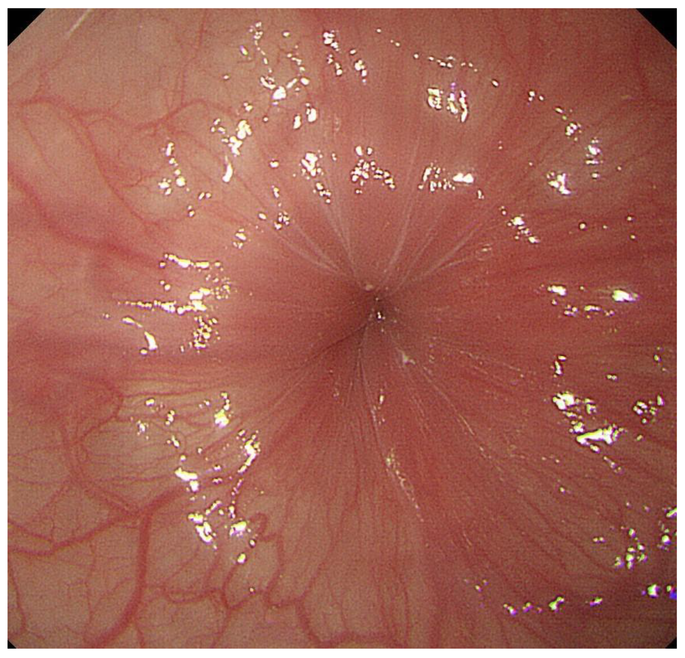
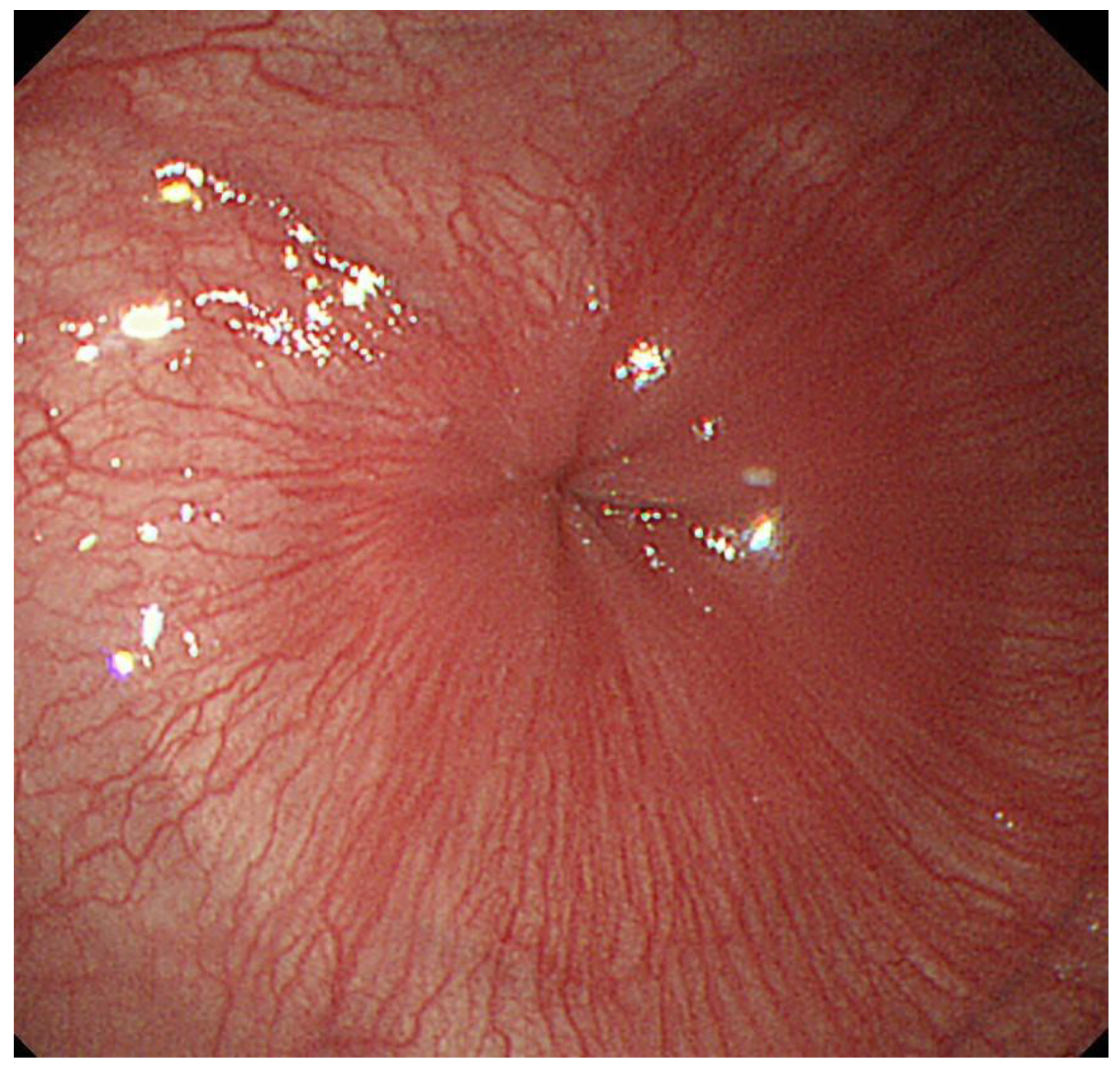
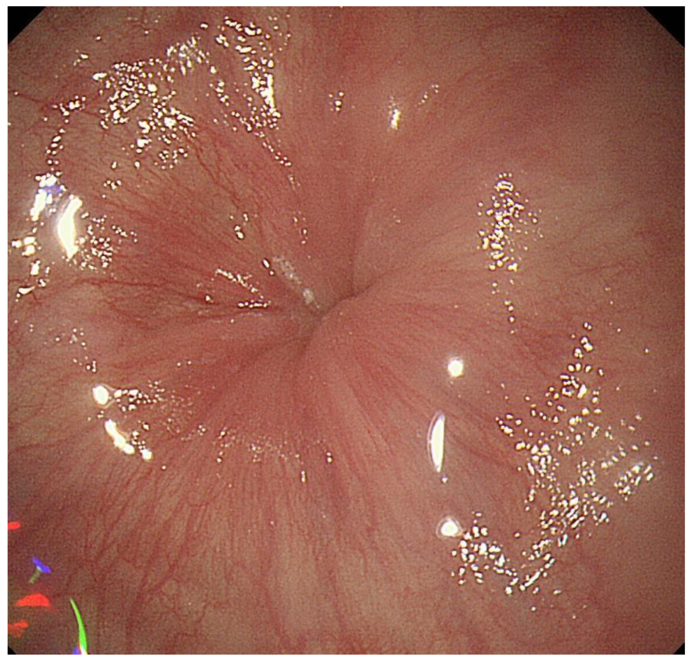
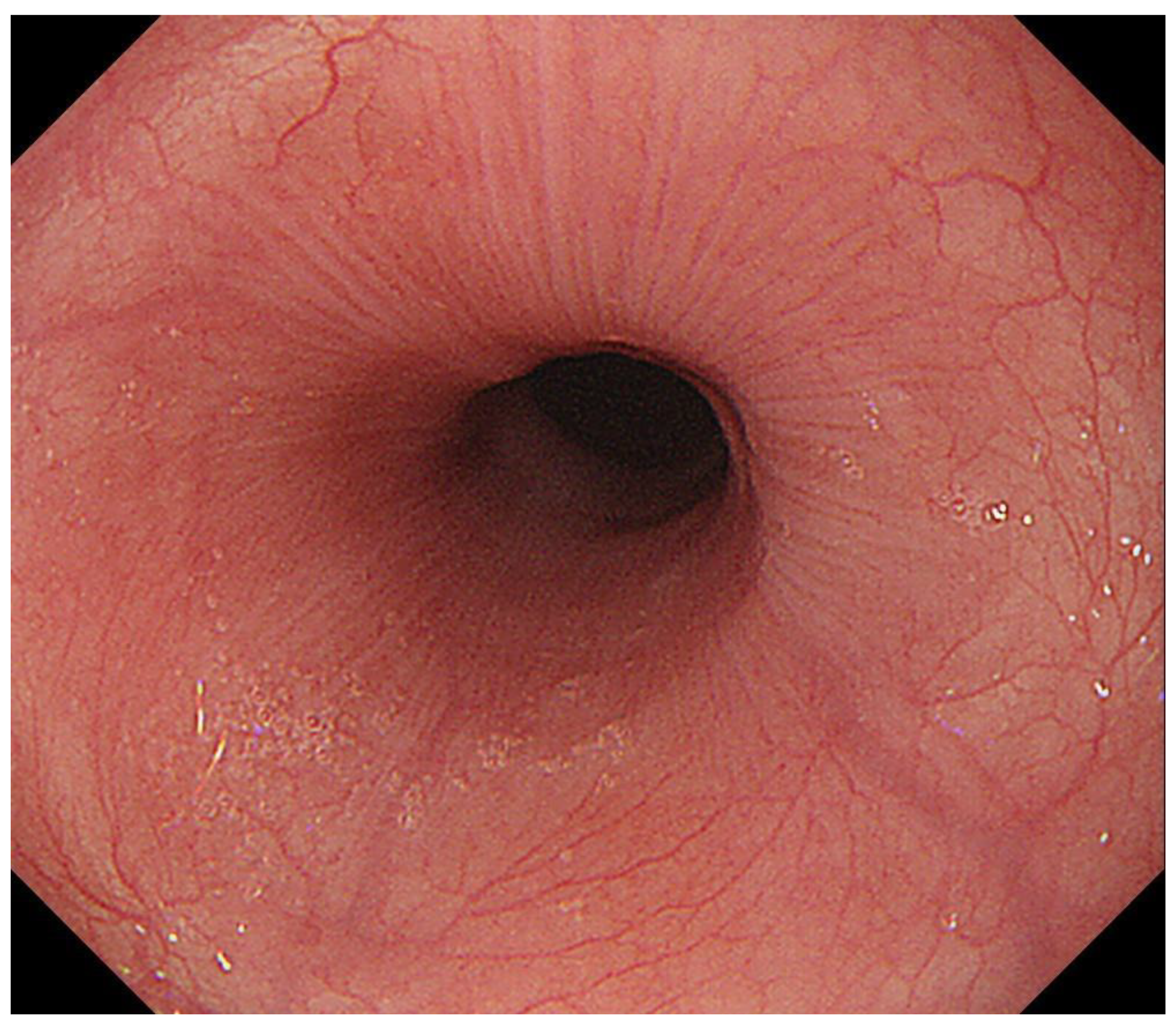
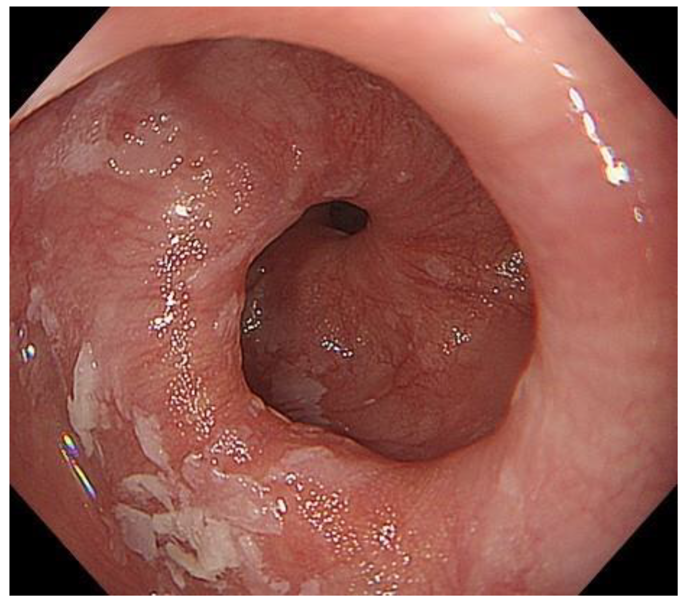
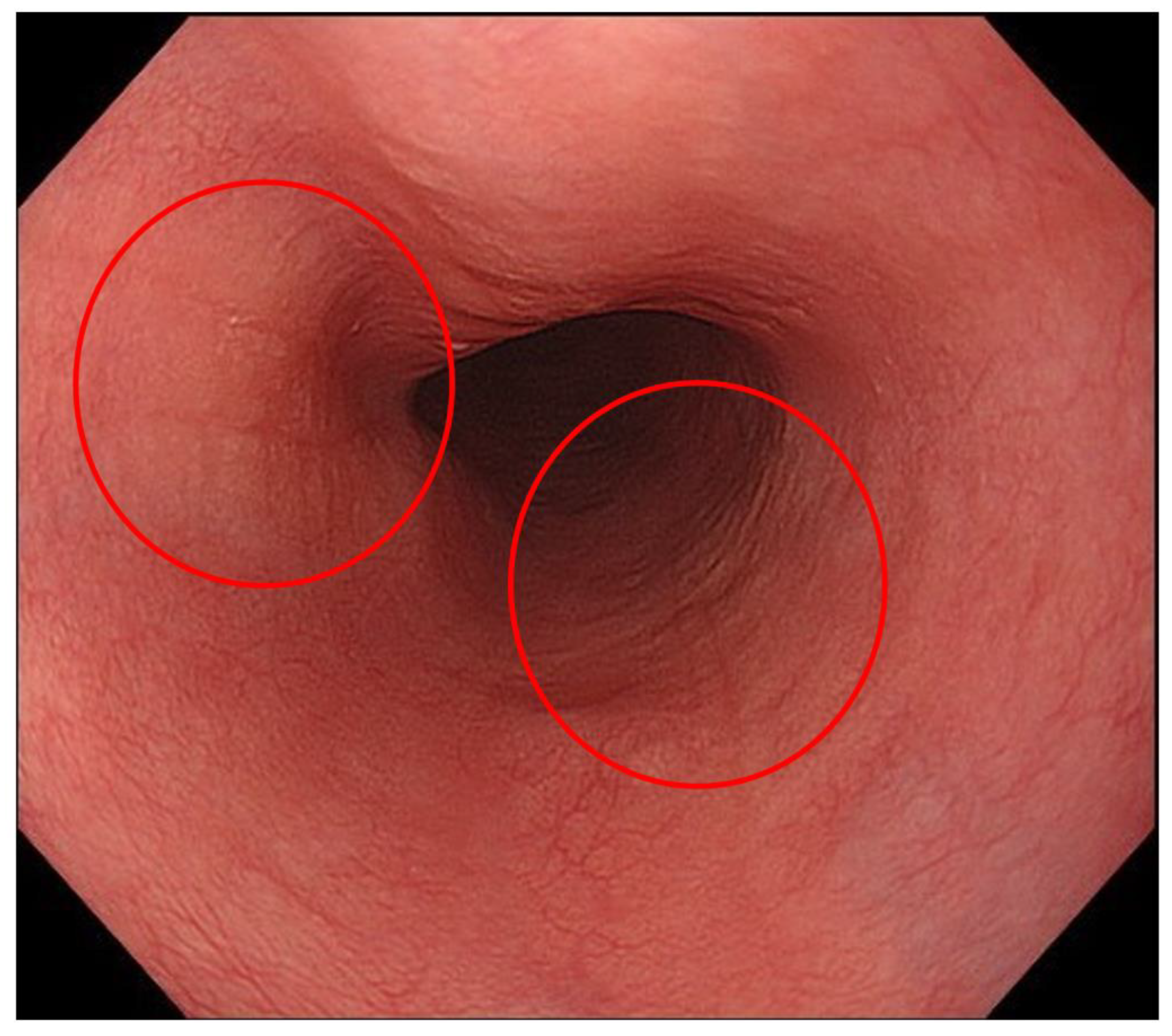
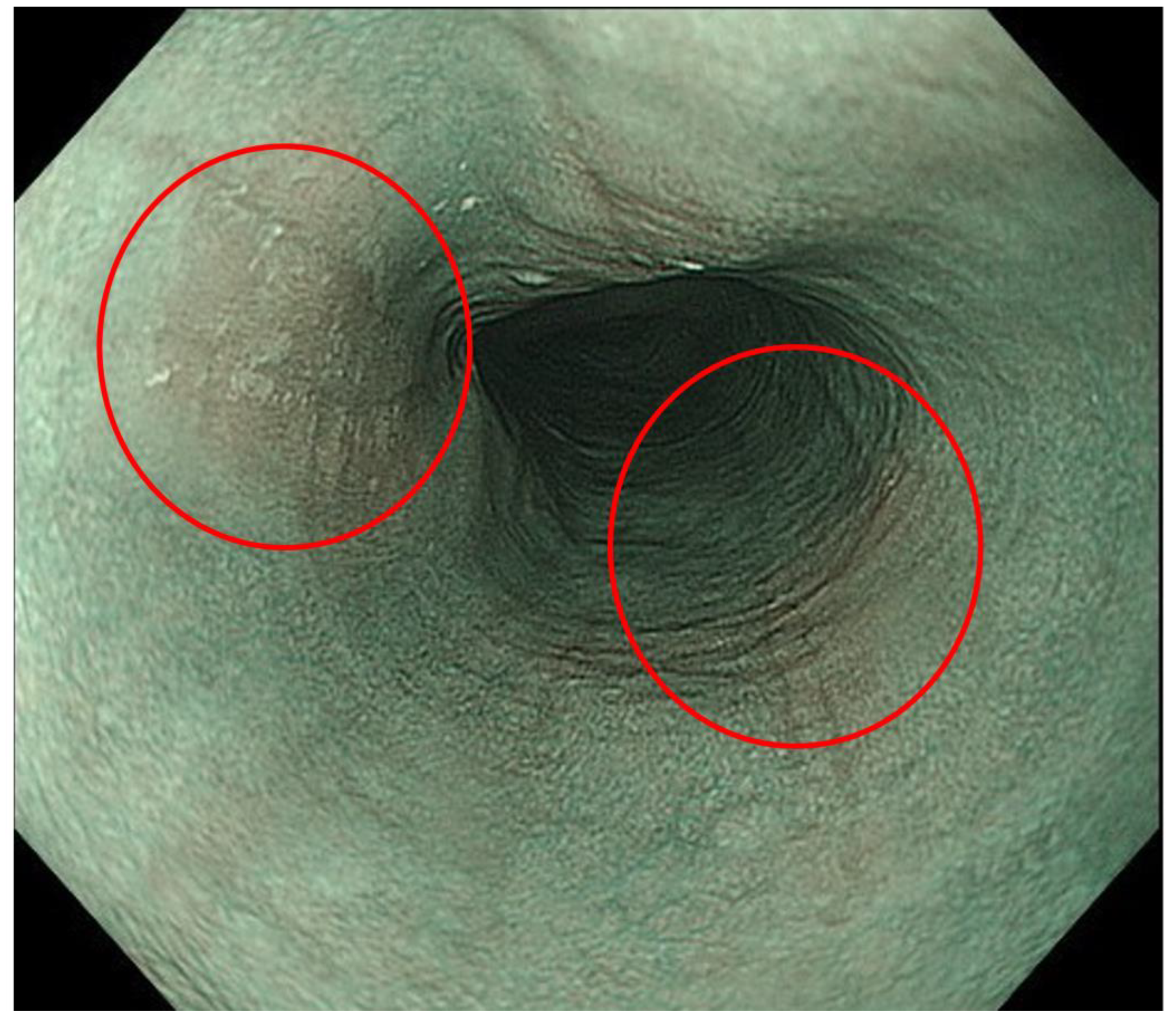
2.1. Endoscopic Findings Related to Primary EMDs
2.2. Endoscopic Findings That Could be Associated with EMDs
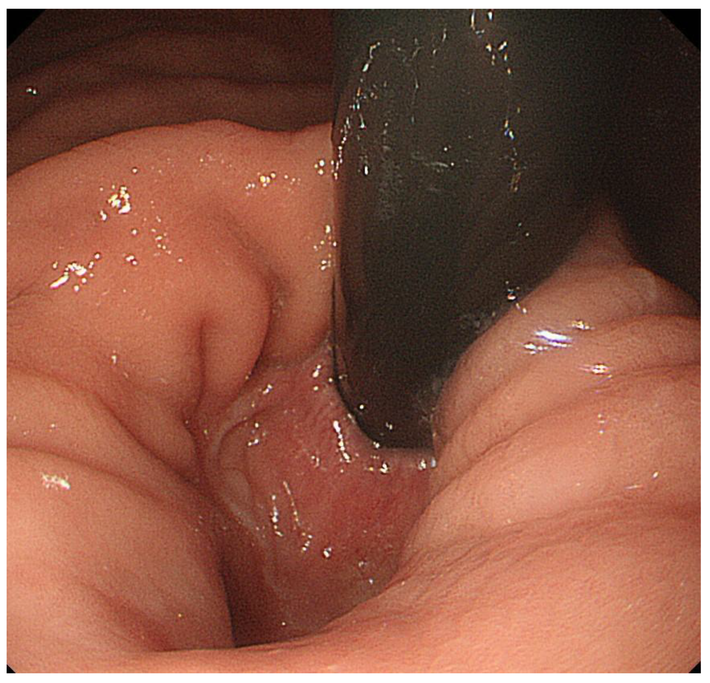
2.2.1. Diverticulum or Esophageal Intramural Pseudodiverticulosis
2.2.2. Candida Esophagitis
2.3. Identification of Primary EMDs during EGD
2.4. Endoscopic Findings of Diseases That Could Be Related to Abnormal Esophageal Motility
2.4.1. GERD
2.4.2. Eosinophilic Esophagitis
2.4.3. Systemic Sclerosis or Other Autoimmune Diseases
2.4.4. Systemic Amyloidosis
2.4.5. Von Recklinghausen’s Neurofibromatosis
3. Conclusions
Author Contributions
Funding
Institutional Review Board Statement
Informed Consent Statement
Data Availability Statement
Acknowledgments
Conflicts of Interest
References
- Patel, D.A.; Yadlapati, R.; Vaezi, M.F. Esophageal Motility Disorders: Current Approach to Diagnostics and Therapeutics. Gastroenterology 2022, 162, 1617–1634. [Google Scholar] [CrossRef] [PubMed]
- Yadlapati, R.; Kahrilas, P.J.; Fox, M.R.; Bredenoord, A.J.; Prakash Gyawali, C.; Roman, S.; Babaei, A.; Mittal, R.K.; Rommel, N.; Savarino, E.; et al. Esophageal motility disorders on high-resolution manometry: Chicago classification version 4.0((c)). Neurogastroenterol. Motil. 2021, 33, e14058. [Google Scholar] [CrossRef] [PubMed]
- Muto, M.; Minashi, K.; Yano, T.; Saito, Y.; Oda, I.; Nonaka, S.; Omori, T.; Sugiura, H.; Goda, K.; Kaise, M.; et al. Early detection of superficial squamous cell carcinoma in the head and neck region and esophagus by narrow band imaging: A multicenter randomized controlled trial. J. Clin. Oncol. 2010, 28, 1566–1572. [Google Scholar] [CrossRef]
- Katada, C.; Tanabe, S.; Koizumi, W.; Higuchi, K.; Sasaki, T.; Azuma, M.; Katada, N.; Masaki, T.; Nakayama, M.; Okamoto, M.; et al. Narrow band imaging for detecting superficial squamous cell carcinoma of the head and neck in patients with esophageal squamous cell carcinoma. Endoscopy 2010, 42, 185–190. [Google Scholar] [CrossRef]
- Goda, K.; Tajiri, H.; Ikegami, M.; Yoshida, Y.; Yoshimura, N.; Kato, M.; Sumiyama, K.; Imazu, H.; Matsuda, K.; Kaise, M.; et al. Magnifying endoscopy with narrow band imaging for predicting the invasion depth of superficial esophageal squamous cell carcinoma. Dis. Esophagus 2009, 22, 453–460. [Google Scholar] [CrossRef] [PubMed]
- Kawahara, Y.; Uedo, N.; Fujishiro, M.; Goda, K.; Hirasawa, D.; Lee, J.H.; Miyahara, R.; Morita, Y.; Singh, R.; Takeuchi, M.; et al. The usefulness of NBI magnification on diagnosis of superficial esophageal squamous cell carcinoma. Dig. Endosc. 2011, 23 (Suppl. S1), 79–82. [Google Scholar] [CrossRef]
- Takahashi, A.; Oyama, T. Barrett’s esophageal adenocarcinoma diagnosed by narrow-band imaging magnifying endoscopy. Dig. Endosc. 2013, 25 (Suppl. S2), 184–189. [Google Scholar] [CrossRef] [PubMed]
- Arantes, V.; Albuquerque, W.; Salles, J.M.; Freitas Dias, C.A.; Alberti, L.R.; Kahaleh, M.; Ferrari, T.C.; Coelho, L.G. Effectiveness of unsedated transnasal endoscopy with white-light, flexible spectral imaging color enhancement, and lugol staining for esophageal cancer screening in high-risk patients. J. Clin. Gastroenterol. 2013, 47, 314–321. [Google Scholar] [CrossRef]
- Li, Y.; Yangjin, C.; Shi, Y.; Wen, Y.; Jin, Z.; Cui, R.; Zhang, H.; Ding, S. The Significance of a Pale Area Via Flexible Spectral Imaging Color Enhancement in the Diagnosis of Esophageal Precancerous Lesions and Early-stage Squamous Cancer. J. Clin. Gastroenterol. 2019, 53, e400–e404. [Google Scholar] [CrossRef] [PubMed]
- Ueda, T.; Dohi, O.; Naito, Y.; Yoshida, T.; Azuma, Y.; Ishida, T.; Matsumura, S.; Kitae, H.; Takayama, S.; Mizuno, N.; et al. Diagnostic performance of magnifying blue laser imaging versus magnifying narrow-band imaging for identifying the depth of invasion of superficial esophageal squamous cell carcinoma. Dis. Esophagus 2021, 34, 1–8. [Google Scholar] [CrossRef] [PubMed]
- Nakamura, K.; Urabe, Y.; Oka, S.; Nagasaki, N.; Yorita, N.; Hata, K.; Masuda, K.; Kurihara, M.; Kotachi, T.; Boda, T.; et al. Usefulness of linked color imaging in the early detection of superficial esophageal squamous cell carcinomas. Esophagus 2021, 18, 118–124. [Google Scholar] [CrossRef]
- Kodashima, S.; Fujishiro, M. Novel image-enhanced endoscopy with i-scan technology. World J. Gastroenterol. 2010, 16, 1043–1049. [Google Scholar] [CrossRef]
- Inoue, H.; Minami, H.; Kobayashi, Y.; Sato, Y.; Kaga, M.; Suzuki, M.; Satodate, H.; Odaka, N.; Itoh, H.; Kudo, S. Peroral endoscopic myotomy (POEM) for esophageal achalasia. Endoscopy 2010, 42, 265–271. [Google Scholar] [CrossRef]
- Inoue, H.; Sato, H.; Ikeda, H.; Onimaru, M.; Sato, C.; Minami, H.; Yokomichi, H.; Kobayashi, Y.; Grimes, K.L.; Kudo, S.E. Per-Oral Endoscopic Myotomy: A Series of 500 Patients. J. Am. Coll. Surg. 2015, 221, 256–264. [Google Scholar] [CrossRef] [PubMed]
- Bechara, R.; Ikeda, H.; Inoue, H. Peroral endoscopic myotomy: An evolving treatment for achalasia. Nat. Rev. Gastroenterol. Hepatol. 2015, 12, 410–426. [Google Scholar] [CrossRef] [PubMed]
- Onimaru, M.; Inoue, H.; Fujiyoshi, Y.; Abad, M.R.A.; Nishikawa, Y.; Toshimori, A.; Shimamura, Y.; Tanabe, M.; Sumi, K.; Ikeda, H. Long-term clinical results of per-oral endoscopic myotomy (POEM) for achalasia: First report of more than 10-year patient experience as assessed with a questionnaire-based survey. Endosc. Int. Open 2021, 9, E409–E416. [Google Scholar] [CrossRef]
- Weusten, B.; Barret, M.; Bredenoord, A.J.; Familiari, P.; Gonzalez, J.M.; van Hooft, J.E.; Ishaq, S.; Lorenzo-Zuniga, V.; Louis, H.; van Meer, S.; et al. Endoscopic management of gastrointestinal motility disorders—Part 1: European Society of Gastrointestinal Endoscopy (ESGE) Guideline. Endoscopy 2020, 52, 498–515. [Google Scholar] [CrossRef] [PubMed]
- Pandolfino, J.E.; Gawron, A.J. Achalasia: A systematic review. JAMA 2015, 313, 1841–1852. [Google Scholar] [CrossRef] [PubMed]
- Japan Esophageal, S. Descriptive Rules for Achalasia of the Esophagus, June 2012: 4th Edition. Esophagus 2017, 14, 275–289. [Google Scholar] [CrossRef]
- Iwakiri, K.; Hoshihara, Y.; Kawami, N.; Sano, H.; Tanaka, Y.; Umezawa, M.; Kotoyori, M.; Nomura, T.; Miyashita, M.; Sakamoto, C. The appearance of rosette-like esophageal folds (“esophageal rosette”) in the lower esophagus after a deep inspiration is a characteristic endoscopic finding of primary achalasia. J. Gastroenterol. 2010, 45, 422–425. [Google Scholar] [CrossRef]
- Hoshikawa, Y.; Hoshino, S.; Kawami, N.; Tanabe, T.; Hanada, Y.; Takenouchi, N.; Goto, O.; Kaise, M.; Iwakiri, K. Possible new endoscopic finding in patients with achalasia: “Gingko leaf sign”. Esophagus 2020, 17, 208–213. [Google Scholar] [CrossRef]
- Gomi, K.; Inoue, H.; Ikeda, H.; Bechara, R.; Sato, C.; Ito, H.; Onimaru, M.; Kitamura, Y.; Suzuki, M.; Nakamura, J.; et al. New endoscopic classification of the cardiac orifice in esophageal achalasia: Champagne glass sign. Dig. Endosc. 2016, 28, 645–649. [Google Scholar] [CrossRef]
- Minami, H.; Isomoto, H.; Miuma, S.; Kobayashi, Y.; Yamaguchi, N.; Urabe, S.; Matsushima, K.; Akazawa, Y.; Ohnita, K.; Takeshima, F.; et al. New endoscopic indicator of esophageal achalasia: “pinstripe pattern”. PLoS ONE 2015, 10, e0101833. [Google Scholar] [CrossRef] [PubMed]
- Shiwaku, H.; Yamashita, K.; Ohmiya, T.; Nimura, S.; Shiwaku, Y.; Inoue, H.; Hasegawa, S. New endoscopic finding of esophageal achalasia with ST Hood short type: Corona appearance. PLoS ONE 2018, 13, e0199955. [Google Scholar] [CrossRef]
- Misra, S.P.; Dwivedi, M.; Gupta, S.C. Endoscopic diagnosis of corkscrew esophagus. Gastrointest. Endosc. 2001, 53, 779. [Google Scholar] [CrossRef]
- Cammarota, G.; Certo, M. Images in clinical medicine. Corkscrew esophagus. N. Engl. J. Med. 2005, 352, 816. [Google Scholar] [CrossRef] [PubMed]
- Kato, T.; Naiki, T.; Araki, H.; Nagaki, M.; Moriwaki, H. Diffuse esophageal spasm. Gastrointest. Endosc. 2004, 60, 428. [Google Scholar] [CrossRef]
- Hosaka, H.; Kawami, N.; Manabe, N.; Kuribayashi, S.; Sato, H.; Funaki, Y.; Ayaki, M.; Hara, K.; Ueda, C.; Matsumura, T.; et al. Clinical presentation and therapeutic outcome of patients with jackhammer esophagus-a multicenter cohort study in Japan. Esophagus 2022, 19, 393–400. [Google Scholar] [CrossRef]
- Cameron, A.J.; Malcolm, A.; Prather, C.M.; Phillips, S.F. Videoendoscopic diagnosis of esophageal motility disorders. Gastrointest. Endosc. 1999, 49, 62–69. [Google Scholar] [CrossRef]
- Matsubara, M.; Manabe, N.; Ayaki, M.; Nakamura, J.; Murao, T.; Fujita, M.; Kuinose, M.; Yamatsuji, T.; Naomoto, Y.; Haruma, K. Clinical significance of esophagogastroduodenoscopy in patients with esophageal motility disorders. Dig. Endosc. 2021, 33, 753–760. [Google Scholar] [CrossRef] [PubMed]
- Tobin, R.W. Esophageal rings, webs, and diverticula. J. Clin. Gastroenterol. 1998, 27, 285–295. [Google Scholar] [CrossRef] [PubMed]
- Nutter, K.M.; Ball, O.G. Esophageal diverticula: Current classification and important complications. J. Miss. State Med. Assoc. 2004, 45, 131–135. [Google Scholar] [PubMed]
- Costantini, M.; Zaninotto, G.; Rizzetto, C.; Narne, S.; Ancona, E. Oesophageal diverticula. Best Pr. Res. Clin. Gastroenterol. 2004, 18, 3–17. [Google Scholar] [CrossRef]
- Sato, H.; Takeuchi, M.; Takahashi, K.; Mizuno, K.I.; Furukawa, K.; Sato, A.; Nakajima, N.; Yokoyama, J.; Terai, S. Esophageal Diverticulum—Indications and Efficacy of Therapeutic Endoscopy. Intern. Med. 2022, 61, 943–949. [Google Scholar] [CrossRef] [PubMed]
- Dua, K.S.; Stewart, E.; Arndorfer, R.; Shaker, R. Esophageal intramural pseudodiverticulosis associated with achalasia. Am. J. Gastroenterol. 1996, 91, 1859–1860. [Google Scholar]
- Mohamed, A.A.; Lu, X.L.; Mounmin, F.A. Diagnosis and Treatment of Esophageal Candidiasis: Current Updates. Can. J. Gastroenterol. Hepatol. 2019, 2019, 3585136. [Google Scholar] [CrossRef]
- Howard, P.J.; Maher, L.; Pryde, A.; Cameron, E.W.; Heading, R.C. Five year prospective study of the incidence, clinical features, and diagnosis of achalasia in Edinburgh. Gut 1992, 33, 1011–1015. [Google Scholar] [CrossRef]
- Abe, H.; Tanaka, S.; Kawara, F.; Toyonaga, T.; Sakaguchi, H.; Nakai, T.; Ikezawa, N.; Ueda, C.; Urakami, S.; Kodama, Y. Esophageal motility disorders missed during endoscopy. Esophagus 2022, 19, 486–492. [Google Scholar] [CrossRef]
- Fouad, Y.M.; Katz, P.O.; Hatlebakk, J.G.; Castell, D.O. Ineffective esophageal motility: The most common motility abnormality in patients with GERD-associated respiratory symptoms. Am. J. Gastroenterol. 1999, 94, 1464–1467. [Google Scholar] [CrossRef]
- Simren, M.; Silny, J.; Holloway, R.; Tack, J.; Janssens, J.; Sifrim, D. Relevance of ineffective oesophageal motility during oesophageal acid clearance. Gut 2003, 52, 784–790. [Google Scholar] [CrossRef]
- Lee, J.; Anggiansah, A.; Anggiansah, R.; Young, A.; Wong, T.; Fox, M. Effects of age on the gastroesophageal junction, esophageal motility, and reflux disease. Clin. Gastroenterol. Hepatol. 2007, 5, 1392–1398. [Google Scholar] [CrossRef]
- Savarino, E.; Gemignani, L.; Pohl, D.; Zentilin, P.; Dulbecco, P.; Assandri, L.; Marabotto, E.; Bonfanti, D.; Inferrera, S.; Fazio, V.; et al. Oesophageal motility and bolus transit abnormalities increase in parallel with the severity of gastro-oesophageal reflux disease. Aliment. Pharmacol. Ther. 2011, 34, 476–486. [Google Scholar] [CrossRef]
- Porter, R.F.; Kumar, N.; Drapekin, J.E.; Gyawali, C.P. Fragmented esophageal smooth muscle contraction segments on high resolution manometry: A marker of esophageal hypomotility. Neurogastroenterol. Motil. 2012, 24, 763–768.e353. [Google Scholar] [CrossRef]
- Iascone, C.; DeMeester, T.R.; Little, A.G.; Skinner, D.B. Barrett’s esophagus. Functional assessment, proposed pathogenesis, and surgical therapy. Arch. Surg. 1983, 118, 543–549. [Google Scholar] [CrossRef]
- Lidums, I.; Holloway, R. Motility abnormalities in the columnar-lined esophagus. Gastroenterol. Clin. N. Am. 1997, 26, 519–531. [Google Scholar] [CrossRef] [PubMed]
- Chow, W.H.; Finkle, W.D.; McLaughlin, J.K.; Frankl, H.; Ziel, H.K.; Fraumeni, J.F., Jr. The relation of gastroesophageal reflux disease and its treatment to adenocarcinomas of the esophagus and gastric cardia. JAMA 1995, 274, 474–477. [Google Scholar] [CrossRef]
- Iwakiri, K.; Sugiura, T.; Hayashi, Y.; Kotoyori, M.; Kawakami, A.; Makino, H.; Nomura, T.; Miyashita, M.; Takubo, K.; Sakamoto, C. Esophageal motility in Japanese patients with Barrett’s esophagus. J. Gastroenterol. 2003, 38, 1036–1041. [Google Scholar] [CrossRef]
- Sanagapalli, S.; Emmanuel, A.; Leong, R.; Kerr, S.; Lovat, L.; Haidry, R.; Banks, M.; Graham, D.; Raeburn, A.; Zarate-Lopez, N.; et al. Impaired motility in Barrett’s esophagus: A study using high-resolution manometry with physiologic challenge. Neurogastroenterol. Motil. 2018, 30, e13330. [Google Scholar] [CrossRef]
- Iwakiri, K.; Hayashi, Y.; Kotoyori, M.; Tanaka, Y.; Kawami, N.; Sano, H.; Takubo, K.; Sakamoto, C.; Holloway, R.H. Defective triggering of secondary peristalsis in patients with non-erosive reflux disease. J. Gastroenterol. Hepatol. 2007, 22, 2208–2211. [Google Scholar] [CrossRef] [PubMed]
- Martinucci, I.; Savarino, E.V.; Pandolfino, J.E.; Russo, S.; Bellini, M.; Tolone, S.; Tutuian, R.; Roman, S.; Furnari, M.; Frazzoni, M.; et al. Vigor of peristalsis during multiple rapid swallows is inversely correlated with acid exposure time in patients with NERD. Neurogastroenterol. Motil. 2016, 28, 243–250. [Google Scholar] [CrossRef] [PubMed]
- Ribolsi, M.; Biasutto, D.; Giordano, A.; Balestrieri, P.; Cicala, M. High-resolution Manometry Findings During Solid Swallows Correlate With Delayed Reflux Clearance and Acid Exposure Time in Non-erosive Reflux Disease Patients. J. Neurogastroenterol. Motil. 2019, 25, 68–74. [Google Scholar] [CrossRef]
- Armstrong, D.; Bennett, J.R.; Blum, A.L.; Dent, J.; De Dombal, F.T.; Galmiche, J.P.; Lundell, L.; Margulies, M.; Richter, J.E.; Spechler, S.J.; et al. The endoscopic assessment of esophagitis: A progress report on observer agreement. Gastroenterology 1996, 111, 85–92. [Google Scholar] [CrossRef] [PubMed]
- Lundell, L.R.; Dent, J.; Bennett, J.R.; Blum, A.L.; Armstrong, D.; Galmiche, J.P.; Johnson, F.; Hongo, M.; Richter, J.E.; Spechler, S.J.; et al. Endoscopic assessment of oesophagitis: Clinical and functional correlates and further validation of the Los Angeles classification. Gut 1999, 45, 172–180. [Google Scholar] [CrossRef]
- Kusano, M.; Ino, K.; Yamada, T.; Kawamura, O.; Toki, M.; Ohwada, T.; Kikuchi, K.; Shirota, T.; Kimura, M.; Miyazaki, M.; et al. Interobserver and intraobserver variation in endoscopic assessment of GERD using the “Los Angeles” classification. Gastrointest. Endosc. 1999, 49, 700–704. [Google Scholar] [CrossRef]
- Pandolfino, J.E.; Vakil, N.B.; Kahrilas, P.J. Comparison of inter- and intraobserver consistency for grading of esophagitis by expert and trainee endoscopists. Gastrointest. Endosc. 2002, 56, 639–643. [Google Scholar] [CrossRef] [PubMed]
- Hoshihara, Y. Diagnosis of GERD (in Japanese). Clin. Gastroenterol. (Rinsho Shokaki Naika) 1996, 11, 1563–1568. [Google Scholar]
- Hoshihara, Y. Endoscopic findings of GERD. Nippon Rinsho 2004, 62, 1459–1464. (In Japanese) [Google Scholar] [PubMed]
- Hongo, M. Minimal changes in reflux esophagitis: Red ones and white ones. J. Gastroenterol. 2006, 41, 95–99. [Google Scholar] [CrossRef]
- Kiesslich, R.; Kanzler, S.; Vieth, M.; Moehler, M.; Neidig, J.; Thanka Nadar, B.J.; Schilling, D.; Burg, J.; Nafe, B.; Neurath, M.F.; et al. Minimal change esophagitis: Prospective comparison of endoscopic and histological markers between patients with non-erosive reflux disease and normal controls using magnifying endoscopy. Dig. Dis. 2004, 22, 221–227. [Google Scholar] [CrossRef]
- Kusano, M. Review article: Diagnosis and investigation of gastro-oesophageal reflux disease in Japanese patients. Aliment. Pharmacol. Ther. 2004, 20 (Suppl. S8), 14–18. [Google Scholar] [CrossRef]
- Miwa, H.; Yokoyama, T.; Hori, K.; Sakagami, T.; Oshima, T.; Tomita, T.; Fujiwara, Y.; Saita, H.; Itou, T.; Ogawa, H.; et al. Interobserver agreement in endoscopic evaluation of reflux esophagitis using a modified Los Angeles classification incorporating grades N and M: A validation study in a cohort of Japanese endoscopists. Dis. Esophagus 2008, 21, 355–363. [Google Scholar] [CrossRef] [PubMed]
- Sharma, P.; Wani, S.; Bansal, A.; Hall, S.; Puli, S.; Mathur, S.; Rastogi, A. A feasibility trial of narrow band imaging endoscopy in patients with gastroesophageal reflux disease. Gastroenterology 2007, 133, 454–464. [Google Scholar] [CrossRef]
- Desai, M.; Srinivasan, S.; Sundaram, S.; Dasari, C.; Andraws, N.; Mathur, S.; Higbee, A.; Miller, J.; Beg, S.; Fateen, W.; et al. Narrow-band imaging for the diagnosis of nonerosive reflux disease: An international, multicenter, randomized controlled trial. Gastrointest. Endosc. 2022, 96, 457–466.e453. [Google Scholar] [CrossRef]
- Lee, Y.C.; Lin, J.T.; Chiu, H.M.; Liao, W.C.; Chen, C.C.; Tu, C.H.; Tai, C.M.; Chiang, T.H.; Chiu, Y.H.; Wu, M.S.; et al. Intraobserver and interobserver consistency for grading esophagitis with narrow-band imaging. Gastrointest. Endosc. 2007, 66, 230–236. [Google Scholar] [CrossRef] [PubMed]
- Fock, K.M.; Teo, E.K.; Ang, T.L.; Tan, J.Y.; Law, N.M. The utility of narrow band imaging in improving the endoscopic diagnosis of gastroesophageal reflux disease. Clin. Gastroenterol. Hepatol. 2009, 7, 54–59. [Google Scholar] [CrossRef] [PubMed]
- Kim, M.S.; Choi, S.R.; Roh, M.H.; Lee, J.H.; Jang, J.S.; Kim, B.G.; Kim, S.O.; Han, J.S.; Hsing, C.T. Efficacy of I-scan endoscopy in the diagnosis of gastroesophageal reflux disease with minimal change. Clin. Endosc. 2011, 44, 27–32. [Google Scholar] [CrossRef]
- Rey, J.W.; Deris, N.; Marquardt, J.U.; Thomaidis, T.; Moehler, M.; Kittner, J.M.; Nguyen-Tat, M.; Dumcke, S.; Tresch, A.; Biesterfeld, S.; et al. High-definition endoscopy with iScan and Lugol’s solution for the detection of inflammation in patients with nonerosive reflux disease: Histologic evaluation in comparison with a control group. Dis. Esophagus 2016, 29, 185–191. [Google Scholar] [CrossRef]
- Netinatsunton, N.; Sottisuporn, J.; Attasaranya, S.; Witeerungrot, T.; Chamroonkul, N.; Jongboonyanuparp, T.; Geater, A.; Ovartlarnporn, B. i-Scan detection of minimal change esophagitis in dyspeptic patients with or without Gastroesophageal Reflux disease. BMC Gastroenterol. 2016, 16, 4. [Google Scholar] [CrossRef]
- Chaiteerakij, R.; Geratikornsupuk, N.; Tangmankongworakoon, N.; Gonlachanvit, S.; Treeprasertsuk, S.; Rerknimitr, R.; Kullavanijaya, P. Efficacy if intelligent chromo endoscopy for detection of minial mucosal breaks in patients with typical reflux symptoms of gastroesophageal reflux disease. Gastrointest. Endosc. 2008, 67, AB86. [Google Scholar] [CrossRef]
- Deng, P.; Min, M.; Dong, T.; Bi, Y.; Tang, A.; Liu, Y. Linked color imaging improves detection of minimal change esophagitis in non-erosive reflux esophagitis patients. Endosc. Int. Open 2018, 6, E1177–E1183. [Google Scholar] [CrossRef]
- Takeda, T.; Asaoka, D.; Abe, D.; Suzuki, M.; Nakagawa, Y.; Sasaki, H.; Inami, Y.; Ikemura, M.; Utsunomiya, H.; Oki, S.; et al. Linked color imaging improves visibility of reflux esophagitis. BMC Gastroenterol. 2020, 20, 356. [Google Scholar] [CrossRef]
- Lee, S.P.; Kae, S.H.; Jang, H.J.; Koh, D.H.; Jung, E.S. Inter-observer variability of experts and trainees for the diagnosis of reflux esophagitis: Comparison of linked color imaging, blue laser imaging, and white light imaging. J. Dig. Dis. 2021, 22, 425–432. [Google Scholar] [CrossRef] [PubMed]
- Fass, R.; Hell, R.W.; Garewal, H.S.; Martinez, P.; Pulliam, G.; Wendel, C.; Sampliner, R.E. Correlation of oesophageal acid exposure with Barrett’s oesophagus length. Gut 2001, 48, 310–313. [Google Scholar] [CrossRef]
- Sharma, P.; Dent, J.; Armstrong, D.; Bergman, J.J.; Gossner, L.; Hoshihara, Y.; Jankowski, J.A.; Junghard, O.; Lundell, L.; Tytgat, G.N.; et al. The development and validation of an endoscopic grading system for Barrett’s esophagus: The Prague C & M criteria. Gastroenterology 2006, 131, 1392–1399. [Google Scholar] [CrossRef] [PubMed]
- Vahabzadeh, B.; Seetharam, A.B.; Cook, M.B.; Wani, S.; Rastogi, A.; Bansal, A.; Early, D.S.; Sharma, P. Validation of the Prague C & M criteria for the endoscopic grading of Barrett’s esophagus by gastroenterology trainees: A multicenter study. Gastrointest. Endosc. 2012, 75, 236–241. [Google Scholar] [CrossRef] [PubMed]
- American Gastroenterological, A.; Spechler, S.J.; Sharma, P.; Souza, R.F.; Inadomi, J.M.; Shaheen, N.J. American Gastroenterological Association medical position statement on the management of Barrett’s esophagus. Gastroenterology 2011, 140, 1084–1091. [Google Scholar] [CrossRef]
- Fitzgerald, R.C.; di Pietro, M.; Ragunath, K.; Ang, Y.; Kang, J.Y.; Watson, P.; Trudgill, N.; Patel, P.; Kaye, P.V.; Sanders, S.; et al. British Society of Gastroenterology guidelines on the diagnosis and management of Barrett’s oesophagus. Gut 2014, 63, 7–42. [Google Scholar] [CrossRef]
- Kuwano, H.; Nishimura, Y.; Oyama, T.; Kato, H.; Kitagawa, Y.; Kusano, M.; Shimada, H.; Takiuchi, H.; Toh, Y.; Doki, Y.; et al. Guidelines for Diagnosis and Treatment of Carcinoma of the Esophagus April 2012 edited by the Japan Esophageal Society. Esophagus 2015, 12, 1–30. [Google Scholar] [CrossRef]
- Shaheen, N.J.; Falk, G.W.; Iyer, P.G.; Gerson, L.B. ACG Clinical Guideline: Diagnosis and Management of Barrett’s Esophagus. Am. J. Gastroenterol. 2016, 111, 30–50. [Google Scholar] [CrossRef]
- Fock, K.M.; Talley, N.; Goh, K.L.; Sugano, K.; Katelaris, P.; Holtmann, G.; Pandolfino, J.E.; Sharma, P.; Ang, T.L.; Hongo, M.; et al. Asia-Pacific consensus on the management of gastro-oesophageal reflux disease: An update focusing on refractory reflux disease and Barrett’s oesophagus. Gut 2016, 65, 1402–1415. [Google Scholar] [CrossRef]
- Weusten, B.; Bisschops, R.; Coron, E.; Dinis-Ribeiro, M.; Dumonceau, J.M.; Esteban, J.M.; Hassan, C.; Pech, O.; Repici, A.; Bergman, J.; et al. Endoscopic management of Barrett’s esophagus: European Society of Gastrointestinal Endoscopy (ESGE) Position Statement. Endoscopy 2017, 49, 191–198. [Google Scholar] [CrossRef]
- Sugano, K.; Spechler, S.J.; El-Omar, E.M.; McColl, K.E.L.; Takubo, K.; Gotoda, T.; Fujishiro, M.; Iijima, K.; Inoue, H.; Kawai, T.; et al. Kyoto international consensus report on anatomy, pathophysiology and clinical significance of the gastro-oesophageal junction. Gut 2022, 71, 1488–1514. [Google Scholar] [CrossRef]
- Codipilly, D.C.; Chandar, A.K.; Singh, S.; Wani, S.; Shaheen, N.J.; Inadomi, J.M.; Chak, A.; Iyer, P.G. The Effect of Endoscopic Surveillance in Patients With Barrett’s Esophagus: A Systematic Review and Meta-analysis. Gastroenterology 2018, 154, 2068–2086.e5. [Google Scholar] [CrossRef]
- Asge Standards Of Practice, C.; Qumseya, B.; Sultan, S.; Bain, P.; Jamil, L.; Jacobson, B.; Anandasabapathy, S.; Agrawal, D.; Buxbaum, J.L.; Fishman, D.S.; et al. ASGE guideline on screening and surveillance of Barrett’s esophagus. Gastrointest. Endosc. 2019, 90, 335–359.e332. [Google Scholar] [CrossRef]
- Committee, A.T.; Thosani, N.; Abu Dayyeh, B.K.; Sharma, P.; Aslanian, H.R.; Enestvedt, B.K.; Komanduri, S.; Manfredi, M.; Navaneethan, U.; Maple, J.T.; et al. ASGE Technology Committee systematic review and meta-analysis assessing the ASGE Preservation and Incorporation of Valuable Endoscopic Innovations thresholds for adopting real-time imaging-assisted endoscopic targeted biopsy during endoscopic surveillance of Barrett’s esophagus. Gastrointest. Endosc. 2016, 83, 684–698.e687. [Google Scholar] [CrossRef]
- Roman, S.; Hirano, I.; Kwiatek, M.A.; Gonsalves, N.; Chen, J.; Kahrilas, P.J.; Pandolfino, J.E. Manometric features of eosinophilic esophagitis in esophageal pressure topography. Neurogastroenterol. Motil. 2011, 23, e111. [Google Scholar] [CrossRef]
- Carlson, D.A.; Shehata, C.; Gonsalves, N.; Hirano, I.; Peterson, S.; Prescott, J.; Farina, D.A.; Schauer, J.M.; Kou, W.; Kahrilas, P.J.; et al. Esophageal Dysmotility Is Associated With Disease Severity in Eosinophilic Esophagitis. Clin. Gastroenterol. Hepatol. 2022, 20, 1719–1728.e1713. [Google Scholar] [CrossRef]
- Lucendo, A.J.; Molina-Infante, J.; Arias, A.; von Arnim, U.; Bredenoord, A.J.; Bussmann, C.; Amil Dias, J.; Bove, M.; Gonzalez-Cervera, J.; Larsson, H.; et al. Guidelines on eosinophilic esophagitis: Evidence-based statements and recommendations for diagnosis and management in children and adults. United Eur. Gastroenterol. J. 2017, 5, 335–358. [Google Scholar] [CrossRef] [PubMed]
- Hirano, I.; Chan, E.S.; Rank, M.A.; Sharaf, R.N.; Stollman, N.H.; Stukus, D.R.; Wang, K.; Greenhawt, M.; Falck-Ytter, Y.T.; Committee, A.G.A.I.C.G.; et al. AGA Institute and the Joint Task Force on Allergy-Immunology Practice Parameters Clinical Guidelines for the Management of Eosinophilic Esophagitis. Gastroenterology 2020, 158, 1776–1786. [Google Scholar] [CrossRef] [PubMed]
- Salek, J.; Clayton, F.; Vinson, L.; Saffari, H.; Pease, L.F., 3rd; Boynton, K.; Fang, J.; Cox, K.; Peterson, K.A. Endoscopic appearance and location dictate diagnostic yield of biopsies in eosinophilic oesophagitis. Aliment. Pharmacol. Ther. 2015, 41, 1288–1295. [Google Scholar] [CrossRef] [PubMed]
- Adachi, K.; Mishiro, T.; Tanaka, S.; Kinoshita, Y. Suitable biopsy site for detection of esophageal eosinophilia in eosinophilic esophagitis suspected cases. Dig. Endosc. 2016, 28, 139–144. [Google Scholar] [CrossRef] [PubMed]
- Kim, H.P.; Vance, R.B.; Shaheen, N.J.; Dellon, E.S. The prevalence and diagnostic utility of endoscopic features of eosinophilic esophagitis: A meta-analysis. Clin. Gastroenterol. Hepatol. 2012, 10, 988–996.e985. [Google Scholar] [CrossRef] [PubMed] [Green Version]
- Hirano, I.; Moy, N.; Heckman, M.G.; Thomas, C.S.; Gonsalves, N.; Achem, S.R. Endoscopic assessment of the oesophageal features of eosinophilic oesophagitis: Validation of a novel classification and grading system. Gut 2013, 62, 489–495. [Google Scholar] [CrossRef]
- Dellon, E.S.; Cotton, C.C.; Gebhart, J.H.; Higgins, L.L.; Beitia, R.; Woosley, J.T.; Shaheen, N.J. Accuracy of the Eosinophilic Esophagitis Endoscopic Reference Score in Diagnosis and Determining Response to Treatment. Clin. Gastroenterol. Hepatol. 2016, 14, 31–39. [Google Scholar] [CrossRef]
- Ayaki, M.; Manabe, N.; Nakamura, J.; Fujita, M.; Kamada, T.; Imamura, K.; Inoue, K.; Haruma, K. The “caterpillar sign”: A novel endoscopic indicator of eosinophilic esophagitis. Esophagus 2021, 18, 156–162. [Google Scholar] [CrossRef]
- Straumann, A.; Rossi, L.; Simon, H.U.; Heer, P.; Spichtin, H.P.; Beglinger, C. Fragility of the esophageal mucosa: A pathognomonic endoscopic sign of primary eosinophilic esophagitis? Gastrointest. Endosc. 2003, 57, 407–412. [Google Scholar] [CrossRef] [PubMed]
- Ichiya, T.; Tanaka, K.; Rubio, C.A.; Hammar, U.; Schmidt, P.T. Evaluation of narrow-band imaging signs in eosinophilic and lymphocytic esophagitis. Endoscopy 2017, 49, 429–437. [Google Scholar] [CrossRef]
- Yasuda, T.; Yagi, N.; Omatsu, T.; Yasuda, Y.; Obora, A.; Kojima, T. Magnifying Endoscopic Characteristics in Eosinophilic Esophagitis Patients. Tech. Innov. Gastrointest. Endosc. 2021, 23, 107–109. [Google Scholar] [CrossRef]
- Ayaki, M.; Manabe, N.; Tomida, A.; Tada, N.; Matsunaga, T.; Murota, M.; Fujita, M.; Katsumata, R.; Kobara, H.; Masaki, T.; et al. Beige mucosa observable under narrow-band imaging indicates the active sites of eosinophilic esophagitis. J. Gastroenterol. Hepatol. 2022, 37, 891–897. [Google Scholar] [CrossRef]
- Abe, Y.; Sasaki, Y.; Kon, T.; Ueno, Y. Linked Color Imaging of Eosinophilic Esophagitis. Intern. Med. 2020, 59, 1573–1574. [Google Scholar] [CrossRef]
- Abe, Y.; Sasaki, Y.; Yagi, M.; Mizumoto, N.; Onozato, Y.; Kon, T.; Shoji, M.; Sakuta, K.; Sakai, T.; Umehara, M.; et al. Linked color imaging improves the diagnostic accuracy of eosinophilic esophagitis. DEN Open 2023, 3, e146. [Google Scholar] [CrossRef]
- Crowell, M.D.; Umar, S.B.; Griffing, W.L.; DiBaise, J.K.; Lacy, B.E.; Vela, M.F. Esophageal Motor Abnormalities in Patients With Scleroderma: Heterogeneity, Risk Factors, and Effects on Quality of Life. Clin. Gastroenterol. Hepatol. 2017, 15, 207–213.e201. [Google Scholar] [CrossRef]
- Kuribayashi, S.; Motegi, S.I.; Hara, K.; Shimoyama, Y.; Hosaka, H.; Sekiguchi, A.; Yamaguchi, K.; Kawamura, O.; Hisada, T.; Ishikawa, O.; et al. Relationship between esophageal motility abnormalities and skin or lung involvements in patients with systemic sclerosis. J. Gastroenterol. 2019, 54, 950–962. [Google Scholar] [CrossRef]
- Murphy, J.R.; McNally, P.; Peller, P.; Shay, S.S. Prolonged clearance is the primary abnormal reflux parameter in patients with progressive systemic sclerosis and esophagitis. Dig. Dis. Sci. 1992, 37, 833–841. [Google Scholar] [CrossRef]
- Stern, E.K.; Carlson, D.A.; Falmagne, S.; Hoffmann, A.D.; Carns, M.; Pandolfino, J.E.; Hinchcliff, M.; Brenner, D.M. Abnormal esophageal acid exposure on high-dose proton pump inhibitor therapy is common in systemic sclerosis patients. Neurogastroenterol. Motil. 2018, 30, e13247. [Google Scholar] [CrossRef]
- Marshall, J.B.; Kretschmar, J.M.; Gerhardt, D.C.; Winship, D.H.; Winn, D.; Treadwell, E.L.; Sharp, G.C. Gastrointestinal manifestations of mixed connective tissue disease. Gastroenterology 1990, 98, 1232–1238. [Google Scholar] [CrossRef] [PubMed]
- Tsianos, E.B.; Chiras, C.D.; Drosos, A.A.; Moutsopoulos, H.M. Oesophageal dysfunction in patients with primary Sjogren’s syndrome. Ann. Rheum. Dis. 1985, 44, 610–613. [Google Scholar] [CrossRef]
- Anselmino, M.; Zaninotto, G.; Costantini, M.; Ostuni, P.; Ianniello, A.; Boccu, C.; Doria, A.; Todesco, S.; Ancona, E. Esophageal motor function in primary Sjogren’s syndrome: Correlation with dysphagia and xerostomia. Dig. Dis. Sci. 1997, 42, 113–118. [Google Scholar] [CrossRef]
- Volter, F.; Fain, O.; Mathieu, E.; Thomas, M. Esophageal function and Sjogren’s syndrome. Dig. Dis. Sci. 2004, 49, 248–253. [Google Scholar] [CrossRef] [PubMed]
- Gutierrez, F.; Valenzuela, J.E.; Ehresmann, G.R.; Quismorio, F.P.; Kitridou, R.C. Esophageal dysfunction in patients with mixed connective tissue diseases and systemic lupus erythematosus. Dig. Dis. Sci. 1982, 27, 592–597. [Google Scholar] [CrossRef] [PubMed]
- Bassotti, G.; Gaburri, M.; Biscarini, L.; Baratta, E.; Pelli, M.A.; Del Favero, A.; Morelli, A. Oesophageal motor activity in rheumatoid arthritis: A clinical and manometric study. Digestion 1988, 39, 144–150. [Google Scholar] [CrossRef]
- Eshraghi, N.; Farahmand, M.; Maerz, L.L.; Campbell, S.M.; Deveney, C.W.; Sheppard, B.C. Adult-onset dermatomyositis with severe gastrointestinal manifestations: Case report and review of the literature. Surgery 1998, 123, 356–358. [Google Scholar] [CrossRef]
- Doria, A.; Bonavina, L.; Anselmino, M.; Ruffatti, A.; Favaretto, M.; Gambari, P.; Peracchia, A.; Todesco, S. Esophageal involvement in mixed connective tissue disease. J. Rheumatol. 1991, 18, 685–690. [Google Scholar] [PubMed]
- Ramirez-Mata, M.; Pena Ancira, F.F.; Alarcon-Segovia, D. Abnormal esophageal motility in primary Sjogren’s syndrome. J. Rheumatol. 1976, 3, 63–69. [Google Scholar]
- Lapadula, G.; Muolo, P.; Semeraro, F.; Covelli, M.; Brindicci, D.; Cuccorese, G.; Francavilla, A.; Pipitone, V. Esophageal motility disorders in the rheumatic diseases: A review of 150 patients. Clin. Exp. Rheumatol. 1994, 12, 515–521. [Google Scholar] [PubMed]
- Sultan, S.M.; Ioannou, Y.; Isenberg, D.A. A review of gastrointestinal manifestations of systemic lupus erythematosus. Rheumatology 1999, 38, 917–932. [Google Scholar] [CrossRef] [PubMed]
- Stevens, M.B.; Hookman, P.; Siegel, C.I.; Esterly, J.R.; Shulman, L.E.; Hendrix, T.R. Aperistalsis of the Esophagus in Patients with Connective-Tissue Disorders and Raynaud’s Phenomenon. N. Engl. J. Med. 1964, 270, 1218–1222. [Google Scholar] [CrossRef]
- Ramirez-Mata, M.; Reyes, P.A.; Alarcon-Segovia, D.; Garza, R. Esophageal motility in systemic lupus erythematosus. Am. J. Dig. Dis. 1974, 19, 132–136. [Google Scholar] [CrossRef] [PubMed]
- Sheehan, N.J. Dysphagia and other manifestations of oesophageal involvement in the musculoskeletal diseases. Rheumatology 2008, 47, 746–752. [Google Scholar] [CrossRef] [PubMed]
- de Merieux, P.; Verity, M.A.; Clements, P.J.; Paulus, H.E. Esophageal abnormalities and dysphagia in polymyositis and dermatomyositis. Arthritis Rheum. 1983, 26, 961–968. [Google Scholar] [CrossRef]
- Rubinow, A.; Burakoff, R.; Cohen, A.S.; Harris, L.D. Esophageal manometry in systemic amyloidosis. A study of 30 patients. Am. J. Med. 1983, 75, 951–956. [Google Scholar] [CrossRef]
- Costigan, D.J.; Clouse, R.E. Achalasia-like esophagus from amyloidosis. Successful treatment with pneumatic bag dilatation. Dig. Dis. Sci. 1983, 28, 763–765. [Google Scholar] [CrossRef] [PubMed]
- Lefkowitz, J.R.; Brand, D.L.; Schuffler, M.D.; Brugge, W.R. Amyloidosis mimics achalasia’s effect on lower esophageal sphincter. Dig. Dis. Sci. 1989, 34, 630–635. [Google Scholar] [CrossRef]
- Suris, X.; Moya, F.; Panes, J.; del Olmo, J.A.; Sole, M.; Munoz-Gomez, J. Achalasia of the esophagus in secondary amyloidosis. Am. J. Gastroenterol. 1993, 88, 1959–1960. [Google Scholar] [PubMed]
- Iida, T.; Yamano, H.; Nakase, H. Systemic amyloidosis with gastrointestinal involvement: Diagnosis from endoscopic and histological views. J. Gastroenterol. Hepatol. 2018, 33, 583–590. [Google Scholar] [CrossRef]
- Nishikawa, K.; Omura, N.; Yuda, M.; Tanaka, Y.; Matsumoto, A.; Tanishima, Y.; Ishibashi, Y.; Yanaga, K.; Ikegami, M. Video-assisted thoracoscopic surgery for localized neurofibroma of the esophagus: Case report and review of the literature. Int. Surg. 2013, 98, 461–465. [Google Scholar] [CrossRef]
- Samat, S.H.; Onyemkpa, C.; Torabi, M.; Oyasiji, T. Understanding esophageal neurofibroma: A case series and systematic review. Int. J. Surg. Case Rep. 2020, 76, 450–457. [Google Scholar] [CrossRef]
- Foster, P.N.; Stewart, M.; Lowe, J.S.; Atkinson, M. Achalasia like disorder of the oesophagus in von Recklinghausen’s neurofibromatosis. Gut 1987, 28, 1522–1526. [Google Scholar] [CrossRef] [PubMed]
- Sica, G.S.; Sujendran, V.; Warren, B.; Maynard, N.D. Neurofibromatosis of the esophagus. Ann. Thorac. Surg. 2006, 81, 1138–1140. [Google Scholar] [CrossRef]


Disclaimer/Publisher’s Note: The statements, opinions and data contained in all publications are solely those of the individual author(s) and contributor(s) and not of MDPI and/or the editor(s). MDPI and/or the editor(s) disclaim responsibility for any injury to people or property resulting from any ideas, methods, instructions or products referred to in the content. |
© 2023 by the authors. Licensee MDPI, Basel, Switzerland. This article is an open access article distributed under the terms and conditions of the Creative Commons Attribution (CC BY) license (https://creativecommons.org/licenses/by/4.0/).
Share and Cite
Kuribayashi, S.; Hosaka, H.; Uraoka, T. Usefulness of Endoscopy for the Detection and Diagnosis of Primary Esophageal Motility Disorders and Diseases Relating to Abnormal Esophageal Motility. Diagnostics 2023, 13, 695. https://doi.org/10.3390/diagnostics13040695
Kuribayashi S, Hosaka H, Uraoka T. Usefulness of Endoscopy for the Detection and Diagnosis of Primary Esophageal Motility Disorders and Diseases Relating to Abnormal Esophageal Motility. Diagnostics. 2023; 13(4):695. https://doi.org/10.3390/diagnostics13040695
Chicago/Turabian StyleKuribayashi, Shiko, Hiroko Hosaka, and Toshio Uraoka. 2023. "Usefulness of Endoscopy for the Detection and Diagnosis of Primary Esophageal Motility Disorders and Diseases Relating to Abnormal Esophageal Motility" Diagnostics 13, no. 4: 695. https://doi.org/10.3390/diagnostics13040695