

An Isolated Intestinal Juvenile Polyp Diagnosed by Abdominal Ultrasonography and Resected by Double-Balloon Endoscopy: A Case Report and Literature Review
 , , , , and
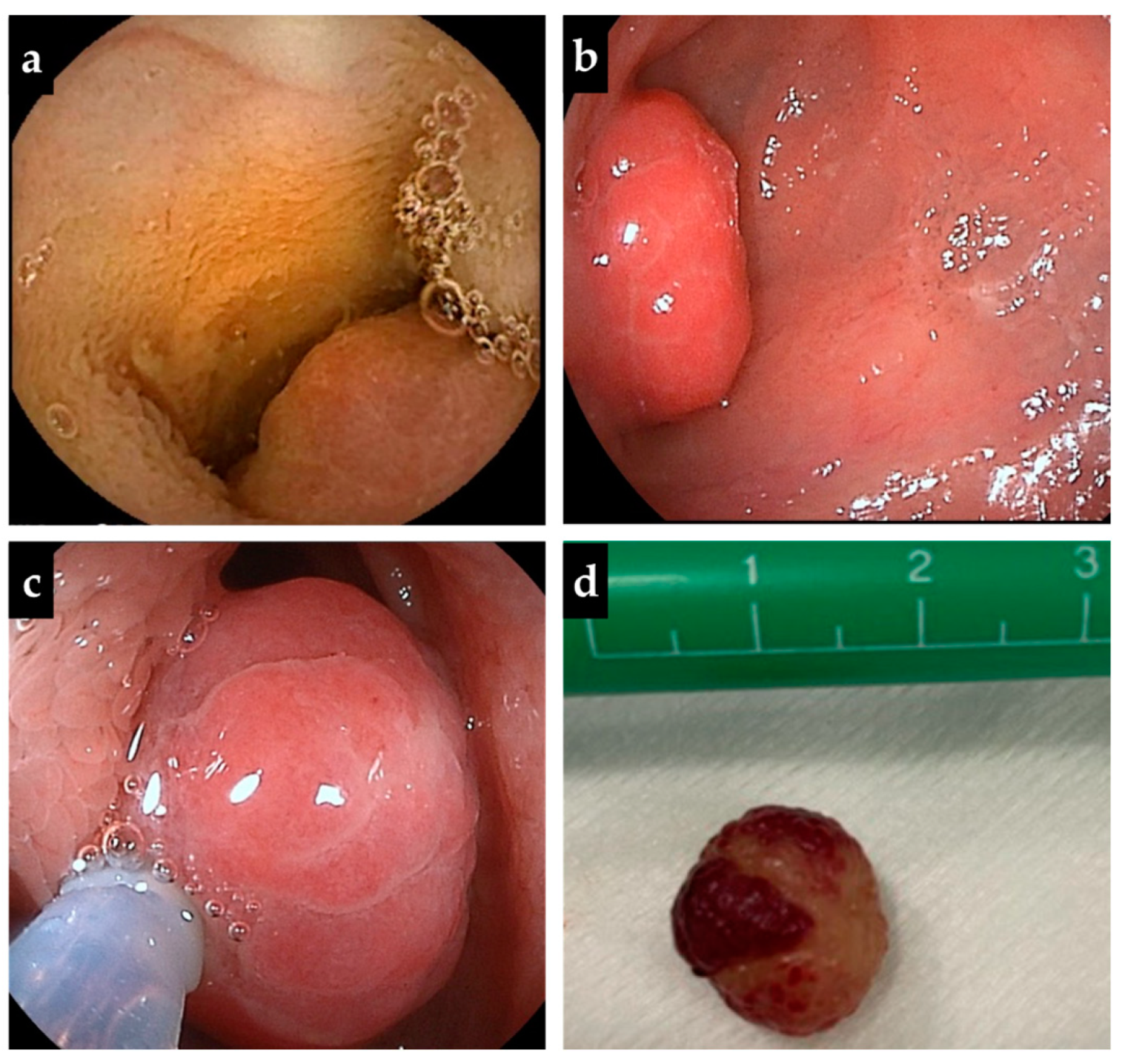
Abstract
:Author Contributions
Funding
Informed Consent Statement
Data Availability Statement
Acknowledgments
Conflicts of Interest
References
- Mandhan, P. Juvenile colorectal polyps in children: Experience in Pakistan. Pediatr. Surg. Int. 2004, 20, 339–342. [Google Scholar] [CrossRef] [PubMed]
- Yan, J.; Shen, Q.; Peng, C.; Pang, W.; Chen, Y. Colocolic intussusception in children: A case series and review of the literature. Front. Surg. 2022, 9, 873624. [Google Scholar] [CrossRef] [PubMed]
- Van Lier, M.G.F.; Mathus-Vliegen, E.M.H.; Wagner, A.; van Leerdam, M.E.; Kuipers, E.J. High cumulative risk of intussusception in patients with Peutz–Jeghers Syndrome: Time to update surveillance guidelines? Am. J. Gastroenterol. 2011, 106, 940–945. [Google Scholar] [CrossRef] [PubMed]
- Honda, W.; Ohmiya, N.; Hirooka, Y.; Nakamura, M.; Miyahara, R.; Ohno, E.; Kawashima, H.; AkihiroItoh; Watanabe, O.; Ando, T.; et al. Enteroscopic and radiologic diagnoses, treatment, and prognoses of small-bowel tumors. Gastrointest. Endosc. 2012, 76, 344–354. [Google Scholar] [CrossRef] [PubMed]
- Krasaelap, A.; Lerner, D.; Southern, J.; Noe, J.; Chugh, A. Endoscopic removal of a single, painless, juvenile polyp in the small intestine causing anemia. J. Pediatr. Gastroenterol. Nutr. 2020, 71, 491–493. [Google Scholar] [CrossRef] [PubMed]
- Kang, S.I.; Kang, J.; Kim, M.J.; Kim, I.K.; Lee, J.; Lee, K.Y. Laparoscopic-assisted resection of jejunojejunal intussusception caused by a juvenile polyp in an adult. Case Rep. Surg. 2014, 2014, 856765. [Google Scholar] [CrossRef] [Green Version]
- Ceccanti, S.; Frediani, S.; Manganaro, F.; Barbato, M.; Marcheggiano, A.; Cozzi, D.A. Laparoscopic-assisted resection of juvenile polyp of the jejunum in a 3-year-old girl. J. Pediatr. Surg. 2012, 47, 426–429. [Google Scholar] [CrossRef]
- Sah, S.P.; Agrawal, C.S.; Jha, P.C.; Rani, S. Juvenile polyps in the small intestine presenting as jejunojejunal intussusception in a 10-year-old child: Report of a case. Surg. Today 2002, 32, 828–830. [Google Scholar] [CrossRef] [PubMed]
- Garcia Crespo, J.M.; Martin Pinto, F.; Dominguez Vallejo, J. Intestinal polyp of infrequent localization: Presentation of 2 cases. An. Esp. Pediatr. 1984, 21, 855–857. [Google Scholar] [PubMed]
- Zimmermann, H.; Stauch, G.; Kamran, D. Juvenile polyp in the small bowel (author’s transl). Z. Kinderchir. 1981, 33, 89–93. [Google Scholar] [CrossRef] [PubMed]
- Das, S.R.; Karim, A.; RukonUzzaman, M.; Mazumder, M.W.; Alam, R.; Benzamin, M.; Marjan, P.; Sarker, N.; Akther, H.; Mondal, M. Juvenile polyps in Bangladeshi children and their association with fecal calprotectin as a biomarker. Pediatr. Gastroenterol. Hepatol. Nutr. 2022, 25, 52–60. [Google Scholar] [CrossRef] [PubMed]
- Hestvik, E.; Tumwine, J.K.; Tylleskar, T.; Grahnquist, L.; Ndeezi, G.; Kaddu-Mulindwa, D.H.; Aksnes, L.; Olafsdottir, E. Faecal calprotectin concentrations in apparently healthy children aged 0-12 years in urban Kampala, Uganda: A community-based survey. BMC. Pediatr. 2011, 11, 9. [Google Scholar] [CrossRef] [PubMed] [Green Version]
- Roca, M.; Rodriguez Varela, A.; Donat, E.; Cano, F.; Hervas, D.; Armisen, A.; Ana, A.; Maria, J.V.; Ander, S.; Ribes-Koninckx, C. Fecal calprotectin and eosinophil-derived neurotoxin in healthy children between 0 and 12 years. J. Pediatr. Gastroenterol. Nutr. 2017, 65, 394–398. [Google Scholar] [CrossRef] [PubMed]


Disclaimer/Publisher’s Note: The statements, opinions and data contained in all publications are solely those of the individual author(s) and contributor(s) and not of MDPI and/or the editor(s). MDPI and/or the editor(s) disclaim responsibility for any injury to people or property resulting from any ideas, methods, instructions or products referred to in the content. |
© 2023 by the authors. Licensee MDPI, Basel, Switzerland. This article is an open access article distributed under the terms and conditions of the Creative Commons Attribution (CC BY) license (https://creativecommons.org/licenses/by/4.0/).
Share and Cite
Nagata, M.; Jimbo, K.; Arai, N.; Kashiwagi, K.; Tokushima, K.; Suzuki, M.; Kudo, T.; Shimizu, T. An Isolated Intestinal Juvenile Polyp Diagnosed by Abdominal Ultrasonography and Resected by Double-Balloon Endoscopy: A Case Report and Literature Review. Diagnostics 2023, 13, 494. https://doi.org/10.3390/diagnostics13030494
Nagata M, Jimbo K, Arai N, Kashiwagi K, Tokushima K, Suzuki M, Kudo T, Shimizu T. An Isolated Intestinal Juvenile Polyp Diagnosed by Abdominal Ultrasonography and Resected by Double-Balloon Endoscopy: A Case Report and Literature Review. Diagnostics. 2023; 13(3):494. https://doi.org/10.3390/diagnostics13030494
Chicago/Turabian StyleNagata, Masumi, Keisuke Jimbo, Nobuyasu Arai, Kosuke Kashiwagi, Kaori Tokushima, Mitsuyoshi Suzuki, Takahiro Kudo, and Toshiaki Shimizu. 2023. "An Isolated Intestinal Juvenile Polyp Diagnosed by Abdominal Ultrasonography and Resected by Double-Balloon Endoscopy: A Case Report and Literature Review" Diagnostics 13, no. 3: 494. https://doi.org/10.3390/diagnostics13030494