
A Complete Review of Automatic Detection, Segmentation, and Quantification of Neovascularization in Optical Coherence Tomography Angiography Images


Abstract
:1. Introduction


- A foundation of knowledge about CNV and RNV;
- Reviewed techniques in a tabular fashion for the reader to easily digest. The works are sorted by published years. It provides achieved accuracy and other evaluations on different NV analyses for research objectives;
- For each NV type, reviews and analyses of the used techniques are carried out from three main approaches: deep learning, image processing, and a hybrid of image processing and machine learning, in a tree fashion to conveniently visualize which areas are the most and the least popular;
- An analysis of the frameworks of techniques, which shows similarities and differences quickly. Detailed methods used by each step in the approach are also summarized in a flowchart fashion;
- Procedural details of each reviewed method;
- A summary of the problems with each type of NV that are still open for future work.
2. Reviews on Methods, Used Datasets, and Performance

| Authors | Tasks and Purposes | Methods | Datasets | Best Performance |
|---|---|---|---|---|
| Deshpande et al. (2023) [48] | CNV segmentation and quantification | Gaussian kernel, Frangi filter [49], local adaptive thresholding [50], Mexican hat filter [51], skeletonization | 26 manually cropped OCTA images with CNV | Results are evaluated qualitatively; no numerical results are provided for NV detection. |
| Feng et al. (2023) [52] | CNV segmentation | U-Net with ResNeSt blocks [53] and spatial pyramid pooling [54] | 116 OCTA images with neovascular AMD | Accuracy of 98.91% with AUC of 94.76%, a specificity of 99.50%, a sensitivity of 72.71%, IOU of 58.67%, dice of 72.99%, and F1 score of 65.05%. |
| Wang et al. (2023) [24] | CNV detection, segmentation, and quantification | Dense-Net, U-Net [55], Parallelized-Net, and Res-Net [56] | 4701 OCTA images with CNV and 5865 images without CNV | AROC of 97.00%, Sensitivity of 95.00% for CNV diagnosis, and IOU of 66.24%, F1 Score of 78% for CNV segmentation. |
| Vali et al. (2023) [44] | CNV segmentation and morphological classification | U-Net [55], binarization, color conversion, dilation and region growing, and VGG16 [57] | 130 OCTA Images with CNV | A dice coefficient of 90.00%. Accuracy of 84%, 85%, 82%, 81%, and 86%, respectively, for classification of branch, shape, anastomosis and loops, peripheral arcade, and dark halo. |
| Li et al. (2023) [25] | RNV detection and segmentation | ResNet 101 [56] classifier + 2D V-Net [58] segmentation | 109 UW-OCTA training images (35 RNV images and 74 images of other lesions) and 95 UW-OCTA test images | A dice coefficient of 55.66% for RNV segmentation. |
| Taibouni et al. (2021) [59] | CNV detection for AMD severity grading | VGG19 [57] | 391 no AMD, 459 non-neovascular AMD, 548 neovascular AMD images | Accuracy of 89.74%, precision of 96.00%, ROC-AUC of 99.00%, and F1 Score of 84.00%. |
| Thakoor et al. (2021) [26] | CNV detection for AMD severity grading | 3D CNN | 97 non-AMD, 169 non-neovascular AMD, and 80 neovascular AMD images | Precision of 40.20% and 42.30%, recall of 70.00% and 72.00%, and F1 Score of 51.10% and 53.30%, respectively for the OCTA + OCT model and the OCTA + OCT + B-scan model. |
| Wu et al. (2021) [60] | RNV segmentation and quantification | Color space conversion, partial line detection to detect vessels, regional connectivity for vessel extraction, Otsu’s binarization [61] for optimization, morphological operations for noise/artifact reduction | 14 eyes with PDR | Results are evaluated qualitatively; no numerical results are provided for NV detection. |
| Wang et al. (2020) [23] | CNV detection and segmentation for AMD grading | Simplified CNN | 1676 images with NV AMD, non-NV AMD-DR | Sensitivity of 100.00% and specificity of 95.00% for CNV diagnosis, Jaccard’s similarity of 88.00% for CNV segmentation with precision of 95.00%, recall and F1 score of 93.00%. |
| Cheng et al. (2019) [27] | CNV detection and segmentation | Color space transformation, Otsu’s method [61], the majority method for blood vessel extraction, eccentricity features for CNV judgment, and morphological thinning [62] for CNV recognition | 17 eyes with CNV | Results were evaluated qualitatively, and no numerical result was provided for NV detection. However, the proposed method can detect NV in only some images. |
| Taibouni et al. (2019) [28] | CNV segmentation and quantification | Median filter for contrast enhancement, Frangi’s filter [49], or Gabor wavelet filtering [63] for vessel enhancement, hysteresis thresholding via Fuzzy C mean classification [64] for NV vessels detection, and morphological operations for mask generation | 54 eyes with neovascular AMD (Type 1 and Type 2 CNV) | Jaccard’s similarity of 87.50%. |
| Coscas et al., (2018) [65] | CNV segmentation and quantification | Median filter, Phansalkar thresholding [66] for binarization, connected component thresholding for noise removal, density map, custom region growing algorithm for CNV shape detection, morphology analysis for blood flow calculation, and box counting and skeletonization for quantification | 104 eyes with neovascular AMD (72 eyes under treatment and 32 eyes under remission) | No direct performance evaluation was reported for segmentation and quantification. |
| Xue et al. (2018) [29] | CNV segmentation | Gaussian filter for contrast enhancement, Gaussian distribution, DBSCAN method [67] with P Systems [68], and morphological operations for CNV membrane mask | 22 eyes with wet AMD | Jaccard’s similarity of 87.20%. |
| Zhang et al. (2017) [69] | CNV segmentation | Contrast adjustment, morphology analysis, and border detection [70] for NV membrane area extraction | 27 eyes from 23 patients with CNV | Correlation coefficients: 89.80% using SS-OCTA images, 82.20% on SD-OCTA images. |
| Gao et al. (2016) [30] | CNV segmentation and quantification | Saliency method [71] for CNV separation, thresholding for quantification, a level set method [72] for NV vessel segmentation within detected NV membrane, skeletonization [73] for vessel length quantification | 9 images of 9 neovascular AMD eyes | Jaccard’s similarity of 69.00%. |
| Liu et al. (2015) [17] | CNV segmentation and quantification | Gaussian filtering for contrast enhancement, saliency method [71], Laplacian and bilateral filter [74], Otsu’s thresholding [61] for rough CNV region extraction, morphological operations for mask generation | 7 images of 7 neovascular AMD eyes | Jaccard’s similarity of 83.40%. |

- The earliest work of automatic NV analysis was CNV segmentation and quantification in 2015.
- Automatic NV detection was used in several applications, such as DR-stage grading and AMD-type classification.
- No standard data sets were used in NV detection and segmentation. Most of the data sets used were local images obtained from organizations or the Internet.
- Most works used NV images from AMD patients. Two groups of researchers used images from DR cases. Algorithm performances depended on data sets and used techniques.
- The developed algorithms were primarily in CNV detection. Very few works were conducted for RNV.
- The accuracies of NV detection reported were around 90%. Some work achieved sensitivity as high as 100%. The best Jaccard’s similarity value reported for NV segmentation was 88.00%.
- For NV detection, only deep learning techniques were explored. These are 2D and 3D convolution networks, ResNet, ResNest 101, VGG19, 2D CNN, and 3D CNN. Modified U-Net with ResNeSt blocks and PSPNet, 2D and 3D convolution networks, U-Net, 2D V-Net, and CNN were employed in the NV segmentation. On the other hand, image-processing methods were largely used for segmentation and quantification.
- Morphological operations, OTSU binarization, and thresholding were the most popular image-processing methods in segmentation and quantification.


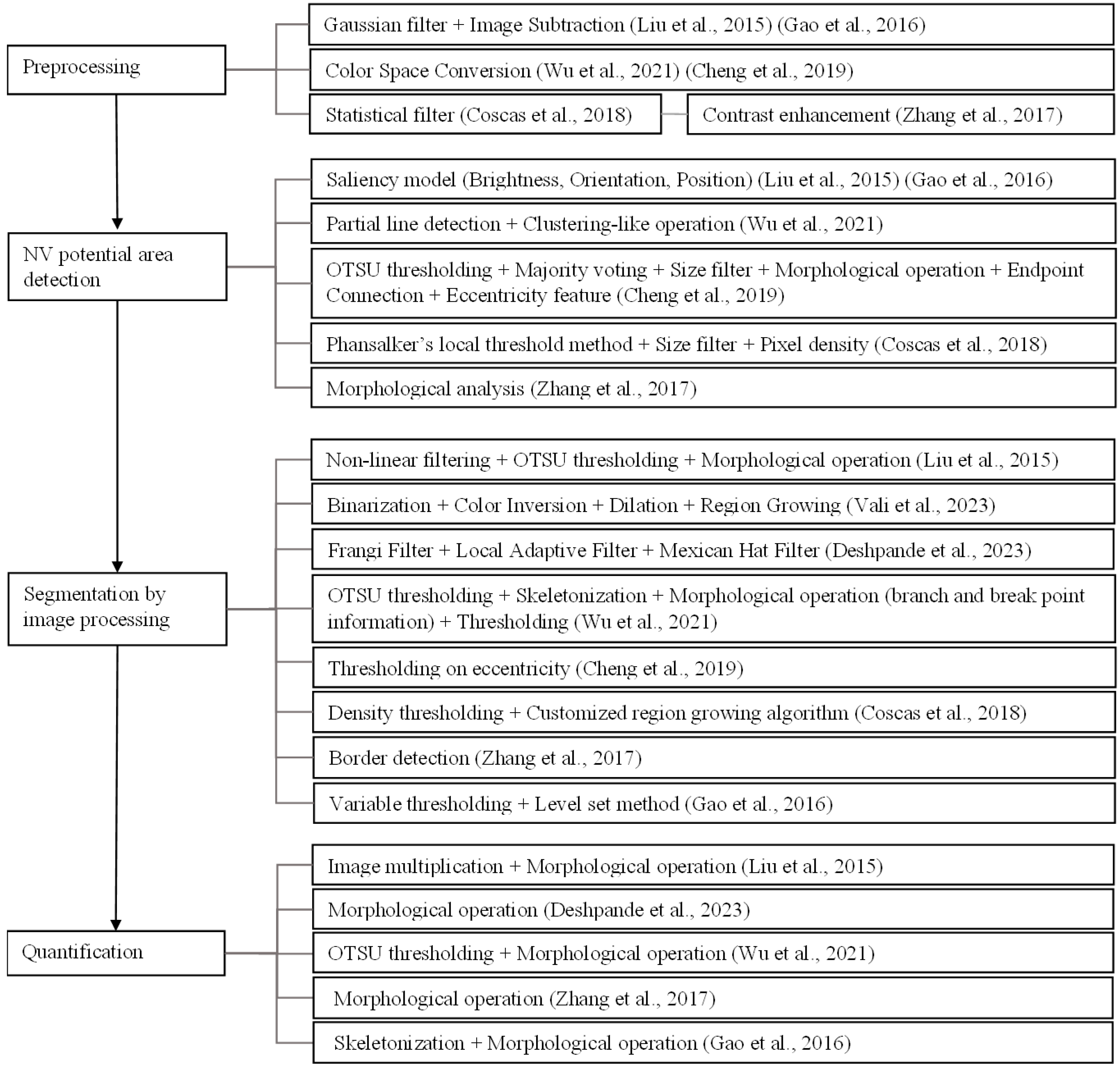
- In the image-processing approach, the NV detection, segmentation, and quantification frameworks overlap. All tasks usually start by preprocessing and then detecting potential NV areas. Additional segmentation methods in the image-processing approach were applied to obtain the region. For quantification, morphological operations were commonly used to assess the NV regions.
- Some works used both image processing and machine learning. Image processing techniques were applied to preprocess the image and obtain the features of regions of interest; then, machine learning methods were used on the extracted features to segment the NV boundary.
- Image subtraction, color space conversion, contrast enhancement, statistical filters, and Gaussian filters were used in the preprocessing.
- Depending on the target NV shape, texture, or size, different features were extracted using various techniques. The saliency method [71] was used for the CNVs that are salient in the input image in terms of brightness, orientation, and position [17,30] to generate a saliency map. A vascular network extraction method with partial line detection was used for NV of curvilinear structures [60]. Binarization and morphological analysis were applied to the preprocessed image in the works of Cheng et al. [27], Coscas et al. [65], and Zhang et al. [69]. Then, thresholding by the size of connected components was carried out in the works of Cheng et al. [27] and Coscas et al. [65] for the NV that is prominent by size. Eccentricity features were extracted for each candidate region in the work of Cheng et al. [27]. Taibouni et al. [28] used the Frangi filter and Gabor wavelet to classify vessels by thickness to identify the potential NV regions. The distances between non-zero intensity pixels were used as features for a clustering task in the work of Xue et al. [29].
- The resulting features, feature maps, or candidate regions were further processed to obtain the final ROI. Thresholding followed by morphological operations was the general process for the final candidate extraction. In some cases, the feature map went through some filters before thresholding [17]. In one case, the region growing method was used after obtaining the final ROI to achieve a more accurate segmentation of the CNV region [65]. Hysteresis thresholding via fuzzy C mean classification was employed by Taibouni et al. [28] to obtain the CNV region. DBSCAN clustering [67] with membrane systems [68] was used by Xue et al. [29] in the extraction of NV pixels.
- Morphological operations were mostly used in the quantification process. Quantifying methods such as box counting and skeletonization were used on the segmented regions to assess the NV region. Different quantifiers such as the vessel’s area, vessel length, vessel width, vessel tortuosity, fractal dimension, bifurcation density, vessel density, lacunary measure, and aspect ratio were reported.
- For the deep learning approach, different CNN architectures were widely used for segmentation.
Deep Learning Architectures and Settings
3. Discussions
4. Conclusions
Funding
Institutional Review Board Statement
Informed Consent Statement
Data Availability Statement
Conflicts of Interest
References
- Cole, E.D.; Novais, E.A.; Louzada, R.N.; Waheed, N.K. Contemporary retinal imaging techniques in Diabetic Retinopathy: A Review. J. Clin. Exp. Ophthalmol. 2016, 44, 289–299. [Google Scholar] [CrossRef]
- Hee, M.R. Optical coherence tomography of the human retina. Arch. Ophthalmol. 1995, 113, 325. [Google Scholar] [CrossRef]
- Spaide, R.F.; Fujimoto, J.G.; Waheed, N.K.; Sadda, S.R.; Staurenghi, G. Optical coherence tomography angiography. Prog. Retin. Eye Res. 2018, 64, 1–55. [Google Scholar] [CrossRef] [PubMed]
- Zhu, Y.; Cui, Y.; Wang, J.C.; Lu, Y.; Zeng, R.; Katz, R.; Wu, D.M.; Eliott, D.; Vavvas, D.G.; Husain, D.; et al. Different scan protocols affect the detection rates of diabetic retinopathy lesions by wide-field swept-source optical coherence tomography angiography. Am. J. Ophthalmol. 2020, 215, 72–80. [Google Scholar] [CrossRef] [PubMed]
- Campochiaro, P.A. Ocular neovascularization. J. Mol. Med. 2013, 91, 311–321. [Google Scholar] [CrossRef]
- Crawford, T.; Alfaro, D., III; Kerrison, J.; Jablon, E. Diabetic retinopathy and angiogenesis. Curr. Diabetes Rev. 2009, 5, 8–13. [Google Scholar] [CrossRef]
- Ehlers, J.P. Proliferative Diabetic Retinopathy. In OCT and OCTA in Retinal Disorders, 1st ed.; Modi, Y., Srivastava, S.K., Kaiser, P.K., Eds.; LWW: Beijing, China, 2020; ISBN 978-1-975144-22-7. [Google Scholar]
- Khalid, H.; Schwartz, R.; Nicholson, L.; Huemer, J.; El-Bradey, M.H.; Sim, D.A.; Patel, P.J.; Balaskas, K.; Hamilton, R.D.; Keane, P.A.; et al. Widefield optical coherence tomography angiography for early detection and objective evaluation of proliferative diabetic retinopathy. Br. J. Ophthalmol. 2020, 105, 118–123. [Google Scholar] [CrossRef] [PubMed]
- Leal, I.; Tan, S.Z.; Aslam, T.; Steeples, L.R.; Jones, N.P.; Chhabra, R. Intra and inter-rater agreement of inflammatory choroidal neovascular membrane measurements using optical coherence tomography angiography. Graefe’s Arch. Clin. Exp. Ophthalmol. 2019, 258, 647–651. [Google Scholar] [CrossRef]
- Menean, M.; Sacconi, R.; Tombolini, B.; Federico, F.; Bandello, F.; Querques, G. Combined Wide-Field Imaging in Grading Diabetic Retinopathy. Eye 2022. online ahead of print. [Google Scholar] [CrossRef] [PubMed]
- Vofo, B.N.; Galarza, P.; Chowers, I.; Levy, J. Interest of widefield-optical coherence tomography angiography for diagnosis and follow-up of retinal neovascularisation in proliferative diabetic retinopathy. J. Ophthalmol. 2022, 2022, 5746238. [Google Scholar] [CrossRef]
- Yang, Y.; Li, F.; Liu, T.; Jiao, W.; Zhao, B. Comparison of widefield swept-source optical coherence tomographic angiography and fluorescein fundus angiography for detection of retinal neovascularization with diabetic retinopathy. BMC Ophthalmol. 2023, 23, 315. [Google Scholar] [CrossRef]
- Nzakimuena, C.B. Automated Analysis of Retinal and Choroidal OCT and OCTA Images in AMD. Master’s Thesis, Polytechnique Montréal, Montréal, QC, Canada, 2020. Available online: https://www.kaggle.com/datasets/cnzakimuena/retinal-oct-and-octa-data-4 (accessed on 27 October 2023).
- Ishibazawa, A.; Nagaoka, T.; Yokota, H.; Takahashi, A.; Omae, T.; Song, Y.-S.; Takahashi, T.; Yoshida, A. Characteristics of retinal neovascularization in proliferative diabetic retinopathy imaged by optical coherence tomography angiography. Investig. Ophthalmol. Vis. Sci. 2016, 57, 6247–6255. [Google Scholar] [CrossRef]
- Karacorlu, M.; Sayman Muslubas, I.; Arf, S.; Hocaoglu, M.; Ersoz, M.G. Membrane patterns in eyes with choroidal neovascularization on optical coherence tomography angiography. Eye 2019, 33, 1280–1289. [Google Scholar] [CrossRef] [PubMed]
- Zhao, X.; Luo, M.; Chen, Y. Age-Related Macular Degeneration. In Atlas of Swept Source OCT and OCT Angiography, 1st ed.; Chen, Y., Peng, X., Eds.; Scientific and Technical Documentation Press: Beijing, China; Springer: Singapore, 2023; pp. 100–111. ISBN 978-9811943904. [Google Scholar]
- Liu, L.; Gao, S.S.; Bailey, S.T.; Huang, D.; Li, D.; Jia, Y. Automated choroidal neovascularization detection algorithm for optical coherence tomography angiography. Biomed. Opt. Express 2015, 6, 3564. [Google Scholar] [CrossRef] [PubMed]
- Li, M.; Huang, K.; Xu, Q.; Yang, J.; Zhang, Y.; Ji, Z.; Xie, K.; Yuan, S.; Liu, Q.; Chen, Q. OCTA-500: A Retinal Dataset for Optical Coherence Tomography Angiography Study. arXiv 2022, arXiv:2012.07261. [Google Scholar]
- Vaz-Pereira, S.; Silva, J.J.; Freund, K.B.; Engelbert, M. Optical coherence tomography angiography features of neovascularization in proliferative diabetic retinopathy. Clin. Ophthalmol. 2020, 14, 3351–3362. [Google Scholar] [CrossRef] [PubMed]
- Huemer, J.; Khalid, H.; Wagner, S.K.; Nicholson, L.; Fu, D.J.; Sim, D.A.; Patel, P.J.; Balaskas, K.; Rajendram, R.; Keane, P.A. Phenotyping of retinal neovascularization in ischemic retinal vein occlusion using wide field oct angiography. Eye 2020, 35, 2812–2819. [Google Scholar] [CrossRef]
- Ong, C.J.; Wong, M.Y.; Cheong, K.X.; Zhao, J.; Teo, K.Y.; Tan, T.-E. Optical coherence tomography angiography in retinal vascular disorders. Diagnostics 2023, 13, 1620. [Google Scholar] [CrossRef]
- Yang, J.; Chen, Y. Diabetic Retinopathy. In Atlas of Swept Source OCT and OCT Angiography, 1st ed.; Chen, Y., Peng, X., Eds.; Scientific and Technical Documentation Press: Beijing, China; Springer: Singapore, 2023; pp. 137–146. ISBN 978-9811943904. [Google Scholar]
- Wang, J.; Hormel, T.T.; Gao, L.; Zang, P.; Guo, Y.; Wang, X.; Bailey, S.T.; Jia, Y. Automated diagnosis and segmentation of choroidal neovascularization in OCT angiography using Deep Learning. Biomed. Opt. Express 2020, 11, 927. [Google Scholar] [CrossRef]
- Wang, J.; Hormel, T.T.; Tsuboi, K.; Wang, X.; Ding, X.; Peng, X.; Huang, D.; Bailey, S.T.; Jia, Y. Deep learning for diagnosing and segmenting choroidal neovascularization in OCT angiography in a large real-world data set. Transl. Vis. Sci. Technol. 2023, 12, 15. [Google Scholar] [CrossRef]
- Li, Y.; Zeghlache, R.; Brahim, I.; Xu, H.; Tan, Y.; Conze, P.-H.; Lamard, M.; Quellec, G.; El Habib Daho, M. Segmentation, classification, and quality assessment of UW-octa images for diagnosis of diabetic retinopathy. In Mitosis Domain Generalization and Diabetic Retinopathy Analysis; MIDOG DRAC 2022 2022; Lecture Notes in Computer Science; Bin Sheng, B., Aubreville, M., Eds.; Springer: Cham, Switzerland; Singapore, 2023; pp. 146–160. ISBN 978-3-031-33657-7. [Google Scholar]
- Thakoor, K.; Bordbar, D.; Yao, J.; Moussa, O.; Chen, R.; Sajda, P. Hybrid 3D-2D deep learning for detection of neovascularage-related macular degeneration using optical coherence tomography B-scans and angiography volumes. In Proceedings of the 2021 IEEE 18th International Symposium on Biomedical Imaging (ISBI), Nice, France, 13–16 April 2021; pp. 1600–1604. [Google Scholar] [CrossRef]
- Cheng, Y.-S.; Lin, S.-H.; Hsiao, C.-Y.; Chang, C.-J. Detection of choroidal neovascularization by optical coherence tomography angiography with assistance from use of the image segmentation method. Appl. Sci. 2019, 10, 137. [Google Scholar] [CrossRef]
- Taibouni, K.; Chenoune, Y.; Miere, A.; Colantuono, D.; Souied, E.; Petit, E. Automated quantification of choroidal neovascularization on optical coherence tomography angiography images. Comput. Biol. Med. 2019, 114, 103450. [Google Scholar] [CrossRef]
- Xue, J.; Camino, A.; Bailey, S.T.; Liu, X.; Li, D.; Jia, Y. Automatic quantification of choroidal neovascularization lesion area on OCT angiography based on density cell-like P systems with active membranes. Biomed. Opt. Express 2018, 9, 3208. [Google Scholar] [CrossRef] [PubMed]
- Gao, S.S.; Liu, L.; Bailey, S.T.; Flaxel, C.J.; Huang, D.; Li, D.; Jia, Y. Quantification of choroidal neovascularization vessel length using optical coherence tomography angiography. J. Biomed. Opt. 2016, 21, 076010. [Google Scholar] [CrossRef]
- Amato, A.; Nadin, F.; Borghesan, F.; Cicinelli, M.V.; Chatziralli, I.; Sadiq, S.; Mirza, R.; Bandello, F. Widefield optical coherence tomography angiography in diabetic retinopathy. J. Diabetes Res. 2020, 2020, 8855709. [Google Scholar] [CrossRef]
- Arya, M.; Sorour, O.; Chaudhri, J.; Alibhai, Y.; Waheed, N.K.; Duker, J.S.; Baumal, C.R. Distinguishing intraretinal microvascular abnormalities from retinal neovascularization using optical coherence tomography angiography. Retina 2019, 40, 1686–1695. [Google Scholar] [CrossRef] [PubMed]
- Pan, J.; Chen, D.; Yang, X.; Zou, R.; Zhao, K.; Cheng, D.; Huang, S.; Zhou, T.; Yang, Y.; Chen, F. Characteristics of neovascularization in early stages of proliferative diabetic retinopathy by optical coherence tomography angiography. Am. J. Ophthalmol. 2018, 192, 146–156. [Google Scholar] [CrossRef]
- Hirano, Y.; Suzuki, N.; Tomiyasu, T.; Kurobe, R.; Yasuda, Y.; Esaki, Y.; Yasukawa, T.; Yoshida, M.; Ogura, Y. Multimodal imaging of microvascular abnormalities in retinal vein occlusion. J. Clin. Med. 2021, 10, 405. [Google Scholar] [CrossRef]
- Khadamy, J.; Aghdam, K.; Falavarjani, K. An update on optical coherence tomography angiography in diabetic retinopathy. J. Ophthalmic Vis. Res. 2018, 13, 487. [Google Scholar] [CrossRef] [PubMed]
- Novais, E.A.; Roisman, L.; de Oliveira, P.R.; Louzada, R.N.; Cole, E.D.; Lane, M.; Filho, M.B.; Romano, A.; de Oliveira Dias, J.R.; Regatieri, C.V.; et al. Optical Coherence Tomography Angiography of Chorioretinal Diseases. Ophthalmic Surg. Lasers Imaging Retin. 2016, 47, 848–861. [Google Scholar] [CrossRef]
- Ouederni, M.; Sassi, H.; Chelly, Z.; Nefaa, F.; Cheour, M. Optical coherence tomography angiography in idiopathic retinal vasculitis, aneurysms and neuroretinitis (Irvan) syndrome: A case report. Eur. J. Ophthalmol. 2020, 32, NP144–NP148. [Google Scholar] [CrossRef] [PubMed]
- Li, J.; Wei, D.; Mao, M.; Li, M.; Liu, S.; Li, F.; Chen, L.; Liu, M.; Leng, H.; Wang, Y.; et al. Ultra-widefield color fundus photography combined with high-speed ultra-widefield swept-source optical coherence tomography angiography for non-invasive detection of lesions in diabetic retinopathy. Front. Public Health 2022, 10, 1047608. [Google Scholar] [CrossRef] [PubMed]
- Stino, H.; Niederleithner, M.; Iby, J.; Sedova, A.; Schlegl, T.; Steiner, I.; Sacu, S.; Drexler, W.; Schmoll, T.; Leitgeb, R.; et al. Detection of diabetic neovascularisation using single-capture 65°-widefield optical coherence tomography angiography. Br. J. Ophthalmol. 2022. online ahead of print. [Google Scholar] [CrossRef] [PubMed]
- Zhang, X.; Wu, C.; Zhou, L.; Dai, R. Observation of optic disc neovascularization using OCT angiography in proliferative diabetic retinopathy after Intravitreal CONBERCEPT injections. Sci. Rep. 2018, 8, 3972. [Google Scholar] [CrossRef]
- Hormel, T.T.; Jia, Y. OCT angiography and its retinal biomarkers [invited]. Biomed. Opt. Express 2023, 14, 4542. [Google Scholar] [CrossRef] [PubMed]
- Hirano, T.; Hoshiyama, K.; Hirabayashi, K.; Wakabayashi, M.; Toriyama, Y.; Tokimitsu, M.; Murata, T. Vitreoretinal interface slab in OCT angiography for detecting diabetic retinal neovascularization. Ophthalmol. Retin. 2020, 4, 588–594. [Google Scholar] [CrossRef]
- Rosenfeld, P.J.; Zheng, F. SD- vs. SS-Octa for CNV in NVAMD—Retina Specialist. Available online: https://www.retina-specialist.com/article/sd-vs-ssocta-for-cnv-in-nvamd (accessed on 15 August 2023).
- Vali, M.; Nazari, B.; Sadri, S.; Pour, E.K.; Riazi-Esfahani, H.; Faghihi, H.; Ebrahimiadib, N.; Azizkhani, M.; Innes, W.; Steel, D.H.; et al. CNV-net: Segmentation, classification and activity score measurement of choroidal neovascularization (CNV) using optical coherence tomography angiography (OCTA). Diagnostics 2023, 13, 1309. [Google Scholar] [CrossRef]
- Giavarina, D. Understanding bland altman analysis. Biochem. Med. 2015, 25, 141–151. [Google Scholar] [CrossRef]
- Schneider, A.; Hommel, G.; Blettner, M. Linear regression analysis. Dtsch. Arztebl. Int. 2010, 107, 776–782. [Google Scholar] [CrossRef]
- Carrington, A.M.; Fieguth, P.W.; Qazi, H.; Holzinger, A.; Chen, H.H.; Mayr, F.; Manuel, D.G. A new concordant partial AUC and partial C statistic for imbalanced data in the evaluation of Machine Learning Algorithms. BMC Med. Inform. Decis. Mak. 2020, 20, 4. [Google Scholar] [CrossRef]
- Deshpande, A.; Raman, S.; Dubey, A.; Susvar, P.; Raman, R. An ImageJ macro tool for OCTA-based quantitative analysis of Myopic Choroidal neovascularization. PLoS ONE 2023, 18, e0283929. [Google Scholar] [CrossRef]
- Frangi, A.F.; Niessen, W.J.; Vincken, K.L.; Viergever, M.A. Multiscale vessel enhancement filtering. In Medical Image Computing and Computer-Assisted Intervention—MICCAI’98; Springer: Berlin/Heidelberg, Germany, 1998; pp. 130–137. [Google Scholar] [CrossRef]
- Landini, G. Auto Local Threshold-ImageJ. Available online: https://imagej.net/plugins/autolocal-threshold (accessed on 4 October 2023).
- Jin, F.; Feng, D. Image registration algorithm using Mexican hat function-based operator and grouped feature matching strategy. PLoS ONE 2014, 9, e95576. [Google Scholar] [CrossRef]
- Feng, W.; Duan, M.; Wang, B.; Du, Y.; Zhao, Y.; Wang, B.; Zhao, L.; Ge, Z.; Hu, Y. Automated segmentation of choroidal neovascularization on optical coherence tomography angiography images of neovascular age-related macular degeneration patients based on Deep Learning. J. Big Data 2023, 10, 111. [Google Scholar] [CrossRef]
- Zhang, H.; Wu, C.; Zhang, Z.; Zhu, Y.; Lin, H.; Zhang, Z.; Sun, Y.; He, T.; Mueller, J.; Manmatha, R.; et al. Resnest: Split-attention networks. In Proceedings of the 2022 IEEE/CVF Conference on Computer Vision and Pattern Recognition Workshops (CVPRW), New Orleans, LA, USA, 19–20 June 2022; pp. 2735–2745. [Google Scholar] [CrossRef]
- Zhao, H.; Shi, J.; Qi, X.; Wang, X.; Jia, J. Pyramid Scene Parsing Network. In Proceedings of the 2017 IEEE Conference on Computer Vision and Pattern Recognition (CVPR), Honolulu, HI, USA, 21–26 July 2017; pp. 6230–6239. [Google Scholar] [CrossRef]
- Ronneberger, O.; Fischer, P.; Brox, T. U-Net: Convolutional Networks for Biomedical Image Segmentation. In Medical Image Computing and Computer-Assisted Intervention—MICCAI 2015; Lecture Notes in Computer Science; Navab, N., Hornegger, J., Wells, W., Frangi, A., Eds.; Springer: Cham, Switzerland; Munich, Germany, 2015; pp. 234–241. ISBN 978-3-319-24573-7. [Google Scholar]
- He, K.; Zhang, X.; Ren, S.; Sun, J. Deep Residual Learning for Image Recognition. In Proceedings of the 2016 IEEE Conference on Computer Vision and Pattern Recognition (CVPR), Las Vegas, NV, USA, 27–30 June 2016; pp. 770–778. [Google Scholar] [CrossRef]
- Simonyan, K.; Zisserman, A. Very deep convolutional networks for large-scale image recognition. arXiv 2014, arXiv:1409.1556. [Google Scholar]
- Milletari, F.; Navab, N.; Ahmadi, S.-A. V-net: Fully convolutional neural networks for volumetric medical image segmentation. In Proceedings of the 2016 Fourth International Conference on 3D Vision (3DV), Stanford, CA, USA, 25–28 October 2016; pp. 565–571. [Google Scholar] [CrossRef]
- Taibouni, K.; Miere, A.; Samake, A.; Souied, E.; Petit, E.; Chenoune, Y. Choroidal neovascularization screening on Oct-angiography choriocapillaris images by Convolutional Neural Networks. Appl. Sci. 2021, 11, 9313. [Google Scholar] [CrossRef]
- Wu, S.; Wu, S.; Feng, H.; Hu, Z.; Xie, Y.; Su, Y.; Feng, T.; Li, L. An optimized segmentation and quantification approach in microvascular imaging for OCTA-based neovascular regression monitoring. BMC Med. Imaging 2021, 21, 13. [Google Scholar] [CrossRef]
- Otsu, N. A threshold selection method from gray-level histograms. IEEE Trans. Syst. Man Cybern. 1979, 9, 62–66. [Google Scholar] [CrossRef]
- Kong, T.Y.; Rosenfeld, A. Topological algorithms for digital image processing. In Machine Intelligence and Pattern Recognition 19; Elsevier: Amsterdam, The Netherlands, 1996. [Google Scholar] [CrossRef]
- Gabor, D. Theory of communication. J. Electr. Eng. Technol. 1946, 93, 58. [Google Scholar] [CrossRef]
- Xiong, G. Fuzzy C-Means Thresholding. Available online: https://fr.mathworks.com/matlabcentral/fileexchange/8351-fuzzy-c-means-thresholding (accessed on 15 August 2023).
- Coscas, F.; Cabral, D.; Pereira, T.; Geraldes, C.; Narotamo, H.; Miere, A.; Lupidi, M.; Sellam, A.; Papoila, A.; Coscas, G.; et al. Quantitative optical coherence tomography angiography biomarkers for neovascular age-related macular degeneration in remission. PLoS ONE 2018, 13, e0205513. [Google Scholar] [CrossRef]
- Phansalkar, N.; More, S.; Sabale, A.; Joshi, M. Adaptive local thresholding for detection of nuclei in diversity stained cytology images. In Proceedings of the 2011 International Conference on Communications and Signal Processing, Kerala, India, 10–12 February 2011; pp. 218–220. [Google Scholar] [CrossRef]
- Birant, D.; Kut, A. ST-DBSCAN: An algorithm for clustering spatial–temporal data. Data Knowl. Eng. 2007, 60, 208–221. [Google Scholar]
- Xue, J.; Liu, X. Lattice based communication P systems with applications in cluster analysis. Soft Comput. 2013, 18, 1425–1440. [Google Scholar] [CrossRef]
- Zhang, Q.; Chen, C.-L.; Chu, Z.; Zheng, F.; Miller, A.; Roisman, L.; Rafael de Oliveira Dias, J.; Yehoshua, Z.; Schaal, K.B.; Feuer, W.; et al. Automated quantitation of choroidal neovascularization: A comparison study between spectral-domain and swept-source OCT angiograms. Investig. Ophthalmol. Vis. Sci. 2017, 58, 1506. [Google Scholar] [CrossRef] [PubMed]
- Canny, J. A computational approach to edge detection. IEEE Trans. Pattern Anal. Mach. Intell. 1987, PAMI-8, 184–203. [Google Scholar]
- Goferman, S.; Zelnik-Manor, L.; Tal, A. Context-aware saliency detection. In Proceedings of the 2010 IEEE Computer Society Conference on Computer Vision and Pattern Recognition, San Francisco, CA, USA, 13–18 June 2010; pp. 2376–2383. [Google Scholar] [CrossRef]
- Li, C.; Huang, R.; Ding, Z.; Gatenby, J.C.; Metaxas, D.N.; Gore, J.C. A level set method for image segmentation in the presence of intensity inhomogeneities with application to MRI. IEEE Trans. Image Process. 2011, 20, 2007–2016. [Google Scholar] [CrossRef] [PubMed]
- Zhang, T.Y.; Suen, C.Y. A fast parallel algorithm for thinning digital patterns. Commun. ACM 1984, 27, 236–239. [Google Scholar] [CrossRef]
- Tomasi, C.; Manduchi, R. Bilateral filtering for gray and color images. In Proceedings of the Sixth International Conference on Computer Vision (IEEE Cat. No.98CH36271), Bombay, India, 7 January 1998; pp. 839–846. [Google Scholar] [CrossRef]
- He, K.; Zhang, X.; Ren, S.; Sun, J. Delving deep into rectifiers: Surpassing human-level performance on ImageNet Classification. In Proceedings of the 2015 IEEE International Conference on Computer Vision (ICCV), Santiago, Chile, 7–13 December 2015; pp. 1026–1034. [Google Scholar] [CrossRef]
- Chen, L.-C.; Papandreou, G.; Kokkinos, I.; Murphy, K.; Yuille, A.L. DeepLab: Semantic image segmentation with deep convolutional nets, atrous convolution, and fully connected crfs. IEEE Trans. Pattern. Anal. Mach. Intell. 2018, 40, 834–848. [Google Scholar] [CrossRef]
- Chen, L.-C.; Papandreou, G.; Schroff, F.; Adam, H. Rethinking atrous convolution for semantic image segmentation. arXiv 2017. [Google Scholar] [CrossRef]
- Zhang, Q.; Zhang, A.; Lee, C.S.; Lee, A.Y.; Rezaei, K.A.; Roisman, L.; Miller, A.; Zheng, F.; Gregori, G.; Durbin, M.K.; et al. Projection artifact removal improves visualization and quantitation of macular neovascularization imaged by optical coherence tomography angiography. Ophthalmol. Retin. 2017, 1, 124–136. [Google Scholar] [CrossRef]
- Wang, J.; Zhang, M.; Hwang, T.S.; Bailey, S.T.; Huang, D.; Wilson, D.J.; Jia, Y. Reflectance-based projection-resolved optical coherence tomography angiography [invited]. Biomed. Opt. Express 2017, 8, 1536. [Google Scholar] [CrossRef]
- Zhang, A.; Zhang, Q.; Wang, R.K. Minimizing projection artifacts for accurate presentation of choroidal neovascularization in Oct micro-angiography. Biomed. Opt. Express 2015, 6, 4130. [Google Scholar] [CrossRef]
Disclaimer/Publisher’s Note: The statements, opinions and data contained in all publications are solely those of the individual author(s) and contributor(s) and not of MDPI and/or the editor(s). MDPI and/or the editor(s) disclaim responsibility for any injury to people or property resulting from any ideas, methods, instructions or products referred to in the content. |
© 2023 by the authors. Licensee MDPI, Basel, Switzerland. This article is an open access article distributed under the terms and conditions of the Creative Commons Attribution (CC BY) license (https://creativecommons.org/licenses/by/4.0/).
Share and Cite
Tun, Y.Z.; Aimmanee, P. A Complete Review of Automatic Detection, Segmentation, and Quantification of Neovascularization in Optical Coherence Tomography Angiography Images. Diagnostics 2023, 13, 3407. https://doi.org/10.3390/diagnostics13223407
Tun YZ, Aimmanee P. A Complete Review of Automatic Detection, Segmentation, and Quantification of Neovascularization in Optical Coherence Tomography Angiography Images. Diagnostics. 2023; 13(22):3407. https://doi.org/10.3390/diagnostics13223407
Chicago/Turabian StyleTun, Yar Zar, and Pakinee Aimmanee. 2023. "A Complete Review of Automatic Detection, Segmentation, and Quantification of Neovascularization in Optical Coherence Tomography Angiography Images" Diagnostics 13, no. 22: 3407. https://doi.org/10.3390/diagnostics13223407