
Klotho in Cancer: Potential Diagnostic and Prognostic Applications

 , , , , and
Abstract
:1. Introduction
2. Materials and Methods
3. Results
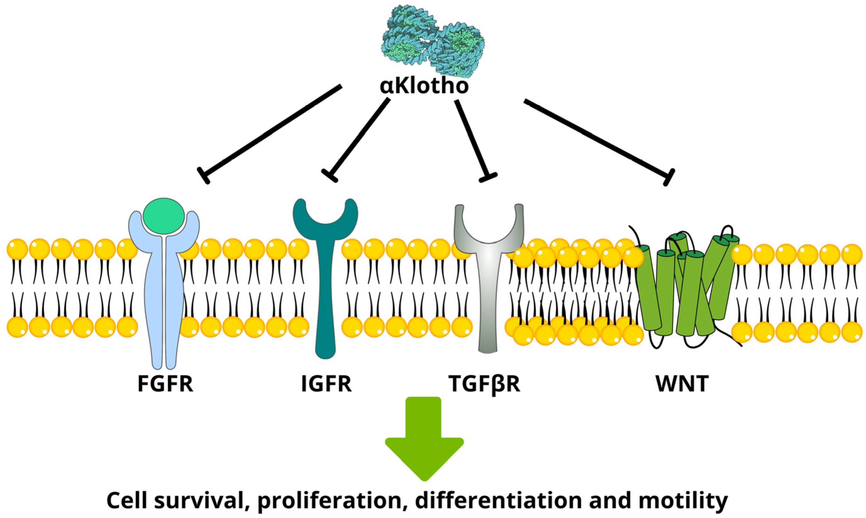
3.1. αKlotho
3.1.1. Clinicopathological Characteristics
3.1.2. Diagnosis
3.1.3. Survival and Treatment Response
3.2. βKlotho
3.2.1. Clinicopathological Characteristics
3.2.2. Survival and Treatment Response
3.3. γKlotho
3.3.1. Clinicopathological Characteristics
3.3.2. Survival and Treatment Response
4. Discussion
5. Conclusions
Author Contributions
Funding
Institutional Review Board Statement
Informed Consent Statement
Data Availability Statement
Conflicts of Interest
References
- Kuro, O.M.; Matsumura, Y.; Aizawa, H.; Kawaguchi, H.; Suga, T.; Utsugi, T.; Ohyama, Y.; Kurabayashi, M.; Kaname, T.; Kume, E.; et al. Mutation of the mouse klotho gene leads to a syndrome resembling ageing. Nature 1997, 390, 45–51. [Google Scholar] [CrossRef] [PubMed]
- Matsumuraab, Y.; Aizawaab, H.; Shiraki-Iida, T.; Nagaibd, R.; Kuro-O, M.; Nabeshima, Y.-I. Identification of the HumanKlothoGene and Its Two Transcripts Encoding Membrane and SecretedKlothoProtein. Biochem. Biophys. Res. Commun. 1998, 242, 626–630. [Google Scholar] [CrossRef] [PubMed]
- Ito, S.; Kinoshita, S.; Shiraishi, N.; Nakagawa, S.; Sekine, S.; Fujimori, T.; Nabeshima, Y.-I. Molecular cloning and expression analyses of mouse betaβklotho, which encodes a novel Klotho family protein. Mech. Dev. 2000, 98, 115–119. [Google Scholar] [CrossRef] [PubMed]
- Chen, C.-D.; Podvin, S.; Gillespie, E.; Leeman, S.E.; Abraham, C.R. Insulin stimulates the cleavage and release of the extracellular domain of Klotho by ADAM10 and ADAM17. Proc. Natl. Acad. Sci. USA 2007, 104, 19796–19801. [Google Scholar] [CrossRef]
- Kuro-O, M. The Klotho proteins in health and disease. Nat. Rev. Nephrol. 2019, 15, 27–44. [Google Scholar] [CrossRef]
- Bednarska, S.; Fryczak, J.; Siejka, A. Serum β-Klotho concentrations are increased in women with polycystic ovary syndrome. Cytokine 2020, 134, 155188. [Google Scholar] [CrossRef]
- Ito, S.; Fujimori, T.; Hayashizaki, Y.; Nabeshima, Y.-I. Identification of a novel mouse membrane-bound family 1 glycosidase-like protein, which carries an atypical active site structure. Biochim. Biophys. Acta (BBA)-Gene Struct. Expr. 2002, 1576, 341–345. [Google Scholar] [CrossRef]
- Kurosu, H.; Ogawa, Y.; Miyoshi, M.; Yamamoto, M.; Nandi, A.; Rosenblatt, K.P.; Baum, M.G.; Schiavi, S.; Hu, M.-C.; Moe, O.W.; et al. Regulation of Fibroblast Growth Factor-23 Signaling by Klotho. J. Biol. Chem. 2006, 281, 6120–6123. [Google Scholar] [CrossRef]
- Urakawa, I.; Yamazaki, Y.; Shimada, T.; Iijima, K.; Hasegawa, H.; Okawa, K.; Fujita, T.; Fukumoto, S.; Yamashita, T. Klotho converts canonical FGF receptor into a specific receptor for FGF23. Nature 2006, 444, 770–774. [Google Scholar] [CrossRef]
- Lee, S.; Choi, J.; Mohanty, J.; Sousa, L.P.; Tome, F.; Pardon, E.; Steyaert, J.; Lemmon, M.A.; Lax, I.; Schlessinger, J. Structures of β-klotho reveal a ‘zip code’-like mechanism for endocrine FGF signalling. Nature 2018, 553, 501–505. [Google Scholar] [CrossRef]
- Lin, B.C.; Wang, M.; Blackmore, C.; Desnoyers, L.R. Liver-specific Activities of FGF19 Require Klotho beta. J. Biol. Chem. 2007, 282, 27277–27284. [Google Scholar] [CrossRef] [PubMed]
- Ogawa, Y.; Kurosu, H.; Yamamoto, M.; Nandi, A.; Rosenblatt, K.P.; Goetz, R.; Eliseenkova, A.V.; Mohammadi, M.; Kuro-O, M. βKlotho is required for metabolic activity of fibroblast growth factor 21. Proc. Natl. Acad. Sci. USA 2007, 104, 7432–7437. [Google Scholar] [CrossRef] [PubMed]
- Kurosu, H.; Choi, M.; Ogawa, Y.; Dickson, A.S.; Goetz, R.; Eliseenkova, A.V.; Mohammadi, M.; Rosenblatt, K.P.; Kliewer, S.A.; Kuro-O, M. Tissue-specific Expression of βKlotho and Fibroblast Growth Factor (FGF) Receptor Isoforms Determines Metabolic Activity of FGF19 and FGF21. J. Biol. Chem. 2007, 282, 26687–26695. [Google Scholar] [CrossRef] [PubMed]
- Kharitonenkov, A.; Dunbar, J.D.; Bina, H.A.; Bright, S.; Moyers, J.S.; Zhang, C.; Ding, L.; Micanovic, R.; Mehrbod, S.F.; Knierman, M.D.; et al. FGF-21/FGF-21 receptor interaction and activation is determined by βKlotho. J. Cell. Physiol. 2007, 215, 1–7. [Google Scholar] [CrossRef]
- Brownstein, C.A.; Adler, F.; Nelson-Williams, C.; Iijima, J.; Li, P.; Imura, A.; Nabeshima, Y.-I.; Reyes-Mugica, M.; Carpenter, T.O.; Lifton, R.P. A translocation causing increased α-Klotho level results in hypophosphatemic rickets and hyperparathyroidism. Proc. Natl. Acad. Sci. USA 2008, 105, 3455–3460. [Google Scholar] [CrossRef]
- Stenvinkel, P.; Painer, J.; Kuro-O, M.; Lanaspa, M.; Arnold, W.; Ruf, T.; Shiels, P.G.; Johnson, R.J. Novel treatment strategies for chronic kidney disease: Insights from the animal kingdom. Nat. Rev. Nephrol. 2018, 14, 265–284. [Google Scholar] [CrossRef]
- Ligumsky, H.; Merenbakh-Lamin, K.; Keren-Khadmy, N.; Wolf, I.; Rubinek, T. The role of α-klotho in human cancer: Molecular and clinical aspects. Oncogene 2022, 41, 4487–4497. [Google Scholar] [CrossRef]
- Ye, X.; Guo, Y.; Zhang, Q.; Chen, W.; Hua, X.; Liu, W.; Yang, Y.; Chen, G. βKlotho Suppresses Tumor Growth in Hepatocellular Carcinoma by Regulating Akt/GSK-3β/Cyclin D1 Signaling Pathway. PLoS ONE 2013, 8, e55615. [Google Scholar] [CrossRef]
- Liu, Z.; Qi, S.; Zhao, X.; Li, M.; Ding, S.; Lu, J.; Zhang, H. Metformin inhibits 17β-estradiol-induced epithelial-to-mesenchymal transition via βKlotho-related ERK1/2 signaling and AMPKα signaling in endometrial adenocarcinoma cells. Oncotarget 2016, 7, 21315–21331. [Google Scholar] [CrossRef]
- Cui, G.; Martin, R.C.; Jin, H.; Liu, X.; Pandit, H.; Zhao, H.; Cai, L.; Zhang, P.; Li, W.; Li, Y. Up-regulation of FGF15/19 signaling promotes hepatocellular carcinoma in the background of fatty liver. J. Exp. Clin. Cancer Res. 2018, 37, 136. [Google Scholar] [CrossRef]
- Trošt, N.; Peña-Llopis, S.; Koirala, S.; Stojan, J.; Potts, P.R.; Tacer, K.F.; Martinez, E.D. γKlotho is a novel marker and cell survival factor in a subset of triple negative breast cancers. Oncotarget 2016, 7, 2611–2628. [Google Scholar] [CrossRef] [PubMed]
- Hori, S.; Miyake, M.; Tatsumi, Y.; Morizawa, Y.; Nakai, Y.; Onishi, S.; Onishi, K.; Iida, K.; Gotoh, D.; Tanaka, N.; et al. Gamma-Klotho exhibits multiple roles in tumor growth of human bladder cancer. Oncotarget 2018, 9, 19508–19524. [Google Scholar] [CrossRef] [PubMed]
- Rubinek, T.; Wolf, I. The Role of Alpha-Klotho as a Universal Tumor Suppressor. Vitam. Horm. 2016, 101, 197–214. [Google Scholar] [CrossRef]
- Kurosu, H.; Yamamoto, M.; Clark, J.D.; Pastor, J.V.; Nandi, A.; Gurnani, P.; McGuinness, O.P.; Chikuda, H.; Yamaguchi, M.; Kawaguchi, H.; et al. Suppression of Aging in Mice by the Hormone Klotho. Science 2005, 309, 1829–1833. [Google Scholar] [CrossRef] [PubMed]
- Sachdeva, A.; Gouge, J.; Kontovounisios, C.; Nikolaou, S.; Ashworth, A.; Lim, K.; Chong, I. Klotho and the Treatment of Human Malignancies. Cancers 2020, 12, 1665. [Google Scholar] [CrossRef]
- Poh, W.; Wong, W.; Ong, H.; Aung, M.O.; Lim, S.G.; Chua, B.T.; Ho, H.K. Klotho-beta overexpression as a novel target for suppressing proliferation and fibroblast growth factor receptor-4 signaling in hepatocellular carcinoma. Mol. Cancer 2012, 11, 14. [Google Scholar] [CrossRef]
- Feng, S.; Dakhova, O.; Creighton, C.J.; Ittmann, M. Endocrine Fibroblast Growth Factor FGF19 Promotes Prostate Cancer Progression. Cancer Res. 2013, 73, 2551–2562. [Google Scholar] [CrossRef] [PubMed]
- Zhu, Y.; Xu, L.; Zhang, J.; Xu, W.; Liu, Y.; Yin, H.; Lv, T.; An, H.; Liu, L.; He, H.; et al. Klotho suppresses tumor progression via inhibiting PI3K/Akt/GSK3β/Snail signaling in renal cell carcinoma. Cancer Sci. 2013, 104, 663–671. [Google Scholar] [CrossRef]
- Suzuki, H.; Watkins, D.N.; Jair, K.-W.; Schuebel, K.E.; Markowitz, S.D.; Chen, W.D.; Pretlow, T.P.; Yang, B.; Akiyama, Y.; van Engeland, M.; et al. Epigenetic inactivation of SFRP genes allows constitutive WNT signaling in colorectal cancer. Nat. Genet. 2004, 36, 417–422. [Google Scholar] [CrossRef] [PubMed]
- Sun, H.; Gao, Y.; Lu, K.; Zhao, G.; Li, X.; Li, Z.; Chang, H. Overexpression of Klotho suppresses liver cancer progression and induces cell apoptosis by negatively regulating wnt/β-catenin signaling pathway. World J. Surg. Oncol. 2015, 13, 307. [Google Scholar] [CrossRef] [PubMed]
- Pickup, M.; Novitskiy, S.; Moses, H.L. The roles of TGFβ in the tumour microenvironment. Nat. Rev. Cancer 2013, 13, 788–799. [Google Scholar] [CrossRef]
- Doi, S.; Zou, Y.; Togao, O.; Pastor, J.V.; John, G.B.; Wang, L.; Shiizaki, K.; Gotschall, R.; Schiavi, S.; Yorioka, N.; et al. Klotho Inhibits Transforming Growth Factor-β1 (TGF-β1) Signaling and Suppresses Renal Fibrosis and Cancer Metastasis in Mice. J. Biol. Chem. 2011, 286, 8655–8665. [Google Scholar] [CrossRef] [PubMed]
- Hori, S.; Miyake, M.; Onishi, S.; Tatsumi, Y.; Morizawa, Y.; Nakai, Y.; Anai, S.; Tanaka, N.; Fujimoto, K. Clinical significance of α- and β-Klotho in urothelial carcinoma of the bladder. Oncol. Rep. 2016, 36, 2117–2125. [Google Scholar] [CrossRef] [PubMed]
- Zhou, J.; Ben, S.; Xu, T.; Xu, L.; Yao, X. Serum β-klotho is a potential biomarker in the prediction of clinical outcomes among patients with NSCLC. J. Thorac. Dis. 2021, 13, 3137–3150. [Google Scholar] [CrossRef] [PubMed]
- Wang, Y.; Chen, L.; Huang, G.; He, D.; He, J.; Xu, W.; Zou, C.; Zong, F.; Li, Y.; Chen, B.; et al. Klotho Sensitizes Human Lung Cancer Cell Line to Cisplatin via PI3k/Akt Pathway. PLoS ONE 2013, 8, e57391. [Google Scholar] [CrossRef] [PubMed]
- Xie, B.; Zhou, J.; Yuan, L.; Ren, F.; Liu, D.-C.; Li, Q.; Shu, G. Epigenetic silencing of Klotho expression correlates with poor prognosis of human hepatocellular carcinoma. Hum. Pathol. 2013, 44, 795–801. [Google Scholar] [CrossRef]
- Gigante, M.; Lucarelli, G.; Divella, C.; Netti, G.S.; Pontrelli, P.; Cafiero, C.; Grandaliano, G.; Castellano, G.; Rutigliano, M.; Stallone, G.; et al. Soluble Serum αKlotho Is a Potential Predictive Marker of Disease Progression in Clear Cell Renal Cell Carcinoma. Medicine 2015, 94, e1917. [Google Scholar] [CrossRef]
- Lojkin, I.; Rubinek, T.; Orsulic, S.; Schwarzmann, O.; Karlan, B.Y.; Bose, S.; Wolf, I. Reduced expression and growth inhibitory activity of the aging suppressor klotho in epithelial ovarian cancer. Cancer Lett. 2015, 362, 149–157. [Google Scholar] [CrossRef]
- Shibayama, Y.; Kondo, T.; Ohya, H.; Fujisawa, S.-I.; Teshima, T.; Iseki, K. Upregulation of microRNA-126-5p is associated with drug resistance to cytarabine and poor prognosis in AML patients. Oncol. Rep. 2015, 33, 2176–2182. [Google Scholar] [CrossRef]
- Dai, D.; Wang, Q.; Li, X.; Liu, J.; Ma, X.; Xu, W. Klotho inhibits human follicular thyroid cancer cell growth and promotes apoptosis through regulation of the expression of stanniocalcin-1. Oncol. Rep. 2015, 35, 552–558. [Google Scholar] [CrossRef]
- Tang, X.; Fan, Z.; Wang, Y.; Ji, G.; Wang, M.; Lin, J.; Huang, S. Expression of klotho and β-catenin in esophageal squamous cell carcinoma, and their clinicopathological and prognostic significance. Dis. Esophagus 2016, 29, 207–214. [Google Scholar] [CrossRef] [PubMed]
- Ibi, T.; Usuda, J.; Inoue, T.; Sato, A.; Takegahara, K. Klotho expression is correlated to molecules associated with epithelial-mesenchymal transition in lung squamous cell carcinoma. Oncol. Lett. 2017, 14, 5526–5532. [Google Scholar] [CrossRef] [PubMed]
- Yan, Y.; Wang, Y.; Xiong, Y.; Lin, X.; Zhou, P.; Chen, Z. Reduced Klotho expression contributes to poor survival rates in human patients with ovarian cancer, and overexpression of Klotho inhibits the progression of ovarian cancer partly via the inhibition of systemic inflammation in nude mice. Mol. Med. Rep. 2017, 15, 1777–1785. [Google Scholar] [CrossRef] [PubMed]
- Haq, F.; Sung, Y.-N.; Park, I.; Kayani, M.A.; Yousuf, F.; Hong, S.-M.; Ahn, S.-M. FGFR1 expression defines clinically distinct subtypes in pancreatic cancer. J. Transl. Med. 2018, 16, 374. [Google Scholar] [CrossRef] [PubMed]
- Brominska, B.; Gabryel, P.; Jarmołowska-Jurczyszyn, D.; Janicka-Jedyńska, M.; Kluk, A.; Trojanowski, M.; Brajer-Luftmann, B.; Woliński, K.; Czepczyński, R.; Gut, P.; et al. Klotho expression and nodal involvement as predictive factors for large cell lung carcinoma. Arch. Med. Sci. 2019, 15, 1010–1016. [Google Scholar] [CrossRef] [PubMed]
- Liu, Y.; Pan, J.; Pan, X.; Wu, L.; Bian, J.; Lin, Z.; Xue, M.; Su, T.; Lai, S.; Chen, F.; et al. Klotho-mediated targeting of CCL 2 suppresses the induction of colorectal cancer progression by stromal cell senescent microenvironments. Mol. Oncol. 2019, 13, 2460–2475. [Google Scholar] [CrossRef]
- Onishi, K.; Miyake, M.; Hori, S.; Onishi, S.; Iida, K.; Morizawa, Y.; Tatsumi, Y.; Nakai, Y.; Tanaka, N.; Fujimoto, K. γ-Klotho is correlated with resistance to docetaxel in castration-resistant prostate cancer. Oncol. Lett. 2020, 19, 2306–2316. [Google Scholar] [CrossRef]
- Xie, B.; Hu, F.; Li, M.; Mo, L.; Xu, C.; Xiao, Y.; Wang, X.; Nie, J.; Yang, L.; He, Y. FLI-1 mediates tumor suppressor function via Klotho signaling in regulating CRC. Cell Biol. Int. 2020, 44, 1514–1522. [Google Scholar] [CrossRef]
- Yang, L.; Wu, Y.; He, H.; Hu, F.; Li, M.; Mo, L.; Xiao, Y.; Wang, X.; Xie, B. Delivery of BR2-SOX17 fusion protein can inhibit cell survival, proliferation, and invasion in gastric cancer cells through regulating Klotho gene expression. Cell Biol. Int. 2020, 44, 2011–2020. [Google Scholar] [CrossRef]
- GGunes, S.; Soykan, M.N.; Sariboyaci, A.E.; Uysal, O.; Sevimli, T.S. Enhancement of Apo2L/TRAIL signaling pathway receptors by the activation of Klotho gene with CRISPR/Cas9 in Caco-2 colon cancer cells. Med. Oncol. 2021, 38, 146. [Google Scholar] [CrossRef]
- Rubinstein, T.A.; Reuveni, I.; Hesin, A.; Klein-Goldberg, A.; Olauson, H.; Larsson, T.E.; Abraham, C.R.; Zeldich, E.; Bosch, A.; Chillón, M.; et al. A Transgenic Model Reveals the Role of Klotho in Pancreatic Cancer Development and Paves the Way for New Klotho-Based Therapy. Cancers 2021, 13, 6297. [Google Scholar] [CrossRef] [PubMed]
- Wang, Y.; Wang, H.; Zheng, R.; Wu, P.; Sun, Z.; Chen, J.; Zhang, L.; Zhang, C.; Qian, H.; Jiang, J.; et al. Circular RNA ITCH suppresses metastasis of gastric cancer via regulating miR-199a-5p/Klotho axis. Cell Cycle 2021, 20, 522–536. [Google Scholar] [CrossRef] [PubMed]
- Sariboyaci, A.E.; Uysal, O.; Soykan, M.N.; Gunes, S. The potential therapeutic effect of klotho on cell viability in human colorectal adenocarcinoma HT-29 cells. Med. Oncol. 2022, 39, 191. [Google Scholar] [CrossRef] [PubMed]
- Tao, Z.; Cui, Y.; Xu, X.; Han, T. FGFR redundancy limits the efficacy of FGFR4-selective inhibitors in hepatocellular carcinoma. Proc. Natl. Acad. Sci. USA 2022, 119, e2208844119. [Google Scholar] [CrossRef]
- Labrecque, M.P.; Coleman, I.M.; Brown, L.G.; True, L.D.; Kollath, L.; Lakely, B.; Nguyen, H.M.; Yang, Y.C.; Gil da Costa, R.M.; Kaipainen, A.; et al. Molecular profiling stratifies diverse phenotypes of treatment-refractory metastatic castration-resistant prostate cancer. J. Clin. Investig. 2019, 129, 4492–4505. [Google Scholar] [CrossRef]
- Harbeck, N.; Gnant, M. Breast cancer. Lancet 2017, 389, 1134–1150. [Google Scholar] [CrossRef]
- Gil da Costa, R.M.; Levesque, C.; Bianchi-Frias, D.; Chatterjee, P.; Lam, H.; Santos, C.; Coleman, I.M.; Ferreirinha, P.; Vilanova, M.; da Cunha, N.P.; et al. Pharmacological NF-κB inhibition decreases cisplatin chemoresistance in muscle-invasive bladder cancer and reduces cisplatin-induced toxicities. Mol. Oncol. 2023. [Google Scholar] [CrossRef]
- DeLucia, D.C.; Cardillo, T.M.; Ang, L.S.; Labrecque, M.P.; Zhang, A.; Hopkins, J.E.; De Sarkar, N.; Coleman, I.; Gil da Costa, R.M.; Corey, E.; et al. Regulation of CEACAM5 and Therapeutic Efficacy of an Anti-CEACAM5–SN38 Antibody–drug Conjugate in Neuroendocrine Prostate Cancer. Clin. Cancer Res. 2021, 27, 759–774. [Google Scholar] [CrossRef]
- Tannock, I.F.; De Wit, R.; Berry, W.R.; Horti, J.; Pluzanska, A.; Chi, K.N.; Oudard, S.; Théodore, C.; James, N.D.; Turesson, I.; et al. Docetaxel plus Prednisone or Mitoxantrone plus Prednisone for Advanced Prostate Cancer. N. Engl. J. Med. 2004, 351, 1502–1512. [Google Scholar] [CrossRef]
- Rodriguez-Canales, J.; Parra-Cuentas, E.; Wistuba, I.I. Diagnosis and Molecular Classification of Lung Cancer. Cancer Treat. Res. 2016, 170, 25–46. [Google Scholar] [CrossRef]
- Forner, A.; Reig, M.; Bruix, J. Hepatocellular carcinoma. Lancet 2018, 391, 1301–1314. [Google Scholar] [CrossRef] [PubMed]
- Lheureux, S.; Braunstein, M.; Oza, A.M. Epithelial ovarian cancer: Evolution of management in the era of precision medicine. CA A Cancer J. Clin. 2019, 69, 280–304. [Google Scholar] [CrossRef] [PubMed]
- Santos, J.M.O.; Costa, A.C.; Dias, T.R.; Satari, S.; e Silva, M.P.C.; Gil da Costa, R.M.; Medeiros, R. Towards Drug Repurposing in Cancer Cachexia: Potential Targets and Candidates. Pharmaceuticals 2021, 14, 1084. [Google Scholar] [CrossRef] [PubMed]
- Liu, H. Emerging agents and regimens for AML. J. Hematol. Oncol. 2021, 14, 49. [Google Scholar] [CrossRef]
- Bianchini, G.; De Angelis, C.; Licata, L.; Gianni, L. Treatment landscape of triple-negative breast cancer—Expanded options, evolving needs. Nat. Rev. Clin. Oncol. 2022, 19, 91–113. [Google Scholar] [CrossRef]
- Patel, S.G.; Karlitz, J.J.; Yen, T.; Lieu, C.H.; Boland, C.R. The rising tide of early-onset colorectal cancer: A comprehensive review of epidemiology, clinical features, biology, risk factors, prevention, and early detection. Lancet Gastroenterol. Hepatol. 2022, 7, 262–274. [Google Scholar] [CrossRef]
- Compérat, E.; Amin, M.B.; Cathomas, R.; Choudhury, A.; De Santis, M.; Kamat, A.; Stenzl, A.; Thoeny, H.C.; Witjes, J.A. Current best practice for bladder cancer: A narrative review of diagnostics and treatments. Lancet 2022, 400, 1712–1721. [Google Scholar] [CrossRef]


| Reference | Year | Type of Cancer | In Vitro | In Vivo | Number of Patients | Clinical (Observational) |
|---|---|---|---|---|---|---|
| [26] | 2012 | Hepatocellular carcinoma | x | 56 | Retrospective cohort | |
| [27] | 2013 | Prostate cancer | x | 136 | Retrospective cohort | |
| [35] | 2013 | Lung cancer | x | x | - | |
| [36] | 2013 | Hepatocellular carcinoma | x | 64 | Retrospective cohort | |
| [28] | 2013 | Renal cell carcinoma | x | 125 | Retrospective cohort | |
| [37] | 2015 | Renal cell carcinoma | 160 | Retrospective cohort | ||
| [38] | 2015 | Ovarian cancer | x | 265 | Retrospective cohort | |
| [39] | 2015 | Acute myeloid leukemia | x | 109 | Retrospective cohort | |
| [30] | 2015 | Hepatocellular carcinoma | x | - | ||
| [21] | 2015 | Breast cancer | x | 68 | Retrospective cohort | |
| [40] | 2016 | Thyroid cancer | x | - | ||
| [41] | 2016 | Esophageal cancer | x | 160 | Retrospective case–control | |
| [42] | 2017 | Pulmonary squamous cell carcinoma | x | 40 | Retrospective cohort | |
| [43] | 2017 | Ovarian cancer | x | x | 198 | Retrospective cohort |
| [44] | 2018 | Pancreatic adenocarcinoma | x | 313 | Retrospective cohort | |
| [22] | 2018 | Bladder cancer | x | x | 205 | Retrospective cohort |
| [45] | 2019 | Large cell neuroendocrine lung cancer | Retrospective cohort | |||
| [46] | 2019 | Colorectal cancer | x | x | 143 | Retrospective cohort |
| [47] | 2020 | Prostate cancer | x | 36 | Retrospective cohort | |
| [48] | 2020 | Colorectal cancer | x | - | ||
| [49] | 2020 | Gastric adenocarcinoma | x | - | ||
| [50] | 2021 | Colorectal cancer | x | - | ||
| [51] | 2021 | Pancreatic cancer | x | 178 | Retrospective cohort | |
| [52] | 2021 | Gastric cancer | x | 94 | Retrospective cohort | |
| [53] | 2022 | Colorectal cancer | x | - | ||
| [54] | 2022 | Hepatocellular carcinoma | x | x | - |
| Cancer Type | Reference | Type of Sample | Main Findings | Potential Applications |
|---|---|---|---|---|
| Prostate cancer | [27] | Frozen and FFPE cancer tissues. PC3, DU145, VCaP, LNCaP cancer cell lines, PNT1a normal prostate cells | KLA gene expression detected in all cell lines by qRT-PCR and FGF19 stimulates PCa cells in vitro. αKlotho detected by IHC in 50% primary and 90% metastatic PCa samples | Screening of patients who may benefit from anti-FGFR therapies and may be using IHC on tumor tissues |
| Lung cancer | [35] | A549 and H460 tumor cells and xenografts | αKlotho downregulation promotes cisplatin resistance in vitro and in vivo | |
| [42] | FFPE cancer tissues (centrally located early lung cancer and SCC), A549, and SQ5 tumor cell lines | αKlotho expressed in 100% centrally located early lung cancer samples but only in 13% SCC using IHC. Inhibited N-cadherin expression in vitro | ||
| [45] | FFPE cancer tissues (large cell neuroendocrine lung cancer) | αKlotho expressed in ¾ patients and associated with survival | Tissue expression may predict prognosis (survival) | |
| Hepatocellular carcinoma | [36] | Frozen and FFPE tumor and adjacent tissues. HRPG2, BEL-7402, SMMC-7721, HL7702, HUH-7, MHCC-97-H cancer cell lines and L-02 hepatocytes | αKlotho is downregulated at mRNA and protein levels in HCC versus adjacent tissue; promoter methylation and reduced protein expression correlate with reduced survival | αKlotho promoter methylation and protein expression may predict prognosis (survival) |
| [30] | HepG2 and SMMC-7721 cancer cell lines, L-02 hepatocytes | Recombinant αKlotho downregulates Wnt/β-catenin signaling, suppressing proliferation and inducing apoptosis | ||
| Renal cell carcinoma | [28] | 786-O, OS-RC-2, ACHN, Caki-1 and Renca cancer cell lines. Tumor tissue | αKlotho tissue expression (IHC) is inversely correlated with tumor size, TNM stage, and nuclear grade. In vitro blocked EMT via PI3K/Akt/GSK3 β/Snail | Potential IHC marker of tumor aggressiveness |
| [37] | Frozen tumor and adjacent tissue (clear cell RCC). Preoperative blood serum | αKlotho is downregulated in tumor tissue at RNA (qRT-PCR) and protein (IHC) levels. Reduced serum levels (ELISA) associated with higher tumor volume, Fuhrman grade, clinical stage, reduced cancer-specific survival, and progression-free survival | Serum αKlotho levels using ELISA may predict prognosis, including survival. | |
| Ovarian cancer | [38] | Tumor (high-grade papillary-serous adenocarcinoma) and adjacent ovarian tissues. 19 cancer cell lines | αKlotho was reduced in tumor versus adjacent tissues (IHC) and in 16/19 cell lines (qRT-PCR) | |
| [43] | FFPE and frozen tumor and adjacent tissues. 7 cancer cell lines | αKlotho was reduced in tumor versus adjacent tissues (IHC). Reduction correlates with low survival. Tumor xenografts expressing αKlotho had a smaller size. KLA−/− mice showed higher IL-6 levels in response to xenografts | Tissue expression using IHC may predict survival | |
| Acute myeloid leukemia | [39] | KG-1 cells | Exposure to miR-126-5p decreased αKlotho levels and induced Akt phosphorylation and cytarabine resistance | αKlotho may predict cytarabine resistance |
| Breast cancer | [21] | Frozen tumor and adjacent tissues. MDA-MB-231 and H357T cancer cell lines | αKlotho was downregulated in cancer versus adjacent tissue. Undetectable in both cell lines | |
| Follicular thyroid carcinoma | [40] | FTC133 and FTC238 cancer cell lines | αKlotho reduced cell proliferation and induced apoptosis in vitro | |
| Esophageal cancer | [41] | FFPE cancer and adjacent tissues. Blood serum from patients/controls | αKlotho was downregulated in cancer versus adjacent tissue (IHC). Correlates with improved survival inversely correlated with staging, grade, lymph node metastasis, and β-catenin. Serum levels are higher in patients versus controls | Tissue levels (IHC) may predict prognosis, including survival. Serum 327 pg/mL cut-off (ELISA) is diagnostic with a sensitivity of 81% and specificity of 81% |
| Colorectal cancer | [46] | FFPE tumor tissue. RKO and LoVo cancer cell lines, Wi-38, and HUVEC cells | Lower αKlotho (IHC) is associated with lower patient survival. αKlotho prevents pro-tumorigenic effects of senescent cells in vitro and in vivo via NFκB/CCL2 blockade | Tissue levels (IHC) may predict survival |
| [48] | Six cancer cell lines and normal cells | FL-1 regulates αKlotho expression in cancer cells | ||
| [50] | CaCo-2 cells | αKlotho induces apoptosis via the TRAIL death receptor | ||
| [53] | HT29 cancer cell line, CCD841 cells | αKlotho induces apoptosis specifically in cancer cells | ||
| Gastric cancer | [49] | 6 cancer cell lines and normal cells | SOX17 regulates αKlotho expression in cancer cells in vitro | |
| [52] | HGC-27, AGS, MKN-45, MGC-803, HE-293-T cancer cell lines, GES-1 cells | Circular RNA ITCH upregulates αKlotho by sponging out miR-199-5p, inhibiting cell proliferation, migration, invasion, and EMT | ||
| Pancreatic cancer | [51] | TCGA pancreatic ductal adenocarcinoma datasets, 3 mouse models | Promoter methylation and mRNA downregulation are associated with reduced survival. αKlotho knockdown synergized with Kras mutation to promote carcinogenesis. Soluble αKlotho inhibited xenograft growth and promoted the survival of KPC mice | Methylation and expression levels may predict survival |
| Cancer Type | Reference | Type of Sample | Main Findings | Potential Applications |
|---|---|---|---|---|
| Hepatocellular carcinoma | [26] | Tumor and adjacent tissue in Trizol | KLB gene expression is upregulated in cancer tissues. A >2-fold increase correlates with the development of multiple lesions. | Screening of patients who could benefit from anti-FGFR therapies. Prediction of lesion multiplicity. |
| [54] | Cell lines and xenograft mouse model | βKlotho mediates FGF9 pro-survival functions via FGFR3 and FGFR4. Inhibiting βKlotho was more effective than inhibiting FGFR4. | Screening of patients who could benefit from anti-FGFR therapies. | |
| Prostate cancer | [27] | Frozen primary tumor tissue, FFPE metastases. PC3, DU145, VCaP, LnCaP cancer cell lines, PNT1a cells | KLB gene expression observed with qRT-PCR in DU145 and VCaP only, and FGF19 showed stimulatory effects. βKlotho was detected in a majority of primary and metastatic lesions using IHC. | βKlotho IHC may be useful for screening patients who could benefit from anti-FGFR therapy. |
| Breast cancer | [21] | Frozen tumor and adjacent tissue. MDA-MB-231 and HS578T cancer cell lines | βKlotho was downregulated in cancer versus normal tissues and was undetectable in both cell lines, suggesting a tumor-suppressor role. | |
| Pancreatic adenocarcinoma | [44] | Gene expression data from the Gene Expression Omnibus database | High KLB mRNA expression is associated with increased overall survival. | KLB gene expression may be useful in predicting patient survival. |
| Cancer Type | Reference | Type of Sample | Main Findings | Potential Applications |
|---|---|---|---|---|
| Breast cancer | [21] | Frozen tumor and adjacent tissue. MDA-MB-231 and HS578T cancer cell lines. | LCTL gene expression is upregulated in cancer versus normal tissues, especially in triple-negative lesions, using qRT-PCR, correlating with increased cell proliferation, histological grade, TNM stage, and reduced progression-free survival. | LCTL gene expression using qRT-PCR may be useful in predicting patient survival. |
| Prostate | [47] | FFPE tumor tissue from castration-resistant prostate cancer and cell lines. | Higher γKlotho expression observed by IHC in tumor tissue correlates with reduced overall survival and poor response to docetaxel in patients and in a mouse xenograft model. | γKlotho IHC may predict overall survival and response to docetaxel in castration-resistant prostate cancer. |
| Bladder cancer | [22] | FFPE pre-treatment tumor tissue. UMUC3, MGH-U3 and J82 cells. | Higher γKlotho expression observed by IHC in muscle-invasive versus non-muscle-invasive lesions. In non-muscle-invasive lesions, γKlotho levels correlated with poor progression-free survival. | γKlotho IHC may predict overall survival in patients with non-muscle-invasive bladder cancer. |
Disclaimer/Publisher’s Note: The statements, opinions and data contained in all publications are solely those of the individual author(s) and contributor(s) and not of MDPI and/or the editor(s). MDPI and/or the editor(s) disclaim responsibility for any injury to people or property resulting from any ideas, methods, instructions or products referred to in the content. |
© 2023 by the authors. Licensee MDPI, Basel, Switzerland. This article is an open access article distributed under the terms and conditions of the Creative Commons Attribution (CC BY) license (https://creativecommons.org/licenses/by/4.0/).
Share and Cite
Mota, J.; Lima, A.M.M.; Gomes, J.I.S.; Souza de Andrade, M.; Brito, H.O.; Silva, M.M.A.L.; Faustino-Rocha, A.I.; Oliveira, P.A.; Lopes, F.F.; Gil da Costa, R.M. Klotho in Cancer: Potential Diagnostic and Prognostic Applications. Diagnostics 2023, 13, 3357. https://doi.org/10.3390/diagnostics13213357
Mota J, Lima AMM, Gomes JIS, Souza de Andrade M, Brito HO, Silva MMAL, Faustino-Rocha AI, Oliveira PA, Lopes FF, Gil da Costa RM. Klotho in Cancer: Potential Diagnostic and Prognostic Applications. Diagnostics. 2023; 13(21):3357. https://doi.org/10.3390/diagnostics13213357
Chicago/Turabian StyleMota, Jucileide, Alice Marques Moreira Lima, Jhessica I. S. Gomes, Marcelo Souza de Andrade, Haissa O. Brito, Melaine M. A. Lawall Silva, Ana I. Faustino-Rocha, Paula A. Oliveira, Fernanda F. Lopes, and Rui M. Gil da Costa. 2023. "Klotho in Cancer: Potential Diagnostic and Prognostic Applications" Diagnostics 13, no. 21: 3357. https://doi.org/10.3390/diagnostics13213357