
Reliability of Multiparametric Magnetic Resonance Imaging in Patients with a Previous Negative Biopsy: Comparison with Biopsy-Naïve Patients in the Detection of Clinically Significant Prostate Cancer

 ,
,  , , , and
Abstract
:1. Introduction
2. Materials and Methods
2.1. mpMRI Protocol
2.2. Biopsy Protocol
2.3. Pathologic Analysis
2.4. Statistical Analysis
3. Results
4. Discussion
5. Conclusions
Author Contributions
Funding
Institutional Review Board Statement
Informed Consent Statement
Data Availability Statement
Acknowledgments
Conflicts of Interest
References
- Siegel, R.L.; Miller, K.D.; Fuchs, H.E.; Jemal, A. Cancer Statistics. CA Cancer J. Clin. 2022, 72, 7–33. [Google Scholar] [CrossRef]
- Layne, T.M.; Graubard, B.I.; Ma, X.; Mayne, S.T.; Albanes, D. Prostate Cancer Risk Factors in Black and White Men in the NIH-AARP Diet and Health Study. Prostate Cancer Prostatic Dis. 2019, 22, 91–100. [Google Scholar] [CrossRef] [PubMed]
- Crocetto, F.; Barone, B.; Caputo, V.F.; Fontana, M.; de Cobelli, O.; Ferro, M. BRCA Germline Mutations in Prostate Cancer: The Future Is Tailored. Diagnostics 2021, 11, 908. [Google Scholar] [CrossRef]
- Hippisley-Cox, J.; Coupland, C. Predicting the Risk of Prostate Cancer in Asymptomatic Men: A Cohort Study to Develop and Validate a Novel Algorithm. Br. J. Gen. Pract. 2021, 71, e364–e371. [Google Scholar] [CrossRef]
- Loeb, S.; Bjurlin, M.A.; Nicholson, J.; Tammela, T.L.; Penson, D.F.; Carter, H.B.; Carroll, P.; Etzioni, R. Overdiagnosis and Overtreatment of Prostate Cancer. Eur. Urol. 2014, 65, 1046–1055. [Google Scholar] [CrossRef] [PubMed]
- Tikkinen, K.A.O.; Dahm, P.; Lytvyn, L.; Heen, A.F.; Vernooij, R.W.M.; Siemieniuk, R.A.C.; Wheeler, R.; Vaughan, B.; Fobuzi, A.C.; Blanker, M.H.; et al. Prostate Cancer Screening with Prostate-Specific Antigen (PSA) Test: A Clinical Practice Guideline. BMJ 2018, 362, k3581. [Google Scholar] [CrossRef] [PubMed]
- Eastham, J. Prostate Cancer Screening. Investig. Clin. Urol. 2017, 58, 217–219. [Google Scholar] [CrossRef] [PubMed]
- Crocetto, F.; Barone, B.; D’Aguanno, G.; Falcone, A.; de Vivo, R.; Rienzo, M.; Recchia, L.; Di Zazzo, E. Vitamin D, a Regulator of Androgen Levels, Is Not Correlated to PSA Serum Levels in a Cohort of the Middle Italy Region Participating to a Prostate Cancer Screening Campaign. JCM 2023, 12, 1831. [Google Scholar] [CrossRef] [PubMed]
- Jia, Y.; Zhu, L.-Y.; Xian, Y.-X.; Sun, X.-Q.; Gao, J.-G.; Zhang, X.-H.; Hou, S.-C.; Zhang, C.-C.; Liu, Z.-X. Detection Rate of Prostate Cancer Following Biopsy among the Northern Han Chinese Population: A Single-Center Retrospective Study of 1022 Cases. World J. Surg. Oncol. 2017, 15, 165. [Google Scholar] [CrossRef]
- Crocetto, F.; Barone, B.; De Luca, L.; Creta, M. Granulomatous Prostatitis: A Challenging Differential Diagnosis to Take into Consideration. Future Oncol. 2020, 16, 805–806. [Google Scholar] [CrossRef]
- Loeb, S.; Carter, H.B.; Berndt, S.I.; Ricker, W.; Schaeffer, E.M. Is Repeat Prostate Biopsy Associated with a Greater Risk of Hospitalization? Data from SEER-Medicare. J. Urol. 2013, 189, 867–870. [Google Scholar] [CrossRef] [PubMed]
- Bokhorst, L.P.; Lepistö, I.; Kakehi, Y.; Bangma, C.H.; Pickles, T.; Valdagni, R.; Alberts, A.R.; Semjonow, A.; Strölin, P.; Montesino, M.F.; et al. Complications after Prostate Biopsies in Men on Active Surveillance and Its Effects on Receiving Further Biopsies in the Prostate Cancer Research International: Active Surveillance (PRIAS) Study. BJU Int. 2016, 118, 366–371. [Google Scholar] [CrossRef]
- Hakozaki, Y.; Matsushima, H.; Murata, T.; Masuda, T.; Hirai, Y.; Oda, M.; Kawauchi, N.; Yokoyama, M.; Kume, H. Detection Rate of Clinically Significant Prostate Cancer in Magnetic Resonance Imaging and Ultrasonography-Fusion Transperineal Targeted Biopsy for Lesions with a Prostate Imaging Reporting and Data System Version 2 Score of 3–5. Int. J. Urol. 2019, 26, 217–222. [Google Scholar] [CrossRef] [PubMed]
- Rapisarda, S.; Bada, M.; Crocetto, F.; Barone, B.; Arcaniolo, D.; Polara, A.; Imbimbo, C.; Grosso, G. The Role of Multiparametric Resonance and Biopsy in Prostate Cancer Detection: Comparison with Definitive Histological Report after Laparoscopic/Robotic Radical Prostatectomy. Abdom. Radiol. 2020, 45, 4178–4184. [Google Scholar] [CrossRef] [PubMed]
- Massanova, M.; Robertson, S.; Barone, B.; Dutto, L.; Caputo, V.F.; Bhatt, J.R.; Ahmad, I.; Bada, M.; Obeidallah, A.; Crocetto, F. The Comparison of Imaging and Clinical Methods to Estimate Prostate Volume: A Single-Centre Retrospective Study. Urol. Int. 2021, 105, 804–810. [Google Scholar] [CrossRef]
- Massanova, M.; Vere, R.; Robertson, S.; Crocetto, F.; Barone, B.; Dutto, L.; Ahmad, I.; Underwood, M.; Salmond, J.; Patel, A.; et al. Clinical and Prostate Multiparametric Magnetic Resonance Imaging Findings as Predictors of General and Clinically Significant Prostate Cancer Risk: A Retrospective Single-Center Study. Curr. Urol. 2023. Epub ahead of printing. [Google Scholar] [CrossRef]
- Mottet, N.; van den Bergh, R.C.N.; Briers, E.; Van den Broeck, T.; Cumberbatch, M.G.; De Santis, M.; Fanti, S.; Fossati, N.; Gandaglia, G.; Gillessen, S.; et al. EAU-EANM-ESTRO-ESUR-SIOG Guidelines on Prostate Cancer-2020 Update. Part 1: Screening, Diagnosis, and Local Treatment with Curative Intent. Eur. Urol. 2021, 79, 243–262. [Google Scholar] [CrossRef]
- Ahmed, H.U.; El-Shater Bosaily, A.; Brown, L.C.; Gabe, R.; Kaplan, R.; Parmar, M.K.; Collaco-Moraes, Y.; Ward, K.; Hindley, R.G.; Freeman, A.; et al. Diagnostic Accuracy of Multi-Parametric MRI and TRUS Biopsy in Prostate Cancer (PROMIS): A Paired Validating Confirmatory Study. Lancet 2017, 389, 815–822. [Google Scholar] [CrossRef]
- Lujan, M.; Paez, A.; Santonja, C.; Pascual, T.; Fernandez, I.; Berenguer, A. Prostate Cancer Detection and Tumor Characteristics in Men with Multiple Biopsy Sessions. Prostate Cancer Prostatic Dis. 2004, 7, 238–242. [Google Scholar] [CrossRef]
- Hoeks, C.M.A.; Schouten, M.G.; Bomers, J.G.R.; Hoogendoorn, S.P.; de Kaa, C.A.H.-V.; Hambrock, T.; Vergunst, H.; Sedelaar, J.P.M.; Fütterer, J.J.; Barentsz, J.O. Three-Tesla Magnetic Resonance-Guided Prostate Biopsy in Men with Increased Prostate-Specific Antigen and Repeated, Negative, Random, Systematic, Transrectal Ultrasound Biopsies: Detection of Clinically Significant Prostate Cancers. Eur. Urol. 2012, 62, 902–909. [Google Scholar] [CrossRef]
- Loeb, S.; Vellekoop, A.; Ahmed, H.U.; Catto, J.; Emberton, M.; Nam, R.; Rosario, D.J.; Scattoni, V.; Lotan, Y. Systematic Review of Complications of Prostate Biopsy. Eur. Urol. 2013, 64, 876–892. [Google Scholar] [CrossRef]
- Rosenkrantz, A.B.; Verma, S.; Choyke, P.; Eberhardt, S.C.; Eggener, S.E.; Gaitonde, K.; Haider, M.A.; Margolis, D.J.; Marks, L.S.; Pinto, P.; et al. Prostate Magnetic Resonance Imaging and Magnetic Resonance Imaging Targeted Biopsy in Patients with a Prior Negative Biopsy: A Consensus Statement by AUA and SAR. J. Urol. 2016, 196, 1613–1618. [Google Scholar] [CrossRef]
- Sonn, G.A.; Chang, E.; Natarajan, S.; Margolis, D.J.; Macairan, M.; Lieu, P.; Huang, J.; Dorey, F.J.; Reiter, R.E.; Marks, L.S. Value of Targeted Prostate Biopsy Using Magnetic Resonance-Ultrasound Fusion in Men with Prior Negative Biopsy and Elevated Prostate-Specific Antigen. Eur. Urol. 2014, 65, 809–815. [Google Scholar] [CrossRef] [PubMed]
- Abdi, H.; Zargar, H.; Goldenberg, S.L.; Walshe, T.; Pourmalek, F.; Eddy, C.; Chang, S.D.; Gleave, M.E.; Harris, A.C.; So, A.I.; et al. Multiparametric Magnetic Resonance Imaging–Targeted Biopsy for the Detection of Prostate Cancer in Patients with Prior Negative Biopsy Results. Urol. Oncol. Semin. Orig. Investig. 2015, 33, 165.e1–165.e7. [Google Scholar] [CrossRef] [PubMed]
- Hambrock, T.; Somford, D.M.; Hoeks, C.; Bouwense, S.A.W.; Huisman, H.; Yakar, D.; van Oort, I.M.; Witjes, J.A.; Fütterer, J.J.; Barentsz, J.O. Magnetic Resonance Imaging Guided Prostate Biopsy in Men with Repeat Negative Biopsies and Increased Prostate Specific Antigen. J. Urol. 2010, 183, 520–527. [Google Scholar] [CrossRef]
- Portalez, D.; Mozer, P.; Cornud, F.; Renard-Penna, R.; Misrai, V.; Thoulouzan, M.; Malavaud, B. Validation of the European Society of Urogenital Radiology Scoring System for Prostate Cancer Diagnosis on Multiparametric Magnetic Resonance Imaging in a Cohort of Repeat Biopsy Patients. Eur. Urol. 2012, 62. [Google Scholar] [CrossRef]
- Costa, D.N.; Bloch, B.N.; Yao, D.F.; Sanda, M.G.; Ngo, L.; Genega, E.M.; Pedrosa, I.; DeWolf, W.C.; Rofsky, N.M. Diagnosis of Relevant Prostate Cancer Using Supplementary Cores from Magnetic Resonance Imaging-Prompted Areas Following Multiple Failed Biopsies. Magn. Reson. Imaging 2013, 31, 947–952. [Google Scholar] [CrossRef]
- Schoots, I.G.; Roobol, M.J.; Nieboer, D.; Bangma, C.H.; Steyerberg, E.W.; Hunink, M.G.M. Magnetic Resonance Imaging-Targeted Biopsy May Enhance the Diagnostic Accuracy of Significant Prostate Cancer Detection Compared to Standard Transrectal Ultrasound-Guided Biopsy: A Systematic Review and Meta-Analysis. Eur. Urol. 2015, 68, 438–450. [Google Scholar] [CrossRef] [PubMed]
- Patel, H.D.; Koehne, E.L.; Shea, S.M.; Bhanji, Y.; Gerena, M.; Gorbonos, A.; Quek, M.L.; Flanigan, R.C.; Goldberg, A.; Gupta, G.N. Risk of Prostate Cancer for Men with Prior Negative Biopsies Undergoing Magnetic Resonance Imaging Compared with Biopsy-Naive Men: A Prospective Evaluation of the PLUM Cohort. Cancer 2022, 128, 75–84. [Google Scholar] [CrossRef]
- Truong, M.; Wang, B.; Gordetsky, J.B.; Nix, J.W.; Frye, T.P.; Messing, E.M.; Thomas, J.V.; Feng, C.; Rais-Bahrami, S. Multi-Institutional Nomogram Predicting Benign Prostate Pathology on Magnetic Resonance/Ultrasound Fusion Biopsy in Men with a Prior Negative 12-Core Systematic Biopsy. Cancer 2018, 124, 278–285. [Google Scholar] [CrossRef]
- Ryoo, H.; Kang, M.Y.; Sung, H.H.; Jeong, B.C.; Seo, S.I.; Jeon, S.S.; Lee, H.M.; Jeon, H.G. Detection of Prostate Cancer Using Prostate Imaging Reporting and Data System Score and Prostate-Specific Antigen Density in Biopsy-Naive and Prior Biopsy-Negative Patients. Prostate Int. 2020, 8, 125–129. [Google Scholar] [CrossRef] [PubMed]
- Lotan, Y.; Haddad, A.Q.; Costa, D.N.; Pedrosa, I.; Rofsky, N.M.; Roehrborn, C.G. Decision Analysis Model Comparing Cost of Multiparametric Magnetic Resonance Imaging vs. Repeat Biopsy for Detection of Prostate Cancer in Men with Prior Negative Findings on Biopsy. Urol. Oncol. Semin. Orig. Investig. 2015, 33, 266.e9–266.e16. [Google Scholar] [CrossRef] [PubMed]
- Ferro, M.; de Cobelli, O.; Musi, G.; del Giudice, F.; Carrieri, G.; Busetto, G.M.; Falagario, U.G.; Sciarra, A.; Maggi, M.; Crocetto, F.; et al. Radiomics in Prostate Cancer: An up-to-Date Review. Ther. Adv. Urol. 2022, 14, 175628722211090. [Google Scholar] [CrossRef] [PubMed]
- Tătaru, O.S.; Vartolomei, M.D.; Rassweiler, J.J.; Virgil, O.; Lucarelli, G.; Porpiglia, F.; Amparore, D.; Manfredi, M.; Carrieri, G.; Falagario, U.; et al. Artificial Intelligence and Machine Learning in Prostate Cancer Patient Management—Current Trends and Future Perspectives. Diagnostics 2021, 11, 354. [Google Scholar] [CrossRef] [PubMed]
- Gaur, K.; Jagtap, M.M. Role of Artificial Intelligence and Machine Learning in Prediction, Diagnosis, and Prognosis of Cancer. Cureus 2022, 14, e31008. [Google Scholar] [CrossRef]
- Crocetto, F.; Russo, G.; Di Zazzo, E.; Pisapia, P.; Mirto, B.F.; Palmieri, A.; Pepe, F.; Bellevicine, C.; Russo, A.; La Civita, E.; et al. Liquid Biopsy in Prostate Cancer Management-Current Challenges and Future Perspectives. Cancers 2022, 14, 3272. [Google Scholar] [CrossRef]


| Mean | Standard Deviation | |
|---|---|---|
| Age | 68.07 | 7.02 |
| PSA (ng/mL) | 9.02 | 7.12 |
| Number of cores | 15.76 | 1.26 |
| Count | Percentage | |
| Positive DRE | 172 | 44.3 |
| PIRADS score | ||
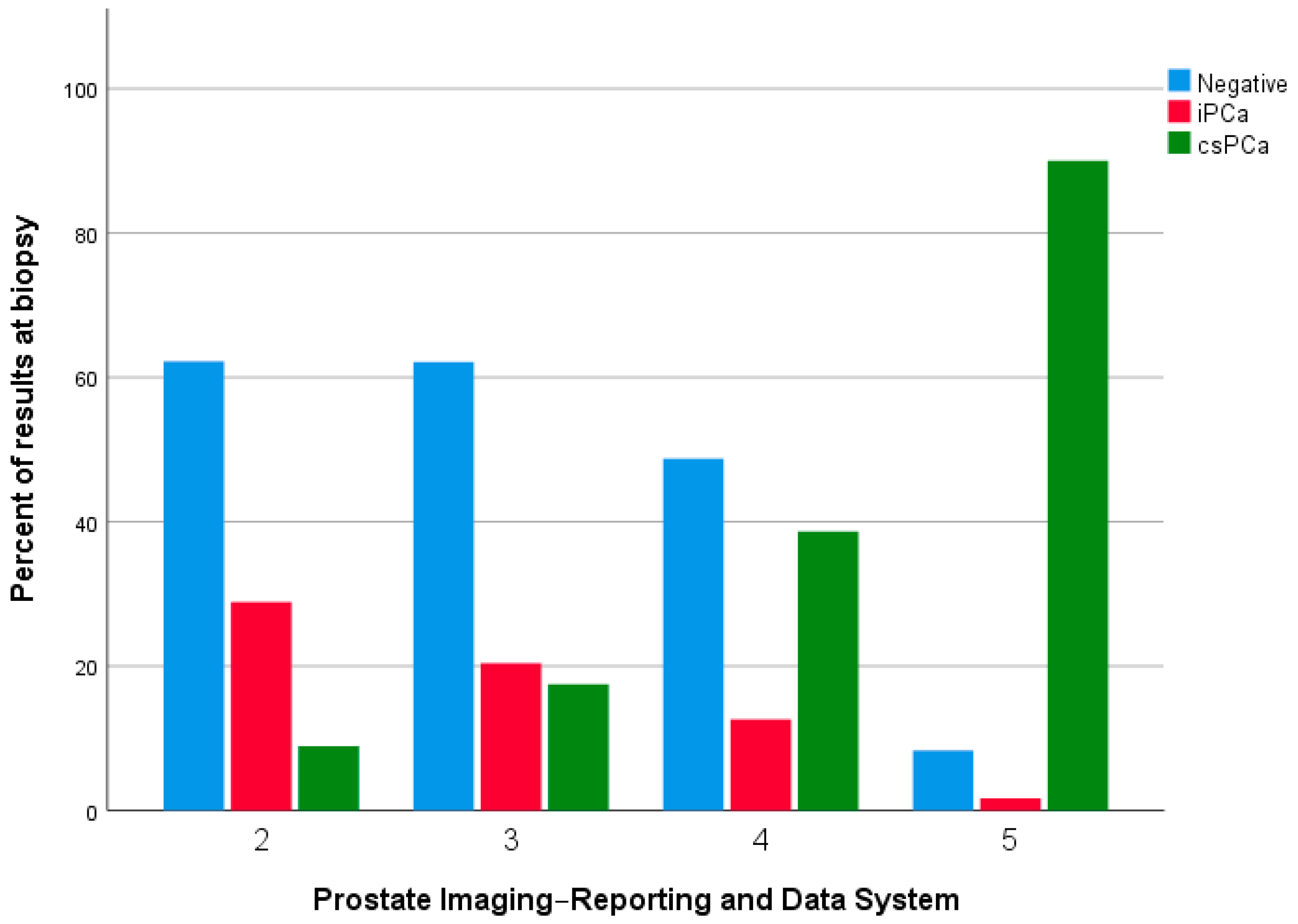
| 2 | 56 | 14.4 |
| 3 | 124 | 31.9 |
| 4 | 137 | 35.2 |
| 5 | 72 | 18.5 |
| Side at mpMRI | ||
| Bilateral | 47 | 12.1 |
| Left | 170 | 43.7 |
| Right | 160 | 41.1 |
| None | 12 | 3.1 |
| Gleason Score | ||
| Negative | 186 | 47.8 |
| PIN | 7 | 1.8 |
| 3 + 3 | 53 | 13.6 |
| 3 + 4 | 22 | 5.7 |
| 4 + 3 | 8 | 2.1 |
| 4 + 4 | 91 | 23.4 |
| 4 + 5 | 7 | 1.8 |
| 5 + 4 | 11 | 2.8 |
| 5 + 5 | 4 | 1 |
| Side at Biopsy | ||
| Bilateral | 57 | 14.7 |
| Left | 72 | 18.5 |
| Right | 74 | 19 |
| None | 186 | 47.8 |
| ISUP Grade | ||
| 1 | 60 | 29.6 |
| 2 | 22 | 10.8 |
| 3 | 8 | 3.9 |
| 4 | 91 | 44.8 |
| 5 | 22 | 10.8 |
| Complications (Clavien-Dindo) | ||
| 1 | 17 | 4.4 |
| 2 | 14 | 3.6 |
| 3 | 8 | 2.1 |
| Group A (Biopsy-Naïve) = 327 | Group B (Re-Biopsy) = 62 | p Value | |
|---|---|---|---|
| Mean ± Standard Deviation | Mean ± Standard Deviation | ||
| Age | 68.05 ± 7.10 | 68.18 ± 6.63 | 0.828 |
| PSA (ng/mL) | 8.91 ± 7.31 | 9.59 ± 5.96 | 0.624 |
| Number of cores | 15.69 ± 1.13 | 16.13 ± 1.76 | 0.661 |
| Count (Percentage) | Count (Percentage) | ||
| Positive DRE | 137 (79.6) | 35 (20.4) | 0.047 * |
| PIRADS score | |||
| 2 | 45 (13.8) | 11 (17.7) | 0.683 |
| 3 | 103 (31.5) | 21 (33.9) | 0.683 |
| 4 | 119(36.4) | 18 (29) | 0.683 |
| 5 | 60 (18.4) | 12 (19.4) | 0.683 |
| Side at mpMRI | |||
| Bilateral | 37 (11.3) | 10 (16.1) | 0.467 |
| Left | 147 (45) | 23 (37.1) | 0.467 |
| Right | 132 (40.4) | 28 (45.2) | 0.467 |
| None | 11 (3.4) | 1 (1.6) | 0.467 |
| Gleason Score | |||
| Negative | 155 (47.4) | 31 (50) | 0.635 |
| PIN | 5 (1.5) | 2 (3.2) | 0.635 |
| 3 + 3 | 45 (13.8) | 8 (12.9) | 0.635 |
| 3 + 4 | 19 (5.8) | 3 (4.8) | 0.635 |
| 4 + 3 | 5 (1.5) | 3 (4.8) | 0.635 |
| 4 + 4 | 78 (23.9) | 13 (21) | 0.635 |
| 4 + 5 | 7 (2.1) | 0 (0) | 0.635 |
| 5 + 4 | 9 (2.8) | 2 (3.2) | 0.635 |
| 5 + 5 | 4 (1.2) | 0 (0) | 0.635 |
| Side at Biopsy | |||
| Bilateral | 49 (15) | 8 (12.9) | 0.472 |
| Left | 64 (19.6) | 8 (12.9) | 0.472 |
| Right | 59 (18) | 15 (24.2) | 0.472 |
| None | 155 (47.4) | 31 (50) | 0.472 |
| ISUP Grade | |||
| 1 | 50 (29.1) | 10 (32.3) | 0.562 |
| 2 | 19 (11) | 3 (9.7) | 0.562 |
| 3 | 5 (2.9) | 3 (9.7) | 0.562 |
| 4 | 78 (45.3) | 13 (41.9) | 0.562 |
| 5 | 20 (11.6) | 2 (6.5) | 0.562 |
| Complications (Clavien-Dindo) | |||
| 1 | 15 (44.1) | 2 (40) | 0.978 |
| 2 | 12 (35.3) | 2 (40) | 0.978 |
| 3 | 7 (20.6) | 1 (20) | 0.978 |
| Negative | PIN | GS 3 + 3 | GS 3 + 4 | GS 4 + 3 | GS 4 + 4 | GS 4 + 5 | GS 5 + 4 | GS 5 + 5 | ||||||||||
|---|---|---|---|---|---|---|---|---|---|---|---|---|---|---|---|---|---|---|
| Group A | Group B | Group A | Group B | Group A | Group B | Group A | Group B | Group A | Group B | Group A | Group B | Group A | Group B | Group A | Group B | Group A | Group B | |
| PIRADS 2 | 28 (62.2) | 10 (90.9) | 0 (0) | 0 (0) | 13 (28.9) | 1 (9.1) | 3 (6.7) | 0 (0) | 0 (0) | 0 (0) | 0 (0) | 0 (0) | 1 (2.2) | 0 (0) | 0 (0) | 0 (0) | 0 (0) | 0 (0) |
| PIRADS 3 | 64 (62.1) | 10 (47.6) | 2 (1.9) | 1 (4.8) | 19 (18.4) | 5 (23.8) | 9 (8.7) | 2 (9.5) | 1 (1) | 1 (4.8) | 7 (6.8) | 2 (9.5) | 1 (1) | 0 (0) | 0 (0) | 0 (0) | 0 (0) | 0 (0) |
| PIRADS 4 | 58 (48.7) | 9 (50) | 3 (2.5) | 1 (5.6) | 12 (10.1) | 1 (5.6) | 3 (2.5) | 0 (0) | 4 (3.4) | 2 (11.2) | 35 (29.4) | 4 (22.2) | 1 (0.8) | 0 (0) | 2 (1.7) | 1 (5.6) | 1 (0.8) | 0 (0) |
| PIRADS 5 | 5 (8.3) | 2 (16.7) | 0 (0) | 0 (0) | 1 (1.7) | 1 (8.3) | 4 (6.7) | 1 (8.3) | 0 (0) | 0 (0) | 36 (60) | 7 (58.3) | 4 (6.7) | 0 (0) | 7 (11.7) | 1 (8.3) | 3 (5) | 0 (0) |
| ISUP | 0 | 1 | 2 | 3 | 4 | 5 | ||||||
|---|---|---|---|---|---|---|---|---|---|---|---|---|
| Group A | Group B | Group A | Group B | Group A | Group B | Group A | Group B | Group A | Group B | Group A | Group B | |
| PIRADS 2 | 28 (62.2) | 10 (90.9) | 13 (28.9) | 1 (9.1) | 3 (6.7) | 0 (0) | 0 (0) | 0 (0) | 0 (0) | 0 (0) | 1 (2.2) | 0 (0) |
| PIRADS 3 | 64 (62.1) | 10 (47.6) | 21 (20.4) | 6 (28.6) | 9 (8.7) | 2 (9.5) | 1 (1) | 1 (9.1) | 7 (6.8) | 2 (9.5) | 1 (1) | 0 (0) |
| PIRADS 4 | 58 (48.7) | 9 (50) | 15 (12.6) | 2 (11.1) | 3 (2.5) | 0 (0) | 4 (3.4) | 2 (22.2) | 35 (29.4) | 4 (22.2) | 4 (3.4) | 1 (5.6) |
| PIRADS 5 | 5 (8.3) | 2 (16.7) | 1 (1.7) | 1 (8.3) | 4 (6.7) | 1 (8.3) | 0 (0) | 0 (0) | 36 (60) | 7 (58.3) | 14 (23.3) | 1 (8.3) |
Disclaimer/Publisher’s Note: The statements, opinions and data contained in all publications are solely those of the individual author(s) and contributor(s) and not of MDPI and/or the editor(s). MDPI and/or the editor(s) disclaim responsibility for any injury to people or property resulting from any ideas, methods, instructions or products referred to in the content. |
© 2023 by the authors. Licensee MDPI, Basel, Switzerland. This article is an open access article distributed under the terms and conditions of the Creative Commons Attribution (CC BY) license (https://creativecommons.org/licenses/by/4.0/).
Share and Cite
Barone, B.; Napolitano, L.; Calace, F.P.; Del Biondo, D.; Napodano, G.; Grillo, M.; Reccia, P.; De Luca, L.; Prezioso, D.; Muto, M.; et al. Reliability of Multiparametric Magnetic Resonance Imaging in Patients with a Previous Negative Biopsy: Comparison with Biopsy-Naïve Patients in the Detection of Clinically Significant Prostate Cancer. Diagnostics 2023, 13, 1939. https://doi.org/10.3390/diagnostics13111939
Barone B, Napolitano L, Calace FP, Del Biondo D, Napodano G, Grillo M, Reccia P, De Luca L, Prezioso D, Muto M, et al. Reliability of Multiparametric Magnetic Resonance Imaging in Patients with a Previous Negative Biopsy: Comparison with Biopsy-Naïve Patients in the Detection of Clinically Significant Prostate Cancer. Diagnostics. 2023; 13(11):1939. https://doi.org/10.3390/diagnostics13111939
Chicago/Turabian StyleBarone, Biagio, Luigi Napolitano, Francesco Paolo Calace, Dario Del Biondo, Giorgio Napodano, Marco Grillo, Pasquale Reccia, Luigi De Luca, Domenico Prezioso, Matteo Muto, and et al. 2023. "Reliability of Multiparametric Magnetic Resonance Imaging in Patients with a Previous Negative Biopsy: Comparison with Biopsy-Naïve Patients in the Detection of Clinically Significant Prostate Cancer" Diagnostics 13, no. 11: 1939. https://doi.org/10.3390/diagnostics13111939