Next Issue
Volume 13, June-2
Previous Issue
Volume 13, May-2


Diagnostics, Volume 13, Issue 11 (June-1 2023) – 157 articles

Cover Story (view full-size image): 
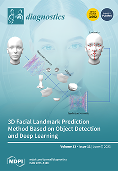
Three-dimensional facial soft tissue landmark prediction plays a crucial role in dentistry. However, existing methods, such as those based on geometric information and machine learning, suffer from a limited number of landmark predictions and low accuracy. To address these issues, this paper presents a novel method based on object detection and deep learning. This method utilizes object detection to locate facial organs, such as the eyes, nose, lips, chin, and face, and employs machine learning algorithms to predict landmarks. As a result, this method achieves an unprecedented prediction of 32 landmarks with a superior precision of 2.62 ± 2.39mm. This groundbreaking technique enhances plastic surgery, facial analysis, animation, virtual reality, and biometric identification. View this paper
  • Issues are regarded as officially published after their release is announced to the table of contents alert mailing list.
  • You may sign up for e-mail alerts to receive table of contents of newly released issues.
  • PDF is the official format for papers published in both, html and pdf forms. To view the papers in pdf format, click on the "PDF Full-text" link, and use the free Adobe Reader to open them.
Order results
Result details
Section
Select all
Export citation of selected articles as:
12 pages, 3947 KiB  
Article
Retrospective Cohort Study of Shear-Wave Elastography and Computed Tomography Enterography in Crohn’s Disease
by Minping Zhang, Enhua Xiao, Minghui Liu, Xilong Mei and Yinghuan Dai
Diagnostics 2023, 13(11), 1980; https://doi.org/10.3390/diagnostics13111980 - 05 Jun 2023
Cited by 6 | Viewed by 1528
Abstract
Distinguishing between inflammatory and fibrotic lesions drastically influences treatment decision-making regarding Crohn’s disease. However, it is challenging to distinguish these two phenotypes before surgery. This study investigates the diagnostic yield of shear-wave elastography and computed tomography enterography to distinguish intestinal phenotypes in Crohn’s [...] Read more.
Distinguishing between inflammatory and fibrotic lesions drastically influences treatment decision-making regarding Crohn’s disease. However, it is challenging to distinguish these two phenotypes before surgery. This study investigates the diagnostic yield of shear-wave elastography and computed tomography enterography to distinguish intestinal phenotypes in Crohn’s disease. Thirty-seven patients (mean age, 29.51 ± 11.52; 31 men) were evaluated with average value of shear-wave elastography (Emean) and computed tomography enterography (CTE) scores. The results demonstrated that a positive correlation between the Emean and fibrosis (Spearman’s r = 0.653, p = 0.000). The cut-off value for fibrotic lesions was 21.30 KPa (AUC: 0.877, sensitivity: 88.90%, specificity: 89.50%, 95% CI:0.755~0.999, p = 0.000). The CTE score showed a positive correlation with inflammation (Spearman’s r = 0.479, p = 0.003), and a 4.5-point grading system was the optimal cut-off value for inflammatory lesions (AUC: 0.766, sensitivity: 73.70%, specificity: 77.80%, 95% CI: 0.596~0.936, p = 0.006). Combining these two metrics improved the diagnostic performance and specificity (AUC: 0.918, specificity: 94.70%, 95% CI: 0.806~1.000, p = 0.000). In conclusion, shear-wave elastography can be used to help detect fibrotic lesions and the computed tomography enterography score emerged as a feasible predictor of inflammatory lesions. The combination of these two imaging techniques is proposed to distinguish intestinal predominant phenotypes. Full article
(This article belongs to the Special Issue Diagnostic Imaging in Gastrointestinal Diseases)
Show Figures

Figure 1

13 pages, 494 KiB  
Article
Role of Neutrophil-to-Lymphocyte Ratio (NLR) in Patients with Mycosis Fungoides
by Cosimo Di Raimondo, Paolo Lombardo, Cristiano Tesei, Fabiana Esposito, Federico Meconi, Roberto Secchi, Flavia Lozzi, Alessandro Monopoli, Maria Grazia Narducci, Enrico Scala, Cecilia Angeloni, Alberto De Stefano, Siavash Rahimi, Luca Bianchi and Maria Cantonetti
Diagnostics 2023, 13(11), 1979; https://doi.org/10.3390/diagnostics13111979 - 05 Jun 2023
Cited by 3 | Viewed by 944
Abstract
Background: The neutrophil/lymphocyte ratio (NLR) at baseline has been demonstrated to correlate with higher stages of disease and to be a prognostic factor in numerous cancers. However, its function as a prognostic factor for mycosis fungoides (MF) has not been yet clarified. Objective: [...] Read more.
Background: The neutrophil/lymphocyte ratio (NLR) at baseline has been demonstrated to correlate with higher stages of disease and to be a prognostic factor in numerous cancers. However, its function as a prognostic factor for mycosis fungoides (MF) has not been yet clarified. Objective: Our work aimed to assess the association of the NLR with different stages of MF and to outline whether higher values of this marker are related to a more aggressive MF. Methods: We retrospectively calculated the NLRs in 302 MF patients at the moment of diagnosis. The NLR was obtained using the complete blood count values. Results: The median NLR among patients with early stage disease (low-grade IA-IB-IIA) was 1.88, while the median NLR for patients with high-grade MF (IIB-IIIA-IIIB) was 2.64. Statistical analysis showed positive associations of advanced MF stages with NLRs higher than 2.3. Conclusions: Our analysis demonstrates that the NLR represents a cheap and easily available parameter functioning as a marker for advanced MF. This might guide physicians in recognizing patients with advanced stages of disease requiring a strict follow-up or an early treatment. Full article
(This article belongs to the Section Pathology and Molecular Diagnostics)
Show Figures

Figure 1

18 pages, 3279 KiB  
Review
Coronary Angiography Upgraded by Imaging Post-Processing: Present and Future Directions
by Benoit Caullery, Laurent Riou and Gilles Barone-Rochette
Diagnostics 2023, 13(11), 1978; https://doi.org/10.3390/diagnostics13111978 - 05 Jun 2023
Viewed by 1446
Abstract
Advances in computer technology and image processing now allow us to obtain from angiographic images a large variety of information on coronary physiology without the use of a guide-wire as a diagnostic information equivalent to FFR and iFR but also information allowing for [...] Read more.
Advances in computer technology and image processing now allow us to obtain from angiographic images a large variety of information on coronary physiology without the use of a guide-wire as a diagnostic information equivalent to FFR and iFR but also information allowing for the performance of a real virtual percutaneous coronary intervention (PCI) and finally the ability to obtain information to optimize the results of PCI. With specific software, it is now possible to have a real upgrading of invasive coronary angiography. In this review, we present the different advances in this field and discuss the future perspectives offered by this technology. Full article
Show Figures

Figure 1

17 pages, 2136 KiB  
Article
An Invasive Ductal Carcinomas Breast Cancer Grade Classification Using an Ensemble of Convolutional Neural Networks
by Eelandula Kumaraswamy, Sumit Kumar and Manoj Sharma
Diagnostics 2023, 13(11), 1977; https://doi.org/10.3390/diagnostics13111977 - 05 Jun 2023
Cited by 11 | Viewed by 1589
Abstract
Invasive Ductal Carcinoma Breast Cancer (IDC-BC) is the most common type of cancer and its asymptomatic nature has led to an increased mortality rate globally. Advancements in artificial intelligence and machine learning have revolutionized the medical field with the development of AI-enabled computer-aided [...] Read more.
Invasive Ductal Carcinoma Breast Cancer (IDC-BC) is the most common type of cancer and its asymptomatic nature has led to an increased mortality rate globally. Advancements in artificial intelligence and machine learning have revolutionized the medical field with the development of AI-enabled computer-aided diagnosis (CAD) systems, which help in determining diseases at an early stage. CAD systems assist pathologists in their decision-making process to produce more reliable outcomes in order to treat patients well. In this work, the potential of pre-trained convolutional neural networks (CNNs) (i.e., EfficientNetV2L, ResNet152V2, DenseNet201), singly or as an ensemble, was thoroughly explored. The performances of these models were evaluated for IDC-BC grade classification using the DataBiox dataset. Data augmentation was used to avoid the issues of data scarcity and data imbalances. The performance of the best model was compared to three different balanced datasets of Databiox (i.e., 1200, 1400, and 1600 images) to determine the implications of this data augmentation. Furthermore, the effects of the number of epochs were analysed to ensure the coherency of the most optimal model. The experimental results analysis revealed that the proposed ensemble model outperformed the existing state-of-the-art techniques in relation to classifying the IDC-BC grades of the Databiox dataset. The proposed ensemble model of the CNNs achieved a 94% classification accuracy and attained a significant area under the ROC curves for grades 1, 2, and 3, i.e., 96%, 94%, and 96%, respectively. Full article
(This article belongs to the Special Issue Machine Learning in Precise and Personalized Diagnosis)
Show Figures

Figure 1

12 pages, 1120 KiB  
Review
Fecal and Circulating Biomarkers for the Non-Invasive Assessment of Intestinal Permeability
by Nuria Perez-Diaz-del-Campo, Gabriele Castelnuovo, Davide Giuseppe Ribaldone and Gian Paolo Caviglia
Diagnostics 2023, 13(11), 1976; https://doi.org/10.3390/diagnostics13111976 - 05 Jun 2023
Cited by 2 | Viewed by 2107
Abstract
The study of intestinal permeability is gaining growing interest due to its relevance in the onset and progression of several gastrointestinal and non-gastrointestinal diseases. Though the involvement of impaired intestinal permeability in the pathophysiology of such diseases is recognized, there is currently a [...] Read more.
The study of intestinal permeability is gaining growing interest due to its relevance in the onset and progression of several gastrointestinal and non-gastrointestinal diseases. Though the involvement of impaired intestinal permeability in the pathophysiology of such diseases is recognized, there is currently a need to identify non-invasive biomarkers or tools that are able to accurately detect alterations in intestinal barrier integrity. On the one hand, promising results have been reported for novel in vivo methods based on paracellular probes, i.e., methods that can directly assess paracellular permeability and, on the other hand, on fecal and circulating biomarkers able to indirectly assess epithelial barrier integrity and functionality. In this review, we aimed to summarize the current knowledge on the intestinal barrier and epithelial transport pathways and to provide an overview of the methods already available or currently under investigation for the measurement of intestinal permeability. Full article
Show Figures

Figure 1

13 pages, 298 KiB  
Article
A Retrospective Study of Staphylococcus aureus Bacteremia in a Tertiary Hospital and Factors Associated with Mortality
by Petros Ioannou, Maria Zacharioudaki, Despoina Spentzouri, Antonia Koutoulakou, Konstantinos Kitsos-Kalyvianakis, Christoforos Chontos, Stamatis Karakonstantis, Sofia Maraki, George Samonis and Diamantis P. Kofteridis
Diagnostics 2023, 13(11), 1975; https://doi.org/10.3390/diagnostics13111975 - 05 Jun 2023
Cited by 1 | Viewed by 1434
Abstract
Staphylococcus aureus bacteremia (SAB) is a severe infection frequently associated with significant morbidity and mortality. Recent studies have shown that SAB mortality has decreased during the last decades. However, about 25% of patients suffering from the disease will ultimately die. Hence, there is [...] Read more.
Staphylococcus aureus bacteremia (SAB) is a severe infection frequently associated with significant morbidity and mortality. Recent studies have shown that SAB mortality has decreased during the last decades. However, about 25% of patients suffering from the disease will ultimately die. Hence, there is an urgent need for more timely and efficient treatment of patients with SAB. The aim of the present study was to retrospectively evaluate a cohort of SAB patients hospitalized in a tertiary hospital and to identify factors independently associated with mortality. All 256 SAB patients hospitalized from January 2005 to December 2021 in the University Hospital of Heraklion, Greece, were evaluated. Their median age was 72 years, while 101 (39.5%) were female. Most SAB patients were cared for in medical wards (80.5%). The infection was community-acquired in 49.5%. Among all strains 37.9% were methicillin-resistant S. aureus (MRSA), however, definite treatment with an antistaphylococcal penicillin was given only in 22% of patients. Only 14.4% of patients had a repeat blood culture after the initiation of antimicrobial treatment. Infective endocarditis was present in 8%. In-hospital mortality has reached 15.9%. Female gender, older age, higher McCabe score, previous antimicrobial use, presence of a central venous catheter, neutropenia, severe sepsis, septic shock, and MRSA SAB were positively associated with in-hospital mortality, while monomicrobial bacteremia was negatively associated. The multivariate logistic regression model identified only severe sepsis (p = 0.05, odds ratio = 12.294) and septic shock (p = 0.007, odds ratio 57.18) to be independently positively associated with in-hospital mortality. The evaluation revealed high rates of inappropriate empirical antimicrobial treatment and non-adherence to guidelines, as shown, by the lack of repeat blood cultures. These data underline the urgent need for interventions with antimicrobial stewardship, increased involvement of infectious diseases physicians, educational sessions, and creation and implementation of local guidelines for improvement of the necessary steps for timely and efficient SAB treatment. Optimization of diagnostic techniques is needed to overcome challenges such as heteroresistance that may affect treatment. Clinicians should be aware of the factors associated with mortality in patients with SAB to identify those who are at a higher risk and optimize medical management. Full article
(This article belongs to the Special Issue Antimicrobial Resistance during and after COVID-19)
29 pages, 30339 KiB  
Review
Peritoneal Carcinosis: What the Radiologist Needs to Know
by Alfonso Reginelli, Giuliana Giacobbe, Maria Teresa Del Canto, Marina Alessandrella, Giovanni Balestrucci, Fabrizio Urraro, Gaetano Maria Russo, Luigi Gallo, Ginevra Danti, Barbara Frittoli, Luca Stoppino, Daria Schettini, Franco Iafrate, Salvatore Cappabianca, Andrea Laghi, Roberto Grassi, Luca Brunese, Antonio Barile and Vittorio Miele
Diagnostics 2023, 13(11), 1974; https://doi.org/10.3390/diagnostics13111974 - 05 Jun 2023
Cited by 2 | Viewed by 5909
Abstract
Peritoneal carcinosis is a condition characterized by the spread of cancer cells to the peritoneum, which is the thin membrane that lines the abdominal cavity. It is a serious condition that can result from many different types of cancer, including ovarian, colon, stomach, [...] Read more.
Peritoneal carcinosis is a condition characterized by the spread of cancer cells to the peritoneum, which is the thin membrane that lines the abdominal cavity. It is a serious condition that can result from many different types of cancer, including ovarian, colon, stomach, pancreatic, and appendix cancer. The diagnosis and quantification of lesions in peritoneal carcinosis are critical in the management of patients with the condition, and imaging plays a central role in this process. Radiologists play a vital role in the multidisciplinary management of patients with peritoneal carcinosis. They need to have a thorough understanding of the pathophysiology of the condition, the underlying neoplasms, and the typical imaging findings. In addition, they need to be aware of the differential diagnoses and the advantages and disadvantages of the various imaging methods available. Imaging plays a central role in the diagnosis and quantification of lesions, and radiologists play a critical role in this process. Ultrasound, computed tomography, magnetic resonance, and PET/CT scans are used to diagnose peritoneal carcinosis. Each imaging procedure has advantages and disadvantages, and particular imaging techniques are recommended based on patient conditions. Our aim is to provide knowledge to radiologists regarding appropriate techniques, imaging findings, differential diagnoses, and treatment options. With the advent of AI in oncology, the future of precision medicine appears promising, and the interconnection between structured reporting and AI is likely to improve diagnostic accuracy and treatment outcomes for patients with peritoneal carcinosis. Full article
Show Figures

Figure 1

11 pages, 839 KiB  
Article
Survival Outcomes of Ewing Sarcoma and Rhabdomyosarcoma by High- versus Low-Volume Cancer Centres in British Columbia, Canada
by Sarah Yeo, Ursula Lee, Ying Hui Xu, Christine Simmons, Alannah Smrke and Ying Wang
Diagnostics 2023, 13(11), 1973; https://doi.org/10.3390/diagnostics13111973 - 05 Jun 2023
Cited by 1 | Viewed by 953
Abstract
Due to the rarity and complexity of treatment for Ewing sarcoma and rhabdomyosarcoma, studies demonstrate improved patient outcomes when managed by a multidisciplinary team at high-volume centres (HVCs). Our study explores the difference in outcomes of Ewing sarcoma and rhabdomyosarcoma patients based on [...] Read more.
Due to the rarity and complexity of treatment for Ewing sarcoma and rhabdomyosarcoma, studies demonstrate improved patient outcomes when managed by a multidisciplinary team at high-volume centres (HVCs). Our study explores the difference in outcomes of Ewing sarcoma and rhabdomyosarcoma patients based on the centre of initial consultation in British Columbia, Canada. This retrospective study assessed adults diagnosed with Ewing sarcoma and rhabdomyosarcoma between 1 January 2000 and 31 December 2020 undergoing curative intent therapy in one of five cancer centres across the province. Seventy-seven patients were included, 46 seen at HVCs and 31 at low-volume centres (LVCs). Patients at HVCs were younger (32.1 vs. 40.8 years, p = 0.020) and more likely to receive curative intent radiation (88% vs. 67%, p = 0.047). The time from diagnosis to first chemotherapy was 24 days shorter at HVCs (26 vs. 50 days, p = 0.120). There was no significant difference in overall survival by treatment centre (HR 0.850, 95% CI 0.448–1.614). Variations in care exist amongst patients treated at HVCs vs. LVCs, which may reflect differences in access to resources, clinical specialists, and varying practice patterns across centres. This study can be used to inform decisions regarding triaging and centralization of Ewing sarcoma and rhabdomyosarcoma patient treatment. Full article
Show Figures

Figure 1

17 pages, 4368 KiB  
Review
COVID-19 Lung Ultrasound Scores and Lessons from the Pandemic: A Narrative Review
by Luigi Maggi, Giulia De Fazio, Riccardo Guglielmi, Flaminia Coluzzi, Silvia Fiorelli and Monica Rocco
Diagnostics 2023, 13(11), 1972; https://doi.org/10.3390/diagnostics13111972 - 05 Jun 2023
Cited by 2 | Viewed by 968
Abstract
The WHO recently declared that COVID-19 no longer constitutes a public health emergency of international concern; however, lessons learned through the pandemic should not be left behind. Lung ultrasound was largely utilized as a diagnostic tool thanks to its feasibility, easy application, and [...] Read more.
The WHO recently declared that COVID-19 no longer constitutes a public health emergency of international concern; however, lessons learned through the pandemic should not be left behind. Lung ultrasound was largely utilized as a diagnostic tool thanks to its feasibility, easy application, and the possibility to reduce the source of infection for health personnel. Lung ultrasound scores consist of grading systems used to guide diagnosis and medical decisions, owning a good prognostic value. In the emergency context of the pandemic, several lung ultrasound scores emerged either as new scores or as modifications of pre-existing ones. Our aim is to clarify the key aspects of lung ultrasound and lung ultrasound scores to standardize their clinical use in a non-pandemic context. The authors searched on PubMed for articles related to “COVID-19”, “ultrasound”, and “Score” until 5 May 2023; other keywords were “thoracic”, “lung”, “echography”, and “diaphragm”. A narrative summary of the results was made. Lung ultrasound scores are demonstrated to be an important tool for triage, prediction of severity, and aid in medical decisions. Ultimately, the existence of numerous scores leads to a lack of clarity, confusion, and an absence of standardization. Full article
(This article belongs to the Section Point-of-Care Diagnostics and Devices)
Show Figures

Figure 1

18 pages, 678 KiB  
Article
A Double-Teacher Model Capable of Exploiting Isomorphic and Heterogeneous Discrepancy Information for Medical Image Segmentation
by Junguo Zou, Zhaohe Wang and Xiuquan Du
Diagnostics 2023, 13(11), 1971; https://doi.org/10.3390/diagnostics13111971 - 05 Jun 2023
Viewed by 846
Abstract
Deep learning, with continuous development, has achieved relatively good results in the field of left atrial segmentation, and numerous semi-supervised methods in this field have been implemented based on consistency regularization to obtain high-performance 3D models by training. However, most semi-supervised methods focus [...] Read more.
Deep learning, with continuous development, has achieved relatively good results in the field of left atrial segmentation, and numerous semi-supervised methods in this field have been implemented based on consistency regularization to obtain high-performance 3D models by training. However, most semi-supervised methods focus on inter-model consistency and ignore inter-model discrepancy. Therefore, we designed an improved double-teacher framework with discrepancy information. Herein, one teacher learns 2D information, another learns both 2D and 3D information, and the two models jointly guide the student model for learning. Simultaneously, we extract the isomorphic/heterogeneous discrepancy information between the predictions of the student and teacher model to optimize the whole framework. Unlike other semi-supervised methods based on 3D models, ours only uses 3D information to assist 2D models, and does not have a fully 3D model, thus addressing the large memory consumption and limited training data of 3D models to some extent. Our approach shows excellent performance on the left atrium (LA) dataset, similar to that of the best performing 3D semi-supervised methods available, compared to existing techniques. Full article
(This article belongs to the Section Machine Learning and Artificial Intelligence in Diagnostics)
Show Figures

Figure 1

4 pages, 1024 KiB  
Interesting Images
Multiple Bone Destruction Secondary to Mycobacterium kansasii Pulmonary Disease: A Case Report
by Lu Dai, Yanyan Wu, Xi Zhou, Sen Liu, Junping Fan, Huaiya Xie, Luo Wang, Xinlun Tian and Wenbing Xu
Diagnostics 2023, 13(11), 1970; https://doi.org/10.3390/diagnostics13111970 - 05 Jun 2023
Viewed by 885
Abstract
Mycobacterium kansasii infections predominantly manifest in immunocompromised people and are primarily responsible for lung disease and systemic disseminated infection. Osteopathy is a rare consequence of M. kansasii infection. Here, we present imaging data from a 44-year-old immunocompetent Chinese woman diagnosed with multiple bone destruction, [...] Read more.
Mycobacterium kansasii infections predominantly manifest in immunocompromised people and are primarily responsible for lung disease and systemic disseminated infection. Osteopathy is a rare consequence of M. kansasii infection. Here, we present imaging data from a 44-year-old immunocompetent Chinese woman diagnosed with multiple bone destruction, particularly of the spine, secondary to M. kansasii pulmonary disease, which is easily misdiagnosed. The patient underwent an emergency operation after experiencing unexpected incomplete paraplegia during hospitalization, indicating an aggravation of bone destruction. Preoperative sputum testing and next-generation sequencing of DNA and RNA of intraoperative samples confirmed the diagnosis of M. kansasii infection. Treatment with anti-tuberculosis therapy and the subsequent patient response supported our diagnosis. Given the rarity of osteopathy secondary to M. kansasii infection in immunocompetent individuals, our case offers some insight into this diagnosis. Full article
(This article belongs to the Section Diagnostic Microbiology and Infectious Disease)
Show Figures

Figure 1

13 pages, 2115 KiB  
Article
A Smartphone Application for Personalized Tooth Shade Determination
by Tomoya Kusayanagi, Sota Maegawa, Shuya Terauchi, Wataru Hashimoto and Shohei Kaneda
Diagnostics 2023, 13(11), 1969; https://doi.org/10.3390/diagnostics13111969 - 05 Jun 2023
Cited by 1 | Viewed by 2066 | Correction
Abstract
Tooth shade determination methods for evaluating the effectiveness of whitening products at home are limited. In this study, an iPhone app for personalized tooth shade determination was developed. While capturing dental photographs in selfie mode before and after whitening, the app can maintain [...] Read more.
Tooth shade determination methods for evaluating the effectiveness of whitening products at home are limited. In this study, an iPhone app for personalized tooth shade determination was developed. While capturing dental photographs in selfie mode before and after whitening, the app can maintain consistent illumination and tooth appearance conditions that affect tooth color measurement. An ambient light sensor was used to standardize the illumination conditions. To maintain consistent tooth appearance conditions determined by appropriately opening the mouth, facial landmark detection, an artificial intelligence technique that estimates key face parts and outlines, was used. The effectiveness of the app in ensuring uniform tooth appearance was investigated through color measurements of the upper incisors of seven participants via photographs captured in succession. The coefficients of variation for incisors L*, a*, and b* were less than 0.0256 (95% CI, 0.0173–0.0338), 0.2748 (0.1596–0.3899), and 0.1053 (0.0078–0.2028), respectively. To examine the feasibility of the app for tooth shade determination, gel whitening after pseudo-staining by coffee and grape juice was performed. Consequently, whitening results were evaluated by monitoring the ∆Eab color difference values (1.3 unit minimum). Although tooth shade determination remains a relative quantification method, the proposed method can support evidence-based selection of whitening products. Full article
(This article belongs to the Section Point-of-Care Diagnostics and Devices)
Show Figures

Figure 1

23 pages, 1218 KiB  
Article
Diagnosis of COVID-19 Using Chest X-ray Images and Disease Symptoms Based on Stacking Ensemble Deep Learning
by Abdulaziz AlMohimeed, Hager Saleh, Nora El-Rashidy, Redhwan M. A. Saad, Shaker El-Sappagh and Sherif Mostafa
Diagnostics 2023, 13(11), 1968; https://doi.org/10.3390/diagnostics13111968 - 05 Jun 2023
Cited by 6 | Viewed by 1488
Abstract
The COVID-19 virus is one of the most devastating illnesses humanity has ever faced. COVID-19 is an infection that is hard to diagnose until it has caused lung damage or blood clots. As a result, it is one of the most insidious diseases [...] Read more.
The COVID-19 virus is one of the most devastating illnesses humanity has ever faced. COVID-19 is an infection that is hard to diagnose until it has caused lung damage or blood clots. As a result, it is one of the most insidious diseases due to the lack of knowledge of its symptoms. Artificial intelligence (AI) technologies are being investigated for the early detection of COVID-19 using symptoms and chest X-ray images. Therefore, this work proposes stacking ensemble models using two types of COVID-19 datasets, symptoms and chest X-ray scans, to identify COVID-19. The first proposed model is a stacking ensemble model that is merged from the outputs of pre-trained models in the stacking: multi-layer perceptron (MLP), recurrent neural network (RNN), long short-term memory (LSTM), and gated recurrent unit (GRU). Stacking trains and evaluates the meta-learner as a support vector machine (SVM) to predict the final decision. Two datasets of COVID-19 symptoms are used to compare the first proposed model with MLP, RNN, LSTM, and GRU models. The second proposed model is a stacking ensemble model that is merged from the outputs of pre-trained DL models in the stacking: VGG16, InceptionV3, Resnet50, and DenseNet121; it uses stacking to train and evaluate the meta-learner (SVM) to identify the final prediction. Two datasets of COVID-19 chest X-ray images are used to compare the second proposed model with other DL models. The result has shown that the proposed models achieve the highest performance compared to other models for each dataset. Full article
(This article belongs to the Special Issue Artificial Intelligence and Machine Learning for Infectious Diseases)
Show Figures

Figure 1

9 pages, 5019 KiB  
Interesting Images
Imaging Criteria for the Diagnosis of Progressive Supranuclear Palsy: Supportive or Mandatory?
by Nicoleta Lupascu, Ioan Cristian Lupescu, Ionuț Caloianu, Florin Naftanaila, Remus Relu Glogojeanu, Carmen Adella Sirbu and Marian Mitrica
Diagnostics 2023, 13(11), 1967; https://doi.org/10.3390/diagnostics13111967 - 05 Jun 2023
Cited by 1 | Viewed by 6314
Abstract
We present the case of a 54-year-old male, without any significant medical history, who insidiously developed speech disturbances and walking difficulties, accompanied by backward falls. The symptoms progressively worsened over time. The patient was initially diagnosed with Parkinson’s disease; however, he failed to [...] Read more.
We present the case of a 54-year-old male, without any significant medical history, who insidiously developed speech disturbances and walking difficulties, accompanied by backward falls. The symptoms progressively worsened over time. The patient was initially diagnosed with Parkinson’s disease; however, he failed to respond to standard therapy with Levodopa. He came to our attention for worsening postural instability and binocular diplopia. A neurological exam was highly suggestive of a Parkinson-plus disease, most likely progressive supranuclear gaze palsy. Brain MRI was performed and revealed moderate midbrain atrophy with the characteristic “hummingbird” and “Mickey mouse” signs. An increased MR parkinsonism index was also noted. Based on all clinical and paraclinical data, a diagnosis of probable progressive supranuclear palsy was established. We review the main imaging features of this disease and their current role in diagnosis. Full article
(This article belongs to the Collection Interesting Images)
Show Figures

Figure 1

15 pages, 2056 KiB  
Article
Impact of Robotic-Assisted Gait Training in Subacute Spinal Cord Injury Patients on Outcome Measure
by Beata Tarnacka, Bogumił Korczyński and Justyna Frasuńska
Diagnostics 2023, 13(11), 1966; https://doi.org/10.3390/diagnostics13111966 - 05 Jun 2023
Cited by 5 | Viewed by 1545
Abstract
The improvement of walking ability is a primary goal for spinal cord injury (SCI) patients. Robotic-assisted gait training (RAGT) is an innovative method for its improvement. This study evaluates the influence of RAGT vs. dynamic parapodium training (DPT) in improving gait motor functions [...] Read more.
The improvement of walking ability is a primary goal for spinal cord injury (SCI) patients. Robotic-assisted gait training (RAGT) is an innovative method for its improvement. This study evaluates the influence of RAGT vs. dynamic parapodium training (DPT) in improving gait motor functions in SCI patients. In this single-centre, single-blinded study, we enrolled 105 (39 and 64 with complete and incomplete SCI, respectively) patients. The investigated subjects received gait training with RAGT (experimental S1-group) and DPT (control S0-group), with six training sessions per week over seven weeks. The American Spinal Cord Injury Association Impairment Scale Motor Score (MS), Spinal Cord Independence Measure, version-III (SCIM-III), Walking Index for Spinal Cord Injury, version-II (WISCI-II), and Barthel Index (BI) were assessed in each patient before and after sessions. Patients with incomplete SCI assigned to the S1 rehabilitation group achieved more significant improvement in MS [2.58 (SE 1.21, p < 0.05)] and WISCI-II [3.07 (SE 1.02, p < 0.01])] scores in comparison with patients assigned to the S0 group. Despite the described improvement in the MS motor score, no progression between grades of AIS (A to B to C to D) was observed. A nonsignificant improvement between the groups for SCIM-III and BI was found. RAGT significantly improved gait functional parameters in SCI patients in comparison with conventional gait training with DPT. RAGT is a valid treatment option in SCI patients in the subacute phase. DPT should not be recommended for patients with incomplete SCI (AIS-C); in those patients, RAGT rehabilitation programs should be taken into consideration. Full article
(This article belongs to the Special Issue Spinal Cord Injury: Diagnostics, Treatment, and Rehabilitation)
Show Figures

Figure 1

11 pages, 1249 KiB  
Article
Assessment of Inspiratory Effort in Spontaneously Breathing COVID-19 ARDS Patients Undergoing Helmet CPAP: A Comparison between Esophageal, Transdiaphragmatic and Central Venous Pressure Swing
by Sergio Lassola, Sara Miori, Andrea Sanna, Ilaria Menegoni, Silvia De Rosa, Giacomo Bellani and Michele Umbrello
Diagnostics 2023, 13(11), 1965; https://doi.org/10.3390/diagnostics13111965 - 05 Jun 2023
Cited by 3 | Viewed by 1073
Abstract
Introduction: The clinical features of COVID-19 are highly variable. It has been speculated that the progression across COVID-19 may be triggered by excessive inspiratory drive activation. The aim of the present study was to assess whether the tidal swing in central venous pressure [...] Read more.
Introduction: The clinical features of COVID-19 are highly variable. It has been speculated that the progression across COVID-19 may be triggered by excessive inspiratory drive activation. The aim of the present study was to assess whether the tidal swing in central venous pressure (ΔCVP) is a reliable estimate of inspiratory effort. Methods: Thirty critically ill patients with COVID-19 ARDS underwent a PEEP trial (0–5–10 cmH2O) during helmet CPAP. Esophageal (ΔPes) and transdiaphragmatic (ΔPdi) pressure swings were measured as indices of inspiratory effort. ΔCVP was assessed via a standard venous catheter. A low and a high inspiratory effort were defined as ΔPes ≤ 10 and  >15 cmH2O, respectively. Results: During the PEEP trial, no significant changes in ΔPes (11 [6–16] vs. 11 [7–15] vs. 12 [8–16] cmH2O, p = 0.652) and in ΔCVP (12 [7–17] vs. 11.5 [7–16] vs. 11.5 [8–15] cmH2O, p = 0.918) were detected. ΔCVP was significantly associated with ΔPes (marginal R2 0.87, p < 0.001). ΔCVP recognized both low (AUC-ROC curve 0.89 [0.84–0.96]) and high inspiratory efforts (AUC-ROC curve 0.98 [0.96–1]). Conclusions: ΔCVP is an easily available a reliable surrogate of ΔPes and can detect a low or a high inspiratory effort. This study provides a useful bedside tool to monitor the inspiratory effort of spontaneously breathing COVID-19 patients. Full article
(This article belongs to the Section Medical Imaging and Theranostics)
Show Figures

Figure 1

15 pages, 2744 KiB  
Article
Federated Machine Learning for Skin Lesion Diagnosis: An Asynchronous and Weighted Approach
by Muhammad Mateen Yaqoob, Musleh Alsulami, Muhammad Amir Khan, Deafallah Alsadie, Abdul Khader Jilani Saudagar and Mohammed AlKhathami
Diagnostics 2023, 13(11), 1964; https://doi.org/10.3390/diagnostics13111964 - 05 Jun 2023
Cited by 5 | Viewed by 1552
Abstract
The accurate and timely diagnosis of skin cancer is crucial as it can be a life-threatening disease. However, the implementation of traditional machine learning algorithms in healthcare settings is faced with significant challenges due to data privacy concerns. To tackle this issue, we [...] Read more.
The accurate and timely diagnosis of skin cancer is crucial as it can be a life-threatening disease. However, the implementation of traditional machine learning algorithms in healthcare settings is faced with significant challenges due to data privacy concerns. To tackle this issue, we propose a privacy-aware machine learning approach for skin cancer detection that utilizes asynchronous federated learning and convolutional neural networks (CNNs). Our method optimizes communication rounds by dividing the CNN layers into shallow and deep layers, with the shallow layers being updated more frequently. In order to enhance the accuracy and convergence of the central model, we introduce a temporally weighted aggregation approach that takes advantage of previously trained local models. Our approach is evaluated on a skin cancer dataset, and the results show that it outperforms existing methods in terms of accuracy and communication cost. Specifically, our approach achieves a higher accuracy rate while requiring fewer communication rounds. The results suggest that our proposed method can be a promising solution for improving skin cancer diagnosis while also addressing data privacy concerns in healthcare settings. Full article
Show Figures

Figure 1

13 pages, 10094 KiB  
Article
Whole-Body Magnetic Resonance Imaging (MRI) for Staging Melanoma Patients in Direct Comparison to Computed Tomography (CT): Results from a Prospective Positron Emission Tomography (PET)/CT and PET/MRI Study
by Christian Philipp Reinert, Cecilia Liang, Matthias Weissinger, Jonas Vogel, Andrea Forschner, Konstantin Nikolaou, Christian la Fougère and Ferdinand Seith
Diagnostics 2023, 13(11), 1963; https://doi.org/10.3390/diagnostics13111963 - 05 Jun 2023
Cited by 1 | Viewed by 1459
Abstract
Purpose: The consideration of radiation exposure is becoming more important in metastatic melanoma due to improved prognoses. The aim of this prospective study was to investigate the diagnostic performance of whole-body (WB) magnetic resonance imaging (MRI) in comparison to computed tomography (CT) with [...] Read more.
Purpose: The consideration of radiation exposure is becoming more important in metastatic melanoma due to improved prognoses. The aim of this prospective study was to investigate the diagnostic performance of whole-body (WB) magnetic resonance imaging (MRI) in comparison to computed tomography (CT) with 18F-FDG positron emission tomography (PET)/CT and 18F-PET/MRI together with a follow-up as the reference standard. Methods: Between April 2014 and April 2018, a total of 57 patients (25 females, mean age of 64 ± 12 years) underwent WB-PET/CT and WB-PET/MRI on the same day. The CT and MRI scans were independently evaluated by two radiologists who were blinded to the patients’ information. The reference standard was evaluated by two nuclear medicine specialists. The findings were categorized into different regions: lymph nodes/soft tissue (I), lungs (II), abdomen/pelvis (III), and bone (IV). A comparative analysis was conducted for all the documented findings. Inter-reader reliability was assessed using Bland–Altman procedures, and McNemar’s test was utilized to determine the differences between the readers and the methods. Results: Out of the 57 patients, 50 were diagnosed with metastases in two or more regions, with the majority being found in region I. The accuracies of CT and MRI did not show significant differences, except in region II where CT detected more metastases compared to MRI (0.90 vs. 0.68, p = 0.008). On the other hand, MRI had a higher detection rate in region IV compared to CT (0.89 vs. 0.61, p > 0.05). The level of agreement between the readers varied depending on the number of metastases and the specific region, with the highest agreement observed in region III and the lowest observed in region I. Conclusions: In patients with advanced melanoma, WB-MRI has the potential to serve as an alternative to CT with comparable diagnostic accuracy and confidence across most regions. The observed limited sensitivity for the detection of pulmonary lesions might be improved through dedicated lung imaging sequences. Full article
(This article belongs to the Section Medical Imaging and Theranostics)
Show Figures

Figure 1

23 pages, 402 KiB  
Review
Salivary Biomarkers of Anti-Epileptic Drugs: A Narrative Review
by Ioana-Andreea Chiș, Vlad Andrei, Alexandrina Muntean, Marioara Moldovan, Anca Ștefania Mesaroș, Mircea Cristian Dudescu and Aranka Ilea
Diagnostics 2023, 13(11), 1962; https://doi.org/10.3390/diagnostics13111962 - 04 Jun 2023
Cited by 1 | Viewed by 1428
Abstract
Saliva is a biofluid that reflects general health and that can be collected in order to evaluate and determine various pathologies and treatments. Biomarker analysis through saliva sampling is an emerging method of accurately screening and diagnosing diseases. Anti-epileptic drugs (AEDs) are prescribed [...] Read more.
Saliva is a biofluid that reflects general health and that can be collected in order to evaluate and determine various pathologies and treatments. Biomarker analysis through saliva sampling is an emerging method of accurately screening and diagnosing diseases. Anti-epileptic drugs (AEDs) are prescribed generally in seizure treatment. The dose–response relationship of AEDs is influenced by numerous factors and varies from patient to patient, hence the need for the careful supervision of drug intake. The therapeutic drug monitoring (TDM) of AEDs was traditionally performed through repeated blood withdrawals. Saliva sampling in order to determine and monitor AEDs is a novel, fast, low-cost and non-invasive approach. This narrative review focuses on the characteristics of various AEDs and the possibility of determining active plasma concentrations from saliva samples. Additionally, this study aims to highlight the significant correlations between AED blood, urine and oral fluid levels and the applicability of saliva TDM for AEDs. The study also focuses on emphasizing the applicability of saliva sampling for epileptic patients. Full article
(This article belongs to the Special Issue Evaluating Novel Biomarkers for Personalized Medicine)
Show Figures

Graphical abstract

11 pages, 5249 KiB  
Article
A Comparison of Clinical Outcomes in Rotator Cuff Re-Tear Patients Who Had Either an Arthroscopic Primary Repair or Arthroscopic Patch Augmentation for Large-to-Massive Rotator Cuff Tears
by Ki-Tae Kim, Gwan-Ho Kim, Dong-Heon Cha, Jae-Hoo Lee and Yong-Beom Lee
Diagnostics 2023, 13(11), 1961; https://doi.org/10.3390/diagnostics13111961 - 04 Jun 2023
Cited by 3 | Viewed by 1205
Abstract
Background and Purpose: Despite the prevalent incidence of re-tear following rotator cuff repair, there is a notable lack of comparative studies investigating the outcomes between patients with re-tear who underwent primary repair versus those who received patch augmentation for large-to-massive tears. We assessed [...] Read more.
Background and Purpose: Despite the prevalent incidence of re-tear following rotator cuff repair, there is a notable lack of comparative studies investigating the outcomes between patients with re-tear who underwent primary repair versus those who received patch augmentation for large-to-massive tears. We assessed clinical outcomes of these techniques through a retrospective, randomized controlled trial. Methods: 134 patients diagnosed with large-to-massive rotator cuff tears from 2018 to 2021 underwent surgery; 65 had primary repair and 69 had patch augmentation. A total of 31 patients with re-tears were included, split into two groups; Group A (primary repair, 12 patients) and Group B (patch augmentation, 19 patients). Outcomes were evaluated using several clinical scales and MRI imaging. Results: Most clinical scores improved postoperatively in both groups. No significant difference in clinical outcomes was observed between groups, except for pain visual analog scale (P-VAS) scores. P-VAS scores showed greater decrease in the patch-augmentation group, a statistically significant difference. Conclusions: for large-to-massive rotator cuff tears, patch augmentation led to greater decreases in pain than primary repair, despite similar radiographic and clinical results. Greater tuberosity coverage of the supraspinatus tendon footprint may impact P-VAS scores. Full article
(This article belongs to the Special Issue Diagnosis and Treatment of Shoulder and Elbow Disease and Trauma 2.0)
Show Figures

Figure 1

14 pages, 2454 KiB  
Article
Fluid-Attenuated Inversion Recovery Sequence with Fat Suppression for Assessment of Ankle Synovitis without Contrast Enhancement: Comparison with Contrast-Enhanced MRI
by Ji Hee Kang, Sung Gyu Moon, Hong-Geun Jung and Eun Young Kwon
Diagnostics 2023, 13(11), 1960; https://doi.org/10.3390/diagnostics13111960 - 04 Jun 2023
Viewed by 1041
Abstract
The purpose of this study was to investigate the feasibility of the fluid-attenuated inversion recovery sequence with fat suppression (FLAIR-FS) for the assessment of ankle synovitis without contrast enhancement. FLAIR-FS and contrast-enhanced, T1-weighted sequences (CE-T1) of 94 ankles were retrospectively reviewed by two [...] Read more.
The purpose of this study was to investigate the feasibility of the fluid-attenuated inversion recovery sequence with fat suppression (FLAIR-FS) for the assessment of ankle synovitis without contrast enhancement. FLAIR-FS and contrast-enhanced, T1-weighted sequences (CE-T1) of 94 ankles were retrospectively reviewed by two radiologists. Grading of synovial visibility (four-point scale) and semi-quantitative scoring of synovial thickness (three-point scale) were performed in four compartments of the ankle in both sequences. Synovial visibility and thickness in FLAIR-FS and CE-T1 images were compared, and agreement between both sequences was assessed. Synovial visibility grades and synovial thickness scores for FLAIR-FS images were lower than those for CE-T1 images (reader 1, p = 0.016, p < 0.001; reader 2, p = 0.009, p < 0.001). Dichotomized synovial visibility grades (partial vs. full visibility) were not significantly different between both sequences. The agreement in synovial thickness scores between the FLAIR-FS and CE-T1 images was moderate to substantial (κ = 0.41–0.65). The interobserver agreement between the two readers was fair for synovial visibility (κ = 0.27–0.32) and moderate to substantial for synovial thickness (κ = 0.54–0.74). In conclusion, FLAIR-FS is a feasible MRI sequence for the evaluation of ankle synovitis without contrast enhancement. Full article
(This article belongs to the Special Issue Advanced MRI Techniques for Musculoskeletal Imaging 2.0)
Show Figures

Figure 1

13 pages, 2794 KiB  
Article
Accurate SARC-F Score in Patients with Liver Disease
by Masahiro Matsui, Akira Asai, Kosuke Ushiro, Saori Onishi, Tomohiro Nishikawa, Keisuke Yokohama, Hideko Ohama, Yusuke Tsuchimoto, Soo Ki Kim and Hiroki Nishikawa
Diagnostics 2023, 13(11), 1959; https://doi.org/10.3390/diagnostics13111959 - 04 Jun 2023
Viewed by 1373
Abstract
SARC-F is a well-accepted screening tool for sarcopenia. A SARC-F value of 1 point is reported to be more discriminating in identifying sarcopenia than 4 points (recommended cutoff point). The prognostic impact of the SARC-F score was investigated in patients with liver disease [...] Read more.
SARC-F is a well-accepted screening tool for sarcopenia. A SARC-F value of 1 point is reported to be more discriminating in identifying sarcopenia than 4 points (recommended cutoff point). The prognostic impact of the SARC-F score was investigated in patients with liver disease (LD, n = 269, median age = 71 years, 96 hepatocellular carcinoma (HCC) cases). Factors associated with SARC-F ≥ 4 points and SARC-F ≥ 1 point were also examined. In the multivariate analysis, age (p = 0.048), and Geriatric Nutritional Risk Index (GNRI) score (p = 0.0365) were significant factors linked to SARC-F ≥ 1 point. In our patients with LD, the SARC-F score is well correlated with the GNRI score. The 1-year cumulative overall survival ratio in patients with SARC-F ≥ 1 (n = 159) and SARC-F 0 (n = 110) was 78.3% and 90.1% (p = 0.0181). After excluding 96 HCC cases, similar tendencies were found (p = 0.0289). In the receiver operating curve (ROC) analysis based on the prognosis for the SARC-F score, the area under the ROC was 0.60. The sensitivity was 0.57, the specificity was 0.62, and the optimal cutoff point of the SARC-F score was 1. In conclusion, sarcopenia in LDs can be affected by nutritional conditions. A SARC-F score of ≥1 is more useful than a score of 4 in predicting the prognosis of patients with LD. Full article
(This article belongs to the Section Pathology and Molecular Diagnostics)
Show Figures

Figure 1

14 pages, 4419 KiB  
Article
Evaluation of Contrast-Enhanced Mammography and Development of Flowchart for BI-RADS Classification of Breast Lesions
by Kristina Klarić, Andrej Šribar, Anuška Budisavljević, Loredana Labinac and Petra Valković Zujić
Diagnostics 2023, 13(11), 1958; https://doi.org/10.3390/diagnostics13111958 - 03 Jun 2023
Viewed by 1295
Abstract
This study aimed to evaluate contrast-enhanced mammography (CEM) and to compare breast lesions on CEM and breast magnetic resonance imaging (MRI) using 5 features. We propose a flowchart for BI-RADS classification of breast lesions on CEM based on the Kaiser score (KS) flowchart [...] Read more.
This study aimed to evaluate contrast-enhanced mammography (CEM) and to compare breast lesions on CEM and breast magnetic resonance imaging (MRI) using 5 features. We propose a flowchart for BI-RADS classification of breast lesions on CEM based on the Kaiser score (KS) flowchart for breast MRI. Sixty-eight subjects (women and men; median age 61.4 ± 11.6 years) who were suspected of having a malignant process in the breast based on digital mammography (MG) findings were included in the study. The patients underwent breast ultrasound (US), CEM, MRI and biopsy of the suspicious lesion. There were 47 patients with malignant lesions confirmed by biopsy and 21 patients with benign lesions, for each of which a KS was calculated. In the patients with malignant lesions, the MRI-derived KS was 9 (IQR 8–9); its CEM equivalent was 9 (IQR 8–9); and BI-RADS was 5 (IQR 4–5). In patients with benign lesions, MRI-derived KS was 3 (IQR 2–3); its CEM equivalent was 3 (IQR 1.7–5); and BI-RADS was 3 (IQR 0–4). There was no significant difference between the ROC-AUC of CEM and MRI (p = 0.749). In conclusion, there were no significant differences in KS results between CEM and breast MRI. The KS flowchart is useful for evaluating breast lesions on CEM. Full article
Show Figures

Figure 1

24 pages, 3235 KiB  
Article
Effective Early Detection of Epileptic Seizures through EEG Signals Using Classification Algorithms Based on t-Distributed Stochastic Neighbor Embedding and K-Means
by Khaled M. Alalayah, Ebrahim Mohammed Senan, Hany F. Atlam, Ibrahim Abdulrab Ahmed and Hamzeh Salameh Ahmad Shatnawi
Diagnostics 2023, 13(11), 1957; https://doi.org/10.3390/diagnostics13111957 - 03 Jun 2023
Cited by 4 | Viewed by 2461
Abstract
Epilepsy is a neurological disorder in the activity of brain cells that leads to seizures. An electroencephalogram (EEG) can detect seizures as it contains physiological information of the neural activity of the brain. However, visual examination of EEG by experts is time consuming, [...] Read more.
Epilepsy is a neurological disorder in the activity of brain cells that leads to seizures. An electroencephalogram (EEG) can detect seizures as it contains physiological information of the neural activity of the brain. However, visual examination of EEG by experts is time consuming, and their diagnoses may even contradict each other. Thus, an automated computer-aided diagnosis for EEG diagnostics is necessary. Therefore, this paper proposes an effective approach for the early detection of epilepsy. The proposed approach involves the extraction of important features and classification. First, signal components are decomposed to extract the features via the discrete wavelet transform (DWT) method. Principal component analysis (PCA) and the t-distributed stochastic neighbor embedding (t-SNE) algorithm were applied to reduce the dimensions and focus on the most important features. Subsequently, K-means clustering + PCA and K-means clustering + t-SNE were used to divide the dataset into subgroups to reduce the dimensions and focus on the most important representative features of epilepsy. The features extracted from these steps were fed to extreme gradient boosting, K-nearest neighbors (K-NN), decision tree (DT), random forest (RF) and multilayer perceptron (MLP) classifiers. The experimental results demonstrated that the proposed approach provides superior results to those of existing studies. During the testing phase, the RF classifier with DWT and PCA achieved an accuracy of 97.96%, precision of 99.1%, recall of 94.41% and F1 score of 97.41%. Moreover, the RF classifier with DWT and t-SNE attained an accuracy of 98.09%, precision of 99.1%, recall of 93.9% and F1 score of 96.21%. In comparison, the MLP classifier with PCA + K-means reached an accuracy of 98.98%, precision of 99.16%, recall of 95.69% and F1 score of 97.4%. Full article
(This article belongs to the Special Issue Artificial Intelligence in Neuroimaging for Diagnosis)
Show Figures

Figure 1

14 pages, 320 KiB  
Review
Alternatives to Polysomnography for the Diagnosis of Pediatric Obstructive Sleep Apnea
by Taylor B. Teplitzky, Audrey J. Zauher and Amal Isaiah
Diagnostics 2023, 13(11), 1956; https://doi.org/10.3390/diagnostics13111956 - 03 Jun 2023
Cited by 1 | Viewed by 1461
Abstract
Diagnosis of obstructive sleep apnea (OSA) in children with sleep-disordered breathing (SDB) requires hospital-based, overnight level I polysomnography (PSG). Obtaining a level I PSG can be challenging for children and their caregivers due to the costs, barriers to access, and associated discomfort. Less [...] Read more.
Diagnosis of obstructive sleep apnea (OSA) in children with sleep-disordered breathing (SDB) requires hospital-based, overnight level I polysomnography (PSG). Obtaining a level I PSG can be challenging for children and their caregivers due to the costs, barriers to access, and associated discomfort. Less burdensome methods that approximate pediatric PSG data are needed. The goal of this review is to evaluate and discuss alternatives for evaluating pediatric SDB. To date, wearable devices, single-channel recordings, and home-based PSG have not been validated as suitable replacements for PSG. However, they may play a role in risk stratification or as screening tools for pediatric OSA. Further studies are needed to determine if the combined use of these metrics could predict OSA. Full article
(This article belongs to the Special Issue Patient Monitoring and Management in Sleep Medicine)
10 pages, 877 KiB  
Article
Post-Operative and Mid-Term Renal Function Impairment Following Elective Fenestrated Endovascular Aortic Repair for Complex Aortic Aneurysms: Incidence and Risk Factors Analysis
by Elda Chiara Colacchio, Mariagiovanna Berton, Franco Grego, Michele Piazza, Mirko Menegolo, Francesco Squizzato and Michele Antonello
Diagnostics 2023, 13(11), 1955; https://doi.org/10.3390/diagnostics13111955 - 03 Jun 2023
Viewed by 952
Abstract
Background. The aim of this study was to assess the incidence of two post-operative acute kidney injury (AKI) stages according to the Risk, Injury, Failure, Loss of function, End-stage (RIFLE) criteria in patients undergoing fenestrated endovascular aortic repair (FEVAR) for complex aortic aneurysms. [...] Read more.
Background. The aim of this study was to assess the incidence of two post-operative acute kidney injury (AKI) stages according to the Risk, Injury, Failure, Loss of function, End-stage (RIFLE) criteria in patients undergoing fenestrated endovascular aortic repair (FEVAR) for complex aortic aneurysms. Furthermore, we analyzed predictors of post-operative AKI and mid-term renal function deterioration and mortality. Methods. We included all patients who underwent elective FEVAR for abdominal and thoracoabdominal aortic aneurysms between January 2014 and September 2021, independently from their preoperative renal function. We registered cases of post-operative acute kidney injury (AKI) both at risk (R-AKI) and injury stage (I-AKI) according to the RIFLE criteria. Estimated glomerular filtration rate (eGFR) was noted preoperatively, at the 48th post-operative hour, at the maximum post-operative peak, at discharge, and then during follow-up approximately every six months. Predictors of AKI were analyzed with univariate and multivariate logistic regression models. Predictors of mid-term chronic kidney disease (CKD) (stage ≥ 3) onset and mortality were analyzed using univariate and multivariate Cox proportional hazard models. Results. Forty-five patients were included in the present study. Mean age was 73.9 ± 6.1 years and 91% of patients were males. Thirteen patients (29%) presented with a preoperative CKD (stage ≥ 3). Post-operative I-AKI was detected in five patients (11.1%). The aneurysm diameter, thoracoabdominal aneurysms and chronic obstructive pulmonary disease were identified as predictors of AKI in univariate analysis (OR 1.05, 95% CI [1.005–1.20], p = 0.030; OR 6.25, 95% CI [1.03–43.97], p = 0.046; OR 7.43, 95% CI [1.20–53.36], p = 0.031; respectively), yet none of these factors were significative on multivariate analysis. Predictors of CKD onset (stage ≥3) during follow-up on multivariate analysis were age (HR 1.16, 95% CI [1.02–1.34], p = 0.023), post-operative I-AKI (HR 26.82, 95% CI [4.18–218.10], p < 0.001) and renal artery occlusion (HR 29.87, 95% CI [2.33–309.05], p = 0.013), while aortic-related reinterventions where not significantly associated with this outcome in univariate analysis (HR 0.66, 95% CI [0.07–2.77], p = 0.615). Mortality was influenced by preoperative CKD (stage ≥3) (HR 5.68, 95% CI [1.63–21.80], p = 0.006) and post-operative AKI (HR 11.60, 95% CI [1.70–97.51], p = 0.012). R-AKI did not represent a risk factor for CKD (stage ≥ 3) onset (HR 1.35, 95% CI [0.45–3.84], p = 0.569) or for mortality (HR 1.60, 95% CI [0.59–4.19], p = 0.339) during follow-up. Conclusions. In-hospital post-operative I-AKI represented the main major adverse event in our cohort, influencing CKD (≥ stage 3) onset and mortality during follow-up, which were not influenced by post-operative R-AKI and aortic-related reinterventions. Full article
(This article belongs to the Collection Vascular Diseases Diagnostics)
Show Figures

Figure 1

49 pages, 34748 KiB  
Article
Ensemble Deep Learning Derived from Transfer Learning for Classification of COVID-19 Patients on Hybrid Deep-Learning-Based Lung Segmentation: A Data Augmentation and Balancing Framework
by Arun Kumar Dubey, Gian Luca Chabert, Alessandro Carriero, Alessio Pasche, Pietro S. C. Danna, Sushant Agarwal, Lopamudra Mohanty, Nillmani, Neeraj Sharma, Sarita Yadav, Achin Jain, Ashish Kumar, Mannudeep K. Kalra, David W. Sobel, John R. Laird, Inder M. Singh, Narpinder Singh, George Tsoulfas, Mostafa M. Fouda, Azra Alizad, George D. Kitas, Narendra N. Khanna, Klaudija Viskovic, Melita Kukuljan, Mustafa Al-Maini, Ayman El-Baz, Luca Saba and Jasjit S. Suriadd Show full author list remove Hide full author list
Diagnostics 2023, 13(11), 1954; https://doi.org/10.3390/diagnostics13111954 - 02 Jun 2023
Cited by 13 | Viewed by 2279
Abstract
Background and motivation: Lung computed tomography (CT) techniques are high-resolution and are well adopted in the intensive care unit (ICU) for COVID-19 disease control classification. Most artificial intelligence (AI) systems do not undergo generalization and are typically overfitted. Such trained AI systems are [...] Read more.
Background and motivation: Lung computed tomography (CT) techniques are high-resolution and are well adopted in the intensive care unit (ICU) for COVID-19 disease control classification. Most artificial intelligence (AI) systems do not undergo generalization and are typically overfitted. Such trained AI systems are not practical for clinical settings and therefore do not give accurate results when executed on unseen data sets. We hypothesize that ensemble deep learning (EDL) is superior to deep transfer learning (TL) in both non-augmented and augmented frameworks. Methodology: The system consists of a cascade of quality control, ResNet–UNet-based hybrid deep learning for lung segmentation, and seven models using TL-based classification followed by five types of EDL’s. To prove our hypothesis, five different kinds of data combinations (DC) were designed using a combination of two multicenter cohorts—Croatia (80 COVID) and Italy (72 COVID and 30 controls)—leading to 12,000 CT slices. As part of generalization, the system was tested on unseen data and statistically tested for reliability/stability. Results: Using the K5 (80:20) cross-validation protocol on the balanced and augmented dataset, the five DC datasets improved TL mean accuracy by 3.32%, 6.56%, 12.96%, 47.1%, and 2.78%, respectively. The five EDL systems showed improvements in accuracy of 2.12%, 5.78%, 6.72%, 32.05%, and 2.40%, thus validating our hypothesis. All statistical tests proved positive for reliability and stability. Conclusion: EDL showed superior performance to TL systems for both (a) unbalanced and unaugmented and (b) balanced and augmented datasets for both (i) seen and (ii) unseen paradigms, validating both our hypotheses. Full article
(This article belongs to the Special Issue Diagnostic AI and Viral or Bacterial Infection)
Show Figures

Figure 1

31 pages, 1649 KiB  
Review
Forestalling Hungry Bone Syndrome after Parathyroidectomy in Patients with Primary and Renal Hyperparathyroidism
by Mara Carsote and Claudiu Nistor
Diagnostics 2023, 13(11), 1953; https://doi.org/10.3390/diagnostics13111953 - 02 Jun 2023
Cited by 5 | Viewed by 4182
Abstract
Hungry bone syndrome (HBS), severe hypocalcemia following parathyroidectomy (PTX) due to rapid drop of PTH (parathormone) after a previous long term elevated concentration in primary (PHPT) or renal hyperparathyroidism (RHPT), impairs the outcome of underlying parathyroid disease. Objective: overview HBS following PTx according [...] Read more.
Hungry bone syndrome (HBS), severe hypocalcemia following parathyroidectomy (PTX) due to rapid drop of PTH (parathormone) after a previous long term elevated concentration in primary (PHPT) or renal hyperparathyroidism (RHPT), impairs the outcome of underlying parathyroid disease. Objective: overview HBS following PTx according to a dual perspective: pre- and post-operative outcome in PHPT and RHPT. This is a case- and study-based narrative review. Inclusion criteria: key research words “hungry bone syndrome” and “parathyroidectomy”; PubMed access; in extenso articles; publication timeline from Inception to April 2023. Exclusion criteria: non-PTx-related HBS; hypoparathyroidism following PTx. We identified 120 original studies covering different levels of statistical evidence. We are not aware of a larger analysis on published cases concerning HBS (N = 14,349). PHPT: 14 studies (N = 1545 patients, maximum 425 participants per study), and 36 case reports (N = 37), a total of 1582 adults, aged between 20 and 72. Pediatric PHPT: 3 studies (N = 232, maximum of 182 participants per study), and 15 case reports (N = 19), a total of 251 patients, aged between 6 and 18. RHPT: 27 studies (N = 12,468 individuals, the largest cohort of 7171) and 25 case reports/series (N = 48), a total of 12,516 persons, aged between 23 and 74. HBS involves an early post-operatory (emergency) phase (EP) followed by a recovery phase (RP). EP is due to severe hypocalcemia with various clinical elements (<8.4 mg/dL) with non-low PTH (to be differentiated from hypoparathyroidism), starting with day 3 (1 to 7) with a 3-day duration (up to 30) requiring prompt intravenous calcium (Ca) intervention and vitamin D (VD) (mostly calcitriol) replacement. Hypophosphatemia and hypomagnesiemia may be found. RP: mildly/asymptomatic hypocalcemia controlled under oral Ca+VD for maximum 12 months (protracted HBS is up to 42 months). RHPT associates a higher risk of developing HBS as compared to PHPT. HBS prevalence varied from 15% to 25% up to 75–92% in RHPT, while in PHPT, mostly one out of five adults, respectively, one out of three children and teenagers might be affected (if any, depending on study). In PHPT, there were four clusters of HBS indicators. The first (mostly important) is represented by pre-operatory biochemistry and hormonal panel, especially, increased PTH and alkaline phosphatase (additional indicators were elevated blood urea nitrogen, and a high serum calcium). The second category is the clinical presentation: an older age for adults (yet, not all authors agree); particular skeleton involvement (level of case reports) such as brown tumors and osteitis fibrosa cystica; insufficient evidence for the patients with osteoporosis or those admitted for a parathyroid crisis. The third category involves parathyroid tumor features (increased weight and diameter; giant, atypical, carcinomas, some ectopic adenomas). The fourth category relates to the intra-operatory and early post-surgery management, meaning an associated thyroid surgery and, maybe, a prolonged PTx time (but this is still an open issue) increases the risk, as opposite to prompt recognition of HBS based on calcium (and PTH) assays and rapid intervention (specific interventional protocols are rather used in RHPT than in PHPT). Two important aspects are not clarified yet: the use of pre-operatory bisphosphonates and the role of 25-hydroxyitamin D assay as pointer of HBS. In RHPT, we mentioned three types of evidence. Firstly, risk factors for HBS with a solid level of statistical evidence: younger age at PTx, pre-operatory elevated bone alkaline phosphatase, and PTH, respectively, normal/low serum calcium. The second group includes active interventional (hospital-based) protocols that either reduce the rate or improve the severity of HBS, in addition to an adequate use of dialysis following PTx. The third category involves data with inconsistent evidence that might be the objective of future studies to a better understanding; for instance, longer pre-surgery dialysis duration, obesity, an elevated pre-operatory calcitonin, prior use of cinalcet, the co-presence of brown tumors, and osteitis fibrosa cystica as seen in PHPT. HBS remains a rare complication following PTx, yet extremely severe and with a certain level of predictability; thus, the importance of being adequately identified and managed. The pre-operatory spectrum of assessments is based on biochemistry and hormonal panel in addition to a specific (mostly severe) clinical presentation while the parathyroid tumor itself might provide useful insights as potential risk factors. Particularly in RHPT, prompt interventional protocols of electrolytes surveillance and replacement, despite not being yet a matter of a unified, HBS-specific guideline, prevent symptomatic hypocalcemia, reduce the hospitalization stay, and the re-admission rates. Full article
(This article belongs to the Section Pathology and Molecular Diagnostics)
Show Figures

Figure 1

15 pages, 2489 KiB  
Article
Validity and Reliability of Point-of-Care Ultrasound for Detecting Moderate- or High-Grade Carotid Atherosclerosis in an Outpatient Department
by Wan-Ling Chang, Pei-Ya Chen, Po-Jen Hsu and Shinn-Kuang Lin
Diagnostics 2023, 13(11), 1952; https://doi.org/10.3390/diagnostics13111952 - 02 Jun 2023
Viewed by 992
Abstract
The prevalence of carotid stenosis is considerably higher in asymptomatic individuals with multiple risk factors than in the general population. We investigated the validity and reliability of carotid point-of-care ultrasound (POCUS) for rapid carotid atherosclerosis screening. We prospectively enrolled asymptomatic individuals with carotid [...] Read more.
The prevalence of carotid stenosis is considerably higher in asymptomatic individuals with multiple risk factors than in the general population. We investigated the validity and reliability of carotid point-of-care ultrasound (POCUS) for rapid carotid atherosclerosis screening. We prospectively enrolled asymptomatic individuals with carotid risk scores of ≥7 who underwent outpatient carotid POCUS and laboratory carotid sonography. Their simplified carotid plaque scores (sCPSs) and Handa’s carotid plaque scores (hCPSs) were compared. Of 60 patients (median age, 81.9 years), 50% were diagnosed as having moderate- or high-grade carotid atherosclerosis. The overestimation and underestimation of outpatient sCPSs were more likely in patients with low and high laboratory-derived sCPSs, respectively. Bland–Altman plots indicated that the mean differences between the participants’ outpatients and laboratory sCPSs were within two standard deviations of their laboratory sCPSs. A Spearman’s rank correlation coefficient revealed a strong positive linear correlation between outpatient and laboratory sCPSs (r = 0.956, p < 0.001). An intraclass correlation coefficient analysis indicated excellent reliability between the two methods (0.954). Both carotid risk score and sCPS were positively and linearly correlated with laboratory hCPS. Our findings indicate that POCUS has satisfactory agreement, strong correlation, and excellent reliability with laboratory carotid sonography, making it suitable for rapid screening of carotid atherosclerosis in high-risk patients. Full article
(This article belongs to the Special Issue Advances in the Diagnosis and Management of Stroke)
Show Figures

Figure 1

6 pages, 329 KiB  
Communication
Establishing Sex-Dependent Reference Intervals for KL-6 in Danish Adults
by Stine Bakkensen Bruun, Jeppe Buur Madsen and Claus Lohman Brasen
Diagnostics 2023, 13(11), 1951; https://doi.org/10.3390/diagnostics13111951 - 02 Jun 2023
Cited by 1 | Viewed by 1042
Abstract
Krebs von den Lungen-6 (KL-6) is a promising biomarker for the diagnosis and prognosis of interstitial lung disease. However, reference intervals in Northern Europeans remain to be established using a latex-particle-enhanced turbidimetric immunoassay. The participants were Danish blood donors subjected to strict health [...] Read more.
Krebs von den Lungen-6 (KL-6) is a promising biomarker for the diagnosis and prognosis of interstitial lung disease. However, reference intervals in Northern Europeans remain to be established using a latex-particle-enhanced turbidimetric immunoassay. The participants were Danish blood donors subjected to strict health requirements. Analyses were performed using the Nanopia KL-6 reagent on the cobas 8000 module c502. Sex-partitioned reference intervals were determined using a parametric quantile approach according to the Clinical and Laboratory Standards Institute guideline EP28-A3c. The study included 240 participants—121 females and 119 males. The common reference interval was 59.4–398.5 U/mL (95% confidence intervals (CI) for the lower and upper limits were 47.3–71.9 and 369.5–430.1, respectively). For females, the reference interval was 56.8–324.0 U/mL (95% CIs for the lower and upper limits were 36.1–77.6 and 303.3–344.7, respectively). For males, the reference interval was 51.5–448.7 U/mL (95% CIs for the lower and upper limits were 32.8–71.2 and 397.3–508.1, respectively). These results emphasize the importance of sex partitioning when evaluating KL-6 reference intervals. The reference intervals increase the clinical applicability of the KL-6 biomarker and provide a basis for future scientific studies of its utility in patient management. Full article
(This article belongs to the Section Clinical Laboratory Medicine)
Show Figures

Figure 1

Previous Issue
Next Issue
Back to TopTop