Article

A Simple, Cost-Effective, and Extraction-Free Molecular Diagnostic Test for Sickle Cell Disease Using a Noninvasive Buccal Swab Specimen for a Limited-Resource Setting

1
CSIR—Institute Genomics and Integrative Biology, Mathura Road, Sukhdev Vihar, New Delhi 110025, India
2
Academy of Scientific and Innovative Research (AcSIR), Ghaziabad 201002, India
3
Indraprastha Apollo Hospitals, Sarita Vihar, New Delhi 110020, India
4
Thalassemia and Sickle Cell Society, Rajendra Nagar, Hyderabad 500052, India
5
Department of Hematology, All India Institute of Medical Sciences, New Delhi 110029, India
*
Authors to whom correspondence should be addressed.
These authors contributed equally to this work.
Diagnostics 2022, 12(7), 1765; https://doi.org/10.3390/diagnostics12071765
Submission received: 8 June 2022 / Revised: 11 July 2022 / Accepted: 12 July 2022 / Published: 21 July 2022
(This article belongs to the Section Diagnostic Microbiology and Infectious Disease)

Abstract

:
Sickle cell disease (SCD) is the most prevalent life-threatening blood monogenic disorder. Currently, there is no cure available, apart from bone marrow transplantation. Early and efficient diagnosis of SCD is key to disease management, which would make considerable strides in alleviating morbidity and reducing mortality. However, the cost and complexity of diagnostic procedures, such as the Sanger sequencing method, impede the early detection of SCD in a resource-limited setting. To address this, the current study demonstrates a simple and efficient proof-of-concept assay for the detection of patients and carriers using extraction-free non-invasive buccal swab samples by isothermal DNA Amplification coupled Restrictase-mediated cleavage (iDAR). This study is a first of its kind reporting the use of buccal swab specimens for iDA in molecular diagnosis of a genetic disease, all the while being cost effective and time saving, with the total assay time of around 150 min at a cost of USD 5. Further, iDAR demonstrates 91.5% sensitivity and 100% specificity for detecting all three alleles: SS, AS, and AA, having a 100% concordance with Sanger sequencing. The applicability of the iDAR assay is further demonstrated with its adaptation to a one-pot reaction format, which simplifies the assay system. Overall, iDAR is a simple, cost-effective, precise, and non-invasive assay for SCD screening, with the potential for use in a limited resource setting.

1. Introduction

Sickle cell disease is a monogenic hematological disorder with varying clinical phenotypes. SCD is caused by a point mutation in the beta globin (HBB) gene found on chromosome 11. This single base pair substitution causes aberrant hemoglobin (HbS), which leads to the characteristic sickle form of the red blood cells (RBCs).
SCD is exceedingly prevalent in Sub-Saharan Africa, Southeast Asia, and the Middle East. According to the World Health Organization (WHO), approximately 300,000 infants are born with SCD in these countries every year [1,2]. Without treatment, many patients born with SCD die before the age of five [3]. Early and efficient diagnosis of SCD would enable better management, alleviate morbidity, and reduce mortality. Molecular based screening specifying the genotype of SCD is crucial, not only for diagnosing patients, but also for genetic counseling to reduce the spread of SCD.
The cost and complexity of current standard diagnostic procedures impede the early detection of SCD in resource-limited settings across the world. Currently, methods such as isoelectric focusing (IEF), electrophoresis, and high-performance liquid chromatography (HPLC) are used to detect the wildtype (AA), sickle cell carrier (AS) and sickle cell disease (SS) genotypes. The above tests are expensive and require highly trained staff to interpret the results [4]. Moreover, HPLC cannot stand alone as a diagnostic test and must be done along with a confirmatory test, such as sequencing-based DNA analysis, before a final diagnosis can be made [4,5]. Although different point-of-care assays are available commercially, most are antibody tests that cannot be applied in individuals who have undergone blood transfusions in the last 90 days, as it may result in misleading interpretations, due to presence of hemoglobin proteins from the donor blood. Antibody tests also prove inconclusive in cases where newborns undergo blood transfusion due to multiple reasons, including hemorrhagic shock and premature anemia, among other birth related complications [6], requiring re-validation 90 days later, further delaying diagnosis and increasing cost. In addition, all newborns express basal level fetal hemoglobin in the first several months of life. As a result, in order to establish a definitive diagnosis, the levels of the different forms of hemoglobin must be compared at a later period [7,8].
Blood samples have long been utilized in clinical diagnosis and genetic investigations, including for newborn screening. However, blood samples are susceptible to clotting, smearing, and oversaturation [9]. Hence, blood samples used for genetic screening must be collected very carefully, by trained professionals. In addition, blood collection is intrusive, especially for pediatric patients, and it may not be essential for molecular diagnosis, where easy-to-collect, less-expensive alternatives, such as the use of buccal swabs and saliva collection, can be attempted.
Allele-specific identification of disease mutation can provide classifications of genetic disease for precise diagnosis. Traditional techniques to identify point mutations are mainly dependent on methods such as gold-standard DNA sequencing [10] and real-time PCR [11], which are often time-consuming and require costly instruments and a clean environment. Isothermal DNA amplification approaches have similar sensitivity and specificity to a conventional PCR, can be set up at constant temperature, and requires only an inexpensive heating block.
Thus far, the majority of the PCR-based genetic tests rely on the use of purified genomic DNA from clinical samples [12], which constitutes a major bottleneck in molecular diagnosis. In the current pilot study, a unique strategy is demonstrated that allows specific discrimination of point mutation using isothermal DNA Amplification coupled 
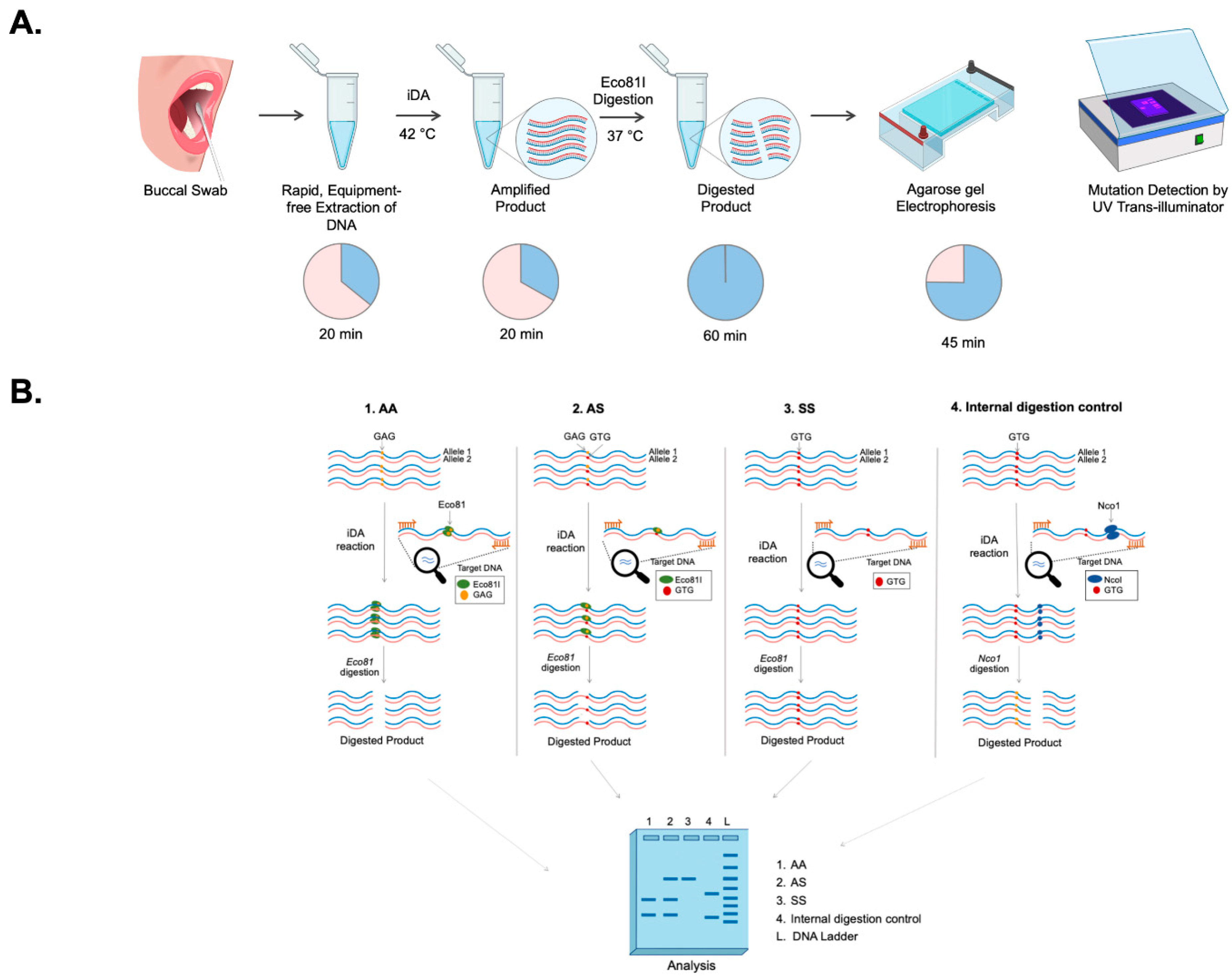
Restrictase-mediated cleavage (iDAR). A simple non-invasive buccal swab specimen has been adopted, which is economical and can be used as a direct sample matrix without the need for a DNA extraction process. Moreover, the assay has been simplified to a one-pot single tube reaction of a complete sample-to-answer workflow which could facilitate the genetic analysis of the clinical sample in less than 150 min. As a proof-of-concept, the robustness of the iDAR assay in the detection of allele-specific SCD genotypes from extraction-free non-invasive crude lysates of buccal swab samples has been shown in patients, carriers, and wild-type samples.

2. Materials and Methods

2.1. Construction of Recombinant HBB Plasmids Carrying HBB Wild-Type (AA), and Sickle Cell Mutation (SS) Sequence

The HBB gene was amplified from blood samples of a healthy individual/wild-type (AA) and SCD patients (SS) using genomic DNA with HBB primers (HBB-F: CTAGGGTTGGCCAATCTACTC and HBB-R: AGTAATGTACTAGGCAGACTGTG). The PCR amplicons were cloned into pJET plasmid (Fermentas, Waltham, MA, USA) following the manufacturer’s instructions. Subsequently, the ligated products were transformed into Escherichia coli DH5α, and colonies were screened using colony PCR. Recombinant plasmids were isolated and purified using Plasmid MiniPrep Kit (Thermo Fisher Scientific, Waltham, MA, USA). Results was further confirmed through Sanger sequencing. The recombinant HBB plasmids were termed pSR-βAA and pSR-βSS, and the concentrations of plasmid DNA were estimated using NanoDrop 2000 (Thermo Fisher Scientific, MA, USA).

2.2. Design of iDA Primers

iDA primers for the target gene HBB were designed as per manufacturer’s recommendations (TwistDx, Cambridge, UK). The primers were designed based on the following parameters: iDA product size between 250 to 300 bp, primer size between 30 to 35 nucleotides, and primer GC content 40–70% using the IDT OligoAnalyser tool. The iDA primers designed were HBB iDA forward primer: GTCAGGGCAGAGCCATCTATTGCTTACATT and HBB iDA reverse primer: ATAGACCAATAGGCAGAGAGAGTCAGTGCC. All the primers used in this study were synthesized from Sigma-Aldrich, Bangalore, India.

2.3. Sensitivity and Specificity Analysis

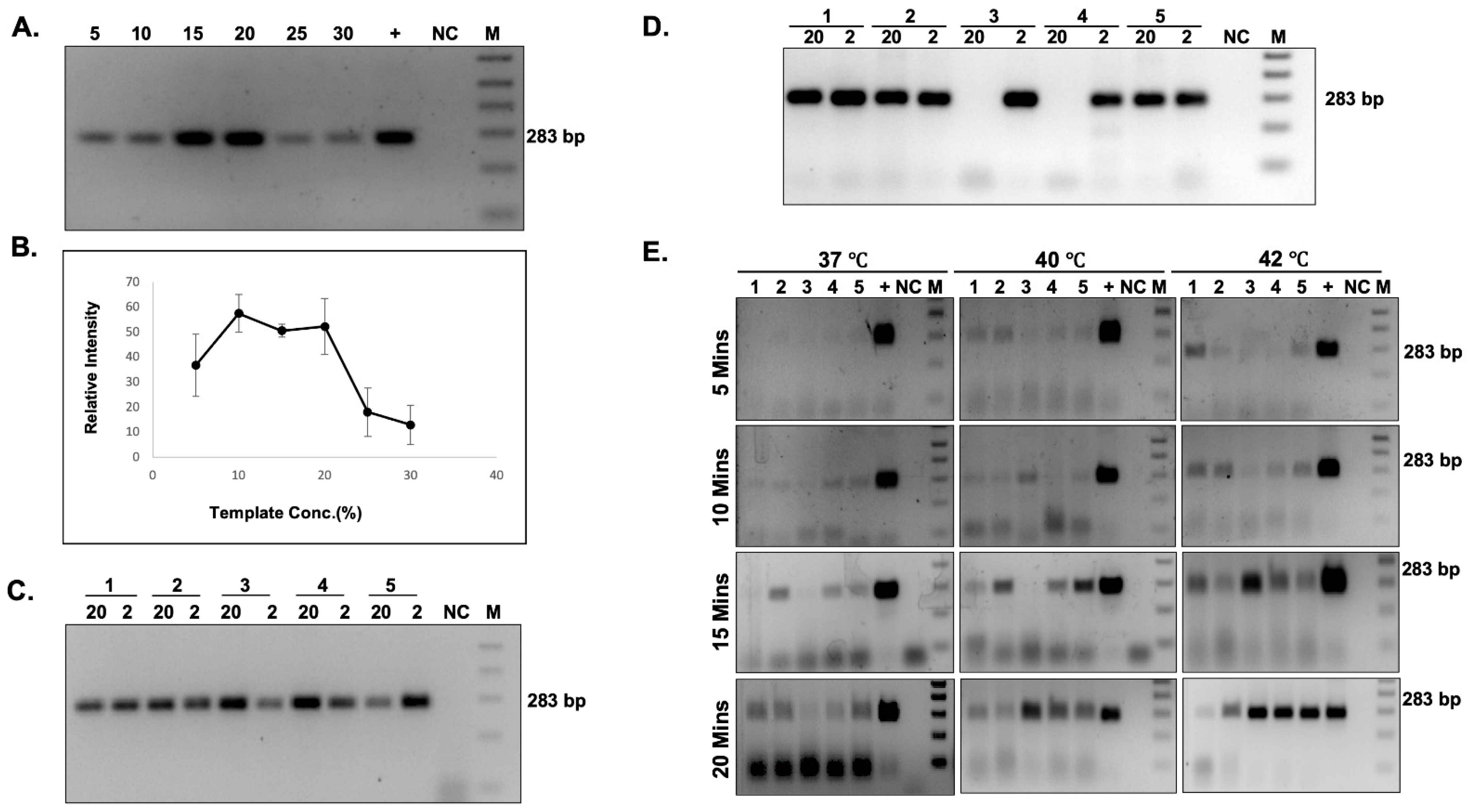
The HBB gene was amplified using HBB-F and HBB-R primer, and the concentration of the amplicon was determined using the Nanodrop 2000 (Thermo Fisher Scientific, MA, USA). The copy number of the amplified product was calculated using the formula: number of copies per µL = ((Amount of ds DNA) × (6.022 × 10²³ molecules per mole))/((length of dsDNA) × 109 ng × (660 g/mole)). The PCR amplified product was serially diluted to obtain the copy number per µL in a range of 108–100. Each dilution was used for iDA. Simultaneously, real time quantitative PCR (qPCR) was also performed for each dilution using the Premix ExTaq Master Mix (Takara Bio Inc. Shiga, Japan), according to the manufacturer’s protocol.

2.4. Buccal Swab Samples Collection

The study was conducted after obtaining approval from the Institutional Ethics Committee at the CSIR-Institute of Genomics and Integrative Biology, New Delhi, and the All India Institute of Medical Sciences (AIIMS, New Delhi, India). Written informed consent was obtained for the study. Participants were requested to provide samples using nylon-flocked buccal swabs (Himedia, Mumbai, India) to swab each inner cheek at least 10–15 times. Buccal swab samples were collected and immediately placed in the lysis buffer.

2.5. Processing of Buccal Swab Specimens in Different Lysis Buffers

Four different lysis buffers, namely, buffer A (custom-made buffer), buffer B [13], buffer C (custom-made buffer) and buffer D [14], were attempted for the direct amplification of clinical buccal swab samples. The details of buffer compositions are given in Table 1 Buccal swab samples collected in lysis buffer A, B, and C were directly used for performing iDA. For lysis buffer D, the samples were vortexed for 10 min and then incubated at 95 °C for 10 min, keeping buccal swabs in the tube itself. Samples were allowed to cool down at room temperature, and the buccal swabs were removed from the tube. Finally, 120 µL 1 M Tris-Cl (pH-8.0) was added. If the iDA was not performed immediately, buccal swab samples collected in respective lysis buffer were stored at 4 °C. The buccal swab samples which were collected and transported as dry swabs were processed in lysis buffer D on arrival and further processed in a similar manner to the processing of fresh buccal swabs.

2.6. iDA Assay Setup and Electrophoresis

iDA reactions were performed using the TwistAmp Basic kit (TwistDx Limited, Cambridge, UK, TABAS03KIT), according to the manufacturer’s instructions. For a 50 µL reaction, 37.5 µL of master mix containing 29.5 µL of rehydration buffer, 2.4 µL of 10 µM HBB iDA forward primer, 2.4 µL of 10 µM HBB iDA reverse primer (all together, called HBB iDA mix); 5–10 ng of recombinant plasmid, or 5–20% of buccal swab lysate, was sequentially added to a reagent pellet and the solution was pipetted up and down 6 to 8 times to mix. Subsequently, 2.4 µL of magnesium acetate (MgOAc) was added to the lid, and the tubes were closed and mixed by brief vortexing. After brief centrifugation, the reaction was immediately initiated by incubation in a heat block, with a constant temperature (either 42 °C or 37 °C) for 20 min. The iDA products were analyzed using 2% agarose gel electrophoresis and documented using a UV transilluminator or gel documentation system.

2.7. Targeted Cleavage of iDA Products

Eco81I-mediated cleavage of iDA products were performed in 30 µL of reaction volume containing 4–8 µL of direct iDA product, 1 × Tango buffer, and 15 U of Eco81I (Thermo Scientific, MA, USA). Alternatively, DdeI enzyme can also be used for iDAR. The reaction was incubated at 37 °C for 1 h under shaking conditions (180 rpm) in an incubator, and the cleaved products were analyzed using 2.5% agarose gel electrophoresis. To ensure the purity of the SCD iDA product, Nco1 enzyme (New England BioLabs, Ipswich, UK) was used as an internal digestion control.

2.8. PCR Amplification and Sanger Sequencing

Clinical buccal swab samples were used for HBB gene amplification using primer HBB-F (seq): GCTGTCATCACTTAGACCTCAC and HBB-R (seq): CATAGACTCACCCTGAAGTTCTCA, which covers SCD mutation using standard PCR with OneTaq DNA polymerase (New England BioLabs, MA, USA)). The PCR products were column purified using a PCR purification kit (Genetix Biotech, Delhi, India) and were subjected to Sanger sequencing. Sequencing was carried out at AgriGenome Labs Pvt. Ltd., Kochi, India

2.9. One-Pot-Reaction

For the one-pot synthesis, 1 reaction of HBB iDA mix was divided into 5 reactions, each containing 8 µL of HBB iDA mix and 5–20% of buccal swab sample. The reaction was activated by addition of 0.8 µL MgOAc and subsequently amplified at 37 °C for 10 min and inactivated at 85 °C for 5 min in the heat block. The digestion premix for the 30 µL reaction was made with 1 × Tango buffer and 15U Eco81I, added to the respective tubes for mutation screening. As an internal control, a digestion premix containing 1 × Cutsmart buffer and 15 U of NcoI was prepared for 30 µL reaction. One-pot products were analyzed using 2.5% agarose gel electrophoresis and documented using a UV transilluminator or gel documentation system.

3. Results

3.1. iDAR Strategy

The experimental strategy used for the development of a simple and efficient allele-specific genetic diagnostic test for detecting SCD mutation in non-invasive buccal swab samples is shown in Figure 1A. Clinical samples were collected in the form of buccal swabs treated with a lysis buffer to release the nucleic acids, followed by isothermal DNA Amplification (iDA). SCD mutation in the HBB gene eliminates the restriction enzyme Eco81I site. The Eco81I restriction enzyme cleaves the amplified product in the sequence CCTNAGG (where N represents any nucleotide). Hence, when thymine (T) is replaced with adenine (A), it alters the target recognition site for Eco81I. There are three possible outcomes of Eco81I cleavage, depending on the genotype of the individual (AA, AS, SS), and the expected cleavage patterns are depicted in Figure 1B. The banding patterns formed by the enzyme cleavage after separation indicate the genotype as follows: (i) In wild-type (AA) healthy individuals with AA, the amplicon is cleaved by Eco81I and produces two bands. (ii) In sickle cell heterozygous (carrier) individuals with AS, no cleavage is expected in the S allele; however, the A allele gets cleaved, resulting in three bands. (iii) In SCD patients with SS, there is no cleavage due to the mutation in both alleles, so a single parent band appears in the gel. In order to ensure the purity of SCD iDA products, an internal control, Nco1 enzyme, was used in the study, which will cleave the iDA product irrespective of its genotype and generate two bands. Therefore, this strategy can differentiate all three possible genotypes through iDA coupled restrictase-mediated cleavage, which is termed iDAR. The genomic structure of the HBB gene and the sequences for iDA primer binding sites are indicated by arrows, as shown in Supplementary Figure S1.

3.2. Optimization of iDA with Different Buccal Swab Lysates

First, iDA based amplification was examined using recombinant plasmid, pSR-βAA, and pSR-βSS, mimicking the genotypes of AA, and SS in 10 µL reactions at 37 °C, 40 °C, and 42 °C. The results demonstrated amplification at all the temperatures, with equal intensity at the expected size (Figure 2A). In order to reduce the processing steps, crude buccal swab lysates were employed for the study. The release of nucleic acid is very important for iDA; hence, the use of an appropriate lysis buffer would be crucial for consistent and efficient iDA using buccal swabs. In this study, previously reported lysis buffers and custom-made lysis buffers were compared for iDA using buccal swabs collected from healthy study participants (AA). All 4 lysates that were used in the study were processed and added to iDA reactions, and the results suggested that iDA performed with Lysis buffer D was consistently the most sensitive (Supplementary Figure S2, Table 1). Hence, for all further experiments and the strategy proposed, the buccal swab samples were processed only in lysis buffer D.

3.3. Limit of Detection

In the present study, the limit of detection was assessed and compared with currently available qRT PCR. A range of 108–100 copies/ul dilution was used to demonstrate the sensitivity of the assay. The results suggested that iDA is capable of amplifying from as low as 10–100 target DNA copies (Figure 2B). In comparison, qRT-PCR, being a more sensitive and sophisticated technique, was able to detect 1–10 DNA copies (Figure 2C–E); however, it is significantly more expensive and requires trained professionals to perform testing. Overall, the above results suggest that iDA is a relatively sensitive technique that can be used as an alternative to the pre-existing sophisticated molecular diagnostic tools in low-resource settings.

3.4. Effect of Temperature, Time, and Concentration of Buccal Swabs Lysates on iDA

To enhance the efficiency of target amplification, different parameters, such as (i) the effect of temperature, (ii) the reaction time, and (iii) the inhibitory effect of the buccal swab lysates on the reaction efficiency were taken into account. First, the inhibitory effect of direct buccal swab lysate on the iDA reaction efficiency was examined in the range from 5% to 30% of buccal swab lysates in the total reaction volume. The results demonstrated successful target amplification at all concentrations of buccal swab lysate (Figure 3A,B). However, beyond a point, the concentration of buccal swab lysate in the total reaction volume inhibited the amplification. Thus, it is suggested that 5–20% of direct buccal swab lysate concentration is optimal for the iDA. We also compared the amplification sensitivity of buccal swab lysate between more sensitive qRT PCR and iDA. A Low (2%) and high (20%) buccal swab lysate concentration of 5 clinical samples with unknown copy numbers were used, and the results demonstrated that iDA worked in all samples at both lysate concentrations (Figure 3C); however, in qRT PCR, few samples at a higher percentage of buccal swab lysate concentrations inhibited the amplification (Figure 3D).
Next, the effect of temperature and time on the amplification efficiency was assessed. The iDA was performed at 37 °C, 40 °C, and 42 °C for 10 min, 15 min, and 20 min each. The results demonstrated target DNA amplification at all three temperatures and reaction times using iDA (Figure 3E, Table 2). However, enhanced amplification of the target DNA was consistently observed at 42 °C, with reaction time of 20 min.
In order to bring down the assay cost, we hypothesized that reducing the iDA per-test reaction volume would lower the assay cost significantly. We investigated reaction volumes ranging from 10 to 50 µL and the appropriate concentration of the buccal swab lysate was added to the reaction and found that amplification was observed in all quantities tested in the above range, without impairing iDA performance (data not shown).

3.5. Successful iDAR Using Clinical Buccal Swab Samples

Detection of the genotype (AA, AS, and SS) analysis is based on the cleavage pattern observed upon Eco81I digestion. Hence, iDA was coupled with restrictase (iDAR) to ultimately deduce the clinical status of the study participants. In this study, first, recombinant plasmids PSR-βAA and pSR-βSS were used to validate iDAR. The results showed complete cleavage in AA and no digestion in SS, as expected (Figure 4A). We then subjected iDA from pSR-βSS plasmid to Nco1 digestion to verify the quality of the iDA product. As expected, Nco1 digestion resulted in complete cleavage (Figure 4A).
Next, to verify the reliability of the optimized iDAR assay, we performed further evaluations using clinical samples. The buccal swab samples were collected with lysis buffer D and subjected to direct iDA amplification at 42 °C for 20 min. Subsequently, 4–8 µL of iDA product was directly subjected to Eco81I digestion at 37 °C for 1 h. However, FastDigest Eco81I requires only 15–30 min for digestion. The major advantage of this iDAR assay is that it provides the qualitative answer in terms of “yes” or “no.” Representative amplification and cleavage from clinical samples of three genotypes, AA, AS, and SS, with two samples each, are shown in Figure 4B. Finally, 15 randomly selected samples from the above clinical pool were further evaluated using Sanger sequencing. Sequencing results corroborated with the iDAR, which proved the accuracy and specificity of our assay. The evaluation of our iDAR assay using 100 clinical samples showed 91.5% sensitivity and 100 % specificity (Table 3). The sensitivity of the assay was increased by modulating the concentration of the buccal swab sample in the total reaction volume. By doing so, we were able to achieve the sensitivity of the assay at almost 100% (data not shown). Storage of the buccal swab lysates at 4 °C for a prolonged period of time before processing did not affect iDA sensitivity. However, it was observed that multiple rounds of processing of the same lysate resulted in reduced iDA sensitivity. Further, a blind iDAR experiment was performed with 15 randomly selected buccal swab lysates from the above pool (Figure 4C). Furthermore, Nco1 digestion was carried out on Eco81I digestion negative samples to conclude the SS genotype (Figure 4D). Genotype deduced based on Eco81I and Nco1 cleavage pattern corroborated with the known genotype of the study participants.
We also evaluated our iDAR assay system with buccal swab samples stored under dry conditions at different temperatures and processed at different time points, as the study aims to provide a confirmatory genetic diagnosis in sickle cell carriers and SCD patients in low- and mid-income countries. It is advantageous, as dry swabs can be collected in non-clinical settings, making the transportation and handling of the samples easy for people living in remote areas, without any requirement for cold storage. In order to assess the effect of dry swabs in prolonged storage, 5 dry swab specimens from each study participant were collected and stored at room temperature for 4 different time points: 12 h, 24 h, 48 h, and 72 h. The iDA with stored dry swab samples produced consistent amplification at all four time points (Supplementary Figure S3).
Next, we checked the impact of storage temperature of buccal swabs samples on iDA sensitivity. This was done, since countries like India experience very high temperatures, which may impact sample integrity. For this evaluation, we collected samples from 5 study participants and stored them at 25 °C, 37 °C, and 42 °C for 5 days. iDA was performed for these samples, and our result suggests successful amplification for all study participants at all of the above temperatures. (Supplementary Figure S4, Table 4).

3.6. Successful Demonstration of One-Pot Reaction

In this study, the genotype analysis of clinical samples using direct buccal swab specimens has been shown. However, independent iDA, followed by restrictase screening separately, may result in cross contamination, if multiple samples are handled together. To avoid any contamination and to ease the workflow, we hypothesized that iDAR could be performed in the same reaction tube (one-pot reaction) (Figure 5A.). Since our previous results had shown successful amplification at 37 °C for both recombinant plasmids and clinical samples (Figure 1A and Figure 3E), we first checked to see if iDAR can be optimized by reducing the reaction volume to 5 μL and the amplification time to 10 min, followed by a 60 min restrictase screening using recombinant plasmids, pSR-βAA and pSR-βSS, at 37 °C in a one-pot reaction. The results showed the expected Eco81I cleavage pattern for AA and SS, with visible intensity. Further, Nco1 internal control digestion also demonstrated the expected cleavage pattern for SS, ensuring the quality of iDA (Figure 5B). We tested the one-pot reaction with clinical buccal swab lysates of known genotype following the above experimental strategy. Initially, 5 μL of HBB iDA mix was used with clinical buccal swab lysates; however, the results showed an inconclusive digestion pattern due to less intense target DNA amplification (data not shown). Hence, one-pot reaction was tested with 10 μL of HBB iDA mix that resulted in the expected Eco81I cleavage pattern, with visible intensity, followed by NcoI digestion of SCD clinical samples (Figure 5C), showing the effectiveness of one-pot synthesis iDAR for the diagnosis of clinical samples.

4. Discussion

Sickle cell disease has emerged as a significant global health burden and to combat it, early diagnosis using genetic confirmation is vital. One way of doing it this is by enabling simple and accurate diagnostic screening in a limited resource setting, leading to early diagnosis, expeditious management in affected newborns, as well as aid in the identification of carriers and subsequent genetic counseling [15].
Currently, in order to make a conclusive diagnosis, there are a series of tests that are performed, including a full blood panel, followed by HPLC and further confirmation of the genetic mutation through Sanger sequencing. Although the latter ensures an accurate diagnosis, it is not quick nor easily deployable, requiring specialized instruments and trained professionals. Hence, developing simple and efficient strategies of diagnosis is crucial.
SCD screening can also be achieved by several point-of-care tests, such as assays based on hemoglobin solubility [16] and monoclonal antibody-based detection of hemoglobin AA or SS [17], which are becoming accessible, reducing the challenges regarding necessary infrastructure and costs. However, any assay based on monoclonal antibodies, such as HemoTypeSC [17], that identifies hemoglobin proteins cannot be utilized in patients who have been transfused recently, since blood transfusions contain globin proteins from the donor, and this leads to misinterpretation of the results [4]. Solubility testing cannot differentiate between SCD and a carrier; hence, it is not recommended due to these limitations, and it is less ideal for SCD screening [16].
In recent years, lateral flow assays (LFAs) have been broadly used as portable molecular diagnostic methods. In 2015, Kanter et al. developed a sickle SCAN scan based on lateral flow immunoassay to detect SCD [8]. However, the assay has several limitations, such as misinterpretation due to visual reading, polyclonal antibody cross-reactivity, inconsistency in the band’s intensity, and false-positive results in identifying HbAS with HbSS. Hence, the authors suggested further confirmatory validation. Another study by Natoli et al. reported a strategy for allele-specific detection of the sickle cell mutation using blood samples [18]. In their assay, two separate reactions for each sample are required to detect AA and SS genotypes, which is the major limitation of the above study.
Isothermal DNA amplification-based molecular diagnosis is an emerging strategy that has been successfully employed for the screening of infectious diseases such as Ebola virus disease (EVD) and COVID-19 [19,20]. Current studies focus on developing an isothermal DNA amplification-based strategy for the screening of SCD. There are, however, certain challenges that had to be addressed in order to develop this assay, such as the nature of sample collected and the quickest way to process the samples without compromising the efficiency and accuracy of the test.
Our iDAR assay system is very simple and easily deployable in a resource limited setting, as it only requires a heat block and a tube prefilled with a mixture containing the essential reagents for amplification and subsequently treated with restrictase in the same tube for screening. Notably, the addition of direct buccal swab samples does not significantly affect the sensitivity of the iDA. It was noted that the sensitivity of iDA was compromised when more than 20% of the buccal swab lysates make up the reaction volume. The present study has demonstrated the sensitivity of iDA to be 91.5%; however, the sensitivity of the assay can be increased by changing the concentration of buccal swab lysate in the reaction volume. Moreover, we were able to achieve 100% amplification by modulating the buccal swab lysate concentration. In addition, we also observed that iDA assay detected the genotype correctly, even in patients who had undergone blood transfusion within a few weeks before sample collection. Hence, the current study has shown 100% specificity in discriminating AA, AS, and SS genotypes. Moreover, the chances of false positive results are highly unlikely using the iDAR assay system, since the enzyme site is lost in SS genotype. It is noteworthy that if digestion is not kept under recommended conditions, the AA genotype can be falsely diagnosed as the AS genotype.
In this study, a single test reaction is required to differentiate all the three genotypes, the SS, AS and AA alleles, which brings down the assay cost. By leveraging Eco81I-mediated sequence-specific cleavage, our iDAR assay system shows a more consistent and precise outcome for heterozygous (AS) and homozygous (SS) samples. The iDAR assay offers an alternative to DNA sequencing or real-time PCR for rapid SCD genotyping, while retaining the advantages of simplicity, affordability, and a faster qualitative readout.
Traditionally, blood samples have been used for clinical diagnostics and genetic studies. However, blood collection is an invasive procedure, and is both expensive and requires trained professionals. Even though blood is the preferred sample for running most of the molecular tests, using it for DNA amplification is not suitable, for multiple reasons [21,22]. The nucleic acid isolated from blood or blood containing samples is usually low; therefore, the sensitivity of isothermal amplification is reduced. A more suitable and less invasive alternative to blood is taking saliva for use in molecular diagnosis. However, certain study participants have difficulty in spitting into a tube, while others struggle to produce enough saliva for genetic testing [23] Dry mouth is a typical medication side effect that can make saliva collection difficult [23]. However, buccal swab sample collection is quick, non-invasive, and inexpensive. Taking all of this into account, this study sought to explore the feasibility of performing SCD genotyping. This type of testing also makes it easy for patients and their family members to collect samples at clinics or at their homes. Biological samples, such as stool, serum, plasma, vaginal, and nasal, have been used for isothermal amplification [24]; however, this is the first report where buccal swab specimens have directly been used for iDA, especially in the molecular diagnosis of a genetic disease.
Developing a molecular diagnostic test that does not require DNA isolation is difficult, since existing genotyping strategies based on DNA amplification necessitate relatively high DNA purity [25,26]. Hence, developing a genetic test that analyzes the samples without DNA isolation and purification has remained elusive. The current study aimed to establish an inexpensive, low-complexity buffer system with routine laboratory chemicals that make direct iDA feasible by utilizing a simple procedure that allows for the efficient release of DNA from buccal swab specimens.
Previously, to aid the release of nucleic acids from human cells, various procedures have been applied, such as using complex solvents and expensive unique materials [27,28]. In the iDAR assay system, we leveraged NaOH to lyse the cell membrane for releasing DNA and Tris-Cl as a crucial pH control agent, providing an appropriate environment for iDA reaction. Thus, the iDAR assay system proposed in this study is capable of genotyping target genes without undertaking the laborious, time-consuming and expensive nucleic acid isolation and purification step, avoiding the possibility of cross contamination.
To date, no information is available on the stability of DNA in “dry swab” for storage and transport at various temperatures and its impact, specifically on the iDA for genetic testing. The storage of biological samples is a major challenge during sample collection at a remote place, as sample integrity is a factor of great importance. Ideally, biological samples must be kept in temperature-controlled, adequately qualified storage [29], and processed as soon as possible after collection. However, in a realistic scenario, the vast majority are stored and transported some distances before processing. In this study, the viability of dry swabs without any transport reagent was tested for storage and transport at various incubation times and temperatures. It was observed that dry swabs can be transported at a maximum of 42 °C and held for up to 72 h without affecting iDA. These findings are significant for low- and middle-income nations that have limited access to rapid refrigerated transport and storage of samples, providing a cost-effective option. Finally, this study reveals the practical and financial benefits of employing dry swabs without a transportation reagent.
In the iDAR assay system, reagents account for the majority of the overall per-test cost. Thus, it was hypothesized that lowering the per-test reaction volume can significantly reduce the assay’s cost. Thus, different reaction volumes were experimented with, and it was observed that amplification using direct buccal swab specimens as templates in all volumes worked efficiently without compromising assay sensitivity and performance. Hence, this brings down the amplification cost by one-fifth and also significantly lowers the overall assay cost to USD 5.
However, we have not shown whether the iDAR assay system can discriminate among AA, hemoglobin type C genotypes AC, CC (HbC), and SC (compound heterozygous for HbC and HbS). Principally, the iDAR assay tool can discriminate between HbC genotypes if the Hpy188III enzyme is used instead of Eco81I. Hpy188III recognises TCNNGA in the AA genotype, whereas in HbC, glutamate (GAG) is changed to lysine (AAG) in the sixth codon of HBB gene. This results in the loss of enzyme recognition site (TCNNAA), and hence, no digestion would be expected with Hpy188II in the CC genotype. Therefore, if the digestion pattern of a study subject shows heterozygosity with both Eco81I and Hpy188III restriction enzymes, it suggests the SC genotype.
One of the limitation of the iDAR assay tool is the differentiation of SS from Sβ thalassemia, which is recognized as the heterozygous AS genotype. Since β thalassemia is caused due to more than 200 mutations and these mutations are spread across the HBB gene, so it becomes difficult to integrate β thalassemia mutations in the iDAR assay.
In summary, the iDAR assay platform has considerable advantages. The results show that iDAR is an efficient assay capable of detecting all three genotypes, SS, AS, and AA. Existing genetic diagnostic tests based on PCR, such as DNA sequencing and qPCR, typically require a complicated operational procedure, as well as expensive and sophisticated instruments that may not be available in many laboratories, whereas the iDAR assay can be done with less complex instruments and provides reliable, inexpensive, easy-to-use, and affordable genetic diagnostics for SCD, facilitating the expansion of SCD screening in low-resource settings.

Supplementary Materials

The following are available online at https://www.mdpi.com/article/10.3390/diagnostics12071765/s1, Figure S1: HBB gene locus showing iDA primer positions; Figure S2: Comparison of iDA sensitivity in different lysis buffers; Figure S3: Comparative analysis of dry buccal swabs processed at different time points; Figure S4: Effects on iDA sensitivity after long-term incubation of buccal swabs at variable temperatures.

Author Contributions

Conceptualization, S.R., P.T. and P.G. (Pragya Gupta); methodology, S.R., P.T. and P.G. (Pragya Gupta); validation, S.R., P.T., P.G. (Pragya Gupta), N.B., R.S., N.V.G., S.G.G. and V.S.; formal analysis, S.R., P.T. and P.G. (Pragya Gupta); investigation, S.R., P.T., P.G. (Pragya Gupta), N.B., R.S., N.V.G., S.G.G. and V.S.; resources, G.K., P.G. (Padma Gunda), S.J., J.D. and M.A.; data curation, P.T. and P.G. (Pragya Gupta); writing—original draft preparation, S.R., P.T., P.G. (Pragya Gupta), N.B. and S.G.G.; writing—review and editing, S.R., P.T., P.G. (Pragya Gupta), N.B., S.G.G. and M.A.; supervision, S.R. and M.A.; project administration, S.R.; funding acquisition, S.R. All authors have read and agreed to the published version of the manuscript.

Funding

This research was funded by the Council for Scientific and Industrial Research (CSIR), grants (MLP2010 and HCP023), the Department of Biotechnology, grant (BT/GET/119/SP31652/2020), and the Government of India (GoI).

Institutional Review Board Statement

The study was conducted according to the guidelines and approval of the Institutional Review Board of the All India Institute of Medical Sciences (IEC-40/14.01.2022 and 17.01.2022).

Informed Consent Statement

Informed consent was obtained from all subjects involved in the study.

Acknowledgments

IPT, PG, SG, and NB are recipients of the Senior Research Fellowship from the Department of Biotechnology, Council of Scientific and Industrial Research, University Grant Commission, and the Indian Council for Medical Research, GoI, respectively.

Conflicts of Interest

The authors declare no conflict of interest.

References

  1. Piel, F.; Hay, S.; Gupta, S.; Weatherall, D.; Williams, T. Global Burden of Sickle Cell Anaemia in Children under Five, 2010–2050: Modelling Based on Demographics, Excess Mortality, and Interventions. PLoS Med. 2013, 10, e1001484. [Google Scholar] [CrossRef] [PubMed] [Green Version]
  2. World Health Organization. Management of Birth Defects and Haemoglobin Disorders: Report of a Joint Who-March Of Dimes Meeting Geneva, Switzerland, 17–19 May 2006; World Health Organization: Geneva, Switzerland, 2006. [Google Scholar]
  3. McGann, P. Time to Invest in Sickle Cell Anemia as a Global Health Priority. Pediatrics 2016, 137, e20160348. [Google Scholar] [CrossRef] [PubMed] [Green Version]
  4. Arishi, W.; Alhadrami, H.; Zourob, M. Techniques for the Detection of Sickle Cell Disease: A Review. Micromachines 2021, 12, 519. [Google Scholar] [CrossRef] [PubMed]
  5. Nair, S. Potential Pithfalls in Using HPLC and its Interpretation in Diagnosing HbS. J. Rare Dis. Res. Treat. 2018, 3, 9–12. [Google Scholar] [CrossRef]
  6. Whyte, R.; Jefferies, A. Red blood cell transfusion in newborn infants. Paediatr. Child Health 2014, 19, 213–217. [Google Scholar] [CrossRef] [Green Version]
  7. Reed, W.; Lane, P.; Lorey, F.; Bojanowski, J.; Glass, M.; Louie, R.R.; Lubin, B.H.; Vichinsky, E.P. Sickle-cell disease not identified by newborn screening because of prior transfusion. J. Pediatr. 2000, 136, 248–250. [Google Scholar] [CrossRef]
  8. Kanter, J.; Telen, M.; Hoppe, C.; Roberts, C.; Kim, J.; Yang, X. Validation of a novel point of care testing device for sickle cell disease. BMC Med. 2015, 13, 225. [Google Scholar] [CrossRef] [Green Version]
  9. Lenicek Krleza, J.; Dorotic, A.; Grzunov, A.; Maradin, M. Capillary blood sampling: National recommendations on behalf of the Croatian Society of Medical Biochemistry and Laboratory Medicine. Biochem. Med. (Zagreb) 2015, 25, 335–358. [Google Scholar] [CrossRef]
  10. Ngo, H.; Gandra, N.; Fales, A.; Taylor, S.; Vo-Dinh, T. Sensitive DNA detection and SNP discrimination using ultrabright SERS nanorattles and magnetic beads for malaria diagnostics. Biosens. Bioelectron. 2016, 81, 8–14. [Google Scholar] [CrossRef] [Green Version]
  11. Chen, Y.; Sun, J.; Xianyu, Y.; Yin, B.; Niu, Y.; Wang, S.; Cao, F.; Zhang, X.; Wang, Y.; Jiang, X. A dual-readout chemiluminescent-gold lateral flow test for multiplex and ultrasensitive detection of disease biomarkers in real samples. Nanoscale 2016, 8, 15205–15212. [Google Scholar] [CrossRef]
  12. Cavanaugh, S.; Bathrick, A. Direct PCR amplification of forensic touch and other challenging DNA samples: A review. Forensic Sci. Int. Genet. 2018, 32, 40–49. [Google Scholar] [CrossRef] [PubMed]
  13. Lever, M.; Torti, A.; Eickenbusch, P.; Michaud, A.; Šantl-Temkiv, T.; Jørgensen, B. A modular method for the extraction of DNA and RNA, and the separation of DNA pools from diverse environmental sample types. Front. Microbiol. 2015, 6, 476. [Google Scholar] [CrossRef] [PubMed] [Green Version]
  14. Walker, A.; Najarian, D.; White, D.; Jaffe, J.; Kanetsky, P.; Rebbeck, T. Collection of genomic DNA by buccal swabs for polymerase chain reaction-based biomarker assays. Environ. Health Perspect. 1999, 107, 517–520. [Google Scholar] [CrossRef] [PubMed]
  15. Pecker, L.; Naik, R. The current state of sickle cell trait: Implications for reproductive and genetic counseling. Blood 2018, 132, 2331–2338. [Google Scholar] [CrossRef] [Green Version]
  16. Keren, D.; Hedstrom, D.; Gulbranson, R.; Ou, C.; Bak, R. Comparison of Sebia Capillarys Capillary Electrophoresis With the Primus High-Pressure Liquid Chromatography in the Evaluation of Hemoglobinopathies. Am. J. Clin. Pathol. 2008, 130, 824–831. [Google Scholar] [CrossRef] [Green Version]
  17. Quinn, C. Minireview: Clinical severity in sickle cell disease: The challenges of definition and prognostication. Exp. Biol. Med. 2016, 241, 679–688. [Google Scholar] [CrossRef]
  18. Natoli, M.; Chang, M.; Kundrod, K.; Coole, J.B.; Airewele, G.E.; Tubman, V.N.; Richards-Kortum, R.R. Allele-Specific Recombinase Polymerase Amplification to Detect Sickle Cell Disease in Low-Resource Settings. Anal. Chem. 2021, 93, 4832–4840. [Google Scholar] [CrossRef]
  19. Euler, M.; Wang, Y.; Heidenreich, D.; Patel, P.; Strohmeier, O.; Hakenberg, S.; Niedrig, M.; Hufert, F.T.; Weidmann, M. Development of a Panel of Recombinase Polymerase Amplification Assays for Detection of Biothreat Agents. J. Clin. Microbiol. 2013, 51, 1110–1117. [Google Scholar] [CrossRef] [Green Version]
  20. Tang, Y.; Wang, Y.; Li, Y.; Zhao, H.; Zhang, S.; Zhang, Y.; Li, J.; Chen, Y.; Wu, X.; Qin, C.; et al. An integrated rapid nucleic acid detection assay based on recombinant polymerase amplification for SARS-CoV-2. Virol. Sin. 2022, 37, 138–141. [Google Scholar] [CrossRef]
  21. Javaid, M.; Ahmed, A.; Durand, R.; Tran, S. Saliva as a diagnostic tool for oral and systemic diseases. J. Oral Biol. Craniofac. Res. 2016, 6, 67–76. [Google Scholar] [CrossRef] [Green Version]
  22. Rohrman, B.; Richards-Kortum, R. Inhibition of Recombinase Polymerase Amplification by Background DNA: A Lateral Flow-Based Method for Enriching Target DNA. Anal. Chem. 2015, 87, 1963–1967. [Google Scholar] [CrossRef] [PubMed]
  23. Ang, J.; Aloise, M.; Dawes, D.; Dempster, M.G.; Fraser, R.; Paterson, A.; Stanley, P.; Suarez-Gonzalez, A.; Dawes, M.; Katzov-Eckert, H. Evaluation of buccal swabs for pharmacogenetics. BMC Res. Notes 2018, 11, 382. [Google Scholar] [CrossRef] [PubMed]
  24. Daher, R.; Stewart, G.; Boissinot, M.; Bergeron, M. Recombinase Polymerase Amplification for Diagnostic Applications. Clin. Chem. 2016, 62, 947–958. [Google Scholar] [CrossRef] [PubMed]
  25. Nanayakkara, I.; Cao, W.; White, I. Simplifying Nucleic Acid Amplification from Whole Blood with Direct Polymerase Chain Reaction on Chitosan Microparticles. Anal. Chem. 2017, 89, 3773–3779. [Google Scholar] [CrossRef]
  26. Ambers, A.; Wiley, R.; Novroski, N.; Budowle, B. Direct PCR amplification of DNA from human bloodstains, saliva, and touch samples collected with microFLOQ ® swabs. Forensic Sci. Int. Genet. 2018, 32, 80–87. [Google Scholar] [CrossRef] [Green Version]
  27. Saavedra-Matiz, C.; Isabelle, J.; Biski, C.; Duva, S.J.; Sweeney, M.L.; Parker, A.L.; Young, A.J.; DiAntonio, L.L.; Krein, L.M.; Nichols, M.J.; et al. Cost-Effective and Scalable DNA Extraction Method from Dried Blood Spots. Clin. Chem. 2013, 59, 1045–1051. [Google Scholar] [CrossRef] [Green Version]
  28. Gong, R.; Li, S. Extraction of human genomic DNA from whole blood using a magnetic microsphere method. Int. J. Nanomed. 2014, 9, 3781–3789. [Google Scholar] [CrossRef] [Green Version]
  29. Szczepek, A.; Frejo, L.; Vona, B.; Trpchevska, N.; Cederroth, C.R.; Caria, H.; Lopez-Escamez, J.A. Recommendations on Collecting and Storing Samples for Genetic Studies in Hearing and Tinnitus Research. Ear Hear. 2019, 40, 219–226. [Google Scholar] [CrossRef] [Green Version]
Figure 1. Experimental design of iDAR assay. (A) Schematics showing the workflow of iDAR. (B) Schematic showing the screening strategy for allele-specific sickle cell mutation detection using iDAR.
Diagnostics 12 01765 g001
Figure 2. Optimization of iDA using recombinant plasmids. (A) iDA of recombinant plasmids pSR-βAA (WT) and βSS (MT) at different temperatures. (B) Minimum copy number required for iDA. (CE) qRT-PCR for n = 3.
Diagnostics 12 01765 g002
Figure 3. Effect of temperature, time, and concentration of buccal swab lysates on iDA. (A) iDA with varying buccal swab lysate concentrations (%) and (B) relative quantification = 3. Amplification sensitivity with high (20%) and low (2%) buccal swab lysate concentration in (C) iDA and (D) qRT-PCR; n = 5. (E) Sensitivity of iDA at different temperatures and timepoints n = 5.
Diagnostics 12 01765 g003
Figure 4. iDAR using recombinant plasmids and patient buccal swabs: (A) iDAR using Eco81I (E) digestion of recombinant plasmids pSR-βAA and pSR-βSS with internal digestion control of NcoI (N). (B) Eco81I screening in individuals with known genotypes AA, ASS, and SSS with internal digestion control of NcoI (N) for n = 2. (C) iDAR of buccal swabs of unknown genotype; n = 15. (D) iDAR with internal control digestion with Nco1(N) of SS samples.
Diagnostics 12 01765 g004
Figure 5. One-pot iDAR. (A) Schematics showing the steps in one-pot reaction. (B) One-pot iDAR using recombinant plasmids pSR-βAA (AA) and SS (SS). (C) One-pot iDAR using Eco81I (E) screening in individuals with known clinical genotypes (AA, AS, and SS) with internal digestion control of NcoI (N) for n = 2.
Diagnostics 12 01765 g005
Table 1. A comparative analysis of different lysis buffers for iDA.
Lysis BufferLysis Buffer CompositioniDA TemperatureiDA SensitivityReference
Buffer A125 mM NaOH
1× Triton-X100
1 mg/mL Proteinase K
NFW
37 °C

42 °C
0/3

0/3
Custom made
Buffer B1% Triton-X100
NFW
37 °C
42 °C
3/3
1/3
[12]
Buffer C0.2 M NaOH
NFW
37 °C
42 °C
2/3
0/3
Custom made
Buffer D50 mM NaOH
1 M Tris-Cl pH8.0
37 °C
42 °C
3/3
3/3
[13]
Table 2. Effect of amplification time and temperature on iDA.
iDA TemperatureiDA Amplification TimeiDA Sensitivity
37 °C5 min0/5
10 min0/5
15 min3/5
20 min4/5
40 °C5 min0/5
10 min1/5
15 min4/5
20 min5/5
42 °C5 min1/5
10 min4/5
15 min5/5
20 min5/5
Table 3. Sensitivity and specificity of the iDAR assay.
GenotypeSensitivity of iDASpecificity of iDAR
Wild-Type (AA)98/10045/45
Sickle cell trait (AS)64/7133/33
Sickle cell disease (SS)43/5322/22
Total205/224100/100
Table 4. Effect of long-term incubation of buccal swabs at variable temperatures on iDA sensitivity.
Storage TemperatureNumber of Days of StorageiDA Sensitivity
25 °C55/5
37 °C55/5
42 °C55/5
Publisher’s Note: MDPI stays neutral with regard to jurisdictional claims in published maps and institutional affiliations.

