
LymphoTrack Is Equally Sensitive as PCR GeneScan and Sanger Sequencing for Detection of Clonal Rearrangements in ALL Patients

 and
and
Abstract
:1. Introduction
2. Materials and Methods
2.1. Sample Preparation
2.2. PCR Amplification and GeneScan Analysis
2.3. Next Generation Sequencing
2.4. Comparison between GeneScan and Sanger Sequencing with LymphoTrack
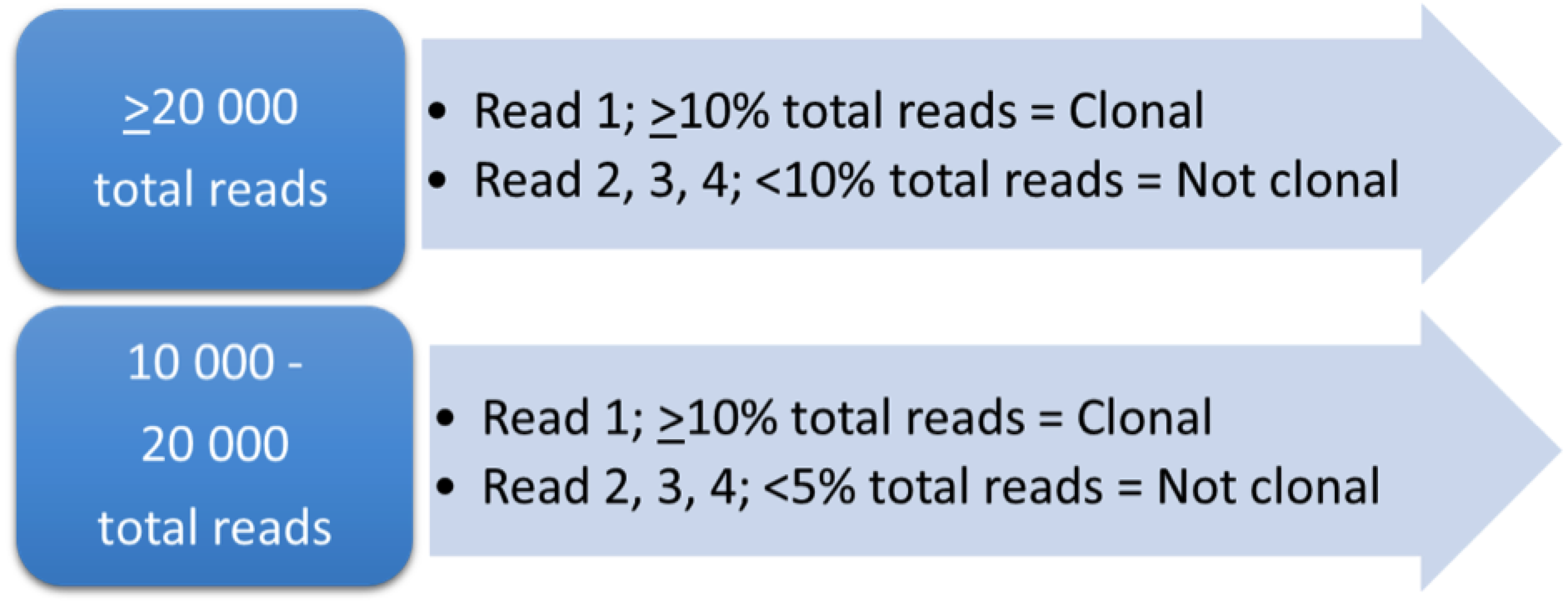
2.5. Comparison between RFU and %Total Reads
3. Results
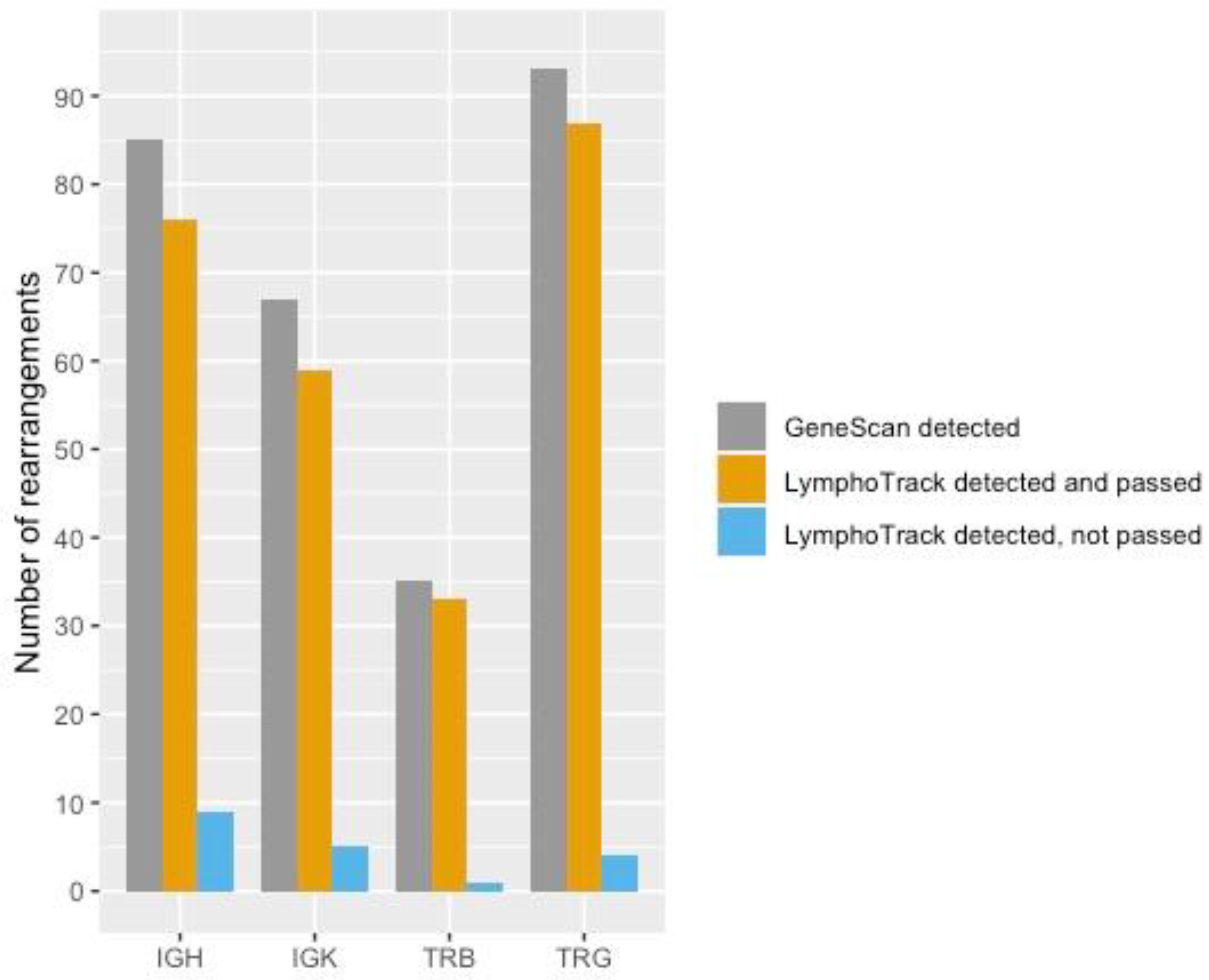
3.1. IGH
3.2. IGK
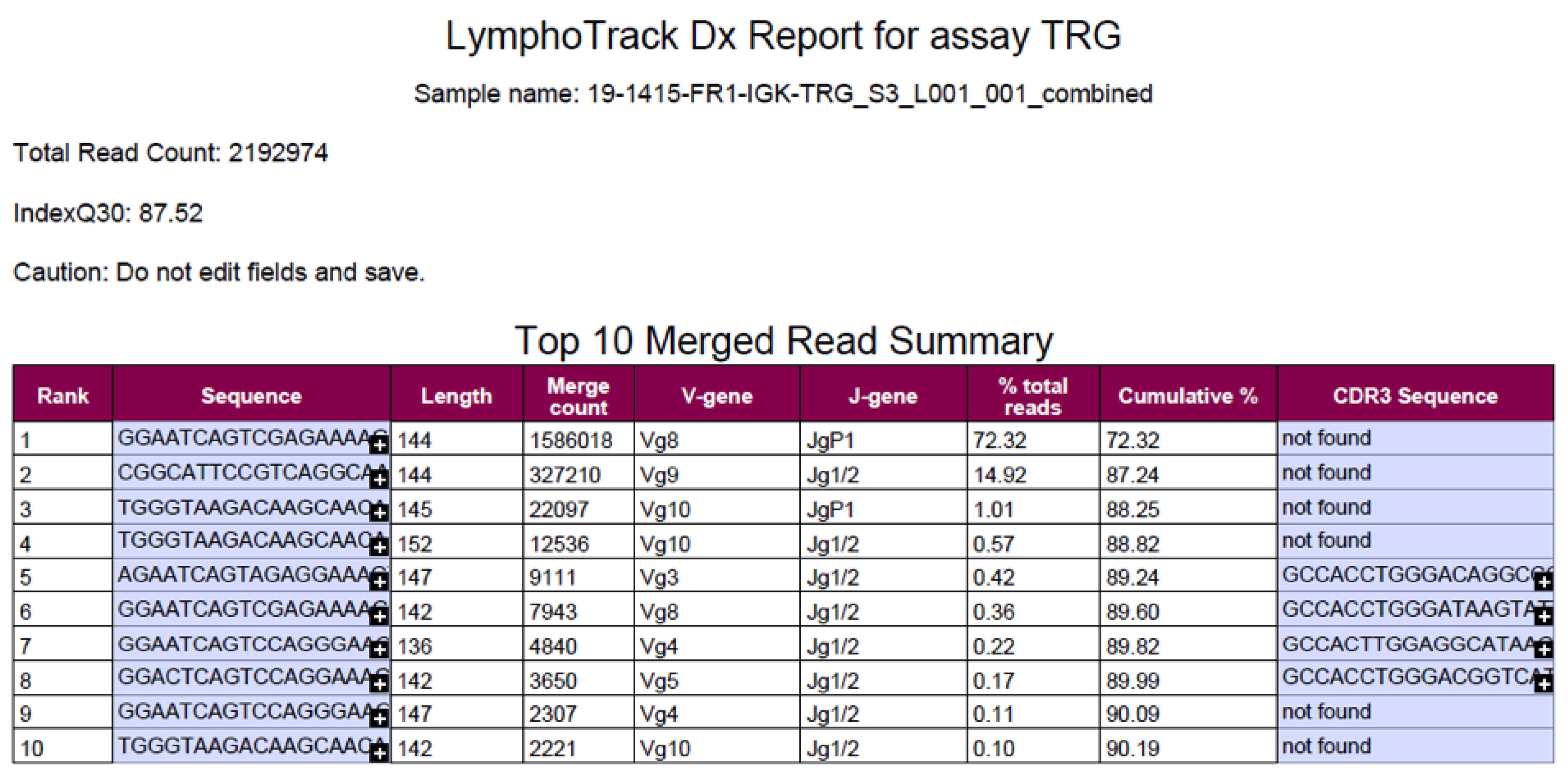
3.3. TRG
3.4. TRB
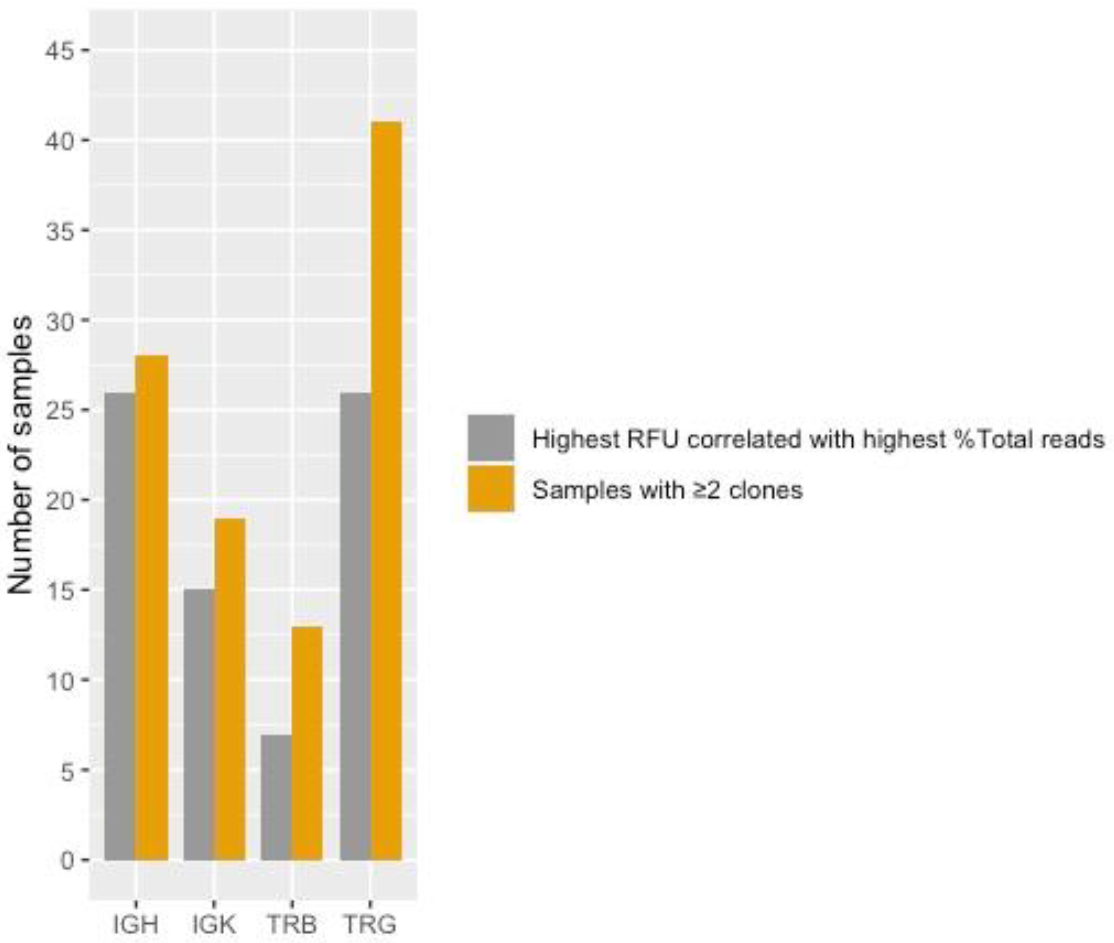
3.5. Correlation between RFU and %Total Reads in Samples with Two or More Rearrangements
3.6. Ambiguous Rearrangements
4. Discussion
4.1. Benefits of LymphoTrack for MRD Screening in ALL
4.2. Considerations When Implementing LymphoTrack in MRD Screening in ALL
4.3. The LymphoTrack Software
4.4. Polyclonal Patterns
4.5. Minor Peaks with Background
4.6. Correlation between RFU and %Total Reads in Samples with Two or More Rearrangements
5. Conclusions
Supplementary Materials
Author Contributions
Funding
Institutional Review Board Statement
Informed Consent Statement
Data Availability Statement
Acknowledgments
Conflicts of Interest
References
- Cave, H.; van der Werff ten Bosch, J.; Suciu, S.; Guidal, C.; Waterkeyn, C.; Otten, J.; Bakkus, M.; Thielemans, K.; Grandchamp, B.; Vilmer, E.; et al. Clinical significance of minimal residual disease in childhood acute lymphoblastic leukemia. N. Engl. J. Med. 1998, 339, 591–598. [Google Scholar] [CrossRef] [PubMed]
- Coustan-Smith, E.; Behm, F.G.; Sanchez, J.; Boyett, J.M.; Hancock, M.L.; Raimondi, S.C.; Rubnitz, J.E.; Rivera, G.K.; Sandlund, J.T.; Pui, C.H.; et al. Immunological detection of minimal residual disease in children with acute lymphoblastic leukaemia. Lancet 1998, 351, 550–554. [Google Scholar] [CrossRef]
- Van Dongen, J.J.; Seriu, T.; Panzer-Grumayer, E.R.; Biondi, A.; Pongers-Willemse, M.J.; Corral, L.; Stolz, F.; Schrappe, M.; Masera, G.; Kamps, W.A.; et al. Prognostic value of minimal residual disease in acute lymphoblastic leukaemia in childhood. Lancet 1998, 352, 1731–1738. [Google Scholar] [CrossRef]
- Biondi, A.; Valsecchi, M.G.; Seriu, T.; D’Aniello, E.; Willemse, M.J.; Fasching, K.; Pannunzio, A.; Gadner, H.; Schrappe, M.; Kamps, W.A.; et al. Molecular detection of minimal residual disease is a strong predictive factor of relapse in childhood B-lineage acute lymphoblastic leukemia with medium risk features. A case control study of the International BFM study group. Leukemia 2000, 14, 1939–1943. [Google Scholar] [CrossRef] [Green Version]
- Zhou, J.; Goldwasser, M.A.; Li, A.; Dahlberg, S.E.; Neuberg, D.; Wang, H.; Dalton, V.; McBride, K.D.; Sallan, S.E.; Silverman, L.B.; et al. Quantitative analysis of minimal residual disease predicts relapse in children with B-lineage acute lymphoblastic leukemia in DFCI ALL Consortium Protocol 95-01. Blood 2007, 110, 1607–1611. [Google Scholar] [CrossRef] [Green Version]
- Borowitz, M.J.; Devidas, M.; Hunger, S.P.; Bowman, W.P.; Carroll, A.J.; Carroll, W.L.; Linda, S.; Martin, P.L.; Pullen, D.J.; Viswanatha, D.; et al. Clinical significance of minimal residual disease in childhood acute lymphoblastic leukemia and its relationship to other prognostic factors: A Children’s Oncology Group study. Blood 2008, 111, 5477–5485. [Google Scholar] [CrossRef] [PubMed] [Green Version]
- Tonegawa, S. Somatic generation of antibody diversity. Nature 1983, 302, 575–581. [Google Scholar] [CrossRef]
- Davis, M.M.; Bjorkman, P.J. T-cell antigen receptor genes and T-cell recognition. Nature 1988, 334, 395–402. [Google Scholar] [CrossRef]
- Van der Velden, V.H.; Hochhaus, A.; Cazzaniga, G.; Szczepanski, T.; Gabert, J.; van Dongen, J.J. Detection of minimal residual disease in hematologic malignancies by real-time quantitative PCR: Principles, approaches, and laboratory aspects. Leukemia 2003, 17, 1013–1034. [Google Scholar] [CrossRef] [PubMed] [Green Version]
- Gazzola, A.; Mannu, C.; Rossi, M.; Laginestra, M.A.; Sapienza, M.R.; Fuligni, F.; Etebari, M.; Melle, F.; Sabattini, E.; Agostinelli, C.; et al. The evolution of clonality testing in the diagnosis and monitoring of hematological malignancies. Ther. Adv. Hematol. 2014, 5, 35–47. [Google Scholar] [CrossRef]
- Germano, G.; del Giudice, L.; Palatron, S.; Giarin, E.; Cazzaniga, G.; Biondi, A.; Basso, G. Clonality profile in relapsed precursor-B-ALL children by GeneScan and sequencing analyses. Consequences on minimal residual disease monitoring. Leukemia 2003, 17, 1573–1582. [Google Scholar] [CrossRef] [PubMed] [Green Version]
- Metzker, M.L. Emerging technologies in DNA sequencing. Genome Res. 2005, 15, 1767–1776. [Google Scholar] [CrossRef] [PubMed] [Green Version]
- Hu, T.; Chitnis, N.; Monos, D.; Dinh, A. Next-generation sequencing technologies: An overview. Hum. Immunol. 2021, 82, 801–811. [Google Scholar] [CrossRef]
- Lay, L.; Stroup, B.; Payton, J.E. Validation and interpretation of IGH and TCR clonality testing by Ion Torrent S5 NGS for diagnosis and disease monitoring in B and T cell cancers. Pract. Lab. Med. 2020, 22, e00191. [Google Scholar] [CrossRef] [PubMed]
- Ho, C.C.; Tung, J.K.; Zehnder, J.L.; Zhang, B.M. Validation of a Next-Generation Sequencing-Based T-Cell Receptor Gamma Gene Rearrangement Diagnostic Assay: Transitioning from Capillary Electrophoresis to Next-Generation Sequencing. J. Mol. Diagn. 2021, 23, 805–815. [Google Scholar] [CrossRef]
- Arcila, M.E.; Yu, W.; Syed, M.; Kim, H.; Maciag, L.; Yao, J.; Ho, C.; Petrova, K.; Moung, C.; Salazar, P.; et al. Establishment of Immunoglobulin Heavy (IGH) Chain Clonality Testing by Next-Generation Sequencing for Routine Characterization of B-Cell and Plasma Cell Neoplasms. J. Mol. Diagn. 2019, 21, 330–342. [Google Scholar] [CrossRef] [PubMed] [Green Version]
- Van Dongen, J.J.; Langerak, A.W.; Bruggemann, M.; Evans, P.A.; Hummel, M.; Lavender, F.L.; Delabesse, E.; Davi, F.; Schuuring, E.; Garcia-Sanz, R.; et al. Design and standardization of PCR primers and protocols for detection of clonal immunoglobulin and T-cell receptor gene recombinations in suspect lymphoproliferations: Report of the BIOMED-2 Concerted Action BMH4-CT98-3936. Leukemia 2003, 17, 2257–2317. [Google Scholar] [CrossRef] [Green Version]
- Boone, E.; Heezen, K.C.; Groenen, P.; Langerak, A.W.; EuroClonality, C. PCR GeneScan and Heteroduplex Analysis of Rearranged Immunoglobulin or T-Cell Receptor Genes for Clonality Diagnostics in Suspect Lymphoproliferations. Methods Mol. Biol. 2019, 1956, 77–103. [Google Scholar] [CrossRef]
- Langerak, A.W.; Groenen, P.J.; Bruggemann, M.; Beldjord, K.; Bellan, C.; Bonello, L.; Boone, E.; Carter, G.I.; Catherwood, M.; Davi, F.; et al. EuroClonality/BIOMED-2 guidelines for interpretation and reporting of Ig/TCR clonality testing in suspected lymphoproliferations. Leukemia 2012, 26, 2159–2171. [Google Scholar] [CrossRef] [PubMed]
- Giudicelli, V.; Brochet, X.; Lefranc, M.P. IMGT/V-QUEST: IMGT standardized analysis of the immunoglobulin (IG) and T cell receptor (TR) nucleotide sequences. Cold Spring Harb. Protoc. 2011, 2011, 695–715. [Google Scholar] [CrossRef] [PubMed]
- Van der Velden, V.H.; Cazzaniga, G.; Schrauder, A.; Hancock, J.; Bader, P.; Panzer-Grumayer, E.R.; Flohr, T.; Sutton, R.; Cave, H.; Madsen, H.O.; et al. Analysis of minimal residual disease by Ig/TCR gene rearrangements: Guidelines for interpretation of real-time quantitative PCR data. Leukemia 2007, 21, 604–611. [Google Scholar] [CrossRef] [PubMed] [Green Version]
- Szczepanski, T.; van der Velden, V.H.; Hoogeveen, P.G.; de Bie, M.; Jacobs, D.C.; van Wering, E.R.; van Dongen, J.J. Vdelta2-Jalpha rearrangements are frequent in precursor-B-acute lymphoblastic leukemia but rare in normal lymphoid cells. Blood 2004, 103, 3798–3804. [Google Scholar] [CrossRef]
- Szczepanski, T.; Beishuizen, A.; Pongers-Willemse, M.J.; Hahlen, K.; Van Wering, E.R.; Wijkhuijs, A.J.; Tibbe, G.J.; De Bruijn, M.A.; Van Dongen, J.J. Cross-lineage T cell receptor gene rearrangements occur in more than ninety percent of childhood precursor-B acute lymphoblastic leukemias: Alternative PCR targets for detection of minimal residual disease. Leukemia 1999, 13, 196–205. [Google Scholar] [CrossRef] [PubMed] [Green Version]
- Szczepanski, T.; Langerak, A.W.; Wolvers-Tettero, I.L.; Ossenkoppele, G.J.; Verhoef, G.; Stul, M.; Petersen, E.J.; de Bruijn, M.A.; van’t Veer, M.B.; van Dongen, J.J. Immunoglobulin and T cell receptor gene rearrangement patterns in acute lymphoblastic leukemia are less mature in adults than in children: Implications for selection of PCR targets for detection of minimal residual disease. Leukemia 1998, 12, 1081–1088. [Google Scholar] [CrossRef] [PubMed] [Green Version]
- Van der Velden, V.H.; van Dongen, J.J. MRD detection in acute lymphoblastic leukemia patients using Ig/TCR gene rearrangements as targets for real-time quantitative PCR. Methods Mol. Biol. 2009, 538, 115–150. [Google Scholar] [CrossRef]


| B-ALL Primer Assays | IGH (V-D-J) | IGK | TCRG | |
|---|---|---|---|---|
| BIOMED2 primers | Tube A, B | Tube A | Tube A, B | |
| IVS IdentiClone | - | Tube B | (G 2.0) | |
| LymphoTrack | IGH FR1 | IGK | TRG | |
| T-ALL Primer Assays | TCRG | TCRB | ||
| BIOMED2 primers | Tube A, B | - | ||
| IVS IdentiClone | (G 2.0) | Tube A, B, C | ||
| LymphoTrack | TRG | TRB |
Publisher’s Note: MDPI stays neutral with regard to jurisdictional claims in published maps and institutional affiliations. |
© 2022 by the authors. Licensee MDPI, Basel, Switzerland. This article is an open access article distributed under the terms and conditions of the Creative Commons Attribution (CC BY) license (https://creativecommons.org/licenses/by/4.0/).
Share and Cite
Paulsen, K.; Marincevic, M.; Cavelier, L.; Hollander, P.; Amini, R.-M. LymphoTrack Is Equally Sensitive as PCR GeneScan and Sanger Sequencing for Detection of Clonal Rearrangements in ALL Patients. Diagnostics 2022, 12, 1389. https://doi.org/10.3390/diagnostics12061389
Paulsen K, Marincevic M, Cavelier L, Hollander P, Amini R-M. LymphoTrack Is Equally Sensitive as PCR GeneScan and Sanger Sequencing for Detection of Clonal Rearrangements in ALL Patients. Diagnostics. 2022; 12(6):1389. https://doi.org/10.3390/diagnostics12061389
Chicago/Turabian StylePaulsen, Karin, Millaray Marincevic, Lucia Cavelier, Peter Hollander, and Rose-Marie Amini. 2022. "LymphoTrack Is Equally Sensitive as PCR GeneScan and Sanger Sequencing for Detection of Clonal Rearrangements in ALL Patients" Diagnostics 12, no. 6: 1389. https://doi.org/10.3390/diagnostics12061389