Article

New Species of Neocosmospora (Ascomycota) from China as Evidenced by Morphological and Molecular Data

State Key Laboratory of Mycology, Institute of Microbiology, Chinese Academy of Sciences, Beijing 100101, China
*
Author to whom correspondence should be addressed.
Life 2023, 13(7), 1515; https://doi.org/10.3390/life13071515
Submission received: 13 June 2023 / Revised: 2 July 2023 / Accepted: 4 July 2023 / Published: 6 July 2023

Abstract

:
Species of Neocosmospora are commonly found in soil, plant debris, and living woody or herbaceous substrates and occasionally found in water and air. Some species are reported as saprobes, endophytes, opportunistic pathogens of plants and animals, or producers of bioactive natural products, cytotoxic compounds, and industrial enzymes. To reveal the species diversity of Neocosmospora, specimens from different provinces of China were investigated. Five new species, Neocosmospora anhuiensis, N. aurantia, N. dimorpha, N. galbana, and N. maoershanica, were introduced based on morphological characteristics and DNA sequence analyses of combined calmodulin (CAM), the internal transcribed spacer (ITS), the second largest subunit of RNA polymerase II (RPB2), and the translation elongation factor 1-α (TEF1) regions. Differences between these new species and their close relatives are compared in detail.

1. Introduction

Neocosmospora E.F. Sm., typified by N. vasinfecta E.F. Sm., is characterized by superficial, globose to pyriform perithecia that are yellow, orange-brown, or red, KOH+, LA+, and coarsely warted; cylindrical to narrowly clavate asci containing eight ascospores; globose to ellipsoidal, 0–1-septate ascospores; oval, ellipsoidal, or subcylindrical, 0–1-septate microconidia; and subcylindrical, multiseptate macroconidia with the tips cell slightly hooked [1,2]. About 129 epithets have been listed under generic names (www.indexfungorum.org), among which 102 species are commonly accepted [2,3,4,5,6,7,8,9]. They are mainly distributed in tropical and subtropical regions and are commonly found in soil, plant debris, and living woody or herbaceous materials, occasionally in air and water, and rarely in human tissues [2,9].
Neocosmospora species are economically important in industrial, agriculture, and human health fields [10]. For example, N. solani (Mart.) L. Lombard & Crous can produce bioactive compounds and various enzymes with industrial utilization including hydrolases and laccases [11,12,13]. On the other hand, many of them are opportunistic phytopathogens that cause cankers, stem and root rot, and cane blight of numerous plants [5,8,10,14,15,16,17,18,19,20,21,22,23,24,25], while a few species were reported as human pathogens [10]. Enhancing and updating our knowledge of Neocosmospora will provide useful information about the maintainable utilization of natural resources and protection against harmful species.
In connection with our recent research on the Chinese fungus flora, five undescribed species of Neocosmospora were encountered based on their perithecial gross morphology, anatomy, and culture characteristics. Their taxonomic placements were confirmed by DNA sequence analyses of combined CAM, ITS, RPB2, and TEF1 loci. Comparisons between the new species and their close relatives were made.

2. Materials and Methods

The collections examined were found on wood substrates from Anhui, Hubei, Hunan Provinces, and the Guangxi Zhuang Autonomous Region of China and are deposited in Herbarium Mycologicum Academiae Sinicae (HMAS) in China. The methods used by Luo and Zhuang [26] were generally followed for morphological observations. Perithecial wall reactions were tested in 3% potassium hydroxide (KOH) and 100% lactic acid (LA). To observe the microscopic characteristics of the perithecial wall, sections were made with a freezing microtome YD-1508-III (Jinhua, China) at a thickness of 6–8 μm. Macroscopic photographs were taken with the digital camera Leica DFC450 (Wetzlar, Germany) attached to the stereomicroscope Leica M125 (Milton Keynes, UK), and microscopic features were recorded using the digital camera Zeiss AxioCam MRc 5 (Jena, Germany) attached to the microscope Zeiss Axio Imager A2 (Göttingen, Germany). Cultures were obtained from fresh ascomata using single ascospore isolation. The colony morphology was observed by growing on potato dextrose agar (PDA) and synthetic nutrient-poor agar (SNA) [27] at 25 °C in an incubator with alternating periods of light and darkness (12 h/12 h). Growth rates of the colony were measured after 7 d.
The genomic DNA was extracted from fresh mycelium following the methods of Wang and Zhuang [28]. Sequences of CAM, ITS, RPB2, and TEF1 were amplified using primer pairs CL1/CL2A [29], ITS5/ITS4 [30], RPB2-5f/RPB2-7cR [31], and EF1/EF2 [32], respectively. Sequences were assembled, aligned, and manually edited by using BioEdit 7.0.5 [33] and altered to NEXUS files by using ClustalX 1.8 [34].
To determine the phylogenetic positions of the Chinese collections, sequences of CAM, ITS, RPB2, and TEF1 were combined and analyzed with Bayesian inference (BI) and maximum likelihood (ML) analyses. The BI analysis was executed by using MrBayes 3.1.2 [35] using a Markov chain Monte Carlo algorithm. The ML analysis was conducted by using IQ-Tree 1.6.12 [36] using the best model for each locus chosen via ModelFinder [37]. Nucleotide substitution models were determined by using MrModeltest 2.3 [38]. Four Markov chains were run simultaneously for 1,000,000 generations with the trees sampled every 100 generations. The Bayesian inference posterior probability (BIPP) was determined from the remaining trees. Trees were examined in TreeView 1.6.6 [39], with BIPP greater than 0.9 and maximum likelihood bootstrap (MLBP) greater than 70% showing at the nodes.

3. Results

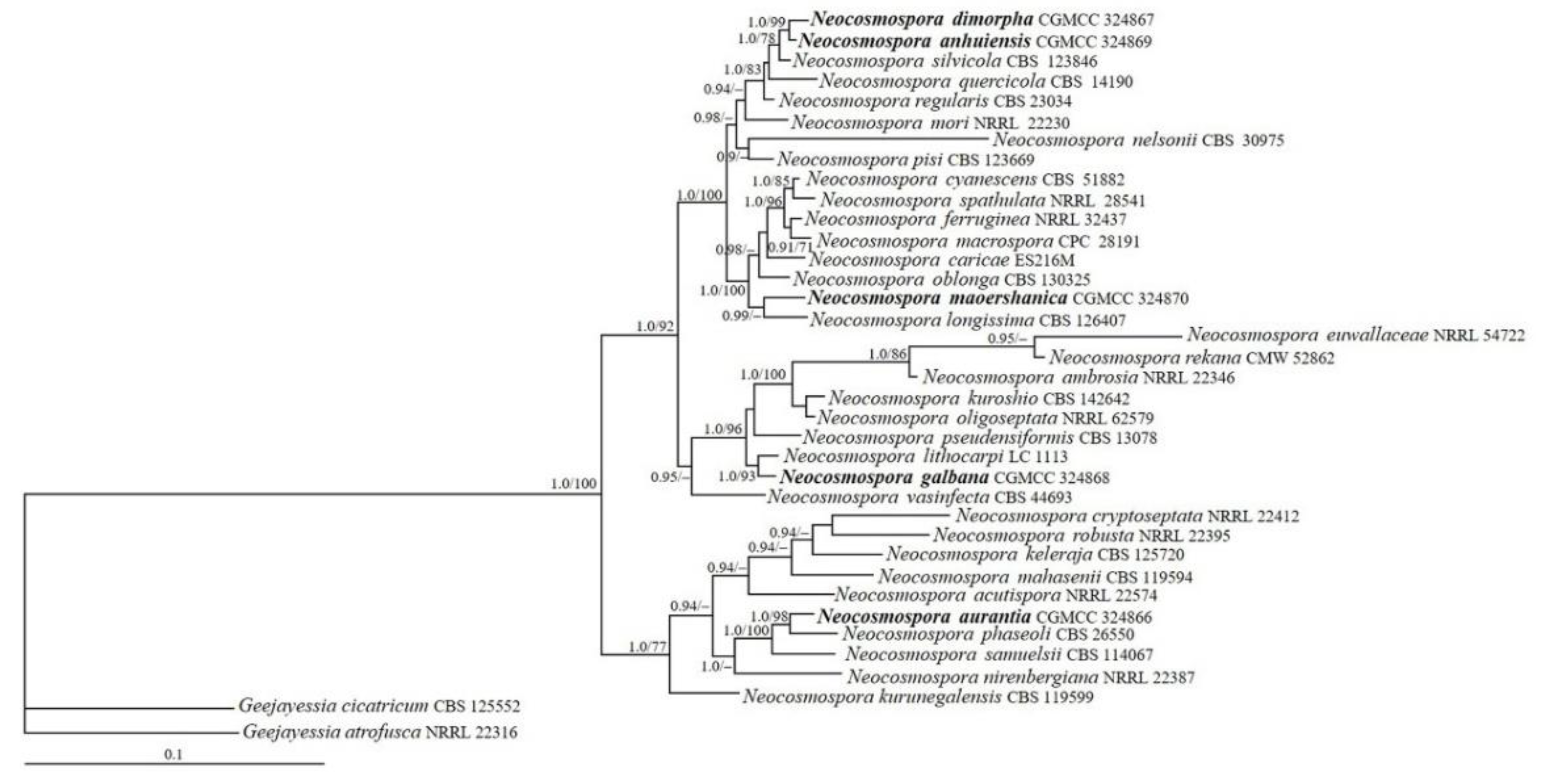
3.1. Phylogenetic Analyses

The sequences of CAM, ITS, RPB2, and TEF1 from 35 representative Neocosmospora species that are closely related to the five new taxa based on BLAST searches were analyzed (Table 1). The resulting BI tree is displayed in Figure 1. The topology of the ML tree was similar to that of the BI tree. The strains CGMCC 3.24866, 3.24867, 3.24868, 3.24869, and 3.24870 were grouped with other members of Neocosmospora receiving high statistical support values (BIPP/MLBP = 1.0/100%). The strain CGMCC 3.24867 and 3.24869 were closely related (BIPP/MLBP = 1.0/99%), which were further grouped with N. silvicola Sand.-Den. & Crous (BIBP/MLBP = 1.0/78%). The strain CGMCC 3.24870 was clustered with N. longissima Sand.-Den. & Crous (BIBP = 0.99), isolate CGMCC 3.24868 was related to N. lithocarpi M.M. Wang & L. Cai (BIBP/MLBP = 1.0/93%), and the remaining strain CGMCC 3.24866 was associated with N. phaseoli (Burkh.) L. Lombard & Crous (BIBP/MLBP = 1.0/98%).

3.2. Taxonomy

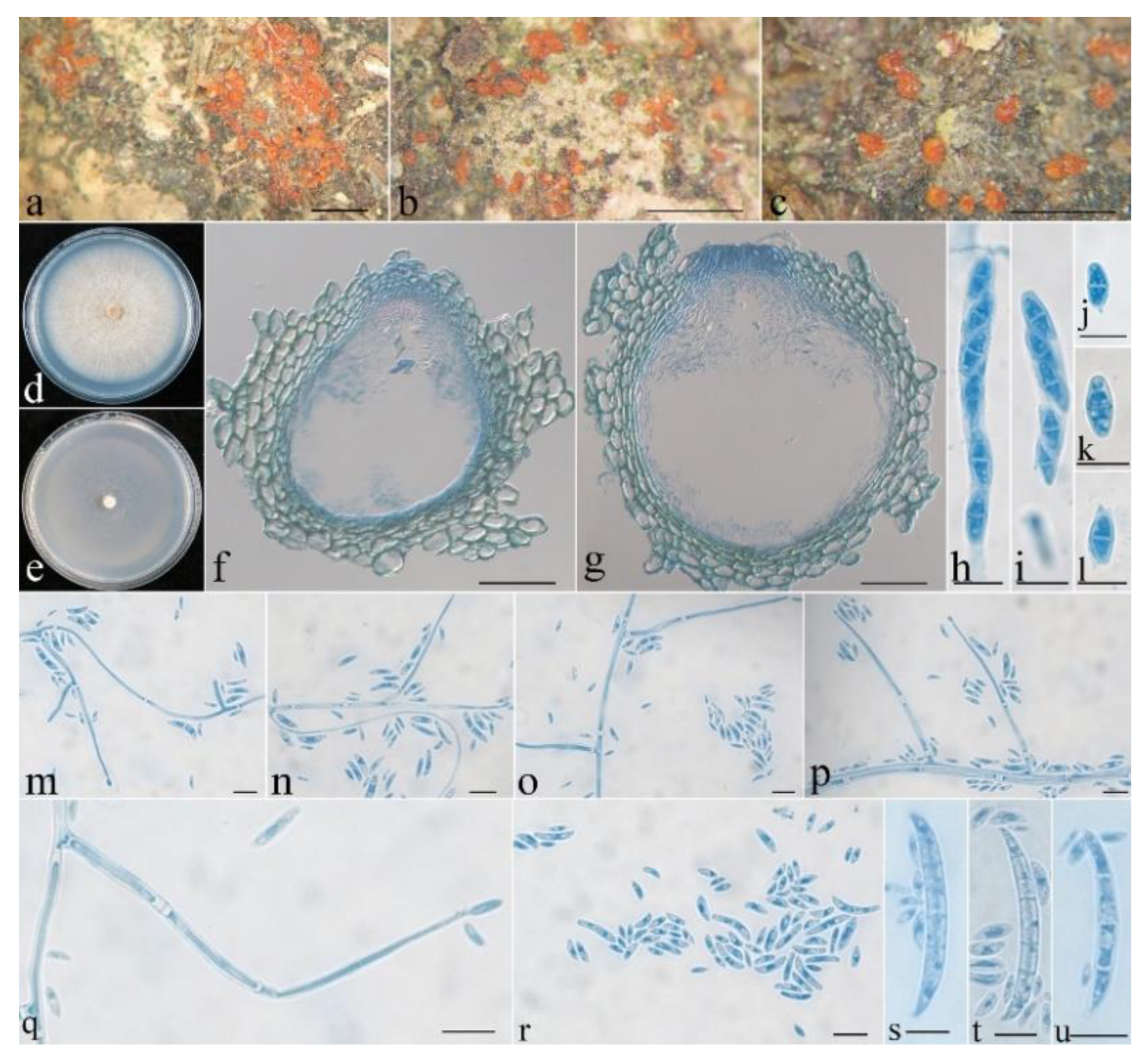
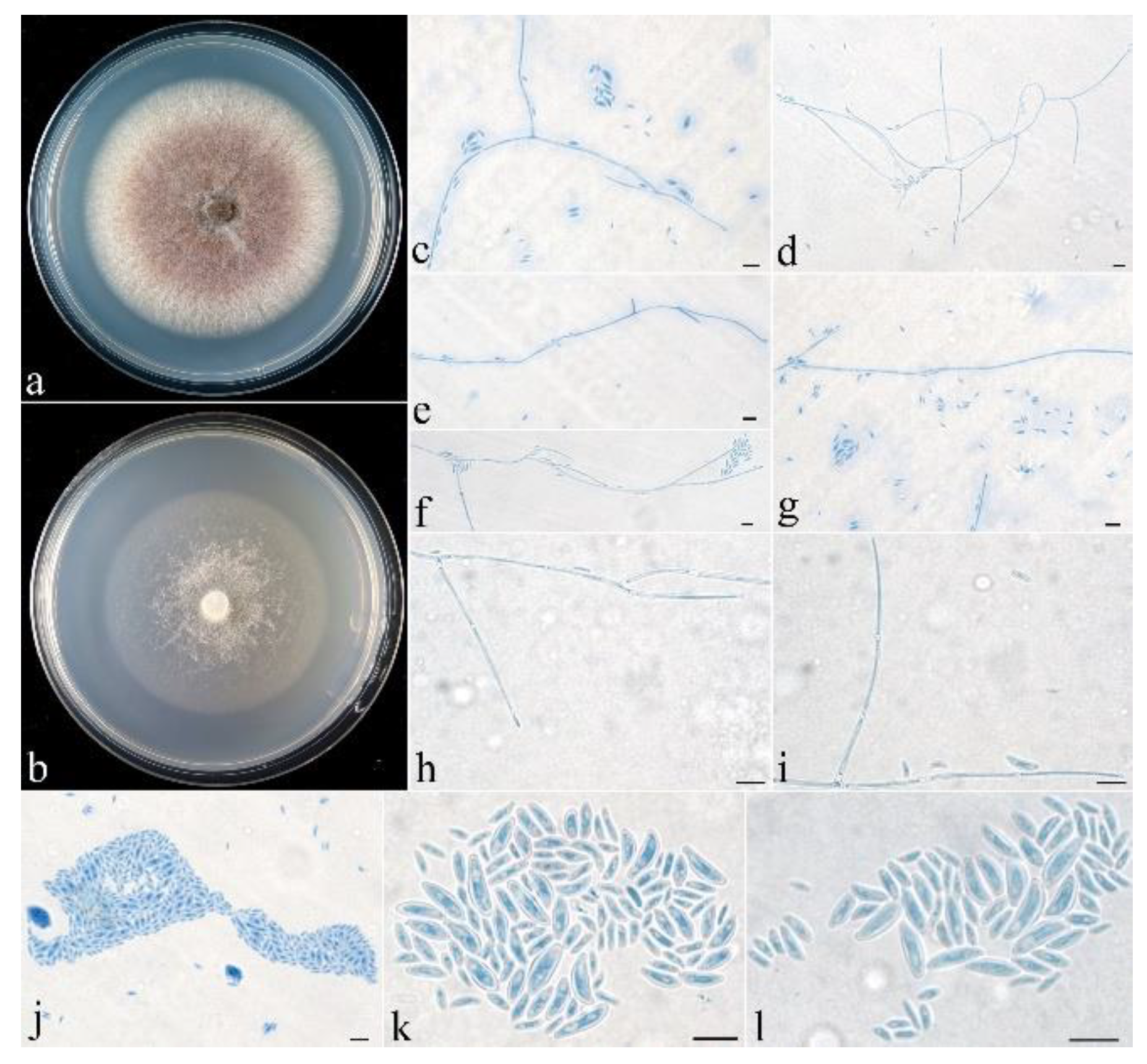
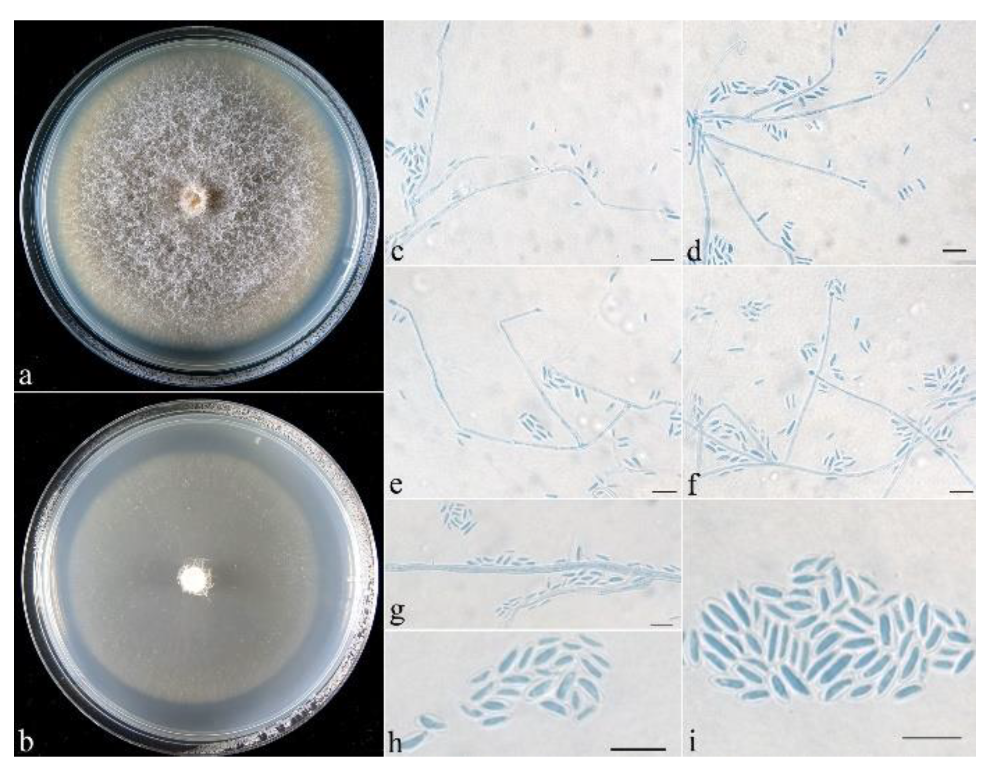
  • Neocosmospora anhuiensis Z.Q. Zeng & W.Y. Zhuang, sp. nov. Figure 2.
Fungal Names: FN 571307.
Etymology: The specific epithet refers to the type locality.
Typification: CHINA. Anhui Province, Huangshan, Yungu Temple, on rotten twigs, 22 June 2019, Z.Q. Zeng & H.D. Zheng 12364 (holotype HMAS 255836, ex-type culture CGMCC 3.24869).
Mycelium was visible neither around ascomata nor on natural substrates. The features of the ascomata were as follows: perithecial, superficial, solitary to gregarious, non-stromatic, subglobose to globose, or pyriform; showing lateral collapse upon drying; orange-red, turning dark red in 3% KOH, becoming light yellow in 100% LA; and 196–245 × 186–255 μm. The features of the perithecial surface were as follows: coarsely warted, of textura angularis to textura globosa, warts 15–40 µm high, cells 10–25 × 8–18 µm, and walls 0.8–1 µm thick. The perithecial wall of two layers was 28–43 µm thick; the outer layer of textura angularis was 23–33 μm thick, the cells were 10–18 × 6–13 μm, the walls were 1–1.2 μm thick; and the inner layer of textura prismatica was 5–10 μm thick, the cells were 8–15 × 2–3 μm, the walls were 0.8–1 μm thick. Asci were cylindrical to clavate, with a round and simple apex, 8-spored and 48–70 × 5–9 µm. Ascospores were ellipsoidal, 1-septate, hyaline, smooth-walled, irregularly biseriate, and 8–15 × 3–5 µm.
On PDA, the colony was 77 mm in diam. after 7 d at 25 °C, and the surface was cottony, with a dense, whitish aerial mycelium forming pale yellow pigments. On SNA, the colony was 70 mm in diam. after 7 d at 25 °C, and the surface was velvety, with a sparse, whitish aerial mycelium. Conidiophores ranged from unbranched to simply branched, with indefinite length. Microconidia ranged from ellipsoidal to rod-shaped and straight to slightly curved and were hyaline, smooth-walled, 0–1-septate, and 4.5–15 × 1.5–2.5 μm. Macroconidia were falcate, 2–6-septate, hyaline, smooth-walled, and 25–58 × 3.5–5 μm. Chlamydospores were not observed.
Notes: Morphologically, N. anhuiensis most resembles N. pseudensiformis Samuels in having subglobose, coarsely warted perithecia, which show lateral collapse upon drying, with clavate asci, ellipsoidal microconidia, and falcate macroconidia and lack the capacity of producing chlamydospores in culture [40]. However, the latter has larger asci (60–100 × 7–11.5 μm); broadly ellipsoidal, pale yellow-brown, and striate ascospores; a tan colony on PDA; and wider macroconidia (4.5–9 μm wide). Phylogenetically, they are remotely related (Figure 1).
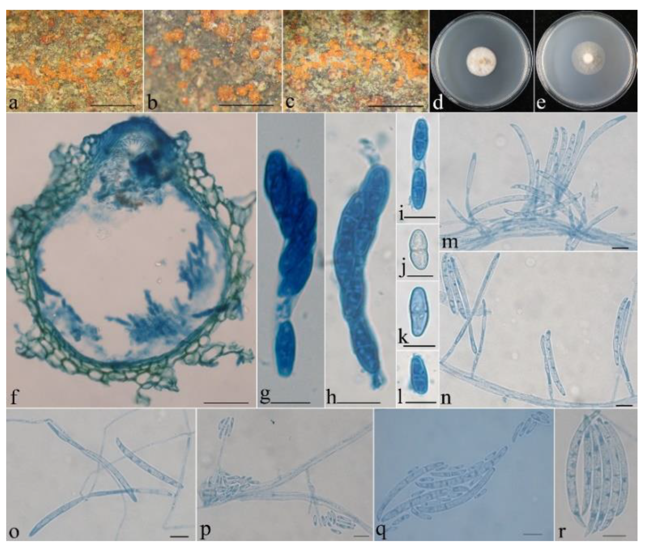
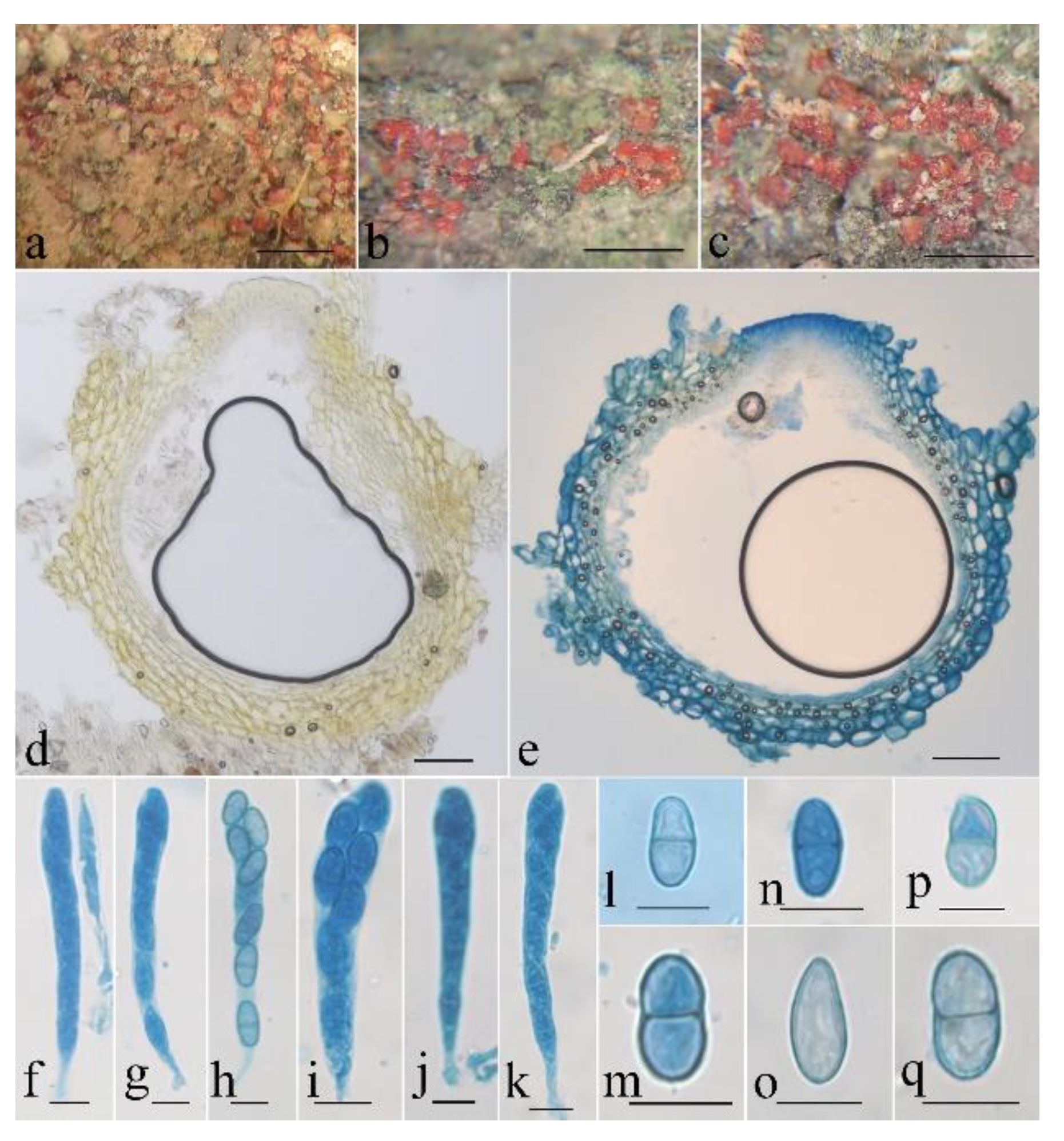
  • Neocosmospora aurantia Z.Q. Zeng & W.Y. Zhuang, sp. nov. Figure 3.
Fungal Names: FN 571308.
Etymology: The specific epithet refers to the orange-colored perithecia.
Typification: CHINA. Hubei Province, Shennongjia forestry district, Muchengshaoqia, on rotten bark, 22 September 2014, Z.Q. Zeng, H.D. Zheng, W.T. Qin & K. Chen 10053 (holotype HMAS 290899, ex-type culture CGMCC 3.24866).
Mycelium was visible neither around ascomata nor on natural substrates. The features of the ascomata were as follows: perithecial, superficial, solitary to gregarious, non-stromatic or with a basal stroma, subglobose to globose, or pyriform; orange-yellow when fresh, yellow-orange when dry, turning dark red in 3% KOH, becoming light yellow in 100% LA; and 235–304 × 206–323 μm. The features of perithecial surface were as follows: slightly warted, of textura angularis to textura globosa, warts 15–63 µm high, cells 10–30 × 8–13 µm, and walls 0.8–1 µm thick. The perithecial wall of two layers was 18–30 µm thick; the outer layer of textura angularis to textura globosa was 13–23 μm thick, the cells were 10–25 × 5–22 μm, the walls were 0.8–1 μm thick; and the inner layer of textura prismatica was 5–8 μm thick, the cells were 5–10 × 2–3 μm, the walls were 0.9–1.2 μm thick. Asci were cylindrical to clavate, with a round and simple apex, 8-spored and 43–75 × 5–12 µm. Ascospores were ellipsoidal to oblong, 1-septate, hyaline, smooth-walled, uniseriate or irregularly biseriate, and 10–16 × 4–5 μm.
On PDA, the colony was 28 mm in diam. after 7 d at 25 °C, and the surface was floccose, with a dense, whitish aerial mycelium forming pale brown pigments. On SNA, the colony was 33 mm in diam. after 7 d at 25 °C, and the surface was floccose, with a sparse, whitish aerial mycelium. Conidiophores were simply branched, 16–83 μm long, and 2–3 μm wide at the base. Microconidia ranged from ellipsoidal to rod-shaped and were hyaline, smooth-walled, 0(–1)-septate, and 4–15 × 1.5–2.5 μm. Macroconidia were mainly falcate, rarely cylindrical, slightly curved, (1–)3–4(–5)-septate, hyaline, smooth-walled, and 43–75 × 5–6 μm.
Notes: The species is morphologically most similar and phylogenetically related to N. phaseoli (BIPP/MLBP = 1.0/98%) (Figure 1) in having dense floccose, white aerial mycelium, sparsely branched conidiophores, ellipsoidal microconidia with 0(–1)-septate, and falcate macroconidia with 3–4-septate [41]. However, the latter differs in its shorter macroconidia (32–58 μm long), larger microconidia (13.5–32.5 × 3.5–6 μm), and the production of subglobose to ellipsoidal chlamydospores [41]. Moreover, the sequence comparisons display that there are 19 bp, 22 bp, 22 bp, and 6 bp differences in the regions of CAM, ITS, RPB2, and TEF1, respectively. They are not conspecific.
Fungal Names: FN 571309.
Etymology: The specific epithet refers to the presence of two types of microconidia.
Typification: CHINA. Hunan Province, Hengyang, Nanyue scenic spot, on rotten twigs, 21 October 2015, Z.Q. Zeng, X.C. Wang, K. Chen & Y.B. Zhang 10144 (holotype HMAS 255837, ex-type culture CGMCC 3.24867).
Mycelium was visible neither around ascomata nor on natural substrates. The features of the ascomata were as follows: perithecial, superficial, solitary to gregarious, non-stromatic, subglobose to globose, or pyriform; orange-red, turning dark red in 3% KOH, becoming light yellow in 100% LA; and 225–294 × 196–254 μm. The features of perithecial surface were as follows: slightly warted, of textura angularis to textura globosa, warts 15–45 µm high, cells 8–28 × 5–20 µm, and walls 0.8–1 µm thick. The perithecial wall of two layers was 15–40 µm thick; the outer layer of textura angularis to textura globosa was 10–30 μm thick, the cells were 5–15 × 4–10 μm, the walls were 1–1.2 μm thick; and the inner layer of textura prismatica was 5–10 μm thick, the cells were 8–14 × 2–3 μm, the walls were 0.8–1 μm thick. Asci were cylindrical to clavate, with a round and simple apex, 8-spored and 63–80 × 5.5–10 µm. Ascospores were ellipsoidal, 1-septate, hyaline, smooth-walled, uniseriate or irregularly biseriate, and 8–15 × 3.5–5 μm.
On PDA, the colony was 67 mm in diam. after 7 d at 25 °C, and the surface was cottony, with a dense, whitish aerial mycelium forming pale brown pigments. On SNA, the colony was 58 mm in diam. after 7 d at 25 °C, and the surface was floccose, with a sparse, whitish aerial mycelium. Conidiophores ranged from unbranched to simply branched, with indefinite length. Microconidia were ellipsoidal or rod-shaped: ellipsoidal microconidia were straight to slightly curved, unseptate, hyaline, smooth-walled, and 4–10.6 × 1.6–4.1 μm; rod-shaped microconidia were slightly curved, 0(–1)-septate, hyaline, smooth-walled, and 4–14.2 × 1.6–5.2 μm. Macroconidia and chlamydospores were not observed.
Notes: Amongst the existing species of Neocosmospora, N. dimorpha is morphologically similar and phylogenetically related to N. anhuiensis in having solitary to gregarious, non-stromatic, subglobose to globose, or pyriform perithecia; cylindrical to clavate asci; ellipsoidal ascospores; and an absence of chlamydospores. However, N. anhuiensis differs in having somewhat shorter asci (48–70 µm long), having faster colony growth rates on PDA and SNA (77 mm and 70 mm in diam.), producing a pale yellow pigment on PDA, and forming falcate, 2–6-septate macroconidia in culture. Additionally, there are 9 bp, 11 bp, and 3 bp differences in the CAM, ITS, and TEF1 regions, respectively, between the type strains (CGMCC 3.24867 and 3.24869).
Fungal Names: FN 571310.
Etymology: The specific epithet refers to the greenish-yellow colony on PDA.
Typification: CHINA. Hubei Province, Shennongjia, Banqiao, on rotten bark, 20 September 2014, Z.Q. Zeng, H.D. Zheng, K. Chen & W.T. Qin 9942 (holotype HMAS 247874, ex-type culture CGMCC 3.24868).
Mycelium was visible neither around ascomata nor on natural substrates. The characteristics of the ascomata are as follows: perithecial, superficial, solitary to gregarious, non-stromatic or with a basal stroma, subglobose to globose, or pyriform; orange-red to brownish-red, turning dark red to violet in 3% KOH, becoming light yellow in 100% LA; and 225–304 × 216–333 μm. The features of perithecial surface were as follows: warted, of textura angularis to textura globosa, warts 12–63 µm high, cells 10–32 × 8–13 µm, and walls 0.8–1 µm thick. The perithecial wall of two layers was 25–45 µm thick; the outer layer of textura angularis to textura globosa was 10–37 μm thick, the cells were 5–15 × 4–8 μm, the walls were 1–1.2 μm thick; the inner layer of textura prismatica was 5–8 μm thick, the cells were 10–20 × 2–3 μm, the walls were 0.8–1 μm thick. Asci were cylindrical to clavate, with a round and simple apex, 8-spored and 63–88 × 7.5–12 μm. Ascospores were ellipsoidal, (0–)1-septate, hyaline, smooth-walled, uniseriate or irregularly biseriate, and 8–13 × 4–5.5 μm.
On PDA, the colony was 84 mm in diam. after 7 d at 25 °C, and the surface was cottony, with a dense, whitish aerial mycelium forming yellowish-green pigments. On SNA, the colony was 58 mm in diam. after 7 d at 25 °C, and the surface was floccose, with a sparse, whitish aerial mycelium. Conidiophores were acremonium- to verticillium-like, with a whorl of 2–4 phialides, and the phialides were subulate to cylindrical, 16–58 μm long, and 1.5–2 μm wide at the base. Microconidia were ellipsoidal to rod-shaped, 0(–1)-septate, hyaline, smooth-walled, and 4–8 × 1.5–2.5 μm. Macroconidia were falcate, 1–6-septate, hyaline, smooth-walled, and 13–73 × 2.5–5 μm.
Notes: Among the existing species of the genus, N. galbana is morphologically most related to N. pseudensiformis Samuels in having subglobose perithecia, clavate asci, acremonium- to verticillium-like conidiophores, and falcate macroconidia [40]. Nevertheless, the latter has pale yellow-brown, striate, and longer ascospores (10–16.2 μm long), a tan colony on PDA, and wider macroconidia (3.5–9 μm wide). Phylogenetically, N. galbana clustered with N. lithocarpi (BIBP/MLBP = 1.0/93%). The sequence comparison of the type cultures indicated that there are 3 bp, 16 bp, and 5 bp differences detected for ITS, RPB2, and TEF1 regions, respectively. Moreover, N. lithocarpi, only known for its asexual stage, differs in having a slower colony growth rate on PDA (57–59 mm in diam.), a greyish-orange pigment in culture, wider macroconidia (3.9–8.1 μm wide) with 5 septa, and larger microconidia (7–24 × 3.5–7 μm) and producing abundant chlamydospores [9].
  • Neocosmospora maoershanica Z.Q. Zeng & W.Y. Zhuang, sp. nov. Figure 8 and Figure 9.
Fungal Names: FN 571311.
Etymology: The specific epithet refers to the type locality.
Typification: CHINA. Guangxi Zhuang Autonomous Region, Guilin, Maoershan, on twigs, 7 December Oct 2019, Z.Q. Zeng, & H.D. Zheng 12500 (holotype HMAS 247875, ex-type culture CGMCC 3.24870).
Mycelium was visible neither around ascomata nor on natural substrates. The characteristics of the ascomata were as follows: perithecial, superficial, solitary to gregarious, non-stromatic, subglobose to globose, or pyriform, with or without an inconspicuous papilla; orange-red when fresh, red when dry, turning dark red in 3% KOH, becoming yellow in 100% LA; and 216–294 × 176–255 μm. The perithecial surface was slightly roughened. The perithecial wall of two layers was 15–55 µm thick; the outer layer of textura angularis was 10–43 μm thick, the cells were 5–10 × 4–8 μm, the walls were 1–1.2 μm thick; the inner layer of textura prismatica was 5–12 μm thick, the cells were 5–12 × 2–3 μm, the walls were 0.8–1 μm thick. Asci were cylindrical to cylindrical-clavate, with a round and simple apex, (6–)8-spored and 55–85 × 5–8 µm. Ascospores were ellipsoidal to oblong, (0–)1-septate, hyaline to light brown, smooth-walled, uniseriate and overlapping obliquely, and 9–16 × 4.5–8 μm.
On PDA, the colony was 80 mm in diam. after 7 d at 25 °C, and the surface was cottony, with a dense, whitish aerial mycelium forming pale violet pigments. On SNA, the colony was 68 mm in diam. after 7 d at 25 °C, and the surface was floccose, with a sparse, whitish aerial mycelium. Conidiophores were acremonium- to verticillium-like, and the phialides were subulate, subcylindrical, acerose, hyaline, smooth-walled, 14–75 μm long, and 1.2–1.6 μm wide at the base. Microconidia were ellipsoidal, rod-shaped, bullet-shaped, hyaline, smooth-walled, 0(–1)-septate, and 3–13 × 2–4 μm. Macroconidia and chlamydospores were not observed.
Notes: Morphologically, N. maoershanica most resembles N. oblonga Sand.-Den. & Crous in having verticillium-like conidiophores and ellipsoidal to bullet-shaped, 0(–1)-septate, hyaline, and smooth-walled microconidia and lacking macroconidia [10]. However, N. oblonga differs in having a white to pale straw colony on PDA, longer microconidia (5–22 μm long), and the production of globose to subglobose chlamydospores. Sequence comparisons revealed that there are 13 bp and 23 bp differences detected for the CAM and RPB2 regions, respectively. Phylogenetically, N. maoershanica is closely associated with N. longissima (BIBP = 0.99) (Figure 1). The latter differs in forming an umber to rust colony on PDA and producing wedge-shaped macroconidia and abundant globose to obpyriform chlamydospores [10].

4. Discussion

Since the monotypic genus Neocosmospora was established [42], many species were described subsequently [43,44,45,46]. Later, the genus Haematonectria Samuels & Nirenberg, typified by H. haematococca (Berk. & Broome) Samuels & Rossman, was introduced by Rossman et al. [1] who stated that its ascomatal morphology and asexual features clearly distinguished it from Neocosmospora. Along with the information accumulated in phylogenetic studies, it has been indicated that the two genera are congeneric [40,47], and Neocosmospora was recommended as the preferable name according to the priority concept [2]. The taxonomic opinion became widely accepted [3,5,6,9,10,18], including the present research.
The phylogenetic tree based on combined analyses of CAM, ITS, RPB2, and TEF1 sequences showed that the five Neocosmospora strains (CGMCC 3.24866, 3.24867, 3.24868, 3.24869, and 3.24870) grouped with the representative species of the genus, which verified their taxonomic placements (Figure 1). Neocosmospora dimorpha (CGMCC 3.24867) clustered with N. anhuiensis (CGMCC 3.24869) (BIPP/MLBP = 1.0/99%), and they further grouped with N. silvicola (CBS 123846) (BIPP/MLBP = 0.99/78%). However, N. silvicola can be easily distinguished from N. dimorpha by its scarlet and ochreous to citrine pigment on PDA and globose to subglobose chlamydospores [10]. Neocosmospora maoershanica (CGMCC 3.24870) is related to N. longissima (CBS 126407) (BIBP = 0.9) but differs by its pale violet pigments produced on PDA and the lack of macroconidia and chlamydospores in culture [10]. Neocosmospora aurantia (CGMCC 3.24866) is phylogenetically related with, but clearly separated from, N. phaseoli (CBS 26550) (BIPP/MLBP = 1.0/98%), and the latter is distinguished by its shorter macroconidia, larger microconidia, and the production of subglobose to ellipsoidal chlamydospores [41]. Neocosmospora galbana (CGMCC 3.24868) grouped with N. lithocarpi (LC 1113) (BIPP/MLBP = 1.0/93%) and differs by its faster growth rate and greenish-yellow colony on PDA, narrower macroconidia, smaller microconidia without septa, and the lack of chlamydospores [9].
Since N. sphaerospora (Q.T. Chen & X.H. Fu) Sand.-Den. & Crous (as Fusarium sphaerosporum Q.T. Chen & X.H. Fu) and N. petroliphila (Q.T. Chen & X.H. Fu) Sand.-Den. & Crous (as Fusarium solani var. petroliphilum Q.T. Chen & X.H. Fu) were first reported by Chen et al. [48], five additional new species of the genus were successively described from different provinces of China [3,9,49,50]. Among these seven species, four taxa, namely, N. lithocarpi, N. pallidimors Tibpromma, Karun., Karasaki & P.E. Mortimer, N. petroliphila, and N. sphaerospora are only known with their asexual stages [3,9,48]. In the present study, five holomorphic novel taxa of the genus were introduced, which significantly increase the species diversity of the genus in the country. Further surveys in the unexplored regions will update our understanding of the species diversity of nectrioid fungi in China and the world [51]. Combining their sexual stages with their asexual stages will give a better comprehension of the whole fungus.

Author Contributions

Conceptualization, Z.-Q.Z. and W.-Y.Z.; methodology, software, validation, formal analysis, investigation, data curation, and visualization, Z.-Q.Z.; resources, W.-Y.Z. and Z.-Q.Z.; writing—original draft preparation, Z.-Q.Z.; writing—review and editing, W.-Y.Z. and Z.-Q.Z.; supervision, Z.-Q.Z. and W.-Y.Z.; project administration, W.-Y.Z.; funding acquisition, W.-Y.Z. and Z.-Q.Z. All authors have read and agreed to the published version of the manuscript.

Funding

This research was funded by the National Natural Science Foundation of China (32270009, 31750001), the Frontier Key Program of the Chinese Academy of Sciences (QYZDY-SSW-SMC029), Biological Resources Programme, Chinese Academy of Sciences (KFJ-BRP-017-082), and the National Project on Scientific Groundwork, Ministry of Science and Technology of China (2019FY100700).

Institutional Review Board Statement

Not applicable.

Informed Consent Statement

Not applicable.

Data Availability Statement

The names of the new species were formally registered in the database Fungal Names (http://www.fungalinfo.net/fungalname/fungalname.html (accessed on 20 February 2023)). Specimens were deposited in the Herbarium Mycologicum Academiae Sini-cae (https://nmdc.cn/fungarium/ (accessed on 18 February 2023)). The newly created sequences were deposited in GenBank (https://www.ncbi.nlm.nih.gov/genbank (accessed on 20 May 2023)).

Acknowledgments

We would like to thank H.D. Zheng, X.C. Wang, W.T. Qin, K. Chen, and Y.B. Zhang for their invaluable help during the field work.

Conflicts of Interest

The authors declare no conflict of interest.

References

  1. Rossman, A.Y.; Samuels, G.J.; Rogerson, C.T.; Lowen, R. Genera of Bionectriaceae, Hypocreaceae and Nectriaceae (Hypocreales, Ascomycetes). Stud. Mycol. 1999, 42, 1–248. [Google Scholar]
  2. Lombard, L.; van der Merwe, N.A.; Groenewald, J.Z.; Crous, P.W. Generic concepts in Nectriaceae. Stud. Mycol. 2015, 80, 189–245. [Google Scholar] [CrossRef] [Green Version]
  3. Karunarathna, S.C.; Dong, Y.; Karasaki, S.; Tibpromma, S.; Hyde, K.D.; Lumyong, S.; Xu, J.C.; Sheng, J.; Mortimer, P.E. Discovery of novel fungal species and pathogens on bat carcasses in a cave in Yunnan Province, China. Emerg. Microbes Infec. 2020, 9, 1554–1566. [Google Scholar] [CrossRef]
  4. Perera, R.H.; Hyde, K.D.; Maharachchikumbura, S.S.N.; Jones, E.B.G.; McKenzie, E.H.C.; Stadler, M.; Lee, H.B.; Samarakoon, M.C.; Ekanayaka, A.H.; Camporesi, E.; et al. Fungi on wild seeds and fruits. Mycosphere 2020, 11, 2108–2480. [Google Scholar] [CrossRef]
  5. Perera, R.H.; Hyde, K.D.; Jones, E.B.G.; Maharachchikumbura, S.S.N.; Bundhun, D.; Camporesi, E.; Akulov, A.; Liu, J.K.; Liu, Z.Y. Profile of Bionectriaceae, Calcarisporiaceae, Hypocreaceae, Nectriaceae, Tilachlidiaceae, Ijuhyaceae fam. nov., Stromatonectriaceae fam. nov. and Xanthonectriaceae fam. nov. Fungal Divers. 2023, 118, 95–271. [Google Scholar] [CrossRef]
  6. Crous, P.W.; Lombard, L.; Sandoval-Denis, M.; Seifert, K.A.; Schroers, H.J.; Chaverri, P.; Gene, J.; Guarro, J.; Hirooka, Y.; Bensch, K.; et al. Fusarium: More than a node or a foot-shaped basal cell. Stud. Mycol. 2021, 98, 1–184. [Google Scholar] [CrossRef]
  7. Crous, P.W.; Sandoval-Denis, M.; Costa, M.M.; Groenewald, J.Z.; van Iperen, A.L.; Starink-Willemse, M.; Hernández-Restrepo, M.; Kandemir, H.; Ulaszewski, B.; de Boer, W.; et al. Fusarium and allied fusarioid taxa (FUSA). 1. Fungal Syst. Evol. 2022, 9, 161–200. [Google Scholar] [CrossRef]
  8. Guarnaccia, V.; van Niekerk, J.; Crous, P.W.; Sandoval-Denis, M. Neocosmospora spp. associated with dry root rot of citrus in South Africa. Phytopathol. Mediterr. 2021, 60, 79–100. [Google Scholar] [CrossRef]
  9. Wang, M.M.; Crous, P.W.; Sandoval-Denis, M.; Han, S.L.; Liu, F.; Liang, J.M.; Duan, W.J.; Cai, L. Fusarium and allied genera from China: Species diversity and distribution. Persoonia 2022, 48, 1–53. [Google Scholar] [CrossRef]
  10. Sandoval-Denis, M.; Lombard, L.; Crous, P.W. Back to the roots: A reappraisal of Neocosmospora. Persoonia 2019, 43, 90–185. [Google Scholar] [CrossRef] [PubMed]
  11. Merlin, J.N.; Christudas, I.V.S.N.; Kumar, P.P.; Agastian, P. Optimization of growth and bioactive metabolite production: Fusarium solani. Asian Journal. Pharm. Clin. Res. 2013, 6, 98–103. [Google Scholar]
  12. Wu, Y.R.; Nian, D.L. Production optimization and molecular structure characterization of a newly isolated novel laccase from Fusarium solani MAS2, an anthracene-degrading fungus. Int. Biodeter. Biodegr. 2014, 86, 382–389. [Google Scholar] [CrossRef]
  13. Jallouli, R.; Othman, H.; Amara, S.; Parsiegla, G.; Carriere, F.; Srairi-abid, N.; Gargouri, Y.; Bezzine, S. The galactolipase activity of Fusarium solani (phospho) lipase. BBA-Mol. Cell. Biol. Lipids 2015, 1851, 282–289. [Google Scholar] [CrossRef]
  14. Baard, S.W.; van Wyk, P.S. Neocosmospora vasinfecta pathogenic to groundnuts in South Africa. Phytophylactica 1985, 17, 49–50. [Google Scholar]
  15. Huang, J.W.; Chen, S.S.; Chung, W.C. Neocosmospora foot rot of peanut in Taiwan. Plant Pathol. Bull. 1992, 1, 203–205. [Google Scholar]
  16. Fuhlbohm, M.F.; Tatnell, J.R.; Ryley, M.J. Neocosmospora vasinfecta is pathogenic on peanut in Queensland. Australas. Plant Dis. Notes 2007, 2, 3–4. [Google Scholar] [CrossRef] [Green Version]
  17. Freeman, S.; Sharon, M.; Maymon, M.; Mendel, Z.; Protasov, A.; Aoki, T.; Eskalen, A.; O’Donnell, K. Fusarium euwallaceae sp. nov.—A symbiotic fungus of Euwallacea sp., an invasive ambrosia beetle in Israel and California. Mycologia 2013, 105, 1595–1606. [Google Scholar] [CrossRef] [Green Version]
  18. Guarnaccia, V.; Sandoval-Denis, M.; Aiello, D.; Polizzi, G.; Crous, P.W. Neocosmospora perseae sp. nov., causing trunk cankers on avocado in Italy. Fungal Syst. Evol. 2018, 1, 131–140. [Google Scholar] [CrossRef] [Green Version]
  19. Guarnaccia, V.; Martino, I.; Brondino, L.; Gullino, M.L. Paraconiothyrium fuckelii, Diaporthe eres and Neocosmospora parceramosa causing cane blight of red raspberry in Northern Italy. J. Plant Pathol. 2022, 104, 683–698. [Google Scholar] [CrossRef]
  20. Sandoval-Denis, M.; Guarnaccia, V.; Polizzi, G.; Crous, P.W. Symptomatic Citrus trees reveal a new pathogenic lineage in Fusarium and two new Neocosmospora species. Persoonia 2018, 40, 1–25. [Google Scholar] [CrossRef] [PubMed] [Green Version]
  21. Zheng, F.; Xu, G.; Zheng, F.Q.; Ding, X.F.; Xie, C.P. Neocosmospora rubicola causing stem rot of pitaya (Hylocereus costaricensis) in China. Plant Dis. 2018, 102, 2653. [Google Scholar] [CrossRef]
  22. Crespo, M.; Lawrence, D.P.; Nouri, M.T.; Doll, D.A.; Trouillas, F.P. Characterization of Fusarium and Neocosmospora species associated with crown rot and stem canker of pistachio rootstocks in California. Plant Dis. 2019, 103, 1931–1939. [Google Scholar] [CrossRef]
  23. Polat, Z.; Gültekin, M.A.; Palacıoğlu, G.; Bayraktar, H.; Özer, N.; Yılmaz, S. First report of Neocosmospora solani causing stem canker on Juglans regia in Turkey. J. Plant Pathol. 2020, 102, 1289. [Google Scholar] [CrossRef]
  24. Riaz, M.; Akhtar, N.; Khan, S.N.; Shakeel, M.; Tahir, A. Neocosmospora rubicola: An unrecorded pathogen from Pakistan causing potato stem rot. Sarhad J. Agric. 2020, 36, 906–912. [Google Scholar] [CrossRef]
  25. Bolboli, Z.; Mostowfizadeh-Ghalamfarsa, R.; Sandoval-Denis, M.; Jafari, M.; Crous, P.W. Neocosmospora caricae sp. nov. and N. metavorans, two new stem and trunk canker pathogens on Ficus carica in Iran. Mycol. Prog. 2022, 21, 89. [Google Scholar] [CrossRef]
  26. Luo, J.; Zhuang, W.Y. Three new species of Neonectria (Nectriaceae, Hypocreales) with notes on their phylogenetic positions. Mycologia 2010, 102, 142–152. [Google Scholar] [CrossRef] [Green Version]
  27. Nirenberg, H.I. Untersuchungen über die morphologische und biologische Differenzierung in der Fusarium-Sektion Liseola. Mitt. Biol. Bundesanst. Land. Forstw. 1976, 169, 1–117. [Google Scholar]
  28. Wang, L.; Zhuang, W.Y. Designing primer sets for amplification of partial calmodulin genes from penicillia. Mycosystema 2004, 23, 466–473. [Google Scholar]
  29. O’Donnell, K.; Kistler, H.C.; Tacke, B.K.; Casper, H.H. Gene genealogies reveal global phylogeographic structure and reproductive isolation among lineages of Fusarium graminearum, the fungus causing wheat scab. Proc. Natl. Acad. Sci. USA 2000, 95, 7905–7910. [Google Scholar] [CrossRef]
  30. White, T.J.; Bruns, T.; Lee, S.; Taylor, J. Amplification and direct sequencing of fungal ribosomal RNA genes for phylogenetics. In PCR Protocols: A Guide to Methods and Applications; Innis, M.A., Gelfland, D.H., Sninsky, J.J., White, T.J., Eds.; Academic Press: San Diego, CA, USA, 1990; pp. 315–322. [Google Scholar] [CrossRef]
  31. Liu, Y.J.; Whelen, S.; Hall, B.D. Phylogenetic relationships among ascomycetes: Evidence from an RNA polymerase II subunit. Mol. Biol. Evol. 1999, 16, 1799–1808. [Google Scholar] [CrossRef] [Green Version]
  32. O’Donnell, K.; Cigelnik, E.; Nirenberg, H.I. Molecular systematics and phylogeography of the Gibberella fujikuroi species complex. Mycologia 1998, 90, 465–493. [Google Scholar] [CrossRef]
  33. Hall, T.A. BioEdit: A user-friendly biological sequence alignment editor and analysis program for Windows 95/98/NT. Nucleic Acids Symp. Ser. 1999, 41, 95–98. [Google Scholar]
  34. Thompson, J.D.; Gibson, T.J.; Plewniak, F.; Jeanmougin, F.; Higgin, D.G. The ClustalX windows interface: Flexible strategies for multiple sequences alignment aided by quality analysis tools. Nucleic Acids Res. 1997, 25, 4876–4882. [Google Scholar] [CrossRef] [PubMed] [Green Version]
  35. Ronquist, F.; Huelsenbeck, J.P. MrBayes 3: Bayesian phylogenetic inference under mixed models. Bioinformatics 2003, 19, 1572–1574. [Google Scholar] [CrossRef] [Green Version]
  36. Nguyen, L.T.; Schmidt, H.A.; von Haeseler, A.; Minh, B.Q. IQ-TREE: A fast and effective stochastic algorithm for estimating maximum likelihood phylogenies. Mol. Biol. Evol. 2015, 32, 268–274. [Google Scholar] [CrossRef] [PubMed]
  37. Chernomor, O.; von Haeseler, A.; Minh, B.Q. Terrace aware data structure for phylogenomic inference from supermatrices. Mol. Syst. Biol. 2016, 65, 997–1008. [Google Scholar] [CrossRef] [Green Version]
  38. Nylander, J.A.A. MrModeltest, v2. Program Distributed by the Author; Evolutionary Biology Centre, Uppsala University: Uppsala, Sweden, 2004. [Google Scholar]
  39. Page, R.D. TreeView: An application to display phylogenetic trees on personal computers. Comp. Appl. Biosci. 1996, 12, 357–358. [Google Scholar] [PubMed] [Green Version]
  40. Nalim, F.A.; Samuels, G.J.; Wijesundera, R.L.; Geiser, D.M. New species from the Fusarium solani species complex derived from perithecia and soil in the old world tropics. Mycologia 2011, 103, 1302–1330. [Google Scholar] [CrossRef]
  41. Aoki, T.; O’Donnell, K.; Homma, Y.; Lattanzi, A.R. Sudden-death syndrome of soybean is caused by two morphologically and phylogenetically distinct species within the Fusarium solani species complex-F. virguliforme in North America and F. tucumaniae in South America. Mycologia 2003, 95, 660–684. [Google Scholar] [CrossRef]
  42. Smith, E.F. Wilt disease of cotton, watermelon and cowpea (Neocosmospora nov. gen.). US. Dep. Agric. Bull. 1899, 17, 1–53. [Google Scholar]
  43. Mahoney, D.P. A new Neocosmospora from Galapagos Island soil. Mycologia 1976, 68, 1111–1116. [Google Scholar] [CrossRef]
  44. Cannon, P.F.; Hawksworth, L. A revision of the genus Neocosmospora (Hypocreales). Trans. Br. Mycol. Soc. 1984, 82, 673–688. [Google Scholar] [CrossRef]
  45. Udagawa, S.I.; Horie, Y.; Cannon, P.F. Two new species of Neocosmospora from Japan, with a key to the currently accepted species. Sydowia 1989, 41, 349–359. [Google Scholar]
  46. Pfenning, L. A new species of Neocosmospora from Brazil. Sydowia 1995, 47, 65–69. [Google Scholar]
  47. Gräfenhan, T.; Schroers, H.J.; Nirenberg, H.I.; Seifert, K.A. An overview of the taxonomy, phylogeny, and typification of nectriaceous fungi in Cosmospora, Acremonium, Fusarium, Stilbella, and Volutella. Stud. Mycol. 2011, 68, 79–113. [Google Scholar] [CrossRef] [Green Version]
  48. Chen, Q.T.; Fu, X.H.; Chen, Y.D. Two petroliphilous taxa of Fusarium. Acta Mycol. Sin. Suppl. 1987, 1, 328–333. [Google Scholar]
  49. Luo, J.; Zhuang, W.Y. Four new species and a new Chinese record of the nectrioid fungi. Sci. China Life Sci. 2010, 53, 909–915. [Google Scholar] [CrossRef]
  50. Zeng, Z.Q.; Zhuang, W.Y. Two new species of Neocosmospora from China. Phytotaxa 2017, 319, 175–183. [Google Scholar] [CrossRef]
  51. Zeng, Z.Q.; Zhuang, W.Y. New species of Nectriaceae (Hypocreales) from China. J. Fungi 2022, 8, 1075. [Google Scholar] [CrossRef]
Figure 1. The Bayesian inference tree created based on CAM, ITS, RPB2, and TEF1 sequences of Neocosmospora species. BIPP (left) above 0.9 and MLBP (right) above 70% are indicated at nodes.
Life 13 01515 g001
Figure 2. Neocosmospora anhuiensis (HMAS 255836). (ac) Perithecia on rotten twigs. (d,e) Colonies after 1 wk at 25 °C ((d) on PDA, (e) on SNA). (f,g) Longitudinal section through perithecium. (h,i) Ascus with ascospores. (jl) Ascospore. (mq) Conidiophores and microconidia. (r) Microconidia. (su) Macroconidia and microconidia. Scale bars: (ac) = 1 mm, (f,g) = 50 μm, (hu) = 10 μm.
Life 13 01515 g002
Figure 3. Neocosmospora aurantia (HMAS 290899). (ac) Perithecia on rotten bark. (d,e) Colonies after 1 wk at 25 °C ((d) on PDA, (e) on SNA). (f) Longitudinal section through perithecium. (g,h) Ascus with ascospores. (il) Ascospore. (mo) Conidiophores and macroconidia. (p) Conidiophores and microconidia. (q) Macroconidia and microconidia. (r) Macroconidia. Scale bars: (ac) = 1 mm, (f) = 50 μm, (gr) = 10 μm.
Life 13 01515 g003
Figure 4. Neocosmospora dimorpha (HMAS 255837). (ac) Perithecia on rotten twigs. (d,e) Longitudinal section through perithecium. (fi) Ascus with ascospores. (jl) Ascospore. Scale bars: (ac) = 0.5 mm, (d,e) = 50 μm, (fl) = 10 μm.
Life 13 01515 g004
Figure 5. Neocosmospora dimorpha (CGMCC 3.24867). (a,b) Colonies after 1 wk at 25 °C ((a) on PDA, (b) on SNA). (ci) Conidiophores and microconidia. (jl) Microconidia. Scale bars: (cl) = 10 μm.
Life 13 01515 g005
Figure 6. Neocosmospora galbana (HMAS 247874). (ac) Perithecia on rotten bark. (d,e) Longitudinal section through perithecium. (fk) Ascus with ascospores. (lq) Ascospore. Scale bars: (ac) = 1 mm, (d,e) = 50 μm, (fq) = 10 μm.
Life 13 01515 g006
Figure 7. Neocosmospora galbana (CGMCC 3.24868). (a,b) Colonies after 1 wk at 25 °C ((a) on PDA, (b) on SNA). (ci) Conidiophores, macroconidia and microconidia. (j,k) Macroconidia and microconidia. Scale bars: (ck) = 10 μm.
Life 13 01515 g007
Figure 8. Neocosmospora maoershanica (HMAS 247875). (ac) Perithecia on twigs. (d,e) Longitudinal section through perithecium. (f,g) Ascus with ascospores. (hp) Ascospore. Scale bars: (ac) = 1 mm, (d,e) = 50 μm, (fp) = 10 μm.
Life 13 01515 g008
Figure 9. Neocosmospora maoershanica (CGMCC 3.24870). (a,b) Colonies after 1 wk at 25 °C ((a) on PDA, (b) on SNA). (cg) Conidiophores and microconidia. (h,i) Microconidia. Scale bars: (ch) = 10 μm.
Life 13 01515 g009
Table 1. Sequences of Neocosmospora species used in this study.
SpeciesStrain/Herbarium NumbersGenBank Accession Numbers
CAMITSRPB2TEF1
Geejayessia atrofuscaNRRL 22316-AF178423JX171609AF178361
Geejayessia cicatricumCBS 125552-MH863560HQ728153HM626644
Neocosmospora acutisporaNRRL 22574MW834122NR169884EU329514AF178345
Neocosmospora ambrosiaNRRL 22346-EU329669EU329503FJ240350
Neocosmospora anhuiensisCGMCC 3.24869OR014310 aOQ842733OQ866525OQ866530
Neocosmospora aurantiaCGMCC 3.24866OR014308OQ842731OQ866523OQ866528
Neocosmospora caricaeES216M-OK422518OK415859OK539518
Neocosmospora cryptoseptataNRRL 22412MW834126NR172368EU329510AF178351
Neocosmospora cyanescensCBS 51882MW218064AB190389LR583826LR583605
Neocosmospora dimorphaCGMCC 3.24867OR014309OQ842732OQ866524OQ866529
Neocosmospora euwallaceaeNRRL 54722KU171422JQ038014JQ038028JQ038007
Neocosmospora ferrugineaNRRL 32437MW834132DQ094446EU329581DQ246979
Neocosmospora galbanaCGMCC 3.24868OR014307OQ842730OQ866527OQ866532
Neocosmospora kelerajaCBS 125720MW834138LR583720LR583834LR583612
Neocosmospora kuroshioCBS 142642MW834140LR583723LR583837KX262216
Neocosmospora kurunegalensisCBS 119599MW834141JF433036LR583838DQ247511
Neocosmospora lithocarpiLC 1113-MW016711MW474697MW620172
Neocosmospora longissimaCBS 126407MW834144NR178144LR583846LR583621
Neocosmospora macrosporaCPC 28191MW218078NR163291LT746331LT746218
Neocosmospora mahaseniiCBS 119594MW834145JF433045LT960563DQ247513
Neocosmospora maoershanicaCGMCC 3.24870OR014311OQ842734OQ866526OQ866531
Neocosmospora moriNRRL 22230MW834149AF178420EU329499AF178358
Neocosmospora nelsoniiCBS 30975MW834152MW827630MW847904MW847907
Neocosmospora nirenbergianaNRRL 22387MW834153NR169883EU329505AF178339
Neocosmospora oblongaCBS 130325MW834154LR583746LR583853LR583631
Neocosmospora oligoseptataNRRL 62579MW834155KC691566LR583854KC691538
Neocosmospora phaseoliCBS 26550KM231380MH856617KM232375HE647964
Neocosmospora pisiCBS 123669MW834159KM231796KM232364KM231925
Neocosmospora pseudensiformisCBS 130.78MW834162LR583759LR583868DQ247635
Neocosmospora quercicolaCBS 14190MW834164NR178125LR583869DQ247634
Neocosmospora regularisCBS 23034MW834168LR583763LR583873LR583643
Neocosmospora rekanaCMW 52862-MN249094MN249137MN249151
Neocosmospora robustaNRRL 22395MW834169NR172367EU329507AF178341
Neocosmospora samuelsiiCBS 114067MW834170NR178127LR583874LR583644
Neocosmospora silvicolaCBS 123846MW834172LR583766LR583876LR583646
Neocosmospora spathulataNRRL 28541MW218091EU329674EU329542DQ246882
Neocosmospora vasinfectaCBS 446.93MW834175LR583791LR583898LR583670
a Numbers in bold indicate the newly generated sequences.
Disclaimer/Publisher’s Note: The statements, opinions and data contained in all publications are solely those of the individual author(s) and contributor(s) and not of MDPI and/or the editor(s). MDPI and/or the editor(s) disclaim responsibility for any injury to people or property resulting from any ideas, methods, instructions or products referred to in the content.

