
TIGIT in Lung Cancer: Potential Theranostic Implications
 , , ,
, , ,
Abstract
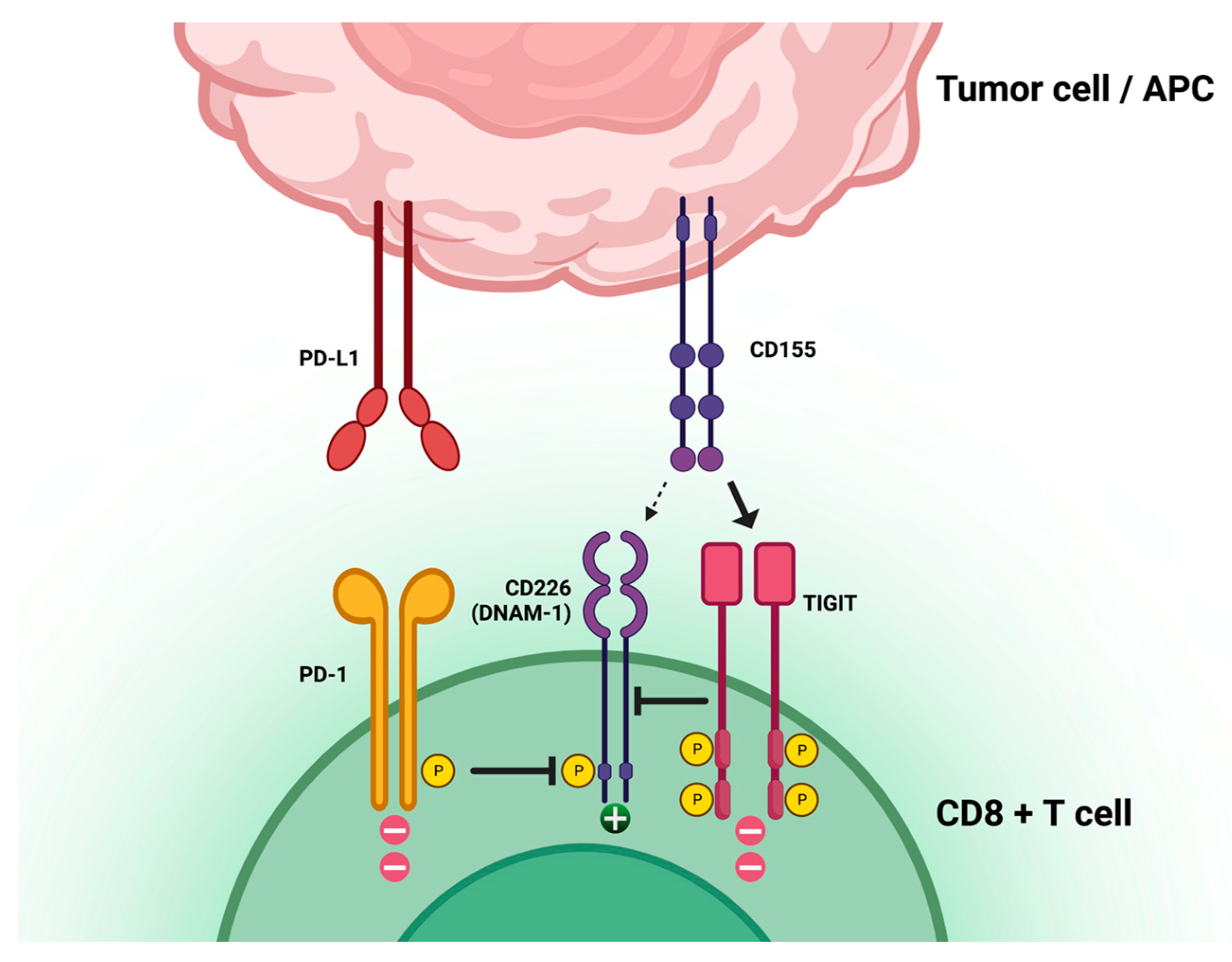
:1. Introduction
2. Materials and Methods
3. Discussion
3.1. Clinical Trials in Lung Cancer Utilizing TIGIT-Blockade

| Agent | Isotype | Company/Sponsor | Clinical Phase | Identifier |
|---|---|---|---|---|
| Tiragolumab | Fully human IgG1/kappa | Roche | II/III | NCT03563716 [11,22,34] NCT04294810 NCT04513925 NCT04619797 NCT04832854 NCT04958811 NCT05034055 NCT03977467 NCT04308785 NCT04256421 |
| Vibostolimab | Fully human IgG1 | Merck Sharp and Dohme | I//II/III | NCT04165798 NCT04725188 NCT04738487 NCT02964013 [23] NCT04165070 |
| Ociperlimab | Humanized IgG1 | BeiGene | II/III | NCT04746924 [26] NCT04866017 NCT04952597 NCT05014815 |
| Domvanalimab | Fully human IgG1 | Arcus Biosciences | I//II/III | NCT04262856 [25] NCT04736173 NCT04791839 NCT03628677 |
| EOS-448 | Fully human IgG1 | iTeos Therapeutics | I/II | NCT05060432 NCT03739710 |
| SEA-TGT | Nonfucosylated human IgG1 | Seagen Inc | Ib/II | NCT04585815 |
| IBI939 | Fully human | Innovent Biologics | I | NCT04672369 NCT04672356 |
| AZD2936 | Humanized IgG1 | AstraZeneca | I/II | NCT04995523 |
| HLX301 | Recombinant Humanized IgG1 | Shanghai Henlius Biotech | I/II | NCT05102214 |
| Etigilimab | Humanized IgG1 | OncoMed Pharmaceuticals | I | NCT03119428 [28] |
| Trial ID | References | Status | Therapy Regimen | Setting | Trial Phase and Type | N |
|---|---|---|---|---|---|---|
| CITYSCAPE NCT03563716 | Cho et al., Lancet Oncol 2022 [11]; Bendell et al., Cancer Res 2020 [22] | Active, non recruiting | Tiragolumab + atezolizumab vs. placebo + atezolizumab | EGFR/ALK wild-type NSCLC with PD-L1 ≥ 1% | Phase II, randomised, double-blinded, placebo-controlled | 67 vs. 68 |
| SKYSCRAPER-01 NCT04294810 | - | Recruiting | Tiragolumab + atezolizumab vs. placebo + atezolizumab | Untreated locally advanced unresectable or metastatic NSCLC with PD-L1 ≥ 50% | Phase III, randomized, double-blinded, placebo-controlled | Estimated 660 |
| SKYSCRAPER-03 NCT04513925 | - | Recruiting | Tiragolumab + atezolizumab vs. Durvalumab | Locally advanced, unresectable stage III NSCLC, after cCRT | Phase III, randomized, open-label | Estimated 800 |
| SKYSCRAPER-06 NCT04619797 | - | Recruiting | Tiragolumab + atezolizumab + pemetrexed + carboplatin or cisplatin vs. pembrolizumab pemetrexed + carboplatin or cisplatin | Previously untreated advanced non-squamous NSCLC | Phase II, randomized, double-blinded, placebo-controlled | Estimated 540 |
| NCT04832854 | - | Recruiting | Neoadjuvant and adjuvant tiragolumab + atezolizumab, with or without platinum-based chemotherapy | Resectable stage II, IIIA, or select III B NSCLC | Phase II, multicenter, open-label | Estimated 82 |
| NCT04958811 | - | Recruiting | Tiragolumab with atezolizumab + bevacizumab | ICI pretreated, PD-L1+, non-squamous NSCLC | Phase II, multi-cohort, open-label | Estimated 42 |
| SKYROCKET NCT05034055 | - | Not yet recruiting | SBRT followed by atezolizumab/tiragolumab | Treatment naïve metastatic NSCLC | Phase II, open-label | Estimated 45 |
| NCT03977467 | - | Recruiting | Atezolizumab + tiragolumab | NSCLC or advanced solid tumors with prior PD-1 inhibitor treatment | Phase II, open-label | Estimated 80 |
| NCT04308785 | - | Active, non recruiting | Atezolizumab ± tiragolumab as consolidation therapy | Limited stage SCLCs who have not progressed to chemoradiotherapy | phase II, randomized, double-blinded, placebo-controlled | 24 |
| SKYSCRAPER-02 NCT04256421 | - | Active, non recruiting | Atezolizumab + carboplatin and etoposide ± tiragolumab | Untreated extensive stage SCLC | Phase III, randomized, double-blinded, placebo-controlled | 490 |
| NCT02964013 | Niu et al., Ann Oncol 2022 [23] | Active, non recruiting | Vibostolimab vs. vibostolimab + pembrolizumab vs. vibostolimab + pembrolizumab | Anti-PD-1/PD-L1-refractory NSCLC | Phase I, multicenter, open-label | 34 vs. 33 vs. 39 |
| Anti-PD-1/PD-L1-refractory NSCLC | ||||||
| Anti-PD-1/PD-L1-naive NSCLC | ||||||
| KEYMAKER-U01 NCT04165798 | - | Recruiting | Vibostolimab + pembrolizumab + chemotherapy vs. vibostolimab + pembrolizumab vs. vibostolimab + pembrolizumab | Treatment naive NSCLC | Phase II, multi-cohort | Estimated 260 |
| Treatment naïve PD-L1 positive NSCLC | ||||||
| NSCLC previously treated with anti-PD-L1 NSCLC | ||||||
| NCT04738487 | - | Recruiting | Pembrolizumab/vibostolimab coformulation (MK-7684°) vs. pembrolizumab | NSCLC with PD-L1 ≥ 1% | Phase III, multicenter, randomized, double-blinded | Estimated 1246 |
| NCT04165070 | - | Recruiting | Pembrolizumab + carboplatin + paclitaxel vs. vibostolimab | Treatment naïve advanced NSCLC | Phase II, open-label | Estimated 360 |
| NCT04725188 | - | Active, non recruiting | Pembrolizumab/vibostolimab coformulation (MK-7684A) or pembrolizumab/vibostolimab coformulation (MK-7684A) + docetaxel vs. docetaxel | ICI and platinum chemotherapy pretreated | Phase II, multicenter, randomized | Estimated 240 |
| ARC-7 NCT04262856 | Catalano et al., Cancers (Basel). 2022 [25] | Active, non recruiting | Domvanalimab + zimberelimab (A2BR antagonist) vs. zimberelimab vs domvanalimab + zimberelimab + etrumadenant (dual adenosine A2a/A2b receptor antagonist) | NSCLC with PD-L1 expression of ≥ 50% | Phase II, open-label, randomized | Estimated 150 |
| ARC-10 NCT04736173 | - | Recruiting | Domvanalimab + zimberelimab vs. zimberelimab vs. chemotherapy | Locally advanced or metastatic NSCLC, with PD-L1 ≥ 1% | Phase III, open-label, randomized | Estimated 625 |
| NCT04791839 | - | Recruiting | Domvanalimab + zimberelimab (anti-PD-1) + etrumadenant (A2R inhibitor) | ICI pretreated, NSCLC with PD-L1 ≥ 1% | Phase II, open-label | Estimated 30 |
| NCT03628677 | - | Active, non recruiting | Domvanalimab ± AB122 (anti PD-1) | Advanced or metastatic NSCLC, SCCHN, RCC, breast cancer, colorectal cancer, melanoma, bladder cancer, ovarian cancer, endometrial cancer, Merkel cell carcinoma, or gastroesophageal cancer | Phase I, open-label | 75 |
| AdvanTIG-302 NCT04746924 | Socinski et al., Clin Oncol. 2021 [26] | Recruiting | Ociperlimab + tislelizumab vs. pembrolizumab + placebo vs. tislelizumab + placebo | NSCLC and PD-L1 tumor cell ≥ 50% expression | Phase III multicenter, randomized, double-blind | Estimated 660 |
| NCT04866017 | - | Recruiting | Ociperlimab + tislelizumab + cCRT → ociperlimab + tislelizumab or tislelizumab + cCRT → tislelizumab vs. cCRT → durvalumab | Untreated, locally advanced, unresectable NSCLC | Phase III, open-label, randomized | Estimated 900 |
| NCT04952597 | - | Active, non recruiting | Ociperlimab + tislelizumab + CRT | Untreated, limited stage SCLC | Phase II, multicenter, randomized, open-label | 126 |
| NCT05014815 | - | Recruiting | Ociperlimab and tislelizumab + CT | Untreated locally advanced, unresectable, or metastatic | Phase II, randomized | Estimated 270 |
| NCT05060432 | - | Recruiting | EOS-448 + SoC and/or investigational therapies | Advanced NSCLC | Phase I/II, multicenter, open-label | Estimated 376 |
| NCT03739710 | - | Recruiting | Feladilimab, ipilimumab (anti-CTLA-4), EOS-448, dostarlimab (various combination) vs. SoC | Relapsed/refractory advanced NSCLC | Phase II, open-label, randomized | Estimated 185 |
| NCT04672369 | - | Active, non recruiting | IBI939 + sintilimab (anti-PD-1) | Advanced LC | Phase I, open-label, randomized | Estimated 42 |
| NCT04672356 | - | Active, non recruiting | IBI939 + sintilimab | Advanced LC | Phase I, open-label | Estimated 20 |
| NCT04585815 | - | Active, non recruiting | SEA-TGT + sasanlimab (anti-PD-1) + Axitinib | Advanced NSCLC | Phase Ib/II, open-label | 23 |
| ARTEMIDE-01 NCT04995523 | - | Recruiting | AZD2936 (anti-TIGIT/anti-PD-1 bispecific antibody) | Locally advanced or metastatic NSCLC | Phase I/II, open-label | Estimated 192 |
| NCT05102214 | - | Recruiting | HLX301 (PDL1/TIGIT bispecific Ab) | Locally advanced or metastatic solid tumors | Phase I/II, open-label | Estimated 150 |
| NCT03119428 | Mettu et al., Clin Cancer Res., 2022 [28] | Terminated (Sponsor decision) | Etigilimab ± nivolumab (anti PD-1 mAb) | Advanced or metastatic solid tumors | Phase I, open-label | 33 |
3.2. TIGIT as an Immunohistochemical Biomarker: Current Knowledge
| Antibody | Publication | Cancer Type | Visualization | Correlations | p-Value |
|---|---|---|---|---|---|
| Abcam, ab243903 Rabbit monoclonal (BLR047F clone) | Wang, P. et al. [46] | Esophageal cancer | H-score | No difference in 3-year OS between TIGIT+ and TIGIT- cases | 0.140 |
| Xu, X. et al. [53] | Esophageal cancer | Multiplex fluorescence immunohistochemistry | TIGIT expression in TME is positively associated with AIF1 expression, a differentially expressed gene that negatively impacts on prognosis. | 0.013 | |
| Steele, NG. et al. [54] | Pancreatic ductal adenocarcinoma | Multiplex fluorescence immunohistochemistry | Validation at the protein level that CD8+ TILs show enriched TIGIT expression | / | |
| Liu, Z. et al. [55] | Urothelial carcinoma | Mean number of positive cells extracted from the view of three HPF | TIGIT+ CD8+ cells high infiltration group possessed inferior OS and RFS compared with the TIGIT+ CD8+ cells low infiltration group | 0.01 | |
| Liu, Z. et al. [56] | Urothelial carcinoma | Mean number of positive cells extracted from the view of three HPF | PD-1+ cells infiltration had no prognostic value in patients with high TIGIT+ cells infiltration. Patients with high TIGIT expression, irrespectively of the number of PD1+ cells, exhibited poorer prognosis | 0.024 | |
| Eichberger, J. et al. [51] | Oral squamous cell carcinoma | Assessment of semiquantitative percentage of TIGIT expression within CD3+ T cells (ranging from 0–100%) | TIGIT expression on CD3+ cells correlates with improved OS | 0.025 | |
| Shi, X. et al. [47] | Medullary thyroid carcinoma | Combined positive score (CPS) algorithm, defined as the percentage of positive tumor cells (total/partial membrane staining) and TILs (membrane/cytoplasm staining) relative to the total number of tumor cells, multiplied by 100. Expression was further stratified into low (1 ≤ CPS < 5), moderate (5 ≤ CPS < 20), and strong (CPS ≥ 20). | TIGIT expression had no impact on prognosis | 0.448 | |
| Guo, C. et al. [57] | Breast cancer | ImageJ analysis of IHC | TIGIT is significantly upregulated in invasive breast tumor TME compared with normal tissues; this finding is confirmed using IHC | / | |
| Duan, X. et al. [58] | Hepatocellular carcinoma | Manual counting | TIGIT expression in TILs gradually increased in liver cancer tissues as the degree of tumor cell differentiation changed from high to low | / | |
| Nakazawa, T. et al. [41] | Thyroid cancer | Semiquantitative evaluation of percentage of positive epithelial cells (0: less than 1%, 1+: 1–49%, and 2+: more than 50%) | Expression in tumor cells was detected in medullary thyroid carcinoma, anaplastic thyroid carcinoma, and poorly differentiated thyroid carcinoma, while it was absent in benign lesions/tumors and differentiated carcinomas. Pleomorphic/giant cell morphology seemed to correlate with TIGIT expression in anaplastic thyroid carcinomas. | <0.05 | |
| Jiang, C. et al. [48] | Non-small cell lung cancer | Inflammatory infiltrates in all the samples were assessed and subclassified semi quantitatively into TIGIT-negative (≤5% stained) or positive (>5% stained) | TIGIT expression in TME had no impact on PFS in patients treated with anti-PD1 therapy | 0.092 | |
| Ishihara, S. et al. [42] | Undifferentiated pleomorphic sarcoma | TIGIT expression was considered low when tumor cells did not express TIGIT or showed a very weak immunopositivity despite immune cells showing strongly positive expression | Expression of TIGIT on tumor cells tended to be associated with poorer OS | 0.555 | |
| Luo, Y. et al. [59] | Advanced thyroid carcinomas | Combined positive score (CPS) algorithm, defined as the percentage of positive tumor cells (total/partial membrane staining) and TILs (membrane/cytoplasm staining) relative to the total number of tumor cells, multiplied by 100. Expression was further stratified into negative (CPS <1), weak (1 ≤ CPS < 10), moderate (10 ≤ CPS < 30), and strong (CPS ≥30) | TIGIT expression had a negative impact on OS | 0.004 | |
| Stålhammar, G. et al. [40] | Choroidal melanoma | Number of TIGIT positive cells per 3 HPF, corresponding to an aggregated area of 0.6 mm2 | Time dependent hazard for metastasis was significantly increased for patients with a number of TIGIT positive cells/mm2 in primary tumor hot spots above the median | 0.03 | |
| TIGIT XP® #99567 Rabbit monoclonal (E5Y1W clone) | Liu, L. et al. [60] | Cervical cancer | Multiplex fluorescence immunohistochemistry | The number of CD8+TIGIT+ cells in cervical cancer tissues was significantly higher than that in adjacent cancer tissues. | <0.01 |
| Liu, H. et al. [45] | Gastric cancer | Dual IHC, counting the number of TIGIT+CD20+ B cells in three representative HPFs (×200 amplification), was calculated for each section and the average of the three values was used as the final counting result | High peritumoral TIGIT+CD20+ B cell infiltration was associated with worse
| < 0.001 0.0252 | |
| Boissière-Michot, F. et al. [52] | Breast cancer | H-score | TIGIT+ cell density in TME tended to be associated with better RFS | 0.079 | |
| Yang, Z. et al. [35] | Lung squamous cell carcinoma | The number of TIGIT+ TILs was counted in six HPFs. TIGIT density was defined as high or low using the median count as the cut-off value. | High TIGIT density was correlated with positive PD-L1 expression, high PD-1 density, and high CD8 density. High TIGIT density correlated with worse prognosis. | / 0.027 | |
| Ducoin, K. et al. [61] | Colorectal cancer | Regions of interest were drawn (tumor glands and peritumoral stroma near the invasive margin). In each region (tumor and stroma), a total number of 5000 cells were counted in the 3 areas per section, and the results are expressed as the mean of the 3 counts | Microsatellite instability correlate with higher expression of TIGIT+CD3+ TILs | 0.0131 | |
| Shen, M. et al. [49] | Lung adenocarcinoma | Inflammatory infiltrates in all samples were assessed and subclassified semi quantitatively into TIGIT-negative (≤5% stained) or positive (>5% stained) | TIGIT expression had no impact on
| 0.564 0.152 | |
| TIGIT antibody Dianova, Hamburg, Germany Rabbit monoclonal (TG1 clone) | Blessin, N.C. et al. [62] | Human cancer TMA | The number of TIGIT+ cells per 0.6 mm tissue spot was manually counted and converted into the density of TIGIT+ cells per square mm | Highest densities of TIGIT+ TILs were found in tumors characterized by high numbers of TILs. In colorectal cancers, expression of TIGIT and PD-1 was considerably higher in T cells located at the invasive margin as compared with T cells in the tumor center, overlapping with PD1 expression | / |
| Li, W. et al. [63] | Hodgkin’s lymphoma | Percentage of stained cells in the lymphocytic background (median value 86%) | Highest staining intensities were found in a case of NLPHL; staining intensity of the T-cell rosettes surrounding malignant cells in NLPHL and in LRCHL appeared stronger | / | |
| Niebel, D. et al. [39] | Melanoma | H-score for cancer cells; TIGIT+ immune cells were assessed as percentage fraction from all cells (TIGIT+ lymphocyte score) | Patients with TIGIT+ lymphocyte scores > 1% had a significant better progression-free survival compared with patients with TIGIT+ lymphocyte scores ≤ 1%. TIGIT was detected also in several melanoma cells | 0.010 | |
| Müller, S. et al. [43] | Lung adenocarcinoma | H-score | TIGIT expression was heterogeneous among cancer cells and TILs. TIGIT expression was observed in malignant and not in benign cells, with increasing proportions from pre-malignant to overtly malignant lesions | / | |
| Scimeca, M. et al. [64] | Prostate adenocarcinoma | TIGIT+ TILs were evaluated with the support of a digital software (Image Viewer, Ventana, Roche) by two blind observers by counting the number of positive prostate cells on 9.42 mm2 prostate tissues | No significant differences were observed in TIGIT+ TILs between prostate adenocarcinoma and benign lesions | 0.9833 | |
| TIGIT antibody Biomatik, Wilmington, DE, USA Rabbit monoclonal (TG1 clone) | Lee, W. J. et al. [37] | Cutaneous melanoma | Staining intensity on TILs was determined on a scale of 0–3, with zero indicating <5%, 1 indicating 5–20%, 2 indicating >20–50%, and 3 indicating >50% of TILs. Cases with a score ≥1 were considered positive. | High TIGIT expression in TILs was associated with deeper Breslow thickness, more vertical growth, higher mitotic counts, higher frequency of lymph node involvement and advanced AJCC stage, higher density of TILs, higher expression of PD-1, and poorer OS and PFS | <0.04 for all parameters |
| TIGIT Santa Cruz sc-103349 | Lucca L.E. et al. [65] | Glioblastoma (GBM) and multiple sclerosis (SM) samples | Immunolabeled cells with a lymphocytic morphology were manually quantified and the counts were averaged. The number of TIGIT+ cells was correlated with the number of CD3+ lymphocytes found in each region of interest. |
| 0.04 0.017 |
| Xu, Y. et al. [50] | Lung small cell carcinoma | Positively stained sections were analyzed using the integrated optical density (IOD) and the areas of staining distribution with NIS-Elements Br version 4.60.00; the mean density was obtained by dividing the IOD value by the area, and an average from 5 representative fields was calculated (magnification, ×400) | TIGIT expression did not impact OS | 0.874 | |
| TIGIT MYBioSource #MBS20013451, Rabbit polyclonal | Sun, Y. et al. [36] | Lung adenocarcinoma | Inflammatory infiltrates in all the samples were assessed and subclassified semi quantitatively into TIGIT-negative (≤5% stained) or positive (>5% stained) | TIGIT expression positively correlated with PD-1 and PD-L1 and portended worse OS | 0.024 |
| TIGIT NBP2-79793, Novus, USA Rabbit monoclonal (TIGIT/3017 clone) | Zhao, K. et al. [44] | Esophageal small cell carcinoma | TIGIT expression was assessed manually and semi-quantitatively in tumor cells as follows: ≤5% staining was considered negative and >5% staining was scored as positive |
| <0.001 0.001 0.034 |
| TIGIT Thermo Fisher Scientific, Rabbit monoclonal (MBSA43 clone) | Zhao, J. J. et al. [66] | Esophageal squamous cell carcinoma | Average number of TIGIT+ immune cells was calculated as the final density of each section (cells/mm2) | Patients carrying a high number of TIGIT+ TILs (n = 76/154, 49.4%) tended to exhibit a shorter OS Cancers enriched with PD-1+/TIGIT+ TILs demonstrated significantly lower survival rates than patients with PD-1−/TIGIT− TILs | 0.045 0.005 |
| TIGIT IHC assay Roche Tissue Diagnostics SP410 antibody | Patil, N. et al. [34] | Non-small cell lung cancer (CITYSCAPE TRIAL) | Evaluating immune cells only, ≤5% staining was considered low and >5% staining was scored as high | No association between high TIGIT expression and PFS | / |
4. Conclusions
Author Contributions
Funding
Institutional Review Board Statement
Informed Consent Statement
Data Availability Statement
Conflicts of Interest
References
- Chauvin, J.M.; Zarour, H.M. TIGIT in cancer immunotherapy. J. Immunother. Cancer 2020, 8, e000957. [Google Scholar] [CrossRef] [PubMed]
- He, X.; Xu, C. Immune checkpoint signaling and cancer immunotherapy. Cell Res. 2020, 30, 660–669. [Google Scholar] [CrossRef] [PubMed]
- McGranahan, N.; Rosenthal, R.; Hiley, C.T.; Rowan, A.J.; Watkins, T.B.K.; Wilson, G.A.; Birkbak, N.J.; Veeriah, S.; Van Loo, P.; Herrero, J.; et al. Allele-Specific HLA Loss and Immune Escape in Lung Cancer Evolution. Cell 2017, 171, 1259–1271.e1211. [Google Scholar] [CrossRef]
- Iwai, Y.; Ishida, M.; Tanaka, Y.; Okazaki, T.; Honjo, T.; Minato, N. Involvement of PD-L1 on tumor cells in the escape from host immune system and tumor immunotherapy by PD-L1 blockade. Proc. Natl. Acad. Sci. USA 2002, 99, 12293–12297. [Google Scholar] [CrossRef]
- Borghaei, H.; Paz-Ares, L.; Horn, L.; Spigel, D.R.; Steins, M.; Ready, N.E.; Chow, L.Q.; Vokes, E.E.; Felip, E.; Holgado, E.; et al. Nivolumab versus Docetaxel in Advanced Nonsquamous Non-Small-Cell Lung Cancer. N. Engl. J. Med. 2015, 373, 1627–1639. [Google Scholar] [CrossRef] [PubMed]
- Ribas, A.; Wolchok, J.D. Cancer immunotherapy using checkpoint blockade. Science 2018, 359, 1350–1355. [Google Scholar] [CrossRef] [PubMed]
- Das, M.; Zhu, C.; Kuchroo, V.K. Tim-3 and its role in regulating anti-tumor immunity. Immunol. Rev. 2017, 276, 97–111. [Google Scholar] [CrossRef]
- Andrews, L.P.; Marciscano, A.E.; Drake, C.G.; Vignali, D.A. LAG3 (CD223) as a cancer immunotherapy target. Immunol. Rev. 2017, 276, 80–96. [Google Scholar] [CrossRef]
- Zhao, Y.; Yang, W.; Huang, Y.; Cui, R.; Li, X.; Li, B. Evolving Roles for Targeting CTLA-4 in Cancer Immunotherapy. Cell. Physiol. Biochem. 2018, 47, 721–734. [Google Scholar] [CrossRef]
- Boles, K.S.; Vermi, W.; Facchetti, F.; Fuchs, A.; Wilson, T.J.; Diacovo, T.G.; Cella, M.; Colonna, M. A novel molecular interaction for the adhesion of follicular CD4 T cells to follicular DC. Eur. J. Immunol. 2009, 39, 695–703. [Google Scholar] [CrossRef]
- Cho, B.C.; Abreu, D.R.; Hussein, M.; Cobo, M.; Patel, A.J.; Secen, N.; Lee, K.H.; Massuti, B.; Hiret, S.; Yang, J.C.H.; et al. Tiragolumab plus atezolizumab versus placebo plus atezolizumab as a first-line treatment for PD-L1-selected non-small-cell lung cancer (CITYSCAPE): Primary and follow-up analyses of a randomised, double-blind, phase 2 study. Lancet Oncol. 2022, 23, 781–792. [Google Scholar] [CrossRef] [PubMed]
- Freed-Pastor, W.A.; Lambert, L.J.; Ely, Z.A.; Pattada, N.B.; Bhutkar, A.; Eng, G.; Mercer, K.L.; Garcia, A.P.; Lin, L.; Rideout, W.M.; et al. The CD155/TIGIT axis promotes and maintains immune evasion in neoantigen-expressing pancreatic cancer. Cancer Cell 2021, 39, 1342–1360.e1314. [Google Scholar] [CrossRef] [PubMed]
- Ozmadenci, D.; Shankara Narayanan, J.S.; Andrew, J.; Ojalill, M.; Barrie, A.M.; Jiang, S.; Iyer, S.; Chen, X.L.; Rose, M.; Estrada, V.; et al. Tumor FAK orchestrates immunosuppression in ovarian cancer via the CD155/TIGIT axis. Proc. Natl. Acad. Sci. USA 2022, 119, e2117065119. [Google Scholar] [CrossRef] [PubMed]
- Zou, Y.; Ye, F.; Kong, Y.; Hu, X.; Deng, X.; Xie, J.; Song, C.; Ou, X.; Wu, S.; Wu, L.; et al. The Single-Cell Landscape of Intratumoral Heterogeneity and The Immunosuppressive Microenvironment in Liver and Brain Metastases of Breast Cancer. Adv. Sci. 2023, 10, e2203699. [Google Scholar] [CrossRef] [PubMed]
- He, W.; Zhang, H.; Han, F.; Chen, X.; Lin, R.; Wang, W.; Qiu, H.; Zhuang, Z.; Liao, Q.; Zhang, W.; et al. CD155T/TIGIT Signaling Regulates CD8+ T-cell Metabolism and Promotes Tumor Progression in Human Gastric Cancer. Cancer Res. 2017, 77, 6375–6388. [Google Scholar] [CrossRef]
- Harjunpää, H.; Guillerey, C. TIGIT as an emerging immune checkpoint. Clin. Exp. Immunol. 2020, 200, 108–119. [Google Scholar] [CrossRef]
- Shao, Q.; Wang, L.; Yuan, M.; Jin, X.; Chen, Z.; Wu, C. TIGIT Induces (CD3+) T Cell Dysfunction in Colorectal Cancer by Inhibiting Glucose Metabolism. Front. Immunol. 2021, 12, 3937. [Google Scholar] [CrossRef]
- Liu, S.; Zhang, H.; Li, M.; Hu, D.; Li, C.; Ge, B.; Jin, B.; Fan, Z. Recruitment of Grb2 and SHIP1 by the ITT-like motif of TIGIT suppresses granule polarization and cytotoxicity of NK cells. Cell Death Differ. 2013, 20, 456–464. [Google Scholar] [CrossRef]
- Shibuya, A.; Shibuya, K. DNAM-1 versus TIGIT: Competitive roles in tumor immunity and inflammatory responses. Int. Immunol. 2021, 33, 687–692. [Google Scholar] [CrossRef]
- Bolm, L.; Petruch, N.; Sivakumar, S.; Annels, N.E.; Frampton, A.E. Gene of the month: T-cell immunoreceptor with immunoglobulin and ITIM domains (TIGIT). J. Clin. Pathol. 2022, 75, 217–221. [Google Scholar] [CrossRef]
- ClinicalTrials.gov. Available online: https://clinicaltrials.gov/ (accessed on 21 March 2023).
- Bendell, J.C.; Bedard, P.; Bang, Y.-J.; LoRusso, P.; Hodi, S.; Gordon, M.; D’Angelo, S.; Desai, J.; Garralda, E.; Italiano, A.; et al. Abstract CT302: Phase Ia/Ib dose-escalation study of the anti-TIGIT antibody tiragolumab as a single agent and in combination with atezolizumab in patients with advanced solid tumors. Cancer Res. 2020, 80, CT302. [Google Scholar] [CrossRef]
- Niu, J.; Maurice-Dror, C.; Lee, D.H.; Kim, D.W.; Nagrial, A.; Voskoboynik, M.; Chung, H.C.; Mileham, K.; Vaishampayan, U.; Rasco, D.; et al. First-in-human phase 1 study of the anti-TIGIT antibody vibostolimab as monotherapy or with pembrolizumab for advanced solid tumors, including non-small-cell lung cancer. Ann. Oncol. 2022, 33, 169–180. [Google Scholar] [CrossRef] [PubMed]
- Attili, I.; Passaro, A.; de Marinis, F. Anti-TIGIT to overcome resistance to immune checkpoint inhibitors in lung cancer: Limits and potentials. Ann. Oncol. 2022, 33, 119–122. [Google Scholar] [CrossRef]
- Catalano, M.; Shabani, S.; Venturini, J.; Ottanelli, C.; Voltolini, L.; Roviello, G. Lung Cancer Immunotherapy: Beyond Common Immune Checkpoints Inhibitors. Cancers 2022, 14, 6145. [Google Scholar] [CrossRef] [PubMed]
- Socinski, M.; Spira, A.; Paz-Ares, L. AdvanTIG-302: Anti-TIGIT monoclonal antibody (mAb) ociperlimab (OCI) plus tislelizumab (TIS) versus pembrolizumab (PEM) in programmed death ligand-1 (PD-L1) selected, previously untreated, locally advanced, unresectable or metastatic non-small cell lung cancer (NSCLC). J. Clin. Oncol. 2021, 39, TPS9128. [Google Scholar]
- De Giglio, A.; Di Federico, A.; Nuvola, G.; Deiana, C.; Gelsomino, F. The Landscape of Immunotherapy in Advanced NSCLC: Driving Beyond PD-1/PD-L1 Inhibitors (CTLA-4, LAG3, IDO, OX40, TIGIT, Vaccines). Curr. Oncol. Rep. 2021, 23, 126. [Google Scholar] [CrossRef]
- Mettu, N.B.; Ulahannan, S.V.; Bendell, J.C.; Garrido-Laguna, I.; Strickler, J.H.; Moore, K.N.; Stagg, R.; Kapoun, A.M.; Faoro, L.; Sharma, S. A Phase 1a/b Open-Label, Dose-Escalation Study of Etigilimab Alone or in Combination with Nivolumab in Patients with Locally Advanced or Metastatic Solid Tumors. Clin. Cancer Res. 2022, 28, 882–892. [Google Scholar] [CrossRef]
- Florou, V.; Garrido-Laguna, I. Clinical Development of Anti-TIGIT Antibodies for Immunotherapy of Cancer. Curr. Oncol. Rep. 2022, 24, 1107–1112. [Google Scholar] [CrossRef]
- Johnston, R.J.; Comps-Agrar, L.; Hackney, J.; Yu, X.; Huseni, M.; Yang, Y.; Park, S.; Javinal, V.; Chiu, H.; Irving, B.; et al. The immunoreceptor TIGIT regulates antitumor and antiviral CD8(+) T cell effector function. Cancer Cell 2014, 26, 923–937. [Google Scholar] [CrossRef] [PubMed]
- Dixon, K.O.; Schorer, M.; Nevin, J.; Etminan, Y.; Amoozgar, Z.; Kondo, T.; Kurtulus, S.; Kassam, N.; Sobel, R.A.; Fukumura, D.; et al. Functional Anti-TIGIT Antibodies Regulate Development of Autoimmunity and Antitumor Immunity. J. Immunol. 2018, 200, 3000–3007. [Google Scholar] [CrossRef]
- Sanchez-Correa, B.; Lopez-Sejas, N.; Duran, E.; Labella, F.; Alonso, C.; Solana, R.; Tarazona, R. Modulation of NK cells with checkpoint inhibitors in the context of cancer immunotherapy. Cancer Immunol. Immunother. 2019, 68, 861–870. [Google Scholar] [CrossRef] [PubMed]
- Houssaini, M.S.; Damou, M.; Ismaili, N. Advances in the management of non-small cell lung cancer (NSCLC): A new practice changing data from asco 2020 annual meeting. Cancer Treat. Res. Commun. 2020, 25, 100239. [Google Scholar] [CrossRef] [PubMed]
- Patil, N.; Cho, B.C.; Johnson, M.; Caro, R.B.; Spira, A.; Chiu, C.; Molden, N.; Pham, T.; Yang, X.; Choi, Y.; et al. P77.02 Efficacy of Tiragolumab + Atezolizumab in PD-L1 IHC and TIGIT Subgroups in the Phase II CITYSCAPE Study in First-Line NSCLC. J. Thorac. Oncol. 2021, 16, S635–S636. [Google Scholar] [CrossRef]
- Yang, Z.; Peng, Y.; Xu, J.; Chen, P.; Zhao, Z.; Cai, Q.; Li, L.; Tian, H.; Bai, G.; Liu, L.; et al. PVR/TIGIT and PD-L1/PD-1 expression predicts survival and enlightens combined immunotherapy in lung squamous cell carcinoma. Transl. Oncol. 2022, 24, 101501. [Google Scholar] [CrossRef] [PubMed]
- Sun, Y.; Luo, J.; Chen, Y.; Cui, J.; Lei, Y.; Cui, Y.; Jiang, N.; Jiang, W.; Chen, L.; Chen, Y.; et al. Combined evaluation of the expression status of CD155 and TIGIT plays an important role in the prognosis of LUAD (lung adenocarcinoma). Int. Immunopharmacol. 2020, 80, 106198. [Google Scholar] [CrossRef]
- Lee, W.J.; Lee, Y.J.; Choi, M.E.; Yun, K.A.; Won, C.H.; Lee, M.W.; Choi, J.H.; Chang, S.E. Expression of lymphocyte-activating gene 3 and T-cell immunoreceptor with immunoglobulin and ITIM domains in cutaneous melanoma and their correlation with programmed cell death 1 expression in tumor-infiltrating lymphocytes. J. Am. Acad. Dermatol. 2019, 81, 219–227. [Google Scholar] [CrossRef]
- Sidaway, P. Tiragolumab active in PD-L1(+) NSCLC. Nat. Rev. Clin. Oncol. 2022, 19, 428. [Google Scholar] [CrossRef]
- Niebel, D.; Fröhlich, A.; Zarbl, R.; Fietz, S.; de Vos, L.; Vogt, T.J.; Dietrich, J.; Sirokay, J.; Kuster, P.; Saavedra, G.; et al. DNA methylation regulates TIGIT expression within the melanoma microenvironment, is prognostic for overall survival, and predicts progression-free survival in patients treated with anti-PD-1 immunotherapy. Clin. Epigenetics 2022, 14, 50. [Google Scholar] [CrossRef]
- Stålhammar, G.; Seregard, S.; Grossniklaus, H.E. Expression of immune checkpoint receptors Indoleamine 2,3-dioxygenase and T cell Ig and ITIM domain in metastatic versus nonmetastatic choroidal melanoma. Cancer Med. 2019, 8, 2784–2792. [Google Scholar] [CrossRef]
- Nakazawa, T.; Nagasaka, T.; Yoshida, K.; Hasegawa, A.; Guo, F.; Wu, D.; Hiroshima, K.; Katoh, R. Expression of T-cell immunoreceptor with immunoglobulin and tyrosine-based inhibitory motif domains (TIGIT) in anaplastic thyroid carcinoma. BMC Endocr. Disord. 2022, 22, 204. [Google Scholar] [CrossRef]
- Ishihara, S.; Iwasaki, T.; Kohashi, K.; Kawaguchi, K.; Toda, Y.; Fujiwara, T.; Setsu, N.; Endo, M.; Matsumoto, Y.; Nakashima, Y.; et al. Clinical significance of signal regulatory protein alpha and T cell immunoreceptor with immunoglobulin and immunoreceptor tyrosine-based inhibition motif domain expression in undifferentiated pleomorphic sarcoma. J. Cancer Res. Clin. Oncol. 2022; Epub ahead of print. [Google Scholar] [CrossRef]
- Müller, S.; Mayer, S.; Möller, P.; Barth, T.F.E.; Marienfeld, R. Spatial distribution of immune checkpoint proteins in histological subtypes of lung adenocarcinoma. Neoplasia 2021, 23, 584–593. [Google Scholar] [CrossRef] [PubMed]
- Zhao, K.; Ma, L.; Feng, L.; Huang, Z.; Meng, X.; Yu, J. CD155 Overexpression Correlates With Poor Prognosis in Primary Small Cell Carcinoma of the Esophagus. Front. Mol. Biosci. 2020, 7, 608404. [Google Scholar] [CrossRef] [PubMed]
- Liu, H.; Wu, J.; Xu, X.; Wang, H.; Zhang, C.; Yin, S.; He, Y. Peritumoral TIGIT+CD20+ B cell infiltration indicates poor prognosis but favorable adjuvant chemotherapeutic response in gastric cancer. Int. Immunopharmacol. 2022, 108, 108735. [Google Scholar] [CrossRef] [PubMed]
- Wang, P.; Chen, Y.; Long, Q.; Li, Q.; Tian, J.; Liu, T.; Wu, Y.; Ding, Z. Increased coexpression of PD-L1 and TIM3/TIGIT is associated with poor overall survival of patients with esophageal squamous cell carcinoma. J. Immunother. Cancer 2021, 9, e002836. [Google Scholar] [CrossRef]
- Shi, X.; Li, C.W.; Tan, L.C.; Wen, S.S.; Liao, T.; Zhang, Y.; Chen, T.Z.; Ma, B.; Yu, P.C.; Lu, Z.W.; et al. Immune Co-inhibitory Receptors PD-1, CTLA-4, TIM-3, LAG-3, and TIGIT in Medullary Thyroid Cancers: A Large Cohort Study. J. Clin. Endocrinol. Metab. 2021, 106, 120–132. [Google Scholar] [CrossRef]
- Jiang, C.; Qu, X.; Ma, L.; Yi, L.; Cheng, X.; Gao, X.; Wang, J.; Che, N.; Zhang, H.; Zhang, S. CD155 expression impairs anti-PD1 therapy response in non-small cell lung cancer. Clin. Exp. Immunol. 2022, 208, 220–232. [Google Scholar] [CrossRef]
- Shen, M.; Jiang, K.; Sui, Y.; Xu, Z.; Cui, H.; Wang, Y.; Zhang, H.; Xu, Z.; Xu, W.; Ding, Q.; et al. Characterization of CD66b and its relationship between immune checkpoints and their synergistic impact in the prognosis of surgically resected lung adenocarcinoma. Lung Cancer 2021, 160, 84–91. [Google Scholar] [CrossRef]
- Xu, Y.; Cui, G.; Jiang, Z.; Li, N.; Zhang, X. Survival analysis with regard to PD-L1 and CD155 expression in human small cell lung cancer and a comparison with associated receptors. Oncol. Lett. 2019, 17, 2960–2968. [Google Scholar] [CrossRef]
- Eichberger, J.; Spoerl, S.; Spanier, G.; Erber, R.; Taxis, J.; Schuderer, J.; Ludwig, N.; Fiedler, M.; Nieberle, F.; Ettl, T.; et al. TIGIT Expression on Intratumoral Lymphocytes Correlates with Improved Prognosis in Oral Squamous Cell Carcinoma. Biomedicines 2022, 10, 3236. [Google Scholar] [CrossRef]
- Boissière-Michot, F.; Chateau, M.C.; Thézenas, S.; Guiu, S.; Bobrie, A.; Jacot, W. Correlation of the TIGIT-PVR immune checkpoint axis with clinicopathological features in triple-negative breast cancer. Front. Immunol. 2022, 13, 1058424. [Google Scholar] [CrossRef]
- Xu, X.; Wang, D.; Li, N.; Sheng, J.; Xie, M.; Zhou, Z.; Cheng, G.; Fan, Y. The Novel Tumor Microenvironment-Related Prognostic Gene AIF1 May Influence Immune Infiltrates and is Correlated with TIGIT in Esophageal Cancer. Ann. Surg. Oncol. 2022, 29, 2930–2940. [Google Scholar] [CrossRef] [PubMed]
- Steele, N.G.; Carpenter, E.S.; Kemp, S.B.; Sirihorachai, V.R.; The, S.; Delrosario, L.; Lazarus, J.; Amir, E.D.; Gunchick, V.; Espinoza, C.; et al. Multimodal Mapping of the Tumor and Peripheral Blood Immune Landscape in Human Pancreatic Cancer. Nat. Cancer 2020, 1, 1097–1112. [Google Scholar] [CrossRef] [PubMed]
- Liu, Z.; Zhou, Q.; Wang, Z.; Zhang, H.; Zeng, H.; Huang, Q.; Chen, Y.; Jiang, W.; Lin, Z.; Qu, Y.; et al. Intratumoral TIGIT(+) CD8(+) T-cell infiltration determines poor prognosis and immune evasion in patients with muscle-invasive bladder cancer. J. Immunother. Cancer 2020, 8, e000978. [Google Scholar] [CrossRef] [PubMed]
- Liu, Z.; Zeng, H.; Jin, K.; Yu, Y.; You, R.; Zhang, H.; Liu, C.; Su, X.; Yan, S.; Chang, Y.; et al. TIGIT and PD-1 expression atlas predicts response to adjuvant chemotherapy and PD-L1 blockade in muscle-invasive bladder cancer. Br. J. Cancer 2022, 126, 1310–1317. [Google Scholar] [CrossRef] [PubMed]
- Guo, C.; Luo, Z.; Ismtula, D.; Bi, X.; Kong, H.; Wang, Y.; Yang, Z.; Mao, X. TIGIT is a Novel Prognostic Marker and Correlate for Immune Infiltration in Invasive Breast Cancer. Comb. Chem. High. Throughput. Screen 2023, 26, 639–651. [Google Scholar] [CrossRef] [PubMed]
- Duan, X.; Liu, J.; Cui, J.; Ma, B.; Zhou, Q.; Yang, X.; Lu, Z.; Du, Y.; Su, C. Expression of TIGIT/CD155 and correlations with clinical pathological features in human hepatocellular carcinoma. Mol. Med. Rep. 2019, 20, 3773–3781. [Google Scholar] [CrossRef] [PubMed]
- Luo, Y.; Yang, Y.C.; Shen, C.K.; Ma, B.; Xu, W.B.; Wang, Q.F.; Zhang, Y.; Liao, T.; Wei, W.J.; Wang, Y. Immune Checkpoint Protein Expression Defines the Prognosis of Advanced Thyroid Carcinoma. Front. Endocrinol. 2022, 13, 859013. [Google Scholar] [CrossRef]
- Liu, L.; Wang, A.; Liu, X.; Han, S.; Sun, Y.; Zhang, J.; Guo, L.; Zhang, Y. Blocking TIGIT/CD155 signalling reverses CD8(+) T cell exhaustion and enhances the antitumor activity in cervical cancer. J. Transl. Med. 2022, 20, 280. [Google Scholar] [CrossRef]
- Ducoin, K.; Bilonda-Mutala, L.; Deleine, C.; Oger, R.; Duchalais, E.; Jouand, N.; Bossard, C.; Jarry, A.; Gervois-Segain, N. Defining the Immune Checkpoint Landscape in Human Colorectal Cancer Highlights the Relevance of the TIGIT/CD155 Axis for Optimizing Immunotherapy. Cancers 2022, 14, 4261. [Google Scholar] [CrossRef]
- Blessin, N.C.; Simon, R.; Kluth, M.; Fischer, K.; Hube-Magg, C.; Li, W.; Makrypidi-Fraune, G.; Wellge, B.; Mandelkow, T.; Debatin, N.F.; et al. Patterns of TIGIT Expression in Lymphatic Tissue, Inflammation, and Cancer. Dis. Markers 2019, 2019, 5160565. [Google Scholar] [CrossRef]
- Li, W.; Blessin, N.C.; Simon, R.; Kluth, M.; Fischer, K.; Hube-Magg, C.; Makrypidi-Fraune, G.; Wellge, B.; Mandelkow, T.; Debatin, N.F.; et al. Expression of the immune checkpoint receptor TIGIT in Hodgkin’s lymphoma. BMC Cancer 2018, 18, 1209. [Google Scholar] [CrossRef] [PubMed]
- Scimeca, M.; Bonfiglio, R.; Urbano, N.; Cerroni, C.; Anemona, L.; Montanaro, M.; Fazi, S.; Schillaci, O.; Mauriello, A.; Bonanno, E. Programmed death ligand 1 expression in prostate cancer cells is associated with deep changes of the tumor inflammatory infiltrate composition. Urol. Oncol. 2019, 37, e219–e297. [Google Scholar] [CrossRef] [PubMed]
- Lucca, L.E.; Lerner, B.A.; Park, C.; DeBartolo, D.; Harnett, B.; Kumar, V.P.; Ponath, G.; Raddassi, K.; Huttner, A.; Hafler, D.A.; et al. Differential expression of the T-cell inhibitor TIGIT in glioblastoma and MS. Neurol. Neuroimmunol. Neuroinflamm. 2020, 7, e712. [Google Scholar] [CrossRef] [PubMed]
- Zhao, J.J.; Zhou, Z.Q.; Wang, P.; Chen, C.L.; Liu, Y.; Pan, Q.Z.; Zhu, Q.; Tang, Y.; Weng, D.S.; Xia, J.C. Orchestration of immune checkpoints in tumor immune contexture and their prognostic significance in esophageal squamous cell carcinoma. Cancer Manag. Res. 2018, 10, 6457–6468. [Google Scholar] [CrossRef]

Disclaimer/Publisher’s Note: The statements, opinions and data contained in all publications are solely those of the individual author(s) and contributor(s) and not of MDPI and/or the editor(s). MDPI and/or the editor(s) disclaim responsibility for any injury to people or property resulting from any ideas, methods, instructions or products referred to in the content. |
© 2023 by the authors. Licensee MDPI, Basel, Switzerland. This article is an open access article distributed under the terms and conditions of the Creative Commons Attribution (CC BY) license (https://creativecommons.org/licenses/by/4.0/).
Share and Cite
Pescia, C.; Pini, G.; Olmeda, E.; Ferrero, S.; Lopez, G. TIGIT in Lung Cancer: Potential Theranostic Implications. Life 2023, 13, 1050. https://doi.org/10.3390/life13041050
Pescia C, Pini G, Olmeda E, Ferrero S, Lopez G. TIGIT in Lung Cancer: Potential Theranostic Implications. Life. 2023; 13(4):1050. https://doi.org/10.3390/life13041050
Chicago/Turabian StylePescia, Carlo, Giuditta Pini, Edoardo Olmeda, Stefano Ferrero, and Gianluca Lopez. 2023. "TIGIT in Lung Cancer: Potential Theranostic Implications" Life 13, no. 4: 1050. https://doi.org/10.3390/life13041050