Article

Clinicopathological Characteristics and Prognostic Profiles of Breast Carcinoma with Neuroendocrine Features

1
Graduate School, Chinese PLA Medical School, Beijing 100853, China
2
Department of General Surgery, The First Medical Center, Chinese PLA General Hospital, 28 Fuxing Road, Haidian District, Beijing 100853, China
3
Graduate School, Inner Mongolia Medical University, Hohhot 010110, China
*
Authors to whom correspondence should be addressed.
These authors contributed equally to this work.
Life 2023, 13(2), 532; https://doi.org/10.3390/life13020532
Submission received: 31 January 2023 / Revised: 11 February 2023 / Accepted: 13 February 2023 / Published: 15 February 2023
(This article belongs to the Special Issue Advances in Breast Cancer Research and Treatment)

Abstract

:
Background: Breast carcinoma with neuroendocrine features includes neuroendocrine neoplasm of the breast and invasive breast cancer with neuroendocrine differentiation. This study aimed to investigate the clinicopathological features and prognosis of this disease according to the fifth edition of the World Health Organization classification of breast tumors. Materials and Methods: A total of 87 patients with breast carcinoma with neuroendocrine features treated in the First Medical Center, Chinese PLA General Hospital from January 2001 to January 2022 were retrospectively enrolled in this study. Results: More than half of the patients were postmenopausal patients, especially those with neuroendocrine neoplasm (62.96%). There were more patients with human epidermal growth factor receptor 2 negative and hormone receptor positive tumors, and most of them were Luminal B type (71.26%). The multivariate analysis showed that diabetes and stage IV disease were related to the progression-free survival of breast carcinoma with neuroendocrine features patients (p = 0.004 and p < 0.001, respectively). Conclusion: Breast carcinoma with neuroendocrine features tended to be human epidermal growth factor receptor 2 negative and hormone receptor positive tumors, most of them were Luminal B type, and the related factors of progression-free survival were diabetes and stage IV disease.

1. Introduction

Breast carcinoma with neuroendocrine features consists of a group of diseases with high heterogeneity. It was reported that the incidence rate of breast carcinoma with neuroendocrine features ranged from 0.1% to 20% [1,2,3]. Breast carcinoma with neuroendocrine features included neuroendocrine neoplasm (NEN) of the breast and invasive breast cancer (IBC) with neuroendocrine differentiation. According to the latest World Health Organization (WHO) classification of breast tumors, NEN was divided into neuroendocrine tumor (NET) and neuroendocrine carcinoma (NEC) based on the degree of differentiation [3]. IBC with neuroendocrine differentiation was classed into breast carcinoma of no special type and breast carcinoma of special types, such as solid papillary carcinoma and the hypercellular subtype of mucinous carcinoma.
Since the third edition of the WHO classification of breast tumors was published, the definition and classification of this disease had changed greatly in different editions. As a result, there have been controversies surrounding the definition and classification of breast carcinoma with neuroendocrine features. The diagnostic criteria for subjects included in existing studies were not identical. As well, the research results were not completely consistent or were even contradictory [4,5]. Further, due to the rarity of the disease, few studies had been conducted, and those which have were mainly case reports [6,7,8]. At present, the treatment strategy of IBC of no special type is used directly in breast carcinoma with neuroendocrine features. The general practice guidelines for breast carcinoma with neuroendocrine features are still not formed. The TNM stage of breast carcinoma with neuroendocrine features was defined by the eighth version of the America joint committee on cancer staging systems [9]. According to the guidelines of the Chinese Society of Clinical Oncology published in 2020 [10], the minimum positive threshold of estrogen receptor (ER), progesterone receptor (PR), and Ki-67 are 1%, 1%, and 14%, respectively, and human epidermal growth factor receptor 2 (Her-2) (3+) or ISH positivity meant Her-2 positivity. Breast carcinoma with neuroendocrine features was divided into Luminal A (ER/PR positive, Her-2 negative with low Ki-67 index) disease, Luminal B (ER/PR positive, Her-2 negative with high Ki-67 index, or ER/PR positive, Her-2 positive) disease, Her-2 positive (ER and PR negative, Her-2 positive) disease, and Triple-negative (ER, PR, and Her-2 negative) disease according to molecular subtyping. To investigate the clinicopathological features and prognosis of this disease under the fifth edition of the WHO classification of breast tumors, we designed this study.

2. Materials and Methods

2.1. Study Groups

The data of 87 patients with breast carcinoma with neuroendocrine features treated in the First Medical Center, Chinese PLA General Hospital from January 2001 to January 2022 were retrospectively collected. Patients with breast carcinoma derived from other organs were excluded. Pregnant patients and patients who were breastfeeding were excluded as well. There were no patients without definite pathological diagnosis or without complete medical records. All procedures performed in this study involving human participants were in accordance with the Declaration of Helsinki (as revised in 2013). The study was approved by the ethics committee of the Chinese PLA General Hospital (NO.: S2022-746). Individual consent for this retrospective analysis was waived.

2.2. Study Variables

General information on the patients was collected, such as age at diagnosis, gender, laterality, smoking history, drinking history, body mass index (BMI), family history, menopause status, hypertension, diabetes, hyperlipidemia, T stage, Her-2 status, Ki-67, ER, PR, molecular typing, vessel carcinoma embolus, N stage, skin or chest wall invasion, distant metastasis, and stage.
Unique clinical features of the patients were analyzed, including clinical symptoms and history of thyroid diseases. Pathological characteristics of the patients were also described, such as detailed classification, expression of neuroendocrine markers, and ductal carcinoma in situ composition. The treatment strategy of the patients, such as neoadjuvant chemotherapy, surgery, and adjuvant therapy, was discussed. All patients enrolled in this study were followed up. The 5-year overall survival (OS), 5-year progression-free survival (PFS), and 5-year disease-specific survival (DSS) of patients in the study group were described. Finally, the factors related to 5-year PFS were analyzed.

2.3. Statistical Analysis

All statistical analyses of this study were performed using Stata Statistical Software version 15.1 (StataCorp LLC, College Station, TX, USA). The measurement data were described by median (inter-quartile range, IQR). Frequency was used to show the counting data. Comparison of counting data between two groups was conducted by Pearson chi-square test. Kruskal–Wallis H test was used to examine multiple comparisons of ranked counting data between groups. Kaplan–Meier method was used in survival analysis, and Log-rank test was used to compare different survival curves. Univariate and multivariate analysis were performed using Cox model. A two-tailed p < 0.05 was considered statistically significant, and all confidence intervals (CI) were expressed at 95% confidence level.

3. Results

3.1. General Clinicopathological Characteristics

The median age at diagnosis of the patients in the study group was 53 (42–64) years old. More than half of the patients were postmenopausal patients, especially those with neuroendocrine neoplasm (62.96%). The proportion of patients with Her-2 negative and hormone receptor (HR) positive tumors was high, and most of them were Luminal B type (71.26% vs. 28.74%). Around 49.43% of the patients had stage II disease. There was no difference between the NEN group and IBC with neuroendocrine differentiation group except in diabetes when analyzing general characteristics (p = 0.039) (Table 1).

3.2. Special Clinicopathological Features of Breast Carcinoma with Neuroendocrine Features Patients

A total of 22.99% of patients had clinical symptoms such as pain, nipple discharge, or both. About 21.84% of patients were complicated, with thyroid diseases such as thyroid nodule, diffuse thyroid disease, and thyroid cancer. A total of 34 patients with lymph node metastasis all had axillary lymph node metastasis, two cases also had supraclavicular lymph node metastasis, and one case had subclavian lymph node metastasis at the same time (Table 2).
A total of 87 cases with breast carcinoma with neuroendocrine features were classified into 29 cases of NEN and 58 cases of IBC with neuroendocrine differentiation (Table 3). After H and E staining and immunohistochemical staining, some tumor cells showed typical positive synaptophysin (Syn) and Chromogranin (CgA) staining (Figure 1). There were also some cases that had CD56+ tumors and neuron-specific enolase (NSE)+ tumors (Table 3).

3.3. Treatment and Follow-Up of Breast Carcinoma with Neuroendocrine Features Patients

Only 7 out of 87 patients received neoadjuvant chemotherapy. All patients received surgical treatment of the breast and/or axillary lymph node. There were 75 cases that underwent mastectomy, while the rest of the patients underwent breast-conserving surgery or nipple-areola complex-sparing mastectomy. As for adjuvant therapy, 81.61% of patients received chemotherapy, 31.03% of patients received radiotherapy, 73.56% of patients received endocrine therapy, and only 9.20% of patients received targeted therapy after surgery (Table 4).
A total of 87 patients were followed up, and 2 patients dropped out. The median follow-up time was 57 (25–74) months. During the follow-up period, local recurrence involving the breast, axilla, or chest wall occurred in five cases. There were 12 cases recorded of distant metastasis including bone, lung, liver, brain, retroperitoneal lymph node, and contralateral axillary lymph node. Eight cases died, six of them died of breast cancer and two died of other causes (Table 5).

3.4. Kaplan-Meier Survival Analysis of Breast Carcinoma with Neuroendocrine Features Patients

The five-year PFS, five-year DSS, and five-year OS of all of the patients were 81.19% (95%CI: 0.6964–0.8869), 91.53% (95%CI: 0.8022–0.9651), and 90.25% (95%CI: 0.7901–0.9564), respectively. No significant differences were found in the five-year PFS, five-year DSS, and five-year OS between NEN and IBC with neuroendocrine differentiation (p > 0.05) (Figure 2, Figure 3 and Figure 4).

3.5. Univariate Analysis of PFS in Breast Carcinoma with Neuroendocrine Features Patients

The results of the univariate analysis of PFS showed that smoking history, diabetes, Her-2 positive disease, N3 stage disease, distant metastasis, and targeted therapy were related to the progression-free survival of breast carcinoma in neuroendocrine features patients (p = 0.038, p = 0.008, p = 0.003, p < 0.001, p < 0.001; p = 0.004, respectively). The results of Her-2 status and family history slightly missed the margin of significance (p = 0.097 and p = 0.092, respectively) (Table 6).

3.6. Multivariate Analysis of PFS in Breast Carcinoma with Neuroendocrine Features Patients

The results of the multivariate analysis of PFS showed that diabetes and stage IV disease were related to the progression-free survival of breast carcinoma in neuroendocrine features patients (p = 0.004 and p < 0.001, respectively). The result of targeted therapy showed a barely detectable statistical significance (p = 0.051) (Table 7).

4. Discussion

Breast carcinoma with neuroendocrine features is a group of heterogeneous tumors. Its definitions and diagnostic criteria have varied with the revisions of the WHO classification of breast tumors. As a result, the results of some studies on the clinicopathological characteristics of this disease have been controversial for a long time. Breast carcinoma with neuroendocrine features is a group of tumors that exhibit morphological features similar to those of neuroendocrine tumors of the gastrointestinal tract and lung [11]. Before 2003, there were no criteria for the definition and diagnosis of this disease. With the further study of breast carcinoma, the consensus on breast carcinoma with neuroendocrine features has gradually been formed. The third version of the WHO criteria of breast tumors defined it as >50% tumor cells with neuroendocrine differentiation confirmed by immunohistochemical staining [12]. From then on, it was recognized as single breast carcinoma entities named “neuroendocrine breast carcinomas”. In 2012, the WHO classification used the category of “carcinoma with neuroendocrine features” and described this disease as tumors expressing neuroendocrine markers to any extent [13]. It included well-differentiated NET, poorly differentiated NEC, and carcinoma with neuroendocrine differentiation. In this version, small cell neuroendocrine carcinoma (SCNEC) was brought into the NEC group. The current WHO classification adopted the term “NEN”, including well-differentiated (NET) and poorly-differentiated (NEC) tumors with predominant neuroendocrine differentiation [3]. The main distinction between the latest classification and the past version is that carcinoma with neuroendocrine differentiation without distinct or uniform enough neuroendocrine histological features and neuroendocrine marker expression is no longer classified as NET or NEC. In this version, large cell neuroendocrine carcinoma (LCNEC) was classified into NEC as well. All the criteria mentioned above are used for the classification of primary breast carcinoma with neuroendocrine features; however, before a diagnosis of primary NEN is made, the possibility of metastasis from other organs should be carefully ruled out. Immunohistochemistry staining is conducive to distinction between NEN derived from other organs from invasive mammary carcinoma with neuroendocrine features [14]. This study only discussed primary breast carcinoma with neuroendocrine features.
The results of the previous studies in different periods were different or even contradictory. Previous studies found that most patients were 50 years old or older [15,16], and that the clinical symptoms of this disease were mainly bloody nipple discharge [16], which was consistent with the results of this study. NEN can be divided into functional and non-functional tumors according to whether the tumor has hormone activity, and most NENs are non-functional. Functional NEN produces excessive hormones, leading to clinical symptoms such as diarrhea and facial flushing. Non-functional NENs do not produce enough hormones to cause these symptoms [17]. Paraneoplastic endocrine syndrome may occur in breast cancer with or without neuroendocrine differentiation [18]. There were few reports on paraneoplastic endocrine syndrome related to breast neuroendocrine tumors. One case with hyperprolactinemia was reported in the previous literature [19]. Studies have illustrated that patients often had ER/PR positive and Her-2 negative tumors [5,20], supporting this study. Another study found that neuroendocrine differentiation was more common in Luminal B breast cancer [21], which is consistent with the results of this study. However, among them, Her-2 positive patients were rare, only occasionally seen in case reports [22]. Research showed that breast carcinoma with neuroendocrine features was highly aggressive, with a high rate of local recurrence and distant metastasis, and a poor prognosis [5]. A study has also shown that its general clinical characteristics are not different from other breast cancers, and its biological behavior was not aggressive; on the contrary, it tended to be an independent good-prognosis subgroup [20].
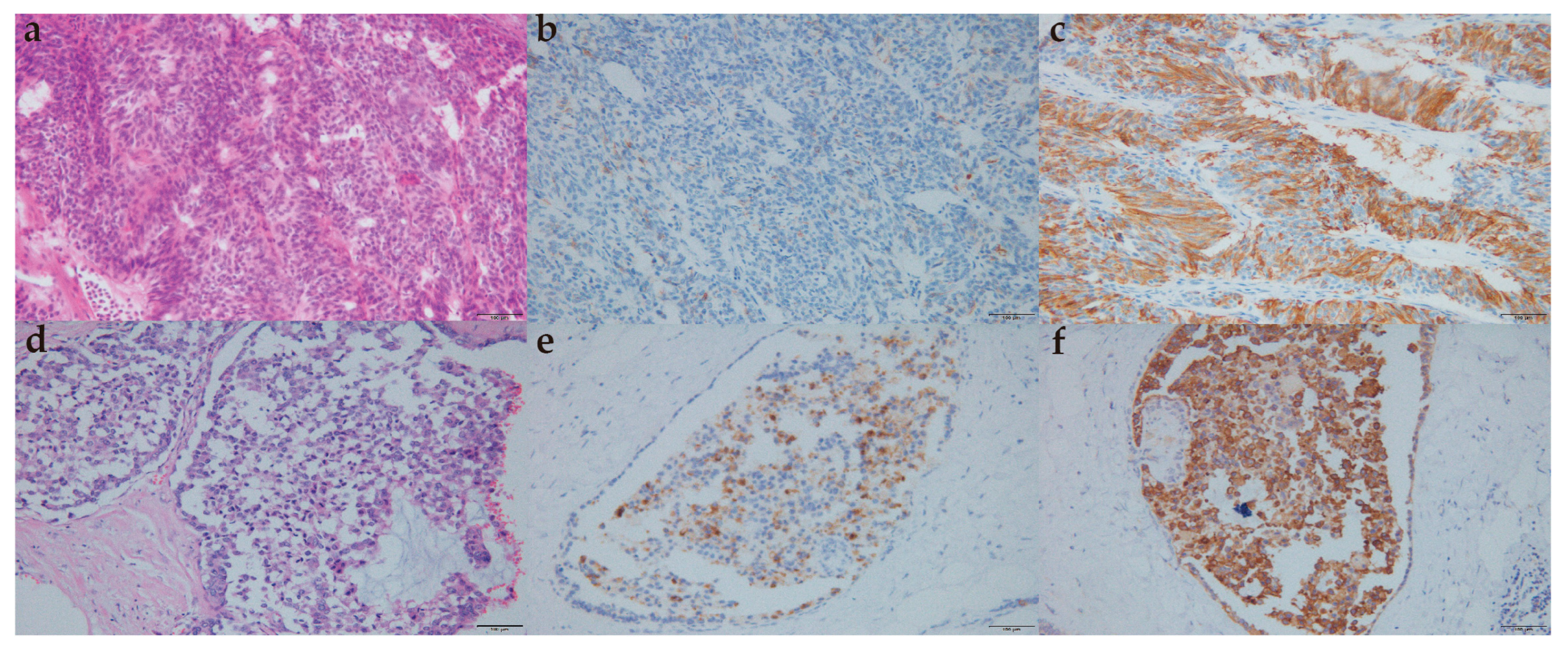
Morphologically, the typical features of the lung/gastrointestinal tract NET, such as ribbons, cords, and rosettes, are not prominent in the breast NET; histological and immunohistochemical features of breast NEC are sometimes difficult to distinguish from lung NEC features [23]. CgA, Syn, NSE, and CD56 were neuroendocrine differentiation markers for breast carcinoma with neuroendocrine features [4]. It has been reported that only 23% of patients were detected with Syn+ and CgA+ at the same time [15]. The expression level of neuroendocrine markers in tumor tissues of patients in this study was also not high. There were significant differences in cytological characteristics between focal and diffuse neuroendocrine differentiated breast carcinomas [24]. However, when breast cancer of no special type with focal neuroendocrine differentiation was regarded as a separate entity, focal neuroendocrine differentiation had no obvious significance for its prognosis [15,25]. In this study, there was no difference between NEN and IBC with neuroendocrine differentiation in five-year OS, five-year DSS, and five-year PFS.
SCNEC was classified into NEC in 2012 and LCNEC was brought into NEC in 2019. A study indicated that approximately half of these patients had triple-negative breast cancer, with a 61.6% five-year DSS rate and 47.7% five-year OS rate [26]. Chemotherapy, surgery, and stage were predictive factors of prognosis. In this study, there was only one case of pure SCNEC and pure LCNEC, respectively, accounting for a relatively low proportion. The special type of breast cancer with neuroendocrine differentiation was rare. According to one study, half of the invasive solid papillary carcinomas were accompanied by neuroendocrine differentiation [7]. In this study, there were four cases of a special type of breast cancer with neuroendocrine differentiation, including two cases of solid papillary carcinoma, one case of invasive papillary carcinoma, and one case of type B mucinous carcinoma.
A study reported that the five-year OS and disease-free survival rates of HR positive/Her-2 negative breast cancer were 93.0% and 92.6%, respectively [27]. The PFS of breast carcinoma with neuroendocrine features was lower than that of the same molecular typing of breast cancer of no special type [28], which was consistent with the results of this study. The overall five-year PFS of patients in this study was 82.37% (95%CI: 0.7084–0.8966), five-year DSS was 91.53% (95%CI: 0.8022–0.9651), and five-year OS was 90.25% (95%CI: 0.7901–0.9564). The study of neoadjuvant therapy for this disease has been limited. In this study, there were seven patients receiving neoadjuvant chemotherapy, and none of them reached a pathological complete response. A study found that endocrine therapy or radiotherapy might improve the prognosis [5]. It was suggested that HR positive NEN patients receive endocrine therapy, especially those with SCNEC with recurrence and metastasis [29]. Endocrine therapy was also found to be effective for liver metastasis of breast cancer with neuroendocrine differentiation [6]. However, surgery was still the main treatment method, and the effect of chemotherapy on prognosis was still uncertain [16].
This study also analyzed the related factors of PFS. A study found that the prognostic factors of NEN of the breast were similar to those of gastrointestinal tract tumors [30], among which lymph node metastasis was an adverse factor of OS [5]. A previous study found that histological grade, pathological stage, ER status, and HER2 status were independent prognostic indicators of OS and disease-free survival [31]. The results of this study showed that diabetes and stage IV disease were related to the PFS of breast carcinoma with neuroendocrine features patients. The influence of diabetes on the PFS of this disease may be related to higher BMI, and there is a lack of relevant research at present.

Limitations

This study was a retrospective study, and the sample size was relatively insufficient. We failed to compare this disease with other types of breast cancer because of lacking a control group. In addition, this study did not describe its imaging characteristics. Because of the rarity of this disease, there was a gap between the length of follow-up time of the patients.

5. Conclusions

Breast carcinoma with neuroendocrine features is relatively rare compared with other types of breast cancer. In this study, it was illustrated that this disease tended to be HR+/Her-2- tumor. In addition, diabetes and stage IV were related to the PFS of patients. These results may provide evidence for the treatment and prognosis prediction of breast carcinoma with neuroendocrine features. Further studies such as large-sample randomized clinical trials are needed to validate the theoretical value and practical significance of these findings and improve understanding of this disease.

Author Contributions

Conceptualization, Y.Q. and J.W.; Methodology, L.Z. (Li Zhu); Investigation, X.H. and Y.D.; Data Collection, L.Z. (Liping Zhang) and B.B.; Writing—Original Draft Preparation, Y.Q. and Y.C.; Writing—Review & Editing, J.W.; Supervision, Y.D. and J.W. All authors have read and agreed to the published version of the manuscript.

Funding

This research was funded by the National Health Commission Capacity Building and Continuing Education Center (grant number: GWJJ2021100303).

Institutional Review Board Statement

All procedures performed in this study involving human participants were in accordance with the Declaration of Helsinki (as revised in 2013). The study was approved by ethics committee of the Chinese PLA General Hospital (NO.: S2022-746 and date of approval 11 January 2023).

Informed Consent Statement

Individual consent for this retrospective analysis was waived.

Data Availability Statement

The data that support the findings of this study are available from the corresponding authors, [J.W. and Y.C.], upon reasonable request.

Conflicts of Interest

The authors declare no conflict of interest.

References

  1. Zhang, Y.; Geng, C.; Xu, B. Controversies and research progress on the diagnosis and treatment of breast neuroendocrine neoplasm. Chin. J. Clin. Oncol. 2022, 49, 968–972. (In Chinese) [Google Scholar] [CrossRef]
  2. Wang, J.; Wei, B.; Albarracin, C.T.; Hu, J.; Abraham, S.C.; Wu, Y. Invasive neuroendocrine carcinoma of the breast: A population-based study from the surveillance, epidemiology and end results (SEER) database. BMC Cancer 2014, 14, 147. [Google Scholar] [CrossRef] [Green Version]
  3. WHO Classification of Tumors Editorial Board. Breast Tumours. In WHO Classification of Tumors, 5th ed.; IARC: Lyon, France, 2019. [Google Scholar]
  4. Rovera, F.; Lavazza, M.; La Rosa, S.; Fachinetti, A.; Chiappa, C.; Marelli, M.; Sessa, F.; Giardina, G.; Gueli, R.; Dionigi, G.; et al. Neuroendocrine breast cancer: Retrospective analysis of 96 patients and review of literature. Int. J. Surg. 2013, 11, S79–S83. [Google Scholar] [CrossRef] [Green Version]
  5. Wei, B.; Ding, T.; Xing, Y.; Wei, W.; Tian, Z.; Tang, F.; Abraham, S.C.; Nayeemuddin, K.M.; Hunt, K.K.; Wu, Y. Invasive neuroendocrine carcinoma of the breast: A distinctive subtype of aggressive mammary carcinoma. Cancer 2010, 116, 4463–4473. [Google Scholar] [CrossRef]
  6. Rischke, H.C.; Staib-Sebler, E.; Mose, S.; Adams, S.W.; Herrmann, G.; Böttcher, H.D.; Lorenz, M. Metastasiertes Mamma-Karzinom neuroendokriner Differenzierung--kombinierte Therapie mit Tamoxifen und dem Somatostatin-Analogon Octreotid [Metastatic breast carcinoma with neuroendocrine differentiation--its combined therapy with tamoxifen and the somatostatin analog octreotide]. DMW-Dtsch. Med. Wochenschr. 1999, 124, 182–186. [Google Scholar] [CrossRef]
  7. Lin, X.; Matsumoto, Y.; Nakakimura, T.; Ono, K.; Umeoka, S.; Torii, M.; Yoshibayashi, H.; Toi, M. Invasive solid papillary carcinoma with neuroendocrine differentiation of the breast: A case report and literature review. Surg. Case Rep. 2020, 6, 143. [Google Scholar] [CrossRef]
  8. Salemis, N.S. Primary neuroendocrine carcinoma of the breast: A rare presentation and review of the literature. Intractable Rare Dis. Res. 2020, 9, 233–246. [Google Scholar] [CrossRef]
  9. Giuliano, A.E.; Connolly, J.L.; Edge, S.B.; Mittendorf, E.A.; Rugo, H.S.; Solin, L.J.; Weaver, D.L.; Winchester, D.J.; Hortobagyi, G.N. Breast Cancer-Major changes in the American Joint Committee on Cancer eighth edition cancer staging manual. CA Cancer J. Clin. 2017, 67, 290–303. [Google Scholar] [CrossRef] [Green Version]
  10. Jiang, Z.; Song, E.; Wang, X.; Wang, H.; Wang, X.; Wu, J.; Yin, Y.; Zhang, Q.; Chen, J.; Che, W.; et al. Guidelines of Chinese Society of Clinical Oncology (CSCO) on Diagnosis and Treatment of Breast Cancer (2020 version). Transl. Breast Cancer Res. 2020, 1, 27. [Google Scholar] [CrossRef]
  11. Rindi, G.; Klimstra, D.S.; Abedi-Ardekani, B.; Asa, S.L.; Bosman, F.T.; Brambilla, E.; Busam, K.J.; De Krijger, R.R.; Dietel, M.; El-Naggar, A.K.; et al. A common classification framework for neuroendocrine neoplasms: An International Agency for Research on Cancer (IARC) and World Health Organization (WHO) expert consensus proposal. Mod. Pathol. 2018, 31, 1770–1786. [Google Scholar] [CrossRef]
  12. Tavassoli, F.A.; Devilee, P. World Health Organization Classification of Tumours: Pathology and Genetics of Tumours of the Breast and Female Genital Organs; IARC Press: Lyon, France, 2003. [Google Scholar]
  13. Lakhani, S.R.; Ellis, I.O.; Schnitt, S.J.; Tan, P.H.; van de Vijver, M.J. WHO Classification of Tumours of the Breast, 4th ed.; IARC Press: Lyon, France, 2012. [Google Scholar]
  14. Mohanty, S.K.; Kim, S.A.; DeLair, D.F.; Bose, S.; Laury, A.R.; Chopra, S.; Mertens, R.B.; Dhall, D. Comparison of metastatic neuroendocrine neoplasms to the breast and primary invasive mammary carcinomas with neuroendocrine differentiation. Mod. Pathol. 2016, 29, 788–798. [Google Scholar] [CrossRef] [Green Version]
  15. Van Krimpen, C.; Elferink, A.; Broodman, C.; Hop, W.; Pronk, A.; Menke, M. The prognostic influence of neuroendocrine differentiation in breast cancer: Results of a long-term follow-up study. Breast 2004, 13, 329–333. [Google Scholar] [CrossRef] [PubMed]
  16. Li, Y.; Du, F.; Zhu, W.; Xu, B. Neuroendocrine carcinoma of the breast: A review of 126 cases in China. Chin. J. Cancer 2017, 36, 1–4. [Google Scholar] [CrossRef] [PubMed]
  17. Ozaki, Y.; Miura, S.; Oki, R.; Morikawa, T.; Uchino, K. Neuroendocrine Neoplasms of the Breast: The Latest WHO Classification and Review of the Literature. Cancers 2021, 14, 196. [Google Scholar] [CrossRef]
  18. Dimitriadis, G.K.; Angelousi, A.; Weickert, M.O.; Randeva, H.S.; Kaltsas, G.; Grossman, A. Paraneoplastic endocrine syndromes. Endocrine-Related Cancer 2017, 24, R173–R190. [Google Scholar] [CrossRef] [Green Version]
  19. Zhang, Q.; He, L.; Lv, W.; Wang, N. Neuroendocrine carcinoma of the breast with hyperprolactinemia: Report of two cases and a minireview. Int. J. Clin. Exp. Pathol. 2020, 13, 1457–1462. [Google Scholar]
  20. Menendez, J.A.; Lopez-Bonet, E.; Alonso-Ruano, M.; Barraza, G.; Vázquez-Martín, A.; Bernadó, L. Solid neuroendocrine breast carcinomas: Incidence, clinico-pathological features and immunohistochemical profiling. Oncol. Rep. 1994, 20, 1369–1374. [Google Scholar] [CrossRef] [PubMed]
  21. Wachter, D.L.; Hartmann, A.; Beckmann, M.W.; Fasching, P.A.; Hein, A.; Bayer, C.M.; Agaimy, A. Expression of Neuroendocrine Markers in Different Molecular Subtypes of Breast Carcinoma. BioMed Res. Int. 2014, 2014, 408459. [Google Scholar] [CrossRef]
  22. Marijanović, I.; Kraljević, M.; Buhovac, T.; Križanac, D.K. Rare Human Epidermal Growth Factor Receptor 2 (HER-2)-Positive Neuroendocrine Carcinoma of the Breast: A Case Report with 9-Year Follow-up. Am. J. Case Rep. 2020, 21, e925895. [Google Scholar] [CrossRef]
  23. Tsang, J.Y.; Tse, G.M. Breast cancer with neuroendocrine differentiation: An update based on the latest WHO classification. Mod. Pathol. 2021, 34, 1062–1073. [Google Scholar] [CrossRef]
  24. Talu, C.K.; Guzelbey, B.; Hacihasanoglu, E.; Cakir, Y.; Nazli, M.A. The effect of the extent of neuroendocrine differentiation on cytopathological findings in primary neuroendocrine neoplasms of the breast. J. Cytol. 2021, 38, 216. [Google Scholar] [CrossRef]
  25. Bogina, G.; Munari, E.; Brunelli, M.; Bortesi, L.; Marconi, M.; Sommaggio, M.; Lunardi, G.; Gori, S.; Massocco, A.; Pegoraro, M.C.; et al. Neuroendocrine differentiation in breast carcinoma: Clinicopathological features and outcome. Histopathology 2015, 68, 422–432. [Google Scholar] [CrossRef]
  26. Zhu, J.; Wu, G.; Zhao, Y.; Yang, B.; Chen, Q.; Jiang, J.; Meng, Y.; Ji, S.; Gu, K. Epidemiology, Treatment and Prognosis Analysis of Small Cell Breast Carcinoma: A Population-Based Study. Front. Endocrinol. 2022, 13, 802339. [Google Scholar] [CrossRef]
  27. Wang, Y.; Zhang, S.; Zhang, H.; Liang, L.; Xu, L.; Cheng, Y.J.; Duan, X.N.; Liu, Y.H.; Li, T. Clinicopathological features and prognosis of hormone receptor-positive/hum an epidermal growth factor receptor 2-negative breast cancer. Beijing Da Xue Xue Bao Yi Xue Ban 2022, 54, 853–862. (In Chinese) [Google Scholar]
  28. Lavigne, M.; Menet, E.; Tille, J.-C.; Lae, M.; Fuhrmann, L.; Bonneau, C.; Deniziaut, G.; Melaabi, S.; Ng, C.C.K.; Marchiò, C.; et al. Comprehensive clinical and molecular analyses of neuroendocrine carcinomas of the breast. Mod. Pathol. 2018, 31, 68–82. [Google Scholar] [CrossRef] [Green Version]
  29. Alkaied, H.; Harris, K.; Brenner, A.; Awasum, M.; Varma, S. Does Hormonal Therapy Have a Therapeutic Role in Metastatic Primary Small Cell Neuroendocrine Breast Carcinoma? Case Report and Literature Review. Clin. Breast Cancer 2012, 12, 226–230. [Google Scholar] [CrossRef]
  30. Roininen, N.; Takala, S.; Haapasaari, K.-M.; Jukkola-Vuorinen, A.; Mattson, J.; Heikkilä, P.; Karihtala, P. Neuroendocrine Breast Carcinomas Share Prognostic Factors with Gastroenteropancreatic Neuroendocrine Tumors: A Putative Prognostic Role of Menin, p27, and SSTR-2A. Oncology 2018, 96, 147–155. [Google Scholar] [CrossRef]
  31. Kwon, S.Y.; Bae, Y.K.; Gu, M.J.; Choi, J.E.; Kang, S.H.; Lee, S.J.; Kim, A.; Jung, H.R.; Kang, S.H.; Oh, H.K.; et al. Neuroendocrine differentiation correlates with hormone receptor expression and decreased survival in patients with invasive breast carcinoma. Histopathology 2013, 64, 647–659. [Google Scholar] [CrossRef]
Figure 1. Morphological and immunohistochemical features. (a). H and E staining of NEC of the breast (×200); (b) positive immunostaining for chromogranin A of NEC of the breast (×200); (c) positive immunostaining for synaptophysin of NEC of the breast (×200); (d) H and E staining of no special type IBC with neuroendocrine differentiation (×200); (e) positive immunostaining for chromogranin A of no special type IBC with neuroendocrine differentiation (×200); (f) positive immunostaining for synaptophysin of no special type IBC with neuroendocrine differentiation (×200). NEC: neuroendocrine carcinoma; IBC: invasive breast cancer; H and E staining: Hematoxylin and Eosin staining.
Life 13 00532 g001
Figure 2. Progression-free survival of breast carcinoma with neuroendocrine features patients (group 1: neuroendocrine neoplasm patients; group 2: invasive breast cancer with neuroendocrine differentiation patients). (a) Progression-free survival of all breast carcinoma with neuroendocrine features patients; (b) progression-free survival of grouped breast carcinoma with neuroendocrine features patients.
Life 13 00532 g002
Figure 3. Disease-specific survival of breast carcinoma with neuroendocrine features patients (group 1: neuroendocrine neoplasm patients; group 2: invasive breast cancer with neuroendocrine differentiation patients). (a) Disease-specific survival of all breast carcinoma with neuroendocrine features patients; (b) disease-specific survival of grouped breast carcinoma with neuroendocrine features patients.
Life 13 00532 g003
Figure 4. Overall survival of breast carcinoma with neuroendocrine features patients (group 1: neuroendocrine neoplasm patients; group 2: invasive breast cancer with neuroendocrine differentiation patients). (a) Overall survival of all breast carcinoma with neuroendocrine features patients; (b) overall survival of grouped breast carcinoma with neuroendocrine features patients.
Life 13 00532 g004
Table 1. General clinicopathological characteristics of breast carcinoma with neuroendocrine features patients.
CategoryBreast Carcinoma with Neuroendocrine Features (n = 87) (%)NEN GroupIBC with Neuroendocrine Differentiation (n = 58) (%)p Value
(n = 29) (%)
Age at diagnosis (years) 0.875
 ≤6055 (63.22%)18 (62.07%)37 (63.79%)
 >6032 (36.78%)11 (37.93%)21 (36.21%)
Gender 0.109
 Female85 (97.7%)27 (93.10%)58 (100.00%)
 Male2 (2.30%)2 (2.30%)0 (0.00%)
Laterality 0.129
 Left44 (50.57%)18 (62.07%)26 (44.83%)
 Right43 (49.43%)11 (37.93%)32 (55.17%)
Smoking history 1
 Yes2 (2.30%)1 (3.45%)1 (1.72%)
 No85 (97.70%)28 (96.55%)57 (98.28%)
Drinking history 0.333
 Yes1 (1.15%)1 (3.45%)0 (0.00%)
 No86 (98.85%)28 (96.55%)58 (100.00%)
BMI (kg/m2) 0.128
 ≥2447 (54.02%)19 (65.52%)28 (48.28%)
 <2440 (45.98%)10 (34.48%)30 (51.72%)
Family history 1
 Yes1 (1.15%)0 (0.00%)1 (1.72%)
 No86 (98.85%)29 (100.00%)57 (98.28%)
Menopausal status a 0.332
 Yes47 (55.29%)17 (62.96%)30 (51.72%)
 No38 (44.71%)10 (37.04%)28 (48.28%)
Hypertension 0.492
 Yes23 (26.44%)9 (31.03%)14 (24.14%)
 No64 (73.56%)20 (68.97%)44 (75.86%)
Diabetes 0.039
 Yes7 (8.05%)5 (17.24%)2 (3.45%)
 No80 (91.95%)24 (82.76%)56 (96.55%)
Hyperlipidemia 0.368
 Yes20 (22.99%)5 (17.24%)15 (25.86%)
 No67 (77.01%)24 (82.76%)43 (74.14%)
T stage (AJCC 8th) 0.2
 145 (51.72%)18 (62.07%)27 (46.55%)
 235 (40.23%)9 (31.03%)26 (44.83%)
 37 (8.05%)2 (6.90%)5 (8.62%)
 40 (0.00%)0 (0.00%)0 (0.00%)
Her-2 status 0.323
 Positive11 (12.64%)2 (6.90%)9 (15.52%)
 Negative76 (87.36%)27 (93.10%)49 (84.48%)
Ki-67 0.229
 Positive72 (82.76%)22 (75.86%)50 (86.21%)
 Negative15 (17.24%)7 (24.14%)8 (13.79%)
ER 0.857
 Positive67 (77.01%)22 (75.86%)45 (77.59%)
 Negative20 (22.99%)7 (24.14%)13 (22.41%)
PR 0.434
 Positive71 (81.61%)25 (86.21%)46 (79.31%)
 Negative16 (18.39%)4 (13.79%)12 (20.69%)
Molecular typing (CSCO 2020) 0.494
 Luminal A13 (14.94%)5 (17.24%)8 (13.79%)
 Luminal B62 (71.26%)21 (72.41%)41 (70.69%)
 Her-2 positive1 (1.15%)0 (0.00%)1 (1.72%)
 Triple negative11 (12.64%)3 (10.34%)8 (13.79%)
Vessel carcinoma embolus 0.702
 Yes17 (19.54%)5 (17.24%)12 (20.69%)
 No70 (80.46%)24 (82.76%)46 (79.31%)
N stage (AJCC 8th) 0.549
 053 (60.92%)19 (65.52%)34 (58.62%)
 118 (20.69%)5 (17.24%)13 (22.41%)
 214 (16.09%)5 (17.24%)9 (15.52%)
 32 (2.30%)0 (0.00%)2 (3.45%)
Skin or chest wall invasion ---
 Yes0 (0.00%)0 (0.00%)0 (0.00%)
 No87 (100.00%)29 (100.00%)58 (100.00%)
Distant metastasis 0.55
 Yes2 (2.30%)0 (0.00%)2 (3.45%)
 No85 (97.70%)29 (100.00%)56 (96.55%)
Stage (AJCC 8th) 0.577
 I26 (29.88%)11 (37.93%)15 (25.86%)
 II43 (49.43%)11 (37.93%)32 (55.17%)
 III16 (18.39%)7 (24.14%)9 (15.52%)
 IV2 (2.30%)0 (0.00%)2 (3.45%)
BMI: body mass index; ER: estrogen receptor; PR: progesterone receptor. Her-2: human epidermal growth factor receptor 2; IBC: invasive breast cancer; AJCC 8th: The eighth version of America joint committee on cancer staging system; CSCO 2020: The guidelines of Chinese Society of Clinical Oncology in 2020. a There were two male breast cancer patients in the study group.
Table 2. Supplement of part clinical features of breast carcinoma with neuroendocrine features patients.
Categoryn = 87
Clinical symptoms
 Yes20 (22.99%)
  Pain14
  Nipple discharge5
  Nipple discharge with pain1
  Paraneoplastic symptoms0
 No67 (77.01%)
Thyroid diseases
 Yes19 (21.84%)
  Thyroid nodule15
  Diffuse thyroid disease3
  Thyroid cancer1
 No68 (78.16%)
Lymph node metastasis
 Yes34 (39.08%) a
  Axillary lymph node metastasis34
  Supraclavicular lymph node metastasis2
  Subclavian lymph node metastasis1
 No53 (60.92%)
a One patient had axillary lymph node metastasis, supraclavicular lymph node metastasis, and subclavian lymph node metastasis at the same time. One patient had axillary lymph node metastasis and supraclavicular lymph node metastasis at the same time.
Table 3. Supplement of part pathological characteristics of breast carcinoma with neuroendocrine features patients.
Categoryn = 87
Classification
 NEN29 (33.34%)
  NET26
   Simple type21
   Mixed type5
  NEC3
   Small cell carcinoma1
   Large cell carcinoma1
   Mixed type1
 IBC with neuroendocrine differentiation58 (66.66%)
  IBC of no special type
   Invasive ductal carcinoma50
  IBC of special type
   Solid papillary carcinoma2
   Invasive papillary carcinoma1
   Type B mucinous carcinoma1
  Mixed type4
Neuroendocrine markers
 Syn
  03 (3.45%)
  1+76 (87.35%)
  2+4 (4.60%)
  3+4 (4.60%)
 CgA
  010 (11.49%)
  1+11 (12.64%)
  2+25 (28.74%)
  3+2 (2.30%)
  NK39 (44.83%)
 CD56
  011 (12.64%)
  1+26 (29.88%)
  2+42 (48.28%)
  3+1 (1.15%)
  NK7 (8.05%)
 NSE
  01 (1.15%)
  1+7 (8.05%)
  2+2 (2.30%)
  3+1 (1.15%)
  NK76 (87.35%)
Ductal carcinoma in situ composition
 Yes29 (33.33%)
 No58 (66.67%)
NEN: neuroendocrine neoplasm; NET: neuroendocrine tumor; NEC: neuroendocrine carcinoma; IBC: invasive breast cancer; Syn: synaptophysin; CgA: chromogranin A; NSE: neuron-specific enolase; NK: not known.
Table 4. Treatment strategy of breast carcinoma with neuroendocrine features patients.
Categoryn = 87
Neoadjuvant chemotherapy
 Yes7 (8.05%)
 No80 (91.95%)
Surgery treatment
 Breast
  Mastectomy75 (86.20%)
  BCS8 (9.20%)
  NSM3 (3.45%)
  None a1 (1.15%)
 Axillary lymph node
  SLNB19 (21.84%)
  ALND67 (77.01%)
  None b1 (1.15%)
Adjuvant therapy
 Chemotherapy
  Yes71 (81.61%)
  No16 (18.39%)
 Radiotherapy
  Yes27 (31.03%)
  No60 (68.97%)
 Endocrine therapy
  Yes64 (73.56%)
  No23 (26.44)
 Targeted therapy
  Yes8 (9.20%)
  No79 (90.80%)
BCS: breast conserving surgery; NSM: nipple-areola complex sparing mastectomy; SLNB: sentinel lymph node biopsy; ALND: axillary lymph node dissection. a One patient had occult breast cancer, receiving ALND only. b One patient who was elderly received breast surgery only.
Table 5. Local recurrence, distant metastasis, and mortality of breast carcinoma with neuroendocrine features patients.
Categoryn = 85 a
Local recurrence5 (5.88%)
 Breast1
 Chest wall3
 Axilla1
Distant metastasis12 (14.14%)
 Bone4
 Lung6
 Liver2
 Brain3
 Retroperitoneal lymph node1
 Contralateral axillary lymph node2
Death8 (9.41%)
 Breast cancer6
 Other causes2
Follow-up time (months, IQR)57 (25–74)
IQR: inter-quartile range. a There were 87 patients totally, and two patients dropped out.
Table 6. Univariate analysis of PFS in breast carcinoma with neuroendocrine features patients.
VariableHR (95% CI)p Value
Age (year)
 >601
 ≤600.9670 (0.3566–2.6224)0.947
Gender
 Female1
 Male1.6943 (0.2138–13.4258)0.618
Laterality
 Right1
 Left0.4458 (0.1587–1.2522)0.125
Smoking history
 No1
 Yes9.1939 (1.1251–75.1266)0.038
Drinking history
 No1
 Yes---1
BMI (kg/m2)
 <241
 ≥241.2394 (0.4535–3.3873)0.676
Family history
 No1
 Yes5.8103 (0.7485–45.1052)0.092
Menopausal status
 No1
 Yes0.7091 (0.2629–1.9125)0.497
Hypertension
 No1
 Yes0.8888 (0.2882–2.7409)0.837
Diabetes
 No1
 Yes4.8491 (1.5089–15.5837)0.008
Hyperlipidemia
 No1
 Yes1.1692 (0.3771–3.6253)0.787
T stage
 11
 21.6177 (0.5770–4.5348)0.36
 31.5048 (0.3172–7.1392)0.607
Her-2 status
 Negative1
 Positive2.6226 (0.8390–8.1975)0.097
Ki-67
 Negative1
 Positive1.1088 (0.3391–3.6256)0.864
ER
 Positive1
 Negative1.6555 (0.5819–4.7100)0.345
PR1
 Positive1.6112 (0.5242–4.9525)
 Negative 0.405
Molecular typing
 Luminal A1
 Luminal B1.7925 (0.3694–8.6984)0.469
 Her-2 positive72.1912 (4.4462–1172.1380)0.003
 Triple negative2.3484 (0.3846–14.3384)0.355
Vessel carcinoma embolus
 No1
 Yes0.4462 (0.1013–1.9650)0.286
N stage
 01
 11.6375 (0.5346–5.0153)0.388
 20.9315 (0.1976–4.3919)0.929
 3150.1349 (12.6995–1774.9190)<0.001
Skin or chest wall invasion
 No1
 Yes------
Distant metastasis
 No1
 Yes133.1195 (11.7712–1505.4410)<0.001
Stage
 I1
 II2.4426 (0.6494–9.1871)0.186
 III1.5589 (0.3132–7.7588)0.588
 IV243.9231 (17.2429–3450.6020)<0.001
Classification
 IBC with neuroendocrine differentiation1
 NEN0.9300 (0.3073–2.8146)0.898
Neoadjuvant chemotherapy
 Yes1
 No0.4378 (0.1241–1.5447)0.199
Breast treatment
 Mastectomy1
 BCS1.1105 (0.2508–4.9179)0.89
 NSM1.1265 (0.1455–8.7217)0.909
Axillary lymph node treatment
 ALND1
 SLNB0.6931 (0.1972–2.4355)0.568
Chemotherapy
 Yes1
 No2.1270 (0.7460–6.0646)0.158
Radiotherapy
 Yes1
 No1.1213 (0.4000–3.1428)0.828
Endocrine therapy
 Yes1
 No1.4339 (0.5213–3.9437)0.485
Targeted therapy
 Yes1
 No0.1838 (0.0579–0.5840)0.004
BMI: body mass index; ER: estrogen receptor; PR: progesterone receptor. Her-2: human epidermal growth factor receptor 2; NEN: neuroendocrine neoplasm; IBC: invasive breast cancer; BCS: breast conserving surgery; NSM: nipple-areola complex sparing mastectomy; SLNB: sentinel lymph node biopsy; ALND: axillary lymph node dissection; PFS: progression-free survival; HR: hazard ratio; CI: confidence interval.
Table 7. Multivariate analysis of PFS in breast carcinoma in neuroendocrine features patients.
VariableHR (95%CI)p Value
Smoking history
 No1
 Yes---1
Diabetes
 No1
 Yes7.2526 (1.8874–27.8691)0.004
Molecular typing
 Luminal A1
 Luminal B0.8695 (0.1472–5.1356)0.877
 Her-2 positive------
 Triple negative1.5148 (0.2420–9.4801)0.657
Stage
 I1
 II1.9789 (0.5132–7.6316)0.322
 III0.8796 (0.1515–5.1080)0.886
 IV2.44 × 1019 (5.72 × 10¹⁷–1.04 × 10²¹)<0.001
Targeted therapy
 Yes1
 No1.7777 (0.0313–1.0085)0.051
PFS: progression-free survival; HR: hazard ratio; CI: confidence interval.
Disclaimer/Publisher’s Note: The statements, opinions and data contained in all publications are solely those of the individual author(s) and contributor(s) and not of MDPI and/or the editor(s). MDPI and/or the editor(s) disclaim responsibility for any injury to people or property resulting from any ideas, methods, instructions or products referred to in the content.

