Article

Molecular Identification of Protozoan Sarcocystis in Different Types of Water Bodies in Lithuania

by
Agnė Baranauskaitė
*,
Živilė Strazdaitė-Žielienė
,
Elena Servienė
,
Dalius Butkauskas
and
Petras Prakas
Nature Research Centre, Akademijos Str. 2, 08412 Vilnius, Lithuania
*
Author to whom correspondence should be addressed.
Life 2023, 13(1), 51; https://doi.org/10.3390/life13010051
Submission received: 1 December 2022 / Accepted: 21 December 2022 / Published: 24 December 2022
(This article belongs to the Special Issue Interactions between Microorganisms, Their Environment and Host)

Abstract

:
Representatives of the genus Sarcocystis are unicellular parasites having a two-host life cycle and infecting mammals, birds, and reptiles. Until now, Sarcocystis spp. have been mainly investigated in definitive and intermediate hosts. Only a few studies have been conducted on the detection of Sarcocystis parasites in water samples. The aim of this research was to examine whether the prevalence of Sarcocystis spp. parasitizing farm animals varies in different types of water bodies. Water samples (n = 150) were collected from the entire territory of Lithuania, dividing water bodies into five groups (lakes, rivers, ponds/canals, swamps, and the inshore zone of the territorial Baltic Sea area). One-liter samples were filtered and subsequently analyzed using nested PCR. At least one of the analyzed Sarcocystis spp. (S. arieticanis, S. bertrami, S. bovifelis, S. capracanis, S. cruzi, S. hirsuta, S. miescheriana, and S. tenella) was determined in all examined samples from water bodies. No significant difference in Sarcocystis spp. prevalence between different types of water sources was detected. Our research proved that selecting appropriate primers is important for the accurate identification of parasites in samples collected from water bodies.

1. Introduction

Unicellular parasites of the genus Sarcocystis are characterized by an obligatory two-host life cycle. These parasites infect mammals, birds, and reptiles. Sarcocysts mostly develop in the muscle tissues of intermediate hosts; meanwhile, sporulation of oocysts occurs in the small intestine of the definitive host [1]. Four of more than 200 species belonging to this genus are zoonotic, S. hominis, S. suihominis, S. heydorni, and S. nesbitti [2]. However, few studies have been carried out to determine the prevalence of these species, as they are often misidentified during morphological or molecular examination [3].
To date, most research has been conducted on Sarcocystis spp. infecting economically important domestic animals. Farm animals can become infected through food or water contaminated with fecal sporocysts of Sarcocystis spp. It has been observed that Sarcocystis species transmitted through canids are more dangerous to farm animals, and acute infections can cause such symptoms as fever, weight loss, anemia, reduction in wool and milk yield, abortion, or even death [4,5,6,7].
Until now, Sarcocystis infection was mainly investigated by performing morphological or molecular analysis of animal carcasses. However, the use of natural environment studies by avoiding animal carcass-based research is increasing. Even so, only a few studies were conducted to test for Sarcocystis spp. in water [8,9,10]. The aim of our previous study was to assess different sample preparation and common PCR methodologies for identifying Sarcocystis species in water. The environmental water samples were collected from ponds (n = 49), lakes (n = 35), rivers (n = 18), canals (n = 10), and lagoons (n = 2). DNA of at least one of eight tested Sarcocystis species (S. arieticanis, S. bertrami, S. bovifelis, S. capracanis, S. cruzi, S. hirsuta, S. miescheriana, and S. tenella) was detected in 111 of 114 (97.4%) water samples using nested PCR targeting cox1 gene [10]. However, Sarcocystis species occurrence rates in different areas and types of water bodies were not compared. Therefore, the aim of this study was to compare detection rates of different Sarcocystis species in five types of water bodies—lakes, rivers, canals/ponds, swamps, and the inshore zone of the Baltic Sea—using molecular methods.

2. Materials and Methods

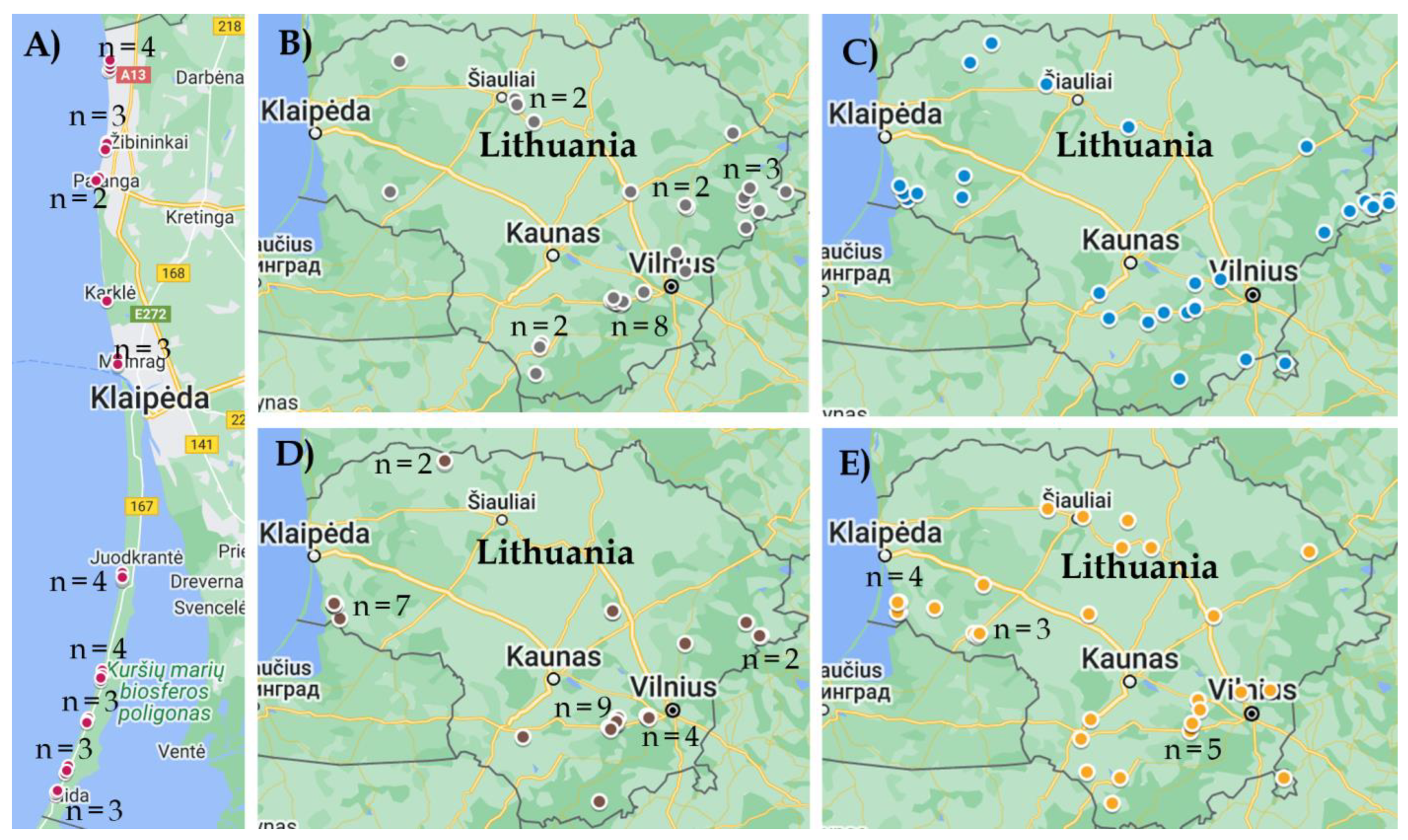
2.1. Sample Collection

Samples (n = 150) were collected from water bodies throughout the territory of Lithuania in the summer of 2021 (Figure 1). Water bodies were divided into five groups with an equal number of samples each—lakes (stagnant water), rivers (flowing water), canals and ponds (small water bodies that are usually close to pastures), swamps (acidic water) and the inshore zone of the Baltic Sea (saline water). Water samples were collected in sterile containers of 1 L capacity and transported in portable coolers with ice batteries. Until further processing, water samples were stored at +4 °C.

2.2. Preparation of Water Samples and Genomic DNA Extraction

First, the water sample was filtered through a metal sieve with 1 mm pores, then through Whatman™ Qualitative Filter Paper Grade 4 and finally filtered using MF-Millipore® 5 μm pore membranes. Two milliliters of distilled water were used for membrane washing and collection of material, which was stored at +4 °C until further processing.
Genomic DNA (gDNA) was isolated from 200 μL of concentrated water samples using the GeneJET Genomic DNA Purification Kit (Thermo Fisher Scientific Baltics, Vilnius, Lithuania), according to the manufacturer’s recommendations. The resulting DNA samples were kept frozen at −20 °C until further analysis.

2.3. Nested PCR-Based Identification of Sarcocystis Parasites

During this work, samples from environmental water bodies were analyzed to distinguish the prevalence of different Sarcocystis spp. (S. arieticanis, S. bertrami, S. bovifelis, S. capracanis, S. cruzi, S. hirsuta, S. miescheriana, and S. tenella) using farm animals (cattle, sheep, goats, horses, and pigs) as intermediate hosts. Since the cox1 gene is considered the most appropriate for the identification of selected Sarcocystis parasites [11,12,13], primers targeting this gene were selected [10]. It was observed that, having selected adequate PCR primer pairs, detection rates of farm animals infecting Sarcocystis spp. in water samples were associated with those observed in the muscles of the intermediate host [10,14,15,16,17]. However, in our earlier research, the prevalence of S. bertrami, S. cruzi, S. miescheriana and S. tenella identified in water samples was significantly lower [10]. Thus, 10 new primers (Table 1; highlighted in bold) were designed for the identification of these species. To detect S. miescheriana and S. bertrami, primers were redesigned to give shorter products, whereas to diagnose S. cruzi and S. tenella, different binding sites of primers were chosen.
During all PCR reactions, both positive (DNA extracted from sarcocysts of the corresponding Sarcocystis species) and negative (distilled water) controls were used. Primers were checked for cross reactions with other Sarcocystis species. The specificity of primer sets was confirmed. To check for possible contamination, distilled water and tap water were examined after the first batch, in the middle of our experiments and after the last batch. Based on the examination of distilled and tap water, PCRs were negative with all primers used in the study.
Preparation of PCR reaction mixtures and cycling conditions were as described previously [10]. The annealing temperatures were modified depending on the primers used (Table 1). Agarose gel electrophoresis was used to visualize PCR amplicons.
The selected PCR products were purified and directly sequenced as described previously [10]. Five positive samples of each species were used for sequencing, except for S. hirsuta, since only three samples were positive for this species. Four positive samples of the species S. tenella, S. cruzi, S. bertrami and S. miescheriana were additionally sequenced with the primers used in the previous study [10].
The editing of resolved sequences was performed manually with subsequent comparative BLAST analysis (http://blast.ncbi.nlm.nih.gov/, accessed on 10 October 2022). Differences in the prevalence of the identified Sarcocystis species were evaluated using the Chi-squared test. The cox1 sequences of Sarcocystis species generated in the present study were deposited in the GenBank under the accession numbers OP681467–OP681524.

3. Results

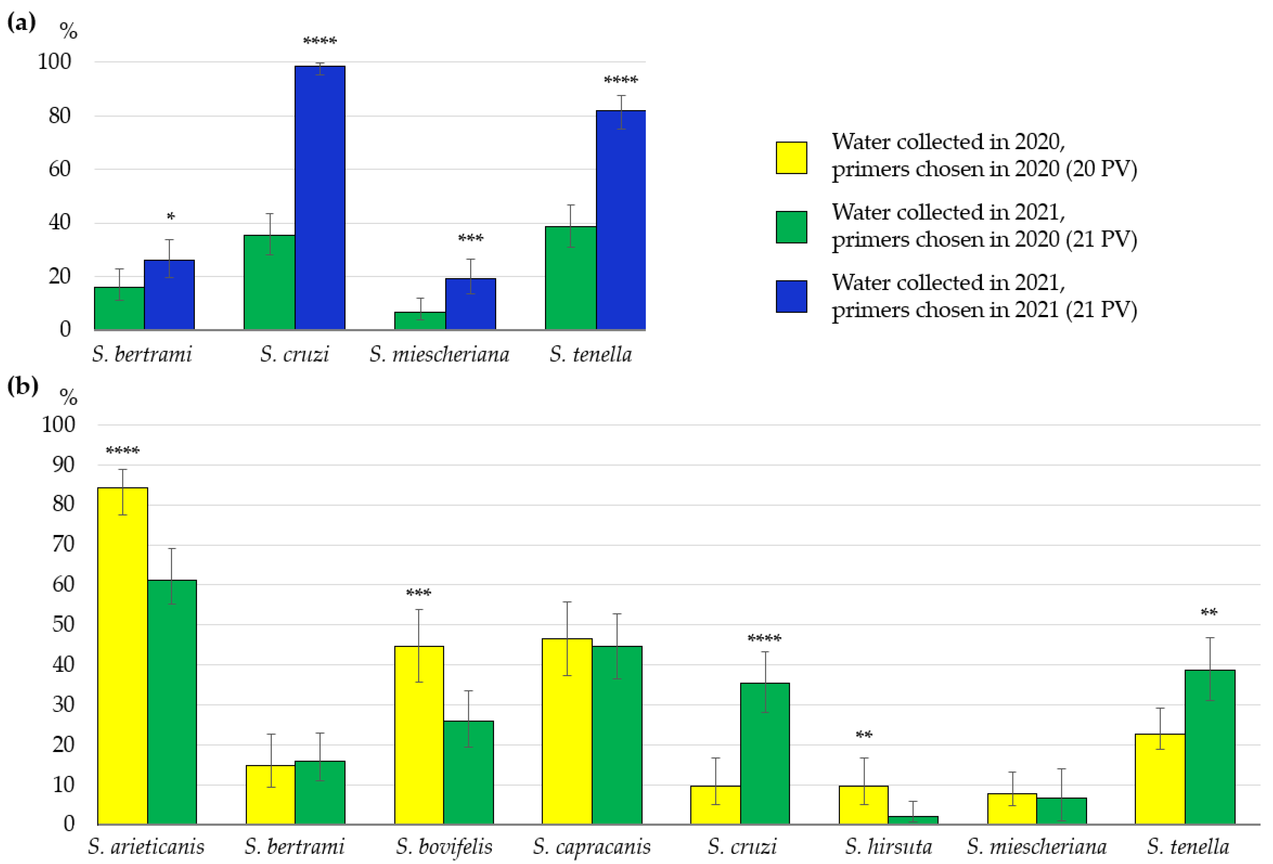
3.1. Identification of Sarcocystis spp. Using Different PCR Primer Sets

GenBank accession numbers, length, and similarity of the obtained cox1 sequences of S. bovifelis, S. cruzi, S. hirsuta, S. arieticanis, S. tenella, S. capracanis, S. bertrami and S. miescheriana are presented in Table 2. In no case did the obtained intraspecific and interspecific genetic differences overlap. Therefore, the primer sets used in this study were appropriate for the identification of Sarcocystis species in water samples examined.
Four of eight investigated Sarcocystis species, S. bertrami, S. cruzi, S. miescheriana and S. tenella, were identified in the same water samples using two different primer combinations, the primer set chosen in our previous study (21 PV) [10] and the primer set selected in the current work (21 PS). The Sarcocystis parasite occurrence rate for the above-mentioned species was significantly higher (p < 0.05) using the primers selected in this study (21 PS) (Figure 2a). Depending on the primers used, the prevalence of S. bertrami was 16.0% and 26.0% (χ2 = 4.52, p < 0.05), whereas the prevalence of S. miescheriana accounted for 6.7% and 19.3% (χ2 = 10.64, p < 0.01). Most significant differences were identified when evaluating the primers tested for the detection of S. cruzi and S. tenella. The prevalence of S. cruzi was 35.3% and 98.7% (χ2 = 136.02, p < 0.00001) and the occurrence of S. tenella was 38.7% and 82.0% (χ2 = 58.85, p < 0.00001), using 21 PV and 21 PS, respectively.
The detection frequency of eight Sarcocystis species was compared in 150 water samples collected during the course of the present study (21 PV) and in 114 water samples collected throughout Lithuania in our previous investigation (20 PV) (Figure 2b). During both studies, Sarcocystis spp. were identified by the same technique (including same primer combinations). Statistically insignificant differences were observed in the cases of S. bertrami (16.0% vs. 14.9%, χ2 = 0.06, p > 0.05), S. capracanis (44.7% vs. 46.5%, χ2 = 0.09, p < 0.05) and S. miescheriana (6.7% vs. 7.9%, χ2 = 0.15, p < 0.05). Significantly higher occurrence rates of S. arieticanis (84.2% vs. 61.3%, χ2 = 16.54, p < 0.001), S. bovifelis (44.7% vs. 26.0%, χ2 = 10.12, p < 0.01) and S. hirsuta (9.6% vs. 2.0%, χ2 = 7.55, p < 0.01) were calculated in previously collected water samples (20 PV), whereas significantly higher detection rates of S. tenella (38.7% vs. 22.8%, χ2 = 7.51, p < 0.01) and S. cruzi (35.3% vs. 9.6%, χ2 = 23.27, p < 0.001) were established in water samples obtained during the current work (21 PV).

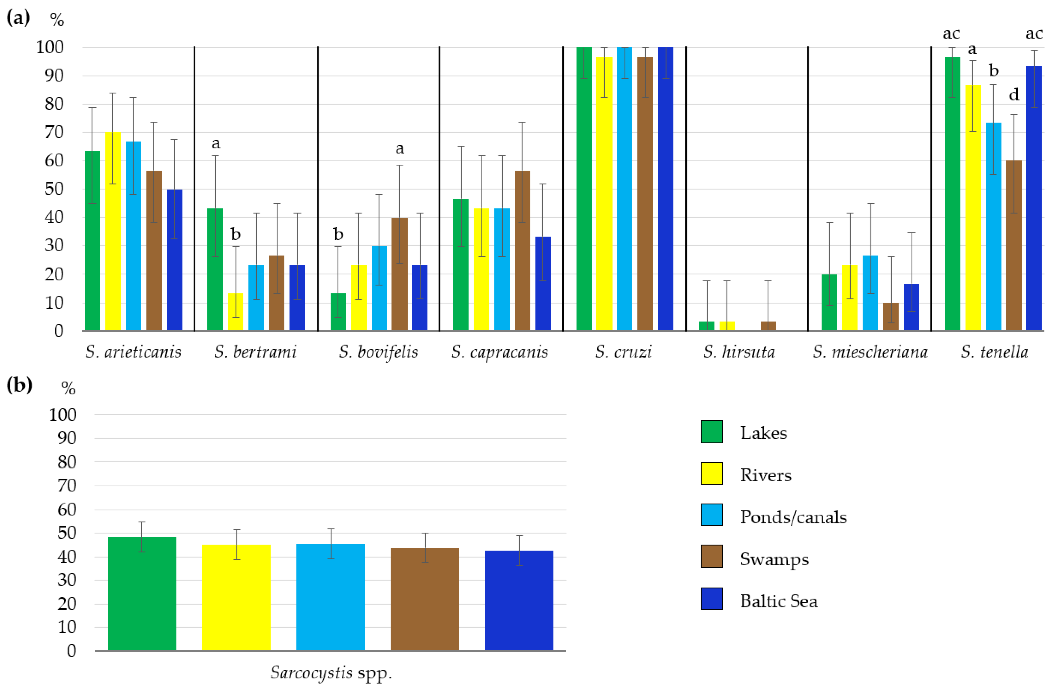
3.2. Sarcocystis spp. Occurrence Rates in Different Types of Water Bodies

The detection rate of Sarcocystis species examined was compared in five types of water bodies—lakes, rivers, ponds/canals, swamps, and the inshore zone of the Baltic Sea (Figure 3a). The comparison showed a significantly higher (χ2 = 6.65, p < 0.01) detection rate of S. bertrami in lakes (43.3%) than that in rivers (13.3%) and a significantly higher (χ2 = 5.45, p < 0.05) detection rate of S. bovifelis in swamps (40.0%) than that in lakes (13.3%). In the case of S. tenella, significantly higher detection rates were calculated in lakes (96.7%) than those in swamps (60.0%) (χ2 = 11.88, p < 0.001) and ponds/canals (73.3%) (χ2 = 6.41, p < 0.05); moreover, they were higher in the inshore zone of the Baltic Sea (93.3%) than the rates in swamps (χ2 = 9.32, p < 0.01) and ponds/canals (χ2 = 4.32, p < 0.05), and finally, they were higher (86.7%) in rivers as compared to those in swamps (χ2 = 5.45, p < 0.05). The overall frequency of Sarcocystis species (calculated by summing up all PCR-positive samples and dividing them by the total number of samples tested) varied depending on water type: from 102 (42.5%) positive cases in the Baltic Sea to 116 (48.3%) positive cases in lakes (Figure 3b). However, the differences observed in occurrence rates of Sarcocystis spp. in five types of water bodies were insignificant (χ2 = 1.85, df = 4, p > 0.05). In summary, the overall prevalence of Sarcocystis species did not depend on the type of water body.

3.3. Distribution of Sarcocystis spp. in Water Samples

Summarizing the results, at least one Sarcocystis species was identified in all 150 examined samples. The number of Sarcocystis species per individual sample was estimated by combining data obtained in the analysis using different primer sets (21 PV and 21 PS). Single species was identified only in three cases (2.0%). The detection of two (18.7%), three (21.3%), four (32.0%) and five (19.3%) Sarcocystis species per sample was more frequent. Finally, six and seven species of Sarcocystis in one water sample were identified in nine cases (6.0%) and one (0.7%) case, respectively.
In the present work, the lowest Sarcocystis species detection rate in water samples analyzed was established for S. hirsuta, and was equal to 2%. The identification rates of S. bertrami (26.7%), S. bovifelis (26.0%) and S. miescheriana (19.3%) did not exceed 30%. Moderate detection frequency was estimated for S. capracanis (44.7%) and S. arieticanis (61.3%), whereas the highest prevalence was revealed for S. tenella (89.3%) and S. cruzi (99.3%).

4. Discussion

Based on nested PCR, we identified eight Sarcocystis species (S. arieticanis, S. bertrami, S. bovifelis, S. capracanis, S. cruzi, S. hirsuta, S. miescheriana, and S. tenella) in different types of water bodies (Table 2, Figure 3). Cattle are intermediate hosts for the first three Sarcocystis species tested, S. arieticanis and S. tenella use sheep as their intermediate hosts, while goats, horses and pigs/wild boar are hosts of S. capracanis, S. bertrami, and S. miescheriana, respectively [5]. Based on current knowledge, the European bison (Bison bonasus) can be an alternative host for Sarcocystis species parasitizing cattle [18]. The sarcocysts of such species as S. arieticanis, S. tenella and S. capracanis can be found in muscle tissues of European mouflon (Ovis aries musimon) [19]. Nevertheless, in the areas under investigation, the mentioned wild animals are rare [20]. Other wild ungulate species that could be intermediate hosts for the tested Sarcocystis species are not free-ranging in Lithuania.
To date, most studies have been conducted on the prevalence of protozoan infection in drinking water treatment facilities [21,22,23,24], whereas only a few studies have investigated the prevalence of parasitic protozoa in environmental water sources, such as rivers [25,26], reservoirs [27], lakes [28] or private wells [29]. Typically, studies cover small regions, basins of a particular river or several nearby villages where an outbreak of parasitic protozoa was identified. The number of studies on other parasitic protozoa is much smaller; for example, only three studies were devoted to the identification of Sarcocystis spp. in water bodies [8,9,10]. The current study is the first attempt to compare the prevalence of eight Sarcocystis species in different types of water bodies. It is noteworthy that equal numbers of samples representing five groups of water bodies (lakes, rivers, ponds/canals, swamps, and the Baltic Sea) were collected throughout the entire territory of the country. The majority of other investigations of parasitic protozoa in water samples were limited to small geographic regions or the location of infection outbreaks [22,27,28,29]. During the present study, it was found that overall detection of the analyzed Sarcocystis species did not depend on the type of water body (Figure 3). The number of positive cases was very similar in all types of water and varied from 102 to 116 positive cases. As a result, it can be assumed that environmental conditions and the location of water bodies had no effect on the prevalence of Sarcocystis parasites in different water sources. However, insignificant differences were noticed when comparing the distribution of eight species in different types of water bodies. Distribution of individual Sarcocystis species may be determined by the abundance of final hosts in a particular area and different characteristics of water bodies, such as water salinity, acidity, or flow turbidity.
Our current research showed that Sarcocystis spp. DNA detection depended on the primer combinations used (Figure 2). The use of different primer pairs for identification of S. bertrami, S. miescheriana, S. cruzi, and S. tenella revealed statistically significant differences. As compared to the prevalence of these four Sarcocystis species obtained using primers selected in the previous study (21 PV) [10], a significantly higher (p < 0.05) prevalence was determined when the primers selected in this study (21 PS) were applied to PCR. Previous studies also showed that amplification success of cox1 fragments of Sarcocystis spp. using ungulates as their intermediate hosts depended on the chosen PCR primers [30,31,32]. It is assumed that some Sarcocystis species exhibit high interspecific genetic variability [17,33]. However, intraspecific genetic variation of Sarcocystis spp. is poorly studied [34,35,36], and selection of suitable primers has been one of the challenges to the diagnosis of Sarcocystis species thus far.
Due to the large morphological and genetic differences between genera or even species of parasitic protozoa, universal methods for identification of these parasites in various environmental samples have not been developed yet. The concentration of many protozoa species in water samples is relatively low [37]; therefore, detection requires very sensitive techniques, such as molecular methods. While some protozoa are undetectable in water samples under a microscope [38], molecular methods for the identification of parasitic protozoa are not fully developed [27,37]. It should be noted that molecular-based techniques are not standardized for the diagnosis of Sarcocystis and other parasitic protozoa in water samples [10,37]. Therefore, a wide variety of methodologies, e.g., conventional PCR and derivatives, DNA hybridization, loop-mediated isothermal amplification (LAMP), or quantitative PCR, are used [10,27,39,40]. During environmental sample testing, discrimination between DNA from a living cell versus DNA from a dead one might be considered impossible. Consequently, the prevalence of parasites in the samples collected can be overestimated [37]. Oocysts or sporocysts of protozoan Sarcocystis parasites are also known to be resistant to various environmental conditions (freezing, low humidity, high temperatures, etc.) and can remain viable in nature for months [5,41]. However, to amplify DNA only from viable sporocysts, water samples can be treated with dyes that penetrate only membrane-damaged cells. After dye has entered the cell, it is covalently cross-linked to DNA. Consequently, PCR amplification is strongly inhibited [42]. To date, fluorescent dyes, such as ethidium monoazide (EMA), propidium monoazide (PMA), ethidium bromide (EB), and propidium iodide (PI), have been successfully used in protozoan studies [43]. Accordingly, further research of Sarcocystis spp. in water samples could focus on additional testing, such as measuring the sensitivity of sporocyst recovery, a spike-and-recovery experiment (controlled contamination of water samples with purified sporocysts) or adaptation of quantitative PCR. However, during the previous studies on animal carcasses, a high prevalence of Sarcocystis spp. infection was determined in some intermediate hosts [14,15,20]. As much as 100% of cattle and sheep bred in Lithuania were found to be infected with Sarcocystis parasites [14,15]. Meanwhile, the prevalence of infection in horses and pigs accounted for 47.2% and 40.2%, respectively [14]. Since it is known that animals can become infected with these parasites through food and water, there seem to be large amounts of viable and infective sporocysts in the environment.

5. Conclusions

The present study is the first attempt to compare detection rates of Sarcocystis parasites using farm animals as their intermediate hosts in five different types of water bodies (lakes, rivers, ponds/canals, swamps, and the inshore zone of the Baltic Sea). The prevalence of the analyzed Sarcocystis species did not vary significantly between the examined water source groups. The environmental conditions of water bodies do not affect the prevalence of Sarcocystis parasites. Based on the nested PCR, eight Sarcocystis species were identified—S. hirsuta (2.0%), S. miescheriana (19.3%), S. bovifelis (26.0%), S. bertrami (26.7%), S. capracanis (44.7%), S. arieticanis (61.3%), S. tenella (89.3%) and S. cruzi (99.3%). Further, it was established that the detection frequency of Sarcocystis species in water samples depended on the combinations of selected primers. The present study showed that in general, Sarcocystis parasites were widespread in water bodies and could easily infect livestock.

Author Contributions

Conceptualization, A.B. and P.P.; methodology, Ž.S.-Ž. and A.B.; software, P.P.; validation, P.P., D.B. and E.S.; formal analysis, P.P.; investigation, A.B.; resources D.B. and E.S.; data curation, P.P.; writing—original draft preparation, A.B. and P.P.; writing—review and editing, P.P., D.B., Ž.S.-Ž. and E.S.; visualization, A.B.; supervision, D.B. and E.S.; project administration, D.B.; funding acquisition, D.B. All authors have read and agreed to the published version of the manuscript.

Funding

This work was funded by the Research Council of Lithuania (grant number S-MIP-20-24). The performed studies are in frame with research networking of COST Action CA18113.

Institutional Review Board Statement

Not applicable.

Informed Consent Statement

Not applicable.

Data Availability Statement

Data supporting the conclusions of this article are included in the article. The sequences generated in the present study were submitted to the GenBank database under Accession Numbers OP681467–OP681524.

Acknowledgments

The authors are grateful to V. Pabrinkis and A. Lengvinas for their support in water sampling. We are also thankful to M. Graužinytė for technical assistance in water filtration.

Conflicts of Interest

The authors declare that they have no conflict of interest.

References

  1. Nimri, L. Unusual case presentation of intestinal Sarcocystis hominis infection in a healthy adult. JMM Case Rep. 2014, 1, e004069. [Google Scholar] [CrossRef] [Green Version]
  2. Castro-Forero, S.P.; Bulla-Castañeda, D.M.B.-C.D.M.; Buitrago, H.A.L.; Anaya, A.M.D.; de Carvalho, L.M.M.; Pulido-Medellín, M.O. Sarcocystis spp., a parasite with zoonotic potential. Bulg. J. Veter.-Med. 2022, 25, 175–186. [Google Scholar] [CrossRef]
  3. Rubiola, S.; Civera, T.; Panebianco, F.; Vercellino, D.; Chiesa, F. Molecular detection of cattle Sarcocystis spp. in North-West Italy highlights their association with bovine eosinophilic myositis. Parasites Vectors 2021, 14, 223. [Google Scholar] [CrossRef]
  4. Yan, W.-C.; Qian, W.; Li, X.; Wang, T.; Ding, K.; Huang, T. Morphological and molecular characterization of Sarcocystis miescheriana from pigs in the central region of China. Parasitol. Res. 2013, 112, 975–980. [Google Scholar] [CrossRef]
  5. Dubey, J.P.; Bernal, R.C.; Rosenthal, B.; Speer, C.; Fayer, R. Sarcocystosis of Animals and Humans, 2nd ed.; CRC Press: Boca Raton, FL, USA, 2016. [Google Scholar]
  6. Dong, H.; Su, R.; Wang, Y.; Tong, Z.; Zhang, L.; Yang, Y.; Hu, J. Sarcocystis species in wild and domestic sheep (Ovis ammon and Ovis aries) from China. BMC Veter.-Res. 2018, 14, 377. [Google Scholar] [CrossRef]
  7. Aráoz, V.; Silveira, C.D.S.; Moré, G.; Banchero, G.; Riet-Correa, F.; Giannitti, F. Fatal Sarcocystis cruzi—induced eosinophilic myocarditis in a heifer in Uruguay. J. Veter.-Diagn. Investig. 2019, 31, 656–660. [Google Scholar] [CrossRef]
  8. Shahari, S.; Tengku-Idris, T.I.N.; Fong, M.Y.; Lau, Y.L. Molecular evidence of Sarcocystis nesbitti in water samples of Tioman Island, Malaysia. Parasites Vectors 2016, 9, 598. [Google Scholar] [CrossRef] [Green Version]
  9. Lee, F.C.H. Finding Sarcocystis spp. on the Tioman Island: 28S rRNA gene next-generation sequencing reveals nine new Sarcocystis species. J. Water Health 2019, 17, 416–427. [Google Scholar] [CrossRef] [Green Version]
  10. Strazdaitė-Žielienė, Ž.; Baranauskaitė, A.; Butkauskas, D.; Servienė, E.; Prakas, P. Molecular Identification of Parasitic Protozoa Sarcocystis in Water Samples. Veter.-Sci. 2022, 9, 412. [Google Scholar] [CrossRef]
  11. Gjerde, B. Molecular characterisation of Sarcocystis bovifelis, Sarcocystis bovini n. sp., Sarcocystis hirsuta and Sarcocystis cruzi from cattle (Bos taurus) and Sarcocystis sinensis from water buffaloes (Bubalus bubalis). Parasitol. Res. 2016, 115, 1473–1492. [Google Scholar] [CrossRef]
  12. Huang, Z.; Ye, Y.; Zhang, H.; Deng, S.; Tao, J.; Hu, J.; Yang, Y. Morphological and molecular characterizations of Sarcocystis miescheriana and Sarcocystis suihominis in domestic pigs (Sus scrofa) in China. Parasitol. Res. 2019, 118, 3491–3496. [Google Scholar] [CrossRef]
  13. Metwally, D.M.; Al-Damigh, M.A.; Al-Turaiki, I.M.; El-Khadragy, M.F. Molecular Characterization of Sarcocystis Species Isolated from Sheep and Goats in Riyadh, Saudi Arabia. Animals 2019, 9, 256. [Google Scholar] [CrossRef] [Green Version]
  14. Januskevicius, V.; Januskeviciene, G.; Prakas, P.; Butkauskas, D.; Petkevicius, S. Prevalence and intensity of Sarcocystis spp. infection in animals slaughtered for food in Lithuania. Veter. Med. 2019, 64, 149–157. [Google Scholar] [CrossRef]
  15. Prakas, P.; Strazdaitė-Žielienė, Ž.; Januškevičius, V.; Chiesa, F.; Baranauskaitė, A.; Rudaitytė-Lukošienė, E.; Servienė, E.; Petkevičius, S.; Butkauskas, D. Molecular identification of four Sarcocystis species in cattle from Lithuania, including S. hominis, and development of a rapid molecular detection method. Parasites Vectors 2020, 13, 610. [Google Scholar] [CrossRef]
  16. Prakas, P.; Kirillova, V.; Dzerkale, A.; Kirjušina, M.; Butkauskas, D.; Gavarāne, I.; Rudaitytė-Lukošienė, E.; Šulinskas, G. First molecular characterization of Sarcocystis miescheriana in wild boars (Sus scrofa) from Latvia. Parasitol. Res. 2020, 119, 3777–3783. [Google Scholar] [CrossRef]
  17. Marandykina-Prakienė, A.; Butkauskas, D.; Gudiškis, N.; Juozaitytė-Ngugu, E.; Januškevičius, V.; Rudaitytė-Lukošienė, E.; Prakas, P. Molecular Identification of Sarcocystis Species in Sheep from Lithuania. Animals 2022, 12, 2048. [Google Scholar] [CrossRef]
  18. Cabaj, W.; Grzelak, S.; Moskwa, B.; Bień-Kalinowska, J. Sarcocystis cruzi infection in free-living European bison (Bison bonasus bonasus L.) from the Białowieża Forest, Poland—A molecular analysis based on the cox1 gene. Int. J. Parasitol. Parasites Wildl. 2021, 16, 59–63. [Google Scholar] [CrossRef]
  19. Prakas, P.; Rehbein, S.; Rudaitytė-Lukošienė, E.; Butkauskas, D. Molecular identification of Sarcocystis species in diaphragm muscle tissue of European mouflon (Ovis gmelini musimon) from Austria. Parasitol. Res. 2021, 120, 2695–2702. [Google Scholar] [CrossRef]
  20. Balčiauskas, L.; Trakimas, G.; Juškaitis, R.; Ulevičius, A.; Balčiauskienė, L. Atlas of Lithuanian Mammals, Amphibians and Reptiles, 2nd ed.; Akstis: Vilnius, Lithuania, 1999. [Google Scholar]
  21. Cacciò, S.M.; De Giacomo, M.; Aulicino, F.A.; Pozio, E. Giardia Cysts in Wastewater Treatment Plants in Italy. Appl. Environ. Microbiol. 2003, 69, 3393–3398. [Google Scholar] [CrossRef] [Green Version]
  22. Gallas-Lindemann, C.; Sotiriadou, I.; Mahmoudi, M.R.; Karanis, P. Detection of Toxoplasma gondii oocysts in different water resources by Loop Mediated Isothermal Amplification (LAMP). Acta Trop. 2013, 125, 231–236. [Google Scholar] [CrossRef]
  23. Ramsay, C.N.; Wagner, A.; Robertson, C.; Smith, H.V.; Pollock, K.G. Effects of Drinking-Water Filtration on Cryptosporidium Seroepidemiology, Scotland. Emerg. Infect. Dis. 2014, 20, 70–76. [Google Scholar] [CrossRef]
  24. Xiao, D.; Lyu, Z.; Chen, S.; Huo, Y.; Fan, W.; Huo, M. Tracking Cryptosporidium in urban wastewater treatment plants in a cold region: Occurrence, species and infectivity. Front. Environ. Sci. Eng. 2022, 16, 112. [Google Scholar] [CrossRef]
  25. Castro-Hermida, J.A.; García-Presedo, I.; González-Warleta, M.; Mezo, M. Cryptosporidium and Giardia detection in water bodies of Galicia, Spain. Water Res. 2010, 44, 5887–5896. [Google Scholar] [CrossRef]
  26. Lass, A.; Kontogeorgos, I.; Ma, L.; Zhang, X.; Li, X.; Karanis, P. Investigation of Toxoplasma gondii in wastewater and surface water in the Qinghai-Tibet Plateau, China using real-time PCR and multilocus genotyping. Sci. Rep. 2022, 12, 5428. [Google Scholar] [CrossRef]
  27. Lass, A.; Szostakowska, B.; Korzeniewski, K.; Karanis, P. Detection of Giardia intestinalis in water samples collected from natural water reservoirs and wells in northern and north-eastern Poland using LAMP, real-time PCR and nested PCR. J. Water Health 2017, 15, 775–787. [Google Scholar] [CrossRef] [Green Version]
  28. Xiao, S.; Zhang, Y.; Zhao, X.; Sun, L.; Hu, S. Presence and molecular characterization of Cryptosporidium and Giardia in recreational lake water in Tianjin, China: A preliminary study. Sci. Rep. 2018, 8, 2353. [Google Scholar] [CrossRef] [Green Version]
  29. Dreelin, E.A.; Ives, R.L.; Molloy, S.; Rose, J.B. Cryptosporidium and Giardia in Surface Water: A Case Study from Michigan, USA to Inform Management of Rural Water Systems. Int. J. Environ. Res. Public Health 2014, 11, 10480–10503. [Google Scholar] [CrossRef] [Green Version]
  30. Gjerde, B.; Luzón, M.; Alunda, J.M.; De La Fuente, C. Morphological and molecular characteristics of six Sarcocystis spp. from red deer (Cervus elaphus) in Spain, including Sarcocystis cervicanis and three new species. Parasitol. Res. 2017, 116, 2795–2811. [Google Scholar] [CrossRef]
  31. Gjerde, B.; Giacomelli, S.; Bianchi, A.; Bertoletti, I.; Mondani, H.; Gibelli, L.R. Morphological and molecular characterization of four Sarcocystis spp., including Sarcocystis linearis n. sp., from roe deer (Capreolus capreolus) in Italy. Parasitol. Res. 2017, 116, 1317–1338. [Google Scholar] [CrossRef]
  32. Gazzonis, A.L.; Gjerde, B.; Villa, L.; Minazzi, S.; Zanzani, S.A.; Riccaboni, P.; Sironi, G.; Manfredi, M.T. Prevalence and molecular characterisation of Sarcocystis miescheriana and Sarcocystis suihominis in wild boars (Sus scrofa) in Italy. Parasitol. Res. 2019, 118, 1271–1287. [Google Scholar] [CrossRef]
  33. Rudaitytė-Lukošienė, E.; Cuevas, G.E.D.D.L.; Prakas, P.; Calero-Bernal, R.; Martínez-González, M.; Strazdaitė-Žielienė, Ž.; Servienė, E.; Habela, M.A.; Butkauskas, D. Sarcocystis spp. diversity in the roe deer (Capreolus capreolus) from Lithuania and Spain. Parasitol. Res. 2020, 119, 1363–1370. [Google Scholar] [CrossRef] [PubMed]
  34. Rosenthal, B.M.; Dunams, D.B.; Pritt, B. Restricted genetic diversity in the ubiquitous cattle parasite, Sarcocystis cruzi. Infect. Genet. Evol. 2008, 8, 588–592. [Google Scholar] [CrossRef] [PubMed]
  35. Kolenda, R.; Ugorski, M.; Bednarski, M. Molecular characterization of Sarcocystis species from Polish roe deer based on ssu rRNA and cox1 sequence analysis. Parasitol. Res. 2014, 113, 3029–3039. [Google Scholar] [CrossRef] [PubMed] [Green Version]
  36. Rudaitytė-Lukošienė, E.; Prakas, P.; Butkauskas, D. Molecular identification of seven Sarcocystis species in red deer (Cervus elaphus) from Lithuania. Parasitol. Int. 2021, 85, 102419. [Google Scholar] [CrossRef] [PubMed]
  37. Fradette, M.-S.; Culley, A.I.; Charette, S.J. Detection of Cryptosporidium spp. and Giardia spp. in Environmental Water Samples: A Journey into the Past and New Perspectives. Microorganisms 2022, 10, 1175. [Google Scholar] [CrossRef]
  38. Karanis, P.; Kourenti, C.; Smith, H. Waterborne transmission of protozoan parasites: A worldwide review of outbreaks and lessons learnt. J. Water Health 2007, 5, 1–38. [Google Scholar] [CrossRef]
  39. Galvani, A.T.; Christ, A.P.G.; Padula, J.A.; Barbosa, M.R.F.; de Araújo, R.S.; Sato, M.I.Z.; Razzolini, M.T.P. Real-time PCR detection of Toxoplasma gondii in surface water samples in São Paulo, Brazil. Parasitol. Res. 2019, 118, 631–640. [Google Scholar] [CrossRef]
  40. Reiling, S.J.; Merks, H.; Zhu, S.; Boone, R.; Corneau, N.; Dixon, B.R. A cloth-based hybridization array system for rapid detection of the food- and waterborne protozoan parasites Giardia duodenalis, Cryptosporidium spp. and Toxoplasma gondii. Food Waterborne Parasitol. 2021, 24, e00130. [Google Scholar] [CrossRef]
  41. Verma, S.K.; Lindsay, D.S.; Grigg, M.E.; Dubey, J.P. Isolation, Culture and Cryopreservation of Sarcocystiss pecies. Curr. Protoc. Microbiol. 2017, 45, 20D.1.1–20D.1.27. [Google Scholar] [CrossRef] [Green Version]
  42. Alonso, J.L.; Amorós, I.; Guy, R.A. Quantification of viable Giardia cysts and Cryptosporidium oocysts in wastewater using propidium monoazide quantitative real-time PCR. Parasitol. Res. 2014, 113, 2671–2678. [Google Scholar] [CrossRef]
  43. Rousseau, A.; La Carbona, S.; Dumètre, A.; Robertson, L.J.; Gargala, G.; Escotte-Binet, S.; Favennec, L.; Villena, I.; Gérard, C.; Aubert, D. Assessing viability and infectivity of foodborne and waterborne stages (cysts/oocysts) of Giardia duodenalis, Cryptosporidium spp., and Toxoplasma gondii: A review of methods. Parasite 2018, 25, 14. [Google Scholar] [CrossRef] [PubMed]
Figure 1. Water sampling sites in Lithuania in 2021. (A)—inshore zone of the Baltic Sea, (B)—lakes, (C)—rivers, (D)—swamps, (E)—ponds/canals.
Life 13 00051 g001
Figure 2. The detection frequency of examined Sarcocystis species in Lithuanian water bodies. (a) Comparison of detection rates of Sarcocystis spp. using different primer sets. (b) Comparison of detection rates of Sarcocystis spp. in 2020 [11] and 2021 (present study). * p < 0.05, ** p < 0.01, *** p < 0.001, **** p < 0.0001.
Life 13 00051 g002
Figure 3. Identification of Sarcocystis species in different water bodies. (a) The detection rate of eight analyzed Sarcocystis species in different types of Lithuanian water bodies. (b) The overall frequency of Sarcocystis spp. in different water bodies. It was calculated by summing up all PCR-positive samples and dividing them by the total number of samples tested. a > b and c > d (p < 0.05).
Life 13 00051 g003
Table 1. List of oligonucleotides used for nested PCR reaction.
Species PrimersTa, °CExT, sProS,
bp
PCR RoundPrimer OriginNameOrientationSequence (5′–3′)
S. bovifelis1stPVV2bo1ForwardAACTTCCTAGGTACAGCGGTATTCG6040556
V2bo2ReverseTGAACAGCAGTACGAAGGCAAC
2ndV2bo3ForwardATATTTACCGGTGCCGTACTTATGTT6030410
V2bo4ReverseGCCACATCATTGGTGCTTAGTCT
S. cruzi1stPVV2cr1ForwardTACAATGTGCTGTTTACGCTCCA5750776
V2cr2ReverseGCAATCATGATAGTTACGGCAGA
2ndV2cr3ForwardACCATCCTGTTCTGTGGTGCTATG6530298
V2cr4ReverseAAACTACTTTACTGCCTACGGTACTC
1stPSV2cr1ForwardTACAATGTGCTGTTTACGCTCCA6355777
V2cr2aReverseCAATCATGATAGTTACGGCAGAGA
2ndV2cr3cForwardTCCAAGTACACGGCATTATTTACC5930268
V2cr4ReverseAAACTACTTTACTGCCTACGGTACTC
S. hirsuta1stPVV2hi5ForwardTATGTTGGTTCTGCCGAAGTCAT6045686
V2hi6ReverseGGTATGGCAATCATTATGGTTACAG
2ndV2hi7ForwardGCACCGTAATATTTCAGGGATGT6030299
V2hi8ReverseAACCTGCTTGCCGGAGTAAGTA
S. arieticanis1stPVV2arie1ForwardCTCTTTGCCGTAGATTCGCTAGTTA6355884
V2arie2ReverseCAAAGATCGGTAGATATCCAATGC
2ndV2arie3ForwardTAGTTCTTGGCCTGGCTATTCTT5930371
V2arie4ReverseCTGACCTCCAAAAACTGGCTTAC
S. tenella1stPVV2te1ForwardGAGCGGTGAACTTCTTAGGAACC6040537
V2te2ReverseCCCAATAATCCGCTGTTAACGTA
2ndV2te3bForwardATTGTAATGCTCCTCGACGATATG5730314
V2te4ReverseATAGTCACGGCAGAGAAGTAGGAC
1stPSV2te1ForwardGAGCGGTGAACTTCTTAGGAACC6040537
V2te2ReverseCCCAATAATCCGCTGTTAACGTA
2ndV2te3cForwardATGTTGATCATAACCATACCGATATTC6130348
V2te4ReverseATAGTCACGGCAGAGAAGTAGGAC
S. capracanis1stPVVocaFForwardGTAAACTTCCTGGGTACTGTGCTGT6040531
VocaR1ReverseCCAGTAATCCGCTGTCAAGATAC
2ndV2cap3ForwardATACCGATCTTTACGGGAGCAGTA6330330
V2cap4ReverseGGTCACCGCAGAGAAGTACGAT
S. bertrami1stPVV2ber1ForwardGTATGAACTGTCAACGGATGGAGTA5860883
V2ber2ReverseAGAAGCCATGTTCGTGACTACC
2ndV2ber3ForwardGTACTACCTCCTTCCAGTCGGTTC5740600
V2ber4ReverseCGGGTATCCACTTCAAGTCCAG
1stPSV2ber3ForwardGTACTACCTCCTTCCAGTCGGTTC5845605
V2ber6ReverseACGACCGGGTATCCACTTCA
2ndV2ber7ForwardCCCCACTCAGTACGAACTCC5930381
V2ber8ReverseACTGCGATATAACTCCAAAACCA
S. miescheriana1stPVV2mie1ForwardTGCTGCGGTATGAACTATCTACCT6160922
V2mie2ReverseGCCCAGAGATCCAAATCCAG
2ndV2mie3ForwardCTTGGTTCAACGTTACTCCTCCA6130474
V2mie4ReverseCTTCGATCCAGCTGAACTAAAGC
1stPSV2mie3ForwardCTTGGTTCAACGTTACTCCTCCA5850701
V2mie2ReverseGCCCAGAGATCCAAATCCAG
2ndV2mie5ForwardTCCTCGGTATTAGCAGCGTACTG5530358
V2mie6ReverseATTGAAGGGCCACCAAACAC
PV are primer pairs selected in the previous study [10], PS are primer combinations selected in the present study. Ta is annealing temperature, Ext is extension time, ProS is product size. Primers designed in the present study are in boldface.
Table 2. Nested PCR-based identification of different Sarcocystis species.
SpeciesAssigned No.
in GenBank
(Length, bp)
Position of cox1
Fragment Corresponding to S. gracilis MN339303
Sequence Similarity, %
Comparison of
Acquired Sequences vs. the Same Species
Accessible in GenBank
Comparison of
Acquired Sequences vs. Greatly Related Species
S. bovifelisOP681482–OP681486 (361)594–95499.5–100S. bovini 93.1–94.5
S. cruziOP681492–OP681501 (248, 218)493–741 a, 523–741 b95.4–100S. levinei 89.9–90.8
S. hirsutaOP681502–OP681504 (254)490–74397.6–100S. buffalonis 92.4–93.2
S. arieticanisOP681467–OP681471 (325)430–75492.6 *–99.4S. hircicanis 86.5–87.4
S. tenella OP681515–OP681524 (263, 296)607–869 c, 574–869 d96.3–100S. capracanis 91.3–93.2
S. capracanisOP681487–OP681491 (284)586–86996.8–99.7S. tenella 90.4–92.9
S. bertramiOP681472–OP681481 (554, 336)294–847 e, 376–711 f96.4–99.8S. matsuoae 77.9–79.7
S. miescherianaOP681505–OP681514 (428, 315)308–739 g, 448–765 h92.4 **–99.4S. rangiferi 76.8–80.4
Primers used for PCR: a V2cr1/V2cr2 and V2cr3/V2cr4, b V2cr1/V2cr2a and V2cr3c/V2cr4, c V2te1/V2te2 and V2te3b/V2te4, d V2te1/V2te2 and V2te3c/V2te4, e V2ber1/V2ber2 and V2ber3/V2ber4, f V2ber3/V2ber6 and V2ber7/V2ber8, g V2mie1/V2mie2 and V2mie3/V2mie4, h V2mie3/V2mie2 and V2mie5/V2mie6. * 98.5–100% similarity with most isolates of S. arieticanis obtained from Europe, and 92.6–93.5% similarity with S. arieticanis isolated from Egypt; ** 96.8–100% similarity with European isolates of S. miescheriana, and 92.4–95.3% similarity with Asian S. miescheriana isolates.
Disclaimer/Publisher’s Note: The statements, opinions and data contained in all publications are solely those of the individual author(s) and contributor(s) and not of MDPI and/or the editor(s). MDPI and/or the editor(s) disclaim responsibility for any injury to people or property resulting from any ideas, methods, instructions or products referred to in the content.

