Article

Characteristics of Aflatoxin B1 Degradation by Stenotrophomonas acidaminiphila and It’s Combination with Black Soldier Fly Larvae

1
College of Animal Science, Xinjiang Agricultural University, Urumqi 830052, China
2
College of Food and Bioengineering, Henan University of Animal Husbandry and Economy, Zhengzhou 450060, China
3
College of Life Sciences, Henan Agricultural University, Zhengzhou 450002, China
4
Henan Key Laboratory of Innovation and Utilization of Unconventional Feed Resources, Henan University of Animal Husbandry and Economy, Zhengzhou 450060, China
5
Animal and Aquatic Products Inspection &Testing Technologies, Henan Institute of Agricultural, Zhengzhou 451450, China
*
Authors to whom correspondence should be addressed.
Life 2023, 13(1), 234; https://doi.org/10.3390/life13010234
Submission received: 21 December 2022 / Revised: 8 January 2023 / Accepted: 11 January 2023 / Published: 14 January 2023
(This article belongs to the Special Issue Insects and Their Biocomposting Technology)

Abstract

:

Simple Summary

Aflatoxin B1 (AFB1), one of the most hazardous mycotoxins commonly present in food and feed, causes great loss in livestock farming and severe safety risks to human health. In this paper, benefiting from using a sterile black soldier fly larvae (BSFL) system, we found that the ability of BSFL to degrade AFB1 was greatly reduced in the absence of gut microbiota, which indicated the important role of gut microbiota in AFB1 degradation. Furthermore, twenty-five AFB1-degrading bacteria were isolated from BSFL gut, and S. acidaminiphila A2 achieved the highest AFB1 degradation, by 94%. When S. acidaminiphila A2 was re-inoculated to BSFL, the detrimental effect of AFB1 on the growth performance of BSFL was alleviated, and complete AFB1 degradation in peanut meal was obtained. The present study may provide a strategy to degrade AFB1 in feedstuff through bioconversion with BSFL in combination with gut-originated AFB1-degrading bacteria, while providing a sustainable insect protein and fat source to animals.

Abstract

Aflatoxin B1 (AFB1) is a common mycotoxin contaminant in cereals that causes severe economic losses and serious risks to the health of humans and animals. In this paper, we investigated the characteristics of AFB1 degradation by black soldier fly larvae (BSFL) combined with commensal intestinal microorganisms. Germ-free BSFL and non-sterile BSFL were reared on peanut meal spiked with AFB1 for 10 days. The result showed that germ-free BSFL and non-sterile BSFL could achieve 31.71% and 88.72% AFB1 degradation, respectively, which indicated the important role of larvae gut microbiota in AFB1 degradation. Furthermore, twenty-five AFB1-degrading bacteria were isolated from BSFL gut, and S. acidaminiphila A2 achieved the highest AFB1 degradation, by 94%. When S. acidaminiphila A2 was re-inoculated to BSFL, the detrimental effect of AFB1 on the growth performance of BSFL was alleviated, and complete AFB1 degradation in peanut meal was obtained. In conclusion, the present study may provide a strategy to degrade AFB1 in feedstuff through bioconversion with BSFL in combination with gut-originated AFB1-degrading bacteria, while providing a sustainable insect protein and fat source to animals.

1. Introduction

Aflatoxins (AFs) are a group of toxic secondary metabolites produced by the Aspergillus species, notably Aspergillus flavus and Aspergillus parasiticus, that frequently contaminate corn, rice, peanuts, nuts, oilseeds and their derived products [1]. Among the twenty AFs identified so far, aflatoxin B1 (AFB1) is the most harmful and is responsible for more than 75% of all AFs contamination in food and feed [2]. Although many prevention strategies have been adopted in the field and during storage, AFB1 contamination in food and feed occurs worldwide [3]. AFB1-related health problems tend to be the most severe in developing countries, and more than 4.5 billion people are chronically exposed owing to the lack of regulation of AFB1 [4]. AFB1 can be activated by hepatic cytochrome P450 enzymes to produce genotoxic intermediate exo-AFB-8, 9-epoxide (AFBO), which can react with the N7 atom of guanine to form pro-mutagenic DNA adducts [5]. The International Agency for Research on Cancer (IARC) has classified AFB1 as a Group I human carcinogen [6]. The global burden of AFB1-driven hepatocellular carcinoma (HCC) or liver cancer is as high as 155,000 cases [7]. In addition to its genotoxic proprieties, AFB1 threatens the health of both humans and animals by inducing hepatotoxicity, nephrotoxicity, teratogenicity and immunotoxicity [8]. Therefore, the development of effective strategies to detoxify AFB1 in contaminated food and feed has attracted tremendous attention.
To date, several biological, physical, and chemical approaches have been developed for the detoxification of AFB1 [8]. Compared with the physical or chemical methods, biodegradation—especially microbial degradation—is usually considered to be low-cost and friendly to the environment, and can transform AFB1 into less toxic or non-toxic metabolites under mild conditions, retaining the palatability and nutritive quality of food commodities [9]. Several studies have reported many bacterial strains with AFB1-degrading ability, such as Pseudomonas putida 1274 [10], Streptomyces aureofaciens ATCC10762 [11], Rhodococcus globerulus AK36 [12] and Bacillus subtilis ANSB060 [13].
As the most abundant number of animals, insects play various roles in human life [14]. In recent years, extensive attention has focused on black soldier fly larvae (BSFL), Hermetia illucens (L.) (Diptera: Stratiomyidae) for the treatment of organic pollutants due to their large food intake rate and high conversion efficiency [15]. BSFL are rich in protein and have a well-balanced essential amino acid profile similar to the amino acids of fishmeal, and thus can provide high-value feedstuff [16]. Moreover, BSFL also have a high fat content, the composition of which will vary with feeding materials [17]. They can partially replace soybean meal, fish meal and fish oil in the fields of animal husbandry and aquaculture [18,19]. Previous studies have reported that BSFL could degrade aflatoxins without accumulation in the body of harvested larvae [20,21]. BSFL carry abundant microbial resources, and their symbiotic and intestinal microbes are of great significance to the life activities of the host. Despite the contribution of BSFL gut microbiota, the roles of microbiota in BSFL-mediated aflatoxins degradation remain poorly understood. In the current study, we investigated the feasibility and capability of BSFL-mediated AFB1 degradation in contaminated peanut meal. The AFB1-degrading bacterial strains were screened and isolated to provide direct evidence on the contribution of the BSFL-associated microbiota to the degradation. Moreover, we further explored the impact of the AFB1-degrading isolate on the degradation performance and growth performance, as well as the nutrient compositions, of BFSL. The findings will provide a novel strategy to degrade AFB1 in feedstuff with BFSL combined with commensal intestinal microorganisms.

2. Materials and Methods

2.1. Chemicals and Medium

AFB1 standard was purchased from Sigma (Shanghai, China), was dissolved in methanol (1 mg mL−1) to prepare a stock solution and was stored in the dark at −20 °C. Methanol (HPLC grade) was obtained from ROE scientific incorporation (Newark, NJ, USA). Water was purified by a Milli-Q Water System (Millipore Corporation, New York, NY, USA). All other reagents were of analytical grade and were obtained from Sinopharm Chemical Reagent Co., Ltd. (Beijing, China). Luria–Bertani (LB) medium contained the following constituents: 5 g L−1 yeast extract, 10 g L−1 NaCl and 10 g L−1 tryptone. LB medium was autoclaved at 121 °C for 15 min. The solid medium was added to 15 g L−1 agar based on the liquid medium.

2.2. Germ-Free Intestinal BSFL Model Construction

Germ-free BSFL was constructed as described in our previous study [22]. Fresh BSFL eggs provided by the BSFL Culturing Center of Henan Agricultural University were placed in a sterile centrifuge tube containing 1 mL of 2.7% NaClO solution and 1 mL of Sporgon (Beijing Mingyangkehua Bio-Technology, Beijing, China) and were shaken gently to disperse the eggs thoroughly. The disinfectant was decanted and the eggs were rinsed with sterile water three times. Disinfected eggs were inoculated into the brain heart infusion (BHI) medium and were incubated at 37 °C for 24 h. The culture was subsequently spread on the LB agar plates to verify the disinfection effect.

2.3. Degradation of AFB1 in Peanut Meal by BSFL

Germ-free BSFL and non-sterile BSFL were reared on autoclaved wheat bran at 30 °C with 70% moisture content until 3rd instar larvae. Subsequently, twenty BSFL were inoculated into 9.0 g autoclaved peanut meal containing 100 ng g−1 AFB1. The BSFL were continuously reared for 10 days at 30 °C with 70% relative humidity. Three layers of gauze were added to the outside the incubator to maintain humidity and prevent larvae from creeping out. The AFB1-contaminated peanut meal without BSFL treatment was also put in the same condition to serve as the control. The BSFL and frass were separated by manual sieving with a 3 mm mesh and were dried at 75 °C for subsequent AFB1 content analysis. The frass refers to the feces of the larvae and the residual substances of the feed. The AFB1 content of all the samples was determined with high-performance liquid chromatography (HPLC) as described in Section 2.8. The AFB1 degradation rate was calculated as: DR = (M1 − M2)/M1 × 100%, where M1 was the total AFB1 content in the peanut meal, and M2 was the total AFB1 content in the frass.

2.4. Screening and Identification of AFB1-Degrading Bacteria from BSFL Gut

Larvae from AFB1-contaminated peanut meal were surfaced cleaned with 75% alcohol and sterile 1% NaCIO solution, and sterile water. The intestines were dissected and washed. One gram of intestine sample was added to 10 mL of peptone water (0.1% w/v), mixed well by vortex and agitated for 30 min. The sample was diluted from 10−1 to 10−7 with sterile distilled water, and 100 μL of each dilution was spread on LB agar medium containing 1 μg mL−1 AFB1 at 37 °C for 48 h. Single colonies were streaked on the same medium in different petri dishes to purify each isolate. Subsequently, the bacterial isolates were cultured in 50 mL of LB medium at 37 °C for 24 h and then tested for AFB1 degradation capacity. An aliquot of 990 μL of bacterial culture was mixed with 10 μL of 10 μg mL−1 AFB1 standard stock solution, and incubated at 37 °C for 48 h. In the control group, the bacterial culture was replaced with a sterile LB medium. The residual AFB1 was extracted and quantified with high-performance liquid chromatography (HPLC). The AFB1 degradation rate was calculated using the following equation: DR = (1 − CT/CC) × 100%, where DR was the degradation rate and CT and CC were the AFB1 concentration of experimental treatments and the control group, respectively.
Taxonomic characterization of the screened AFB1-degrading bacteria was performed by sequence analysis of the 16S rRNA gene. The genomic DNA was extracted using a Rapid Bacteria Genomic DNA Isolation Kit (Sangon Biotech, Shanghai, China). The 16S rRNA gene was amplified with universal primer 27F (5′-AGAGTTTGATCCTGGCTCAG-3′) and 1492R (5′-TACGACTTAACCCCAATCGC-3′). The obtained sequence was analyzed using a BLAST search in the NCBI database (http://blast.ncbi.nlm.nih.gov/blast, accessed on 3 March 2020), and was aligned using the Clustal W program. The phylogenetic tree was constructed by MEGA 7.0 according to the neighbor-joining algorithms with 1000 bootstrapping.

2.5. Characterization of the AFB1 Degradation Ability of Stenotrophomonas acidaminiphila Strain A2

To determine the functional detoxification component, we analyzed the effects of the culture supernatant, cell pellets and cell fragments of the strain A2 on the AFB1 removal ability. The bacterial culture was centrifuged at 12,000× g for 10 min. The culture supernatant and cell pellets were separated and then the supernatant was filtered with a 0.22 μm filter before use. The pellets were washed twice in PBS and then given an equal volume of PBS. A portion of cell suspension was sonicated on ice for 1 min and centrifuged at 12,000× g for 15 min at 4 °C to obtain cell fragments. The bacterial culture, culture supernatant, cell pellets and cell fragments were individually incubated with AFB1 at 37 °C for 24 h. The sterile LB medium with AFB1 was incubated for the same duration of time as a control. The experiment was conducted in triplicate. The effects of incubation time, pH, temperature, metal ions and AFB1 content on AFB1 removal by the strain A2 culture supernatant were further evaluated. The bacterial culture supernatant was incubated with AFB1 for 8, 12, 16, 20, 24, 28, 32 and 36 h. The effect of pH on AFB1 degradation was studied by adjusting the pH of bacterial culture supernatant to 5.0, 6.0, 7.0, 8.0 and 9.0 with 1 M HCI or 1 M NaOH and then incubating the samples at 37 °C for 24 h. The effect of temperature on AFB1 degradation was studied by incubating the bacterial culture supernatant with AFB1 at 20, 30, 40, 50, 60, 70, 80, 90 and 100 °C for 24 h. The effect of metal ions on AFB1 degradation was explored by adding Na+, Cu2+, Mn2+, Mg2+, Ca2+, Zn2+ and Fe3+ to the bacterial culture supernatant at the final concentration of 1 mM, which was then incubated with AFB1 at 37 °C for 24 h. Moreover, the AFB1 degradation capacity of the bacterial culture supernatant was investigated at different initial AFB1 concentrations (100, 200, 300, 400 and 500 ng mL−1).

2.6. The Influence of Stenotrophomonas acidaminiphila Strain A2 on the Growth Performance and AFB1 Degradation Capacity of BSFL

Non-sterile BSFL were first reared on wheat bran until the 3rd instar larvae, and they were then divided into three groups. For the control group, twenty non-sterile BSFL and 9.0 g of autoclaved peanut meal were added. For the AFB1 group, twenty non-sterile BSFL were inoculated into 9.0 g of autoclaved peanut meal containing 100 ng g−1 AFB1. For the AFB1+A2 group, twenty non-sterile BSFL and 1 mL of Stenotrophomonas acidaminiphila strain A2 culture suspension (OD600 = 1.0) were added to 9.0 g of autoclaved peanut meal containing 100 ng g−1AFB1. Moreover, 1 mL of Stenotrophomonas acidaminiphila strain A2 culture suspension (OD600 = 1.0) was inoculated into 9.0 g of autoclaved peanut meal containing 100 ng g−1 AFB1 without BSFL. The experiments were performed for 10 days in an incubator at 37 °C with 70% relative humidity. The survival rate, feed consumption rate and feed conversion rate of BSFL were determined as follows. The survival rate of BSFL (%) = (N1/N2) × 100%, where N1 and N2 were the BSFL survival numbers before and after rearing, respectively. Feed consumption rate of BSFL (%) = (W1 – W2)/W1 × 100%, where W1 was the weight of dry matter of peanut meal, and W2 was the weight of dry matter of frass. Feed conversion rate of BSFL (%) = (W3 − W4)/W1 × 100%, where W1 was the weight of dry matter of peanut meal, and W3 and W4 were the dry weight of BSFL before and after rearing, respectively. The AFB1 degradation rate was determined as described in Section 2.3.
Dried BSFL samples were ground into homogenous powder for the measurement of nutritional compositions. The crude protein content was measured using hte Kjeldahl method based on GB 5009.5-2016 [23]; crude fat content was determined by Soxhlet Extraction based on GB 5009.6-2016 [24]; crude ash content was determined using the burning weighing method based on GB 5009.4-2016 [25]. The amino acid contents of BSFL were determined by liquid chromatography (LC) based on the method described by GB 5009.124-2016 [26].

2.7. Quantification of AFB1 by HPLC

AFB1 quantification was carried out by a high-performance liquid chromatography (HPLC) coupled with a fluorescence detector (Shimadzu, Tokyo, Japan). Detection conditions were: excitation at 360 nm and emission at 440 nm. A Diamonsil® C18 reverse phase column (5 μm, 4.6 × 150 mm) was used for separation with the mobile phase consisting of water: methanol (45:55, v/v) at a flow rate of 1 mL min−1. The sample injection volume was 10 µL. The AFB1 standard samples were prepared in methanol with the concentration gradients (0, 6.25, 12.5, 25, 50, 75 and 100 ng mL−1) to establish the standard curve for AFB1 concentration calculation.

2.8. Statistical Analysis

All experiments were performed with three replications. Student’s t-test was used to determine the statistical significance of AFB1 degradation rate between GF-BSFL and NS-BSFL groups. A one-way analysis of variance (ANOVA) with Tukey’s multiple comparison test was performed to compare survival rate, feed consumption rate, body length, average dry weight and feed conversion rate among CON, AFB1 and AFB1+A2 groups. The difference was regarded as statistically significant when p < 0.05.

3. Results

3.1. Comparison of AFB1 Degradation Ability of Germ-Free BSFL and Non-Sterile BSFL

The AFB1 was not detected in the body of germ-free BSFL and non-sterile BSFL, which suggested that BSFL rapidly excreted or catabolized AFB1 after ingestion. As shown in Table 1, the frass weight, AFB1 concentration in frass and total residual AFB1 content in frass in non-sterile BSFL treatment were significantly lower than those in the germ-free BSFL treatment. The AFB1 degradation rate reached 31.71% and 88.72%, respectively, after treatment of germ-free BSFL and non-sterile BSFL.

3.2. Screening and Identification of AFB1-Degrading Bacteria from BSFL Gut

Individual isolates tested for AFB1 degradation from the larval gut would provide direct evidence for AFB1 degradation by gut microbiota. Two hundred and sixty eight bacteria were isolated from BSFL gut, and twenty-five of them displayed AFB1-degrading ability with a degradation percentage ranging from 28% to 94% (Figure 1A). The colony of the strain A2, which displayed the highest AFB1 degradation rate, was faint yellow with non-smooth edges on LB agar (Figure 1B). Phylogenetic analysis indicated that the strain A2 belonged to the genus Stenotrophomonas in the phylogenetic clade of Stenotrophomonas acidaminiphila (Figure 1C). The 16S rRNA gene of the strain A2 was 99.36% similar to that of S. acidaminiphila CB19. The taxonomic identification of ten other bacterial strains capable of degrading more than 60% of AFB1 is shown in Table S1.

3.3. Characterization of the AFB1 Degradation Ability of S. acidaminiphila Strain A2

In order to confirm the components for the degradation of AFB1 by strain A2, the bacterial culture, cell-free culture supernatant, cell pellets and cell fragments were individually tested for their AFB1-degrading abilities. The cell-free culture supernatant, cell pellets and cell fragments displayed quite different degradation activities. The cell-free culture supernatant achieved 86% AFB1 degradation, while the cell pellets and cell fragments could only degrade AFB1 by less than 20% (Figure 2A). Thus, the major active components of the strain A2 for AFB1 degradation were in the cell-free culture supernatant. The proteinase K treatment could destroy the active components in culture supernatant, and resulted in a decrease of the AFB1 degradation rate to 44%. The AFB1 degradation by the strain A2 cell-free culture supernatant was time-dependent. As shown in Figure 2B, the AFB1 degradation rate was only 43% at 8 h, and increased gradually with the increase of incubation time up to 24 h. The degradation of AFB1 was sensitive to the pH value. The cell-free culture supernatant could effectively degrade AFB1 at neutral and alkaline pH, achieving 77% to 92% AFB1 degradation at pH 7.0 to 9.0 (Figure 2C). However, the percentage of AFB1 degradation decreased to less than 40% at pH 5.0 and 6.0. The effect of temperature on AFB1 degradation by the strain A2 cell-free culture supernatant is shown in Figure 2D. The AFB1 degradation rate ascended rapidly from 50% to 88% as the temperature rose from 20 to 40 °C, and then increased slightly to almost 100% at 70 °C. The further increase of incubation temperature led to a rapid decrease of AFB1 degradation rate. The effects of metal ions showed that the presence of Zn2+ resulted in the reduction of AFB1 degradation rate from 86% to 65%, while Na+, Cu2+, Mn2+, Mg2+, Ca2+ and Fe3+ showed little effect on AFB1 degradation (Figure 2E). Another important factor that can affect the AFB1 degradation rate is the substrate concentration. The result presented in Figure 2F shows that the cell-free culture supernatant was able to degrade more than 78% of AFB1 at an initial concentration of 100 to 500 ng mL−1.

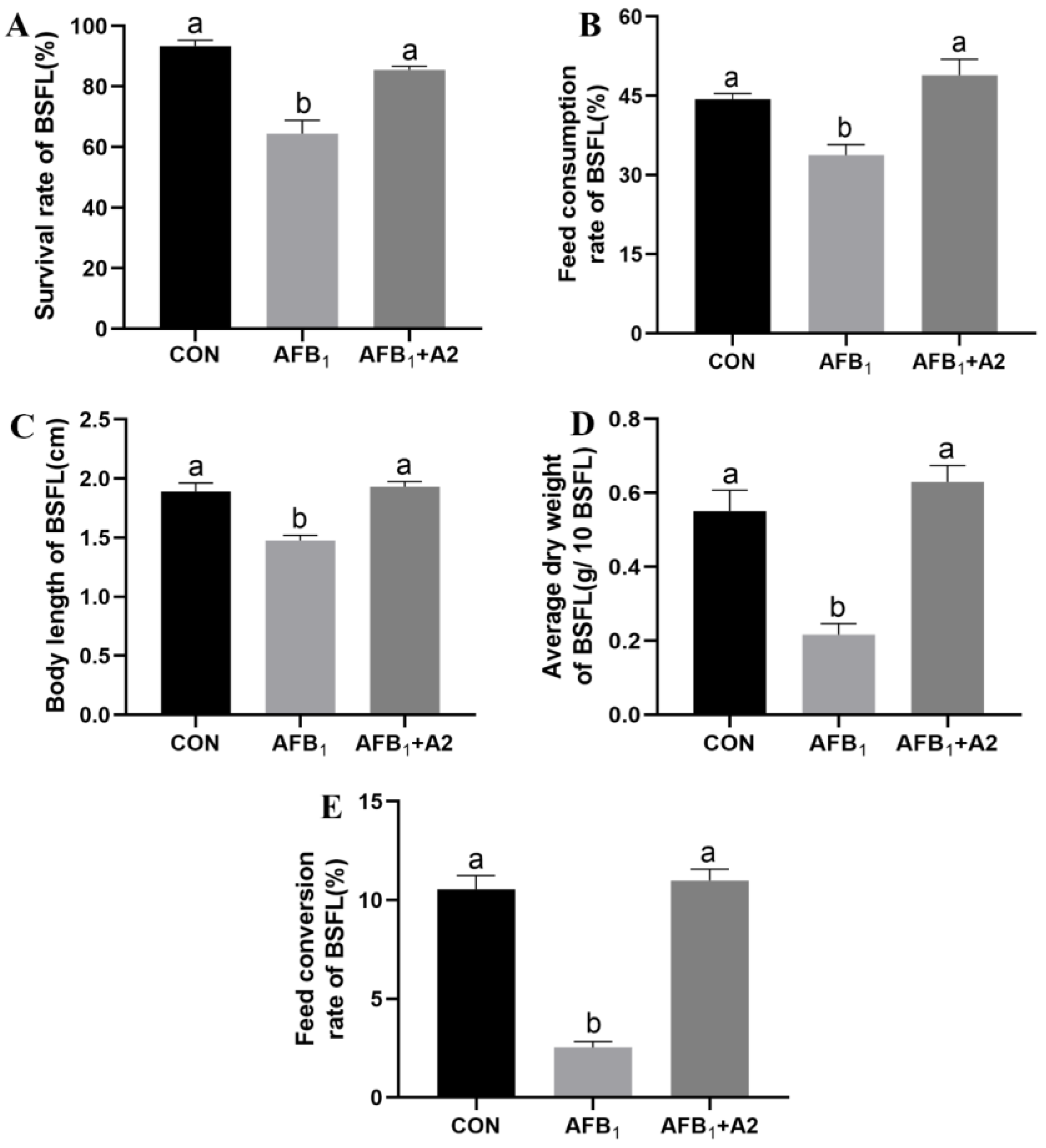
3.4. Effect of Stenotrophomonas acidaminiphila Strain A2 on the Growth Performance of BSFL

It is shown in Figure 3A that feeding the peanut meal without AFB1 resulted in a BSFL survival rate of 93%, indicating that the applied rearing condition was suitable to support its growth and development. However, the survival rate of BSFL reduced to 64% under exposure to AFB1 in the same condition. After the inoculation of S. acidaminiphila A2 into the AFB1-contaminated peanut meal, the survival rate of BSFL increased up to 86%. As presented in Figure 3B–E, the feed consumption rate, body length, average dry weight and feed conversion rate of BSFL were significantly reduced in the AFB1 group in comparison with the CON group, but no remarkable differences were observed between the AFB1+A2 group and the CON group. Thus, S. acidaminiphila A2 could effectively alleviate the toxicity of AFB1 to BSFL. In addition, the nutritional compositions of BSFL were determined (Table 2). The exposure to AFB1 did not significantly influence crude ash and crude protein content, but resulted in an increase of crude fat in the body of BSFL. The addition of S. acidaminiphila A2 in AFB1-contaminated peanut meal slightly decreased the crude protein content in harvested BSFL. A total of seventeen free amino acids, including eight essential amino acids, were found in BSFL. There were no significant differences in thirteen individual free amino acids among the three groups. The Proline and Alanine content in BSFL harvested in the AFB1 and AFB1+A2 groups were lowered in comparison with the CON group. The Serine and Cystine content in BSFL harvested in the AFB1 group were significantly reduced after the addition of S. acidaminiphila A2.

3.5. Degradation of AFB1 by BSFL Combined with Stenotrophomonas acidaminiphila Strain A2

The AFB1 synergistic degradation between BSFL and commensal S. acidaminiphila A2 was explored. As shown in Figure 4, S. acidaminiphila A2 could degrade 41% AFB1 in peanut meal after 10 days, while complete AFB1 degradation was achieved by BSFL in combination with S. acidaminiphila A2.

4. Discussion

Among numerous saprophagous insects, BSFL have become a strikingly good candidate for bioconversion. They can effectively convert various organic wastes, such as animal manure, food waste and agricultural residues, to obtain high-quality insect biomass rich in protein and fat [16]. Previous research has shown that BSFL can biodegrade six mycotoxins (aflatoxins B1/B2/G2, deoxynivalenol, ochratoxin A, zearalenone) and three pesticides (chlorpyrifos, chlorpyrifos-methyl, pirimiphos-methyl) with no bioaccumulation in the larvae harvested [27]. Moreover, Meijer et al. [28] found that cytochrome P450 enzymes and cytoplasmic reductases were involved in the metabolic conversion of AFB1 in BSFL, which could transform AFB1 into aflatoxicol and aflatoxin P1. Benefiting from using a sterile BSFL system, we found that the ability of BSFL to degrade AFB1 was greatly reduced in the absence of microbiota, which indicated the important role of microbiota in the degradation. Indeed, insect gut microbiota can be considered an additional organ with fluidity or malleability. The larval intestinal microbiota is a crucial contributor to the BSFL nutrient metabolic process [22] and helps the host to tolerate heavy metals [29] and degrade antibiotics [30,31]. Lou et al. [30] identified two bacterial strains—Alcaligenes faecalis GLD-1 and Ochrobactrum intermedium GLD-2—capable of degrading lincomycin from BSFL gut. Yang et al. [31] found that ciprofloxacin degradation by BSFL was associated with intestinal microorganisms, and five strains that degrade ciprofloxacin were identified including Klebsiella pneumoniae BSFLG-CIP1, Trichosporon asahii BSFLG-CIP2, Geotrichum sp. BSFLG-CIP3, Pichia kudriavzevii BSFLG-CIP4 and Proteus mirabilis BSFLG-CIP5. In the current study, twenty-five AFB1-degrading bacteria were isolated from the intestine of BSFL reared on AFB1-contaminated peanut meal. Among them, Stenotrophomonas acidaminiphila strain A2 displayed the highest AFB1 degradation rate of 94%. Similarly, Guan et al. [32] previously obtained a Stenotrophomonas Maltophilia from tapir feces, which could reduce AFB1 by 82.5% in the liquid medium at 37 °C for 72 h. Cai et al. [33] reported that Stenotrophomonas sp. CW117 isolated from soils was able to degrade more than 90% of AFB1 at the initial concentration of 40 to 4000 μg L−1 within 24 h.
The active components responsible for AFB1 degradation were located in the culture supernatant of S. acidaminiphila A2. This is in agreement with the report of Cai et al. [33], who studied the AFB1 degradation mechanism by Stenotrophomonas sp. CW117 and revealed that extracellular enzymes and non-protein components were responsible for the degradation activity. In a previous study of Zhao et al. [34], a 32 kDa extracellular enzyme with AFB1 degradation capacity was purified from Myxococcus fulvus. Guo et al. [35] reported that the spore CotA laccase from Bacillus licheniformis could catalyze the C3-hydroxylation of AFB1, resulting in the formation of non-toxic transformation products aflatoxin Q1 and epi-aflatoxin Q1. Further study was needed to elucidate the enzymatic mechanisms for AFB1 degradation in S. acidaminiphila A2. The degradation of AFB1 by S. acidaminiphila A2 depends on factors such as incubation time, pH, temperature, metal ions and substrate concentration. The AFB1 degradation by the culture supernatant of S. acidaminiphila A2 was a relatively rapid and continuous process, with 67% AFB1 degraded in the first 12 h and 87% degraded after 24 h. Similar results were obtained elsewhere. Cai et al. [33] reported a 52% reduction of AFB1 within 6 h by a cell-free supernatant of Stenotrophomonas sp. CW117. Microbacterium proteolyticum B204 isolated from bovine feces could eliminate about 80% of AFB1 after a 12 h treatment [36]. The culture supernatant of S. acidaminiphila A2 could effectively degrade AFB1 under neutral and alkaline pH conditions, but gradually lost its AFB1 degradation capacity at acidic pH. The effect of pH on AFB1 degradation by a culture supernatant of S. maltophilia 35-3 showed a similar trend [32]. Moreover, the highest AFB1 degradation was achieved at pH 7 and 8 by M. proteolyticum B204 [36]. Regarding the effect of temperature, we found that high temperature did not inhibit the AFB1 degradation by a culture supernatant of S. acidaminiphila A2. Indeed, the degradation rate increased with the increase of temperature from 20 °C to 70 °C. Samuel et al. [37] also reported that the percentage of AFB1 degradation by culture supernatant of Pseudomonas aeruginosa N17-1 was elevated from 45% at 20 °C to 90% at 65 °C. The culture supernatant of Trichoderma reesei CGMCC3.5218 could achieve more than 90% AFB1 degradation at a temperature range from 45 °C to 90 °C [38]. These results imply that the enzymes or proteins involved in the AFB1 degradation are thermostable. Various metal ions were examined for their effects on the AFB1 degradation, and it was shown that the presence of Zn2+ led to a decrease of AFB1 degradation rate by a culture supernatant of S. acidaminiphila A2. The inhibitory effect of Zn2+ was commonly observed on AFB1-degrading strains including Stenotrophomonas sp. CW117 [33], P. aeruginosa N17-1 [37] and T. reesei CGMCC3.5218 [38].
The growth performance of BSFL in AFB1-contaminated peanut meal was investigated. The exposure to AFB1 resulted in a decrease of the survival rate and dry weight of BSFL as well as a reduction of feed consumption rate and feed conversion rate. However, Bosh et al. [20] previously found that BSFL displayed a high tolerance to AFB1, and their survival rate and body weight were not influenced when fed with poultry feed spiked with 0.5 mg kg −1 AFB1. BSFL are considered a promising alternative protein and fat source for use in animal feed. Interestingly, we found that the presence of AFB1 in peanut meal could increase crude fat content by 2% but did not influence crude protein content in BSFL. In a recent study by Zhao et al. [39], yellow mealworms reared on AFB1-containing bran could provide the same quality of available larval protein and fat as those fed on bran without AFB1. The effect of AFB1 on fat metabolism in the two different insects is worth further study. The presence of S. acidaminiphila A2 could alleviate the detrimental effect of AFB1 on the growth performance of BSFL. Functional bacteria isolated from the BSFL gut have been found to benefit the growth of BSFL. Pei et al. [22] reported that Bacillus velezensis EEAM 10B could improve the protein synthesis process and digestive enzyme activities in BSFL, thus elevating the substance uptake and protein conversion ability of BSFL. When the intestinal ciprofloxacin-degrading isolates were re-inoculated to the sterile BSFL system, the larvae survival rate and weight were significantly increased in comparison with the sterile BSFL system [31]. Moreover, the addition of ciprofloxacin-degrading isolates could enhance the ciprofloxacin degradation efficiency of the sterile BSFL system. In our study, the AFB1 in peanut meal could be completely degraded by BSFL in combination with S. acidaminiphila A2. The metabolic pathways of AFB1 in BSFL and S. acidaminiphila A2 should be further investigated and clarified in depth. A toxicological test of the AFB1 degradation product is also necessary.

5. Conclusions

The present study indicated that AFB1 in peanut meal could be degraded by 31.71% and 88.72%, respectively, by germ-free BSFL and non-sterile BSFL for 10 days. Twenty-five AFB1-degrading bacteria were isolated from BSFL gut, and S. acidaminiphila A2 achieved the highest AFB1 degradation, by 94%. The exposure to AFB1 had an adverse effect on the growth performance of BSFL, resulting in a decrease of larvae survival rate and body weight. The supplementation of S. acidaminiphila A2 to BSFL could achieve complete AFB1 degradation in peanut meal and alleviate the negative effect of AFB1 on the growth performance of BSFL. Thus, it is promising to degrade AFB1 in contaminated feedstuff by BSFL in combination with commensal AFB1-degrading microorganisms.

Supplementary Materials

The following supporting information can be downloaded at: https://www.mdpi.com/article/10.3390/life13010234/s1, Table S1: Identification of strains capable of degrading more than 60% of AFB1 by 16S rRNA gene analysis.

Author Contributions

Conceptualization, J.S. and T.L.; methodology, J.S. and H.Z.; validation, J.S., K.L. and Q.L.; formal analysis, J.S.; investigation, J.S. and X.L.; resources, K.X.; data curation, J.G.; writing—original draft preparation, J.S. and Q.L.; writing—review and editing, J.S. and S.Y.; visualization, Q.L.; supervision, Q.L. and S.Y.; project administration, J.S.; funding acquisition, J.S. and S.Y. All authors have read and agreed to the published version of the manuscript.

Funding

This research was funded by the National Key Research and Development Program of China (No. 2021YFD1700900), and Foundation of Key Technology Research Project of Henan Province (Grant No. 222102110156 and Grant No. 222102110414).

Institutional Review Board Statement

Not applicable.

Informed Consent Statement

Not applicable.

Data Availability Statement

The data presented in this study are available on request from the corresponding author.

Conflicts of Interest

The authors declare no conflict of interest.

References

  1. Reddy, K.R.N.; Salleh, B.; Saad, B.; Abbas, H.K.; Abel, C.A.; Shier, W.T. An overview of mycotoxin contamination in foods and its implications for human health. Toxin Rev. 2010, 29, 3–26. [Google Scholar] [CrossRef]
  2. My, A.; Ds, S. Dietary factors affecting aflatoxin Bi carcinogenicity. Malays. J. Nutr. 1997, 3, 161–179. [Google Scholar]
  3. Gruber-Dorninger, C.; Jenkins, T.; Schatzmayr, G. Global mycotoxin occurrence in feed: A ten-year survey. Toxins 2019, 11, 375. [Google Scholar] [CrossRef] [PubMed] [Green Version]
  4. Pandey, M.K.; Kumar, R.; Pandey, A.K.; Soni, P.; Gangurde, S.S.; Sudini, H.K.; Fountain, J.C.; Liao, B.; Desmae, H.; Okori, P.; et al. Mitigating aflatoxin contamination in groundnut through a combination of genetic resistance and post-harvest management practices. Toxins 2019, 11, 315. [Google Scholar] [CrossRef] [Green Version]
  5. Bbosa, G.S.; Kitya, D.; Odda, J.; Ogwal-Okeng, J. Aflatoxins metabolism, effects on epigenetic mechanisms and their role in carcinogenesis. Health 2013, 5, 14–34. [Google Scholar] [CrossRef] [Green Version]
  6. Ostry, V.; Malir, F.; Toman, J.; Grosse, Y. Mycotoxins as human carcinogens-the IARC Monographs classification. Mycotoxin Res. 2017, 33, 65–73. [Google Scholar] [CrossRef] [PubMed]
  7. Liu, Y.; Wu, F. Global burden of aflatoxin-induced hepatocellular carcinoma: A risk assessment. Environ. Health Perspect. 2010, 118, 818–824. [Google Scholar] [CrossRef] [Green Version]
  8. Rushing, B.R.; Selim, M.I. Aflatoxin B1: A review on metabolism, toxicity, occurrence in good, occupational exposure, and detoxification methods. Food Chem. Toxicol. 2019, 124, 81–100. [Google Scholar] [CrossRef]
  9. Guo, Y.; Zhao, L.; Ma, Q.; Ji, C. Novel strategies for degradation of aflatoxins in food and feed: A review. Food Res. Int. 2021, 140, 109878. [Google Scholar] [CrossRef]
  10. Samuel, M.S.; Sivaramakrishna, A.; Mehta, A. Degradation and detoxification of aflatoxin B1 by Pseudomonas putida. Int. Biodeterior. Biodegrad. 2014, 86, 202–209. [Google Scholar] [CrossRef]
  11. Eshelli, M.; Harvey, L.; Edrada-Ebel, R.; McNeil, B. Metabolomics of the bio-degradation process of aflatoxin B1 by Actinomycetes at an initial pH of 6.0. Toxins 2015, 7, 439–456. [Google Scholar] [CrossRef] [PubMed]
  12. Cserháti, M.; Kriszt, B.; Krifaton, C.; Szoboszlay, S.; Háhn, J.; Tóth, S.; Nagy, I.; Kukolya, J. Mycotoxin-degradation profile of Rhodococcus strains. Int. J. Food Microbiol. 2013, 166, 176–185. [Google Scholar] [CrossRef] [PubMed]
  13. Gao, X.; Ma, Q.; Zhao, L.; Lei, Y.; Shan, Y.; Ji, C. Isolation of Bacillus subtilis: Screening for aflatoxins B1, M1, and G1 detoxification. Eur. Food Res. Technol. 2011, 232, 957–962. [Google Scholar] [CrossRef]
  14. Allegretti, G.; Talamini, E.; Schmidt, V.; Bogorni, P.C.; Ortega, E. Insect as feed: An emergy assessment of insect meal as a sustainable protein source for the brazilian poultry industry. J. Clean. Prod. 2018, 171, 403–412. [Google Scholar] [CrossRef]
  15. Salam, M.; Shahzadi, A.; Zheng, H.; Alam, F.; Nabi, G.; Dezhi, S.; Ullah, W.; Ammara, S.; Ali, N.; Bilal, M. Effect of different environmental conditions on the growth and development of black soldier fly larvae and its utilization in solid waste management and pollution mitigation. Environ. Technol. Innov. 2022, 28, 102649. [Google Scholar] [CrossRef]
  16. Sánchez-Muros María, J.; Barroso, F.G.; Manzano-Agugliaro, F. Insect meal as renewable source of food for animal feeding: A review. J. Clean. Prod. 2014, 65, 16–27. [Google Scholar] [CrossRef]
  17. Scala, A.; Cammack, J.A.; Salvia, R.; Scieuzo, C.; Franco, A.; Bufo, S.A.; Tomberlin, J.K.; Falabella, P. Rearing substrate impacts growth and macronutrient composition of Hermetia illucens (L.) (Diptera: Stratiomyidae) larvae produced at an industrial scale. Sci. Rep. 2020, 10, 19448. [Google Scholar] [CrossRef]
  18. Schiavone, A.; Dabbou, S.; De Marco, M.; Cullere, M.; Biasato, I.; Biasibetti, E.; Capucchio, M.T.; Bergagna, S.; Dezzutto, D.; Meneguz, M.; et al. Black soldier fly larva fat inclusion in finisher broiler chicken diet as an alternative fat source. Animal 2018, 12, 2032–2039. [Google Scholar] [CrossRef]
  19. Belghit, I.; Liland, N.S.; Waagbø, R.; Biancarosa, I.; Pelusio, N.; Li, Y.; Krogdahl, Å.; Lock, E.-J. Potential of insect-based diets for Atlantic salmon (Salmo salar). Aquaculture 2018, 491, 72–81. [Google Scholar] [CrossRef]
  20. Bosch, G.; Van Der Fels-Klerx, H.J.; De Rijk, T.C.; Oonincx, D.G.A.B. Aflatoxin B1 tolerance and accumulation in black soldier fly larvae (Hermetia illucens) and yellow mealworms (Tenebrio molitor). Toxins 2017, 9, 185. [Google Scholar] [CrossRef]
  21. Camenzuli, L.; Van Dam, R.; de Rijk, T.; Andriessen, R.; Van Schelt, J.; Van der Fels-Klerx, H.J.I. Tolerance and excretion of the mycotoxins aflatoxin B1, zearalenone, deoxynivalenol, and ochratoxin a by Alphitobius diaperinus and Hermetia illucens from contaminated substrates. Toxins 2018, 10, 91. [Google Scholar] [CrossRef] [PubMed]
  22. Pei, Y.; Zhao, S.; Chen, X.; Zhang, J.; Ni, H.; Sun, M.; Lin, H.; Liu, X.; Chen, H.; Yang, S. Bacillus Velezensis EEAM 10b strengthens nutrient metabolic process in black soldier fly larvae (Hermetia illucens) via changing gut microbiome and metabolic pathways. Front. Nutr. 2022, 9, 880488. [Google Scholar] [CrossRef] [PubMed]
  23. GB 5009.5-2016; The Ministry of Health of the People’s Republic of China. National Food Safety Standard: Determination of Protein in Foods. China Standards Press: Beijing, China, 2016.
  24. GB 5009.6-2016; The Ministry of Health of the People’s Republic of China. National Food Safety Standard: Determination of Fat in Foods. China Standards Press: Beijing, China, 2016.
  25. GB 5009.4-2016; The Ministry of Health of the People’s Republic of China. National Food Safety Standard: Determination of Ash in Foods. China Standards Press: Beijing, China, 2016.
  26. GB 5009.124-2016; The Ministry of Health of the People’s Republic of China. National Food Safety Standard: Determination of Amino Acids in Foods. China Standards Press: Beijing, China, 2016.
  27. Purschke, B.; Scheibelberger, R.; Axmann, S.; Adler, A.; Jäger, H. Impact of substrate contamination with mycotoxins, heavy metals and pesticides on the growth performance and composition of black soldier fly larvae (Hermetia illucens) for use in the feed and food value chain. Food Addit. Contam. A 2017, 34, 1410–1420. [Google Scholar] [CrossRef]
  28. Meijer, N.; Stoopen, G.; Van der Fels-Klerx, H.J.; Van Loon, J.J.A.; Carney, J.; Bosch, G. Aflatoxin B1 conversion by black soldier fly (Hermetia illucens) larval enzyme extracts. Toxins 2019, 11, 532. [Google Scholar] [CrossRef] [PubMed] [Green Version]
  29. Wu, N.; Wang, X.; Xu, X.; Cai, R.; Xie, S. Effects of heavy metals on the bioaccumulation, excretion and gut microbiome of black soldier fly larvae (Hermetia illucens). Ecotoxicol. Environ. Saf. 2020, 192, 110323. [Google Scholar] [CrossRef]
  30. Luo, X.; Yang, Q.; Lin, Y.; Tang, Z.; Tomberlin, J.K.; Liu, W.; Huang, Y. Black soldier fly larvae effectively degrade lincomycin from pharmaceutical industry wastes. J. Environ. Manag. 2022, 307, 114539. [Google Scholar] [CrossRef]
  31. Yang, C.; Ma, S.; Li, F.; Zheng, L.; Tomberlin, J.K.; Yu, Z.; Zhang, J.; Yu, C.; Fan, M.; Cai, M. Characteristics and mechanisms of ciprofloxacin degradation by black soldier fly larvae combined with associated intestinal microorganisms. Sci. Total Environ. 2022, 811, 151371. [Google Scholar] [CrossRef]
  32. Guan, S.; Ji, C.; Zhou, T.; Li, J.; Ma, Q.; Niu, T. Aflatoxin B1 degradation by Stenotrophomonas maltophilia and other microbes selected using coumarin medium. Int. J. Mol. Sci. 2008, 9, 1489–1503. [Google Scholar] [CrossRef] [Green Version]
  33. Cai, M.; Qian, Y.; Chen, N.; Ling, T.; Wang, J.; Jiang, H.; Wang, X.; Qi, K.; Zhou, Y. Detoxification of aflatoxin B1 by Stenotrophomonas sp. CW117 and characterization the thermophilic degradation process. Environ. Pollut. 2020, 261, 114178. [Google Scholar] [CrossRef]
  34. Zhao, L.; Guan, S.; Gao, X.; Ma, Q.; Lei, Y.; Bai, X.; Ji, C. Preparation, purification and characteristics of an aflatoxin degradation enzyme from Myxococcus fulvus ANSM068. J. Appl. Microbiol. 2011, 110, 147–155. [Google Scholar] [CrossRef]
  35. Guo, Y.; Qin, X.; Tang, Y.; Ma, Q.; Zhang, J.; Zhao, L. CotA laccase, a novel aflatoxin oxidase from Bacillus licheniformis, transforms aflatoxin B1 to aflatoxin Q1 and epi-aflatoxin Q1. Food Chem. 2020, 325, 126877. [Google Scholar] [CrossRef] [PubMed]
  36. Yan, Y.; Zhang, X.; Chen, H.; Huang, W.; Jiang, H.; Wang, C.; Xiao, Z.; Zhang, Y.; Xu, J. Isolation and aflatoxin B1-degradation characteristics of a Microbacterium proteolyticum B204 Strain from bovine faeces. Toxins 2022, 14, 525. [Google Scholar] [CrossRef] [PubMed]
  37. Sangare, L.; Zhao, Y.; Folly, Y.M.E.; Chang, J.; Li, J.; Selvaraj, J.N.; Xing, F.; Zhou, L.; Wang, Y.; Liu, Y. Aflatoxin B1 degradation by a Pseudomonas strain. Toxins 2014, 6, 3028–3040. [Google Scholar] [CrossRef] [PubMed] [Green Version]
  38. Yue, X.; Ren, X.; Fu, J.; Wei, N.; Altomare, C.; Haidukowski, M.; Logrieco, A.F.; Zhang, Q.; Li, P. Characterization and mechanism of aflatoxin degradation by a novel strain of Trichoderma reesei CGMCC3.5218. Front. Microbiol. 2022, 13, 1003039. [Google Scholar] [CrossRef] [PubMed]
  39. Zhao, D.; Xie, H.; Gao, L.; Zhang, J.; Li, Y.; Mao, G.; Zhang, H.; Wang, F.; Lam, S.S.; Song, A. Detoxication and bioconversion of aflatoxin B1 by yellow mealworms (Tenebrio molitor): A sustainable approach for valuable larval protein production from contaminated grain. Ecotoxicol. Environ. Saf. 2022, 242, 113935. [Google Scholar] [CrossRef]
Figure 1. Isolation and identification of AFB1-degrading bacterial strains from BSFL gut. (A) The AFB1 degradation rate by culture medium of different strains. Values are means with their standard errors represented by vertical bars (n = 3). (B) The colony morphology of the strain A2. (C) The phylogenetic tree of the strain A2.
Life 13 00234 g001
Figure 2. Characterization of the AFB1-degrading capacity of S. acidaminiphila A2. (A) The degradation of AFB1 by different components of the strain A2. 1, Bacterial culture; 2, Culture supernatant; 3, Cell pellets; 4, Cell fragments; 5, Culture supernatant + Proteinase K. The effects of incubation time (B), pH (C), temperature (D), metal ions (E) and substrate concentration (F) on AFB1 degradation by cell-free culture supernatant of S. acidaminiphila A2. Values are means with their standard errors represented by vertical bars (n = 3).
Life 13 00234 g002
Figure 3. Effects of S. acidaminiphila A2 on the growth performance of BSFL in AFB1 contaminated peanut meal. (A) Survival rate, (B) Feed consumption rate, (C) Body length, (D) Average dry weight, and (E) Feed conversion rate. Values are means with their standard errors represented by vertical bars (n = 3). Values with different letters differ significantly (p < 0.05).
Life 13 00234 g003
Figure 4. Degradation of AFB1 in peanut meal by BSFL in combination with S. acidaminiphila A2. Values are means with their standard errors represented by vertical bars (n = 3).
Life 13 00234 g004
Table 1. Comparison of AFB1 degradation in peanut meal by germ-free BSFL and non-sterile BSFL (n = 3).
ItemsGroup 1SEM 2p-Valve
GF-BSFLNS-BSFL
Dried frass weight (g)8.29 a5.96 b0.09<0.001
AFB1 concentration in frass (ng g−1)74.13 a17.24 b2.09<0.001
Total residual AFB1 content in frass (ng)614.63 a101.53 b14.71<0.001
AFB1 degradation rate (%)31.71 a88.72 b1.64<0.001
a,b Values in the same row with no common superscript differ significantly (p < 0.05). 1 GF-BSFL, germ-free BSFL; NS-BSFL, non-sterile BSFL. 2 SEM, pooled standard error of the mean.
Table 2. Nutrient compositions of BSFL(n = 3).
ItemsGroup 1SEM 2p-Valve
CONAFB1AFB1 + A2
Crude fat (%)23.49 b25.47 a25.53 a0.035<0.001
Crude fiber (%)8.908.909.000.0190.125
Crude protein (%)51.86 a51.46 ab51.03 b0.0710.009
Aspartic acid (%)3.903.943.810.0290.253
Glutamate (%)4.174.104.040.0300.252
Serine (%)1.25 a1.26 a1.20 b0.0070.028
Arginine (%)1.561.581.580.0140.720
Glycine (%)3.093.002.940.0200.060
Threonine (%)1.531.501.470.0110.196
Prolin (%)2.77 a2.65 b2.60 b0.0180.025
Alanine (%)4.25 a4.04 b3.94 b0.0270.008
Valine (%)2.682.612.560.0180.087
Methionine (%)0.650.620.610.0070.124
Cystine (%)0.31 b0.34 a0.30 b0.001<0.001
Isoleucine (%)1.981.961.900.0130.086
Leucine (%)2.902.882.780.0200.093
Phenylalanine (%)1.931.931.910.0160.762
Histidine (%)1.111.121.060.0120.173
Lysine (%)2.622.702.590.0150.065
Tyrosine (%)2.502.412.410.0160.116
a,b Values in the same row with no common superscript differ significantly (p < 0.05). 1 CON, non-sterile BSFL reared on peanut meal without AFB1; AFB1, non-sterile BSFL reared on peanut meal with AFB1; AFB1+A2, non-sterile BSFL reared on AFB1 contaminated peanut meal in the presence of S. acidaminiphila A2. 2 SEM, pooled standard error of the mean.
Disclaimer/Publisher’s Note: The statements, opinions and data contained in all publications are solely those of the individual author(s) and contributor(s) and not of MDPI and/or the editor(s). MDPI and/or the editor(s) disclaim responsibility for any injury to people or property resulting from any ideas, methods, instructions or products referred to in the content.

