
Neutrophil-to-Lymphocyte Ratio Predicts Early Neurological Deterioration after Endovascular Treatment in Patients with Ischemic Stroke
 , , , , , and
Abstract
:1. Introduction
2. Methods
2.1. Study Participants
2.2. Statistical Analysis
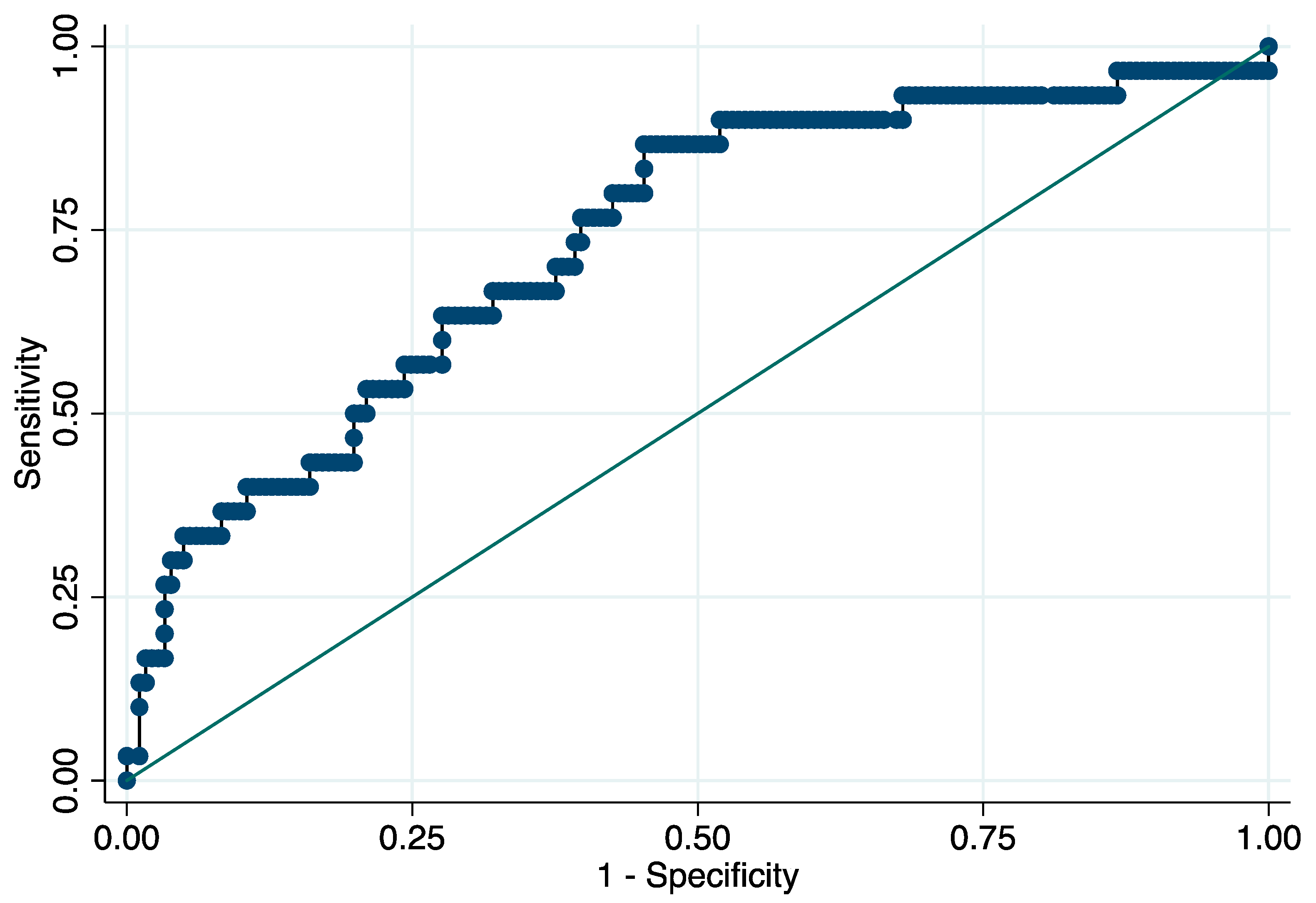
3. Results
4. Discussion
5. Conclusions
Author Contributions
Funding
Institutional Review Board Statement
Informed Consent Statement
Data Availability Statement
Conflicts of Interest
References
- Tsao, C.W.; Aday, A.W.; Almarzooq, Z.I.; Alonso, A.; Beaton, A.Z.; Bittencourt, M.S.; Boehme, A.K.; Buxton, A.E.; Carson, P.A.; Commodore-Mensah, Y.; et al. Heart Disease and Stroke Statistics-2022 Update: A Report from the American Heart Association. Circulation 2022, 145, e153–e639. [Google Scholar] [CrossRef] [PubMed]
- GBD 2019 Stroke Collaborators. Global, regional, and national burden of stroke and its risk factors, 1990-2019: A systematic analysis for the Global Burden of Disease Study 2019. Lancet Neurol. 2021, 20, 795–820. [Google Scholar] [CrossRef]
- Krueger, H.; Koot, J.; Hall, R.E.; O’Callaghan, C.; Bayley, M.; Corbett, D. Prevalence of individuals experiencing the effects of stroke in Canada: Trends and projections. Stroke 2015, 46, 2226–2231. [Google Scholar] [CrossRef] [PubMed]
- Lattanzi, S.; Coccia, M.; Pulcini, A.; Cagnetti, C.; Galli, F.L.; Villani, L.; Campa, S.; Dobran, M.; Polonara, G.; Ceravolo, M.G.; et al. Endovascular treatment and cognitive outcome after anterior circulation ischemic stroke. Sci. Rep. 2020, 10, 18524. [Google Scholar] [CrossRef]
- Lattanzi, S.; Rinaldi, C.; Cagnetti, C.; Foschi, N.; Norata, D.; Broggi, S.; Rocchi, C.; Silvestrini, M. Predictors of pharmaco-resistance in patients with post-stroke epilepsy. Brain Sci. 2021, 11, 418. [Google Scholar] [CrossRef]
- Lattanzi, S.; Trinka, E.; Turcato, G.; Rinaldi, C.; Cagnetti, C.; Foschi, N.; Broggi, S.; Norata, D.; Brigo, F.; Silvestrini, M. Latency of poststroke epilepsy can predict drug resistance. Eur. J. Neurol. 2022, 29, 2481–2485. [Google Scholar] [CrossRef]
- Thanvi, B.; Treadwell, S.; Robinson, T. Early neurological deterioration in acute ischaemic stroke: Predictors, mechanisms and management. Postgrad. Med. J. 2008, 84, 412–417. [Google Scholar] [CrossRef]
- Girot, J.B.; Richard, S.; Gariel, F.; Sibon, I.; Labreuche, J.; Kyheng, M.; Gory, B.; Dargazanli, C.; Maier, B.; Consoli, A.; et al. Predictors of Unexplained Early Neurological Deterioration after Endovascular Treatment for Acute Ischemic Stroke. Stroke 2020, 51, 2943–2950. [Google Scholar] [CrossRef]
- Toni, D.; Fiorelli, M.; Gentile, M.; Bastianello, S.; Sacchetti, M.L.; Argentino, C.; Pozzilli, C.; Fieschi, C. Progressing neurological deficit secondary to acute ischemic stroke. A study on predic-tability, pathogenesis, and prognosis. Arch. Neurol. 1995, 52, 670–675. [Google Scholar] [CrossRef]
- Davalos, A.; Cendra, E.; Teruel, J.; Martinez, M.; Genis, D. Deteriorating ischemic stroke: Risk factors and prognosis. Neurology 1990, 40, 1865–1869. [Google Scholar] [CrossRef]
- Duan, Z.; Guo, W.; Tang, T.; Tao, L.; Gong, K.; Zhang, X. Relationship between high-sensitivity C-reactive protein and early neurological deterioration in stroke patients with and without atrial fibrillation. Heart Lung 2020, 49, 193–197. [Google Scholar] [CrossRef] [PubMed]
- Li, Z.; Zhang, H.; Han, J.; Chu, Z.; Zhao, S.; Yang, Q.; Huang, X.; Zhou, Z. Time Course and Clinical Relevance of Neurological Deterioration after Endovascular Recanalization Therapy for Anterior Circulation Large Vessel Occlusion Stroke. Front. Aging Neurosci. 2021, 13, 651614. [Google Scholar] [CrossRef] [PubMed]
- Zhang, Y.B.; Su, Y.Y.; He, Y.B.; Liu, Y.F.; Liu, G.; Fan, L.L. Early neurological deterioration after recanalization treatment in patients with acute ischemic stroke: A retrospective study. Chin. Med. J. 2018, 131, 137–143. [Google Scholar] [CrossRef] [PubMed]
- Davalos, A.; Toni, D.; Iweins, F.; Lesaffre, E.; Bastianello, S.; Castillo, J. Neurological deterioration in acute ischemic stroke: Potential predictors and associated factors in the European Cooperative Acute Stroke Study (ECASS) I. Stroke 1999, 30, 2631–2636. [Google Scholar] [CrossRef]
- Birschel, P.; Ellul, J.; Barer, D. Progressing stroke: Towards an internationally agreed definition. Cerebrovasc. Dis. 2004, 17, 242–252. [Google Scholar] [CrossRef]
- Weimar, C.; Mieck, T.; Buchthal, J.; Ehrenfeld, C.E.; Schmid, E.; Diener, H.-C. For the German Stroke Study Collaboration. Neurologic worsening during the acute phase of ischemic stroke. Arch. Neurol. 2005, 62, 393–397. [Google Scholar] [CrossRef]
- Kim, J.M.; Bae, J.H.; Park, K.Y.; Lee, W.J.; Byun, J.S.; Ahn, S.-W.; Shin, H.-W.; Han, S.-H.; Yoo, I.-H. Incidence and mechanism of early neurological deterioration after endovascular thrombectomy. J. Neurol. 2019, 266, 609–615. [Google Scholar] [CrossRef]
- Bourcier, R.; Goyal, M.; Muir, K.W.; Desal, H.; Dippel, D.W.J.; Majoie, C.B.L.M.; van Zwam, W.H.; Jovin, T.G.; Mitchell, P.J.; Demchuk, A.M.; et al. HERMES Trialists Collaboration. Risk factors of unexplained early neurological deterioration after treatment for ischemic stroke due to large vessel occlusion: A post hoc analysis of the HERMES study. J. Neurointerv. Surg. 2022. [Google Scholar] [CrossRef]
- Simats, A.; García-Berrocoso, T.; Montaner, J. Neuroinflammatory biomarkers: From stroke diagnosis and prognosis to therapy. Biochim. Biophys. Acta 2016, 1862, 411–424. [Google Scholar] [CrossRef]
- Zangari, R.; Zanier, E.R.; Torgano, G.; Bersano, A.; Beretta, S.; Beghi, E.; Casolla, B.; Checcarelli, N.; Lanfranconi, S.; Maino, A.; et al. Early ficolin-1 is a sensitive prognostic marker for functional out-come in ischemic stroke. J. Neuroinflammation 2016, 13, 16. [Google Scholar] [CrossRef] [Green Version]
- Wang, L.; Song, Q.; Wang, C.; Wu, S.; Deng, L.; Li, Y.; Zheng, L.; Liu, M. Neutrophil to lymphocyte ratio predicts poor outcomes after acute ischemic stroke: A cohort study and systematic review. J. Neurol. Sci. 2019, 406, 116445. [Google Scholar] [CrossRef] [PubMed]
- Zhang, J.; Ren, Q.; Song, Y.; He, M.; Zeng, Y.; Liu, Z.; Xu, J. Prognostic role of neutrophil-lymphocyte ratio in patients with acute ischemic stroke. Medicine 2017, 96, e8624. [Google Scholar] [CrossRef] [PubMed]
- Goyal, N.; Tsivgoulis, G.; Chang, J.J.; Malhotra, K.; Pandhi, A.; Ishfaq, M.F.; Alsbrook, D.; Arthur, A.S.; Elijovich, L.; Alexandrov, A.V. Admission neutrophil-to-lymphocyte ratio as a prognostic biomarker of outcomes in large vessel occlusion strokes. Stroke 2018, 49, 1985–1987. [Google Scholar] [CrossRef] [PubMed]
- Maestrini, I.; Strbian, D.; Gautier, S.; Haapaniemi, E.; Moulin, S.; Sairanen, T.; Dequatre-Ponchelle, N.; Sibolt, G.; Cordonnier, C.; Melkas, S.; et al. Higher neutrophil counts before thrombolysis for cerebral ischemia predict worse outcomes. Neurology 2015, 85, 1408–1416. [Google Scholar] [CrossRef]
- Malhotra, K.; Goyal, N.; Chang, J.J.; Broce, M.; Pandhi, A.; Kerro, A.; Shahripour, R.B.; Alexandrov, A.V.; Tsivgoulis, G. Differential leukocyte counts on admission predict outcomes in patients with acute ischaemic stroke treated with intravenous thrombolysis. Eur. J. Neurol. 2018, 25, 1417–1424. [Google Scholar] [CrossRef]
- Duan, Z.; Wang, H.; Wang, Z.; Hao, Y.; Zi, W.; Yang, D.; Zhou, Z.; Liu, W.; Lin, M.; Shi, Z.; et al. Neutrophil-lymphocyte ratio predicts functional and safety outcomes after endovascular treatment for acute ischemic stroke. Cerebrovasc. Dis. 2018, 45, 221–227. [Google Scholar] [CrossRef]
- Sengeze, N.; Giray, S. The relationship between first pass recanalization of stent retriever-based thrombectomy and neutro-phil to lymphocyte ratio in middle cerebral artery occlusions. Int. J. Neurosci. 2021, 131, 634–640. [Google Scholar] [CrossRef]
- Zhang, R.; Wu, X.; Hu, W.; Zhao, L.; Zhao, S.; Zhang, J.; Chu, Z.; Xu, Y. Neutrophil-to-lymphocyte ratio predicts hemorrhagic transformation in ischemic stroke: A meta-analysis. Brain Behav. 2019, 9, e01382. [Google Scholar] [CrossRef]
- Świtońska, M.; Piekuś-Słomka, N.; Słomka, A.; Sokal, P.; Zekanowska, E.; Lattanzi, S. Neutrophil-to-Lymphocyte Ratio and Symptomatic Hemorrhagic Transformation in Is-chemic Stroke Patients Undergoing Revascularization. Brain Sci. 2020, 10, 771. [Google Scholar] [CrossRef]
- Sharma, D.; Spring, K.J.; Bhaskar, S.M.M. Role of Neutrophil-Lymphocyte Ratio in the Prognosis of Acute Ischaemic Stroke after Reperfusion Therapy: A Systematic Review and Meta-analysis. J. Cent. Nerv. Syst. Dis. 2022, 14, 11795735221092518. [Google Scholar] [CrossRef]
- Lattanzi, S.; Brigo, F.; Trinka, E.; Cagnetti, C.; Di Napoli, M.; Silvestrini, M. Neutrophil-to-Lymphocyte Ratio in Acute Cerebral Hemorrhage: A System Review. Transl. Stroke Res. 2019, 10, 137–145. [Google Scholar] [CrossRef]
- Shi, M.; Yang, C.; Tang, Q.W.; Xiao, L.-F.; Chen, Z.-H.; Zhao, W.-Y. The Prognostic Value of Neutrophil-to-Lymphocyte Ratio in Patients with Aneurysmal Subarachnoid Hemorrhage: A Systematic Review and Meta-Analysis of Observational Studies. Front. Neurol. 2021, 12, 745560. [Google Scholar] [CrossRef] [PubMed]
- Song, S.Y.; Zhao, X.X.; Rajah, G.; Hua, C.; Kang, R.J.; Han, Y.P.; Ding, Y.C.; Meng, R. Clinical significance of baseline neutrophil-to-lymphocyte ratio in patients with ischemic stroke or hemorrhagic stroke: An updated meta-analysis. Front. Neurol. 2019, 10, 1032. [Google Scholar] [CrossRef] [PubMed]
- SPREAD–Stroke Prevention and Educational Awareness Diffusion. Ictus Cerebrale: Linee Guida Italiane di Prevenzione e Trattamento. Available online: http://www.iso-spread.it/capitoli/LINEE_GUIDA_SPREAD_8a_EDIZIONE.pdf (accessed on 1 July 2022).
- Jauch, E.C.; Saver, J.L.; Adams, H.P., Jr.; Bruno, A.; Connors, J.J.; Demaerschalk, B.M.; Khatri, P.; McMullan, P.W., Jr.; Qureshi, A.I.; Rosenfield, K.; et al. Guidelines for the early management of patients with acute ischemic stroke: A guideline for healthcare professionals from the American Heart Association/American Stroke Association. Stroke 2013, 44, 870–947. [Google Scholar] [CrossRef] [PubMed]
- Powers, W.J.; Rabinstein, A.A.; Ackerson, T.; Adeoye, O.M.; Bambakidis, N.C.; Becker, K.; Biller, J.; Brown, M.; Demaerschalk, B.M.; Hoh, B.; et al. 2018 Guidelines for the early management of patients with acute ischemic stroke: A guideline for healthcare professionals from the American Heart Association/American Stroke Association. Stroke 2018, 49, e46–e99. [Google Scholar] [CrossRef]
- Powers, W.J.; Rabinstein, A.A.; Ackerson, T.; Adeoye, O.M.; Bambakidis, N.C.; Becker, K.; Biller, J.; Brown, M.; Demaerschalk, B.M.; Hoh, B.; et al. Guidelines for the Early Management of Patients with Acute Ischemic Stroke: 2019 Update to the 2018 Guidelines for the Early Management of Acute Ischemic Stroke: A Guideline for Healthcare Professionals from the American Heart Association/American Stroke Association. Stroke 2019, 50, e344–e418. [Google Scholar]
- Da Ros, V.; Scaggiante, J.; Sallustio, F.; Lattanzi, S.; Bandettini, M.; Sgreccia, A.; Rolla-Bigliani, C.; Lafe, E.; Sanfilippo, G.; Diomedi, M.; et al. Carotid Stenting and Mechanical Thrombectomy in Patients with Acute Ischemic Stroke and Tandem Occlusions: Antithrombotic Treatment and Functional Outcome. AJNR Am. J. Neuroradiol. 2020, 41, 2088–2093. [Google Scholar] [CrossRef]
- Da Ros, V.; Scaggiante, J.; Pitocchi, F.; Sallustio, F.; Lattanzi, S.; Umana, G.E.; Chaurasia, B.; di Poggio, M.B.; Toscano, G.; Bigliani, C.R.; et al. Mechanical thrombectomy in acute ischemic stroke with tandem occlusions: Impact of extracranial carotid lesion etiology on endovascular management and outcome. Neurosurg. Focus 2021, 51, E6. [Google Scholar] [CrossRef]
- Wityk, R.J.; Pessin, M.S.; Kaplan, R.F.; Caplan, L.R. Serial assessment of acute stroke using the NIH Stroke Scale. Stroke 1994, 25, 362–365. [Google Scholar] [CrossRef]
- Barber, P.A.; Demchuk, A.M.; Zhang, J.; Buchan, A.M. Validity and reliability of a quantitative computed tomography score in predicting outcome of hyperacute stroke before thrombolytic therapy. ASPECTS Study Group. Alberta Stroke Programme Early CT Score. Lancet 2000, 355, 1670–1674. [Google Scholar] [CrossRef]
- Lattanzi, S.; Cagnetti, C.; Pulcini, A.; Morelli, M.; Maffei, S.; Provinciali, L.; Silvestrini, M. The P-wave terminal force in embolic strokes of undetermined source. J. Neurol. Sci. 2017, 375, 175–178. [Google Scholar] [CrossRef] [PubMed]
- Lattanzi, S.; Pulcini, A.; Corradetti, T.; Rinaldi, C.; Zedde, M.L.; Ciliberti, G.; Silvestrini, M. Prediction of Outcome in Embolic Strokes of Undetermined Source. J. Stroke Cerebrovasc. Dis. 2020, 29, 104486. [Google Scholar] [CrossRef] [PubMed]
- Lattanzi, S.; Rinaldi, C.; Pulcini, A.; Corradetti, T.; Angelocola, S.; Zedde, M.L.; Ciliberti, G.; Silvestrini, M. Clinical phenotypes of embolic strokes of undetermined source. Neurol. Sci. 2021, 42, 297–300. [Google Scholar] [CrossRef]
- Hosmer, D.W.; Lemeshow, S. Applied Logistic Regression, 2nd ed.; John Wiley & Sons: New York, NY, USA, 1989; pp. 160–164. [Google Scholar]
- Iadecola, C.; Anrather, J. The immunology of stroke: From mechanisms to translation. Nat. Med. 2011, 17, 796–808. [Google Scholar] [CrossRef] [PubMed]
- Jin, R.; Yang, G.; Li, G. Inflammatory mechanisms in ischemic stroke: Role of inflammatory cells. J. Leukoc. Biol. 2010, 87, 779–789. [Google Scholar] [CrossRef] [PubMed]
- Anrather, J.; Iadecola, C. Inflammation and Stroke: An Overview. Neurotherapeutics 2016, 13, 661–670. [Google Scholar] [CrossRef]
- Lattanzi, S.; Di Napoli, M.; Ricci, S.; Divani, A.A. Matrix Metalloproteinases in Acute Intracerebral Hemorrhage. Neurotherapeutics 2020, 17, 484–496. [Google Scholar] [CrossRef]
- Semerano, A.; Laredo, C.; Zhao, Y.; Rudilosso, S.; Renú, A.; Llull, L.; Amaro, S.; Obach, V.; Planas, A.M.; Urra, X.; et al. Leukocytes, collateral circulation, and reperfusion in ischemic stroke patients treated with mechanical thrombectomy. Stroke 2019, 50, 3456–3464. [Google Scholar] [CrossRef]
- Rosell, A.; Cuadrado, E.; Ortega-Aznar, A.; Hernández-Guillamon, M.; Lo, E.H.; Montaner, J. MMP-9-positive neutrophil infiltration is associated to blood-brain barrier breakdown and basal lamina type IV collagen degradation during hemorrhagic transformation after human ischemic stroke. Stroke 2008, 39, 1121–1126. [Google Scholar] [CrossRef]
- Nie, X.; Pu, Y.; Zhang, Z.; Liu, X.; Duan, W.; Liu, L. Futile Recanalization after Endovascular Therapy in Acute Ischemic Stroke. BioMed. Res. Int. 2018, 2018, 5879548. [Google Scholar] [CrossRef]
- De Meyer, S.F.; Denorme, F.; Langhauser, F.; Geuss, E.; Fluri, F.; Kleinschnitz, C. Thromboinflammation in Stroke Brain Damage. Stroke 2016, 47, 1165–1172. [Google Scholar] [CrossRef] [PubMed] [Green Version]
- Stoll, G.; Nieswandt, B. Thrombo-inflammation in acute ischaemic stroke-implications for treatment. Nat. Rev. Neurol. 2019, 15, 473–481. [Google Scholar] [CrossRef] [PubMed]
- Meisel, C.; Schwab, J.; Prass, K.; Meisel, A.; Dirnagl, U. Central nervous system injury-induced immune deficiency syndrome. Nat. Rev. Neurosci. 2005, 6, 775–786. [Google Scholar] [CrossRef]
- Urra, X.; Cervera, Á.; Villamor, N.; Planas, A.; Chamorro, Á. Harms and benefits of lymphocyte subpopulations in patients with acute stroke. Neuroradio 2009, 158, 1174–1183. [Google Scholar] [CrossRef] [PubMed]
- Chen, S.; Wu, H.; Klebe, D.; Hong, Y.; Zhang, J.; Tang, J. Regulatory T Cell in Stroke: A New Paradigm for Immune Regulation. Clin. Dev. Immunol. 2013, 2013, 689827. [Google Scholar] [CrossRef] [PubMed]
- Brait, V.H.; Arumugam, T.; Drummond, G.; Sobey, C.G. Importance of T Lymphocytes in Brain Injury, Immunodeficiency, and Recovery after Cerebral Ischemia. Br. J. Pharmacol. 2012, 32, 598–611. [Google Scholar] [CrossRef] [PubMed]
- Ferro, D.; Matias, M.; Neto, J.; Dias, R.; Moreira, G.; Petersen, N.; Azevedo, E.; Castro, P. Neutrophil-to-Lymphocyte Ratio Predicts Cerebral Edema and Clinical Worsening Early after Reperfusion Therapy in Stroke. Stroke 2021, 52, 859–867. [Google Scholar] [CrossRef]
- Nam, K.W.; Kim, T.J.; Lee, J.S.; Park, S.H.; Jeong, H.B.; Yoon, B.W.; Ko, S.B. Neutrophil-to-lymphocyte ratio predicts early worsening in stroke due to large vessel disease. PLoS ONE 2019, 14, e0221597. [Google Scholar] [CrossRef]
- Bi, Y.; Shen, J.; Chen, S.C.; Chen, J.X.; Xia, Y.P. Prognostic value of neutrophil to lymphocyte ratio in acute ischemic stroke after reperfusion therapy. Sci. Rep. 2021, 11, 6177. [Google Scholar] [CrossRef]
- Lattanzi, S.; Cagnetti, C.; Provinciali, L.; Silvestrini, M. Neutrophil-to-lymphocyte ratio and neurological deterioration fol-lowing acute cerebral hemorrhage. Oncotarget 2017, 8, 57489–57494. [Google Scholar] [CrossRef]
- Lattanzi, S.; Cagnetti, C.; Rinaldi, C.; Angelocola, S.; Provinciali, L.; Silvestrini, M. Neutrophil-to-lymphocyte ratio improves outcome prediction of acute intracerebral hemorrhage. J. Neurol. Sci. 2018, 387, 98–102. [Google Scholar] [CrossRef] [PubMed]
- Lattanzi, S.; Norata, D.; Divani, A.A.; Di Napoli, M.; Broggi, S.; Rocchi, C.; Ortega-Gutierrez, S.; Mansueto, G.; Silvestrini, M. Systemic Inflammatory Response Index and Futile Recanalization in Patients with Ischemic Stroke Undergoing Endovascular Treatment. Brain Sci. 2021, 11, 1164. [Google Scholar] [CrossRef] [PubMed]
- Fu, Y.; Zhang, N.; Ren, L.; Yan, Y.; Sun, N.; Li, Y.-J.; Han, W.; Xue, R.; Liu, Q.; Hao, J.; et al. Impact of an immune modulator fingolimod on acute ischemic stroke. Proc. Natl. Acad. Sci. USA 2014, 111, 18315–18320. [Google Scholar] [CrossRef] [PubMed]
- Sheth, K.N.; Elm, J.J.; Molyneaux, B.J.; Hinson, H.; Beslow, L.A.; Sze, G.K.; Ostwaldt, A.-C.; del Zoppo, G.J.; Simard, J.M.; Jacobson, S.; et al. Safety and efficacy of intravenous glyburide on brain swelling after large hemispheric infarction (GAMES-RP): A randomised, double-blind, placebo controlled phase 2 trial. Lancet Neurol. 2016, 15, 1160–1169. [Google Scholar] [CrossRef]
- Elkins, J.; Veltkamp, R.; Montaner, J.; Johnston, S.C.; Singhal, A.B.; Becker, K.; Lansberg, M.G.; Tang, W.; Chang, I.; Muralidharan, K.; et al. Safety and efficacy of natalizumab in patients with acute ischaemic stroke (ACTION): A randomised, placebo-controlled, double-blind phase 2 trial. Lancet Neurol. 2017, 16, 217–226. [Google Scholar] [CrossRef]


| Early Neurological Deterioration | p-Value | ||
|---|---|---|---|
| No (n = 181) | Yes (n = 30) | ||
| Demographics | |||
| Age (years) | 72 (10) | 79 (5) | 0.002 a |
| Male sex | 87 (48.1) | 14 (46.7) | 0.887 b |
| Clinical history | |||
| Current smoking | 38 (21.0) | 6 (20.0) | 0.901 b |
| Hypertension | 105 (58.0) | 22 (73.3) | 0.112 b |
| Diabetes mellitus | 23 (12.7) | 3 (10.0) | 0.676 b |
| Dyslipidemia | 80 (44.2) | 12 (40.0) | 0.668 b |
| Coronary artery disease | 30 (16.6) | 5 (16.7) | 0.990 b |
| Prior stroke | 17 (9.4) | 3 (10.0) | 0.916 b |
| Baseline clinical assessment | |||
| NIHSS score | 15 (3) | 14 (2.5) | 0.248 a |
| ASPECT value | 9 (1) | 8 (1) | 0.036 a |
| Location of intracranial occlusion | 0.017 b | ||
| Internal carotid artery | 23 (12.7) | 9 (30.0) | |
| * Internal carotid artery terminus | 9 (5.0) | 4 (13.3) | |
| Middle cerebral artery | |||
| M1 segment | 117 (64.6) | 13 (43.3) | |
| M2 segment | 32 (17.7) | 4 (13.3) | |
| Serum glucose (mg/dL) | 107 (21) | 140 (32.5] | <0.001 a |
| White blood cell count (×109/L) | 9660 (2015) | 13,580 [3345) | <0.001 a |
| Absolute neutrophil count (×109/L) | 7430 (1980) | 11,050 (3795) | <0.001 a |
| Absolute lymphocyte count (×109/L) | 1280 (505) | 975 (450) | 0.004 a |
| NLR | 5.8 (3.5) | 11.8 (6.7) | <0.001 a |
| Treatment | 0.375 b | ||
| Endovascular treatment alone | 69 (38.1) | 14 (46.7) | |
| Intravenous thrombolysis plus endovascular treatment | 112 (61.9) | 16 (53.3) | |
| Dependent Variable | * Adjusted OR (95% CI) | p-Value |
|---|---|---|
| Age | 1.07 (1.02–1.13) | 0.005 |
| Male sex | 0.97 (0.36–2.61) | 0.953 |
| Baseline NIHSS score | 0.91 (0.82–1.02) | 0.106 |
| ASPECT value | 0.78 (0.56–1.07) | 0.120 |
| Location of intracranial occlusion | 0.89 (0.49–1.59) | 0.690 |
| Serum glucose | 1.01 (1.01–1.02) | 0.002 |
| Neutrophil-to-lymphocyte ratio | 1.11 (1.04–1.18) | 0.001 |
Publisher’s Note: MDPI stays neutral with regard to jurisdictional claims in published maps and institutional affiliations. |
© 2022 by the authors. Licensee MDPI, Basel, Switzerland. This article is an open access article distributed under the terms and conditions of the Creative Commons Attribution (CC BY) license (https://creativecommons.org/licenses/by/4.0/).
Share and Cite
Lattanzi, S.; Norata, D.; Broggi, S.; Meletti, S.; Świtońska, M.; Słomka, A.; Silvestrini, M. Neutrophil-to-Lymphocyte Ratio Predicts Early Neurological Deterioration after Endovascular Treatment in Patients with Ischemic Stroke. Life 2022, 12, 1415. https://doi.org/10.3390/life12091415
Lattanzi S, Norata D, Broggi S, Meletti S, Świtońska M, Słomka A, Silvestrini M. Neutrophil-to-Lymphocyte Ratio Predicts Early Neurological Deterioration after Endovascular Treatment in Patients with Ischemic Stroke. Life. 2022; 12(9):1415. https://doi.org/10.3390/life12091415
Chicago/Turabian StyleLattanzi, Simona, Davide Norata, Serena Broggi, Stefano Meletti, Milena Świtońska, Artur Słomka, and Mauro Silvestrini. 2022. "Neutrophil-to-Lymphocyte Ratio Predicts Early Neurological Deterioration after Endovascular Treatment in Patients with Ischemic Stroke" Life 12, no. 9: 1415. https://doi.org/10.3390/life12091415