Review

The Role of Lysine Methyltransferase SET7/9 in Proliferation and Cell Stress Response

1
Institute of Cytology RAS, 194064 St. Petersburg, Russia
2
Children’s Hospital Los Angeles, University of Southern California, Los Angeles, CA 90027, USA
*
Author to whom correspondence should be addressed.
These authors contributed equally to this work.
Life 2022, 12(3), 362; https://doi.org/10.3390/life12030362
Submission received: 26 January 2022 / Revised: 25 February 2022 / Accepted: 26 February 2022 / Published: 2 March 2022
(This article belongs to the Special Issue Structure, Activity, and Function of Protein Methyltransferases)

Abstract

:
Lysine-specific methyltransferase 7 (KMT7) SET7/9, aka Set7, Set9, or SetD7, or KMT5 was discovered 20 years ago, yet its biological role remains rather enigmatic. In this review, we analyze the particularities of SET7/9 enzymatic activity and substrate specificity with respect to its biological importance, mostly focusing on its two well-characterized biological functions: cellular proliferation and stress response.

1. Introduction

Methyltransferases are a compendium of diverse enzymes, most of which use S-adenosyl-methionine (Ado-Met) as a donor of methyl groups. The basic methyl group transfer reaction is the catalytic attack of a nucleophile (carbon, oxygen, nitrogen, or sulfur) on a methyl group to form methylated derivatives of proteins, lipids, polysaccharides, nucleic acids, and various small molecules. This methyl conjugation not only affects the bioconversion pathways of many drugs, but also affects the properties of endogenous neurotransmitters and hormones. Furthermore, methylation is fundamental to the regulation of gene expression. Unlike DNA methylation, which has been known since the middle of the last century, protein methylation was discovered relatively recently. Proteins can be methylated at different amino acids, however, for protein-protein interactions the most relevant and well-studied is methylation on lysine and arginine residues. Gene expression can be regulated by lysine methylation on two levels: methylation of histones and methylation of non-histone proteins that include transcription factors and chromatin modifiers.

2. The History of SET7/9 Discovery

SET7/9, a lysine methyltransferase (PKMT) encoded by the SETD7 gene ((su(var)3–9, enhancer of zeste, trithorax (SET) domain-containing protein 7) was discovered independently by two laboratories in 2001. Reinberg’s lab named this enzyme Set9 and Ye Zhang’s lab called it Set7 [1,2]. Later, these two names were unified as SET7/9. Studies from both groups identified SET7/9 as specific lysine 4 (K4) of histone H3 (H3K4) methyltransferase. Zhang’s group indicated that Set7 was able to di-methylate H3K4, which led to transcriptional activation by counteracting SuVar39h1-mediated H3K9 methylation. However, the caveat in the interpretation of the in vitro methylation data was that these experiments were done on free histones, whereas it is well known that the basic unit of chromatin is the nucleosome that is formed by a histone octamer wrapped by 157 nucleotides of DNA. Moreover, the in vivo experiments relied on the modification-specific antibodies, which are notoriously famous for their off-target recognition. Numerous experiments from different groups, including ours, have clearly demonstrated that SET7/9 failed to methylate nucleosomal histones. Furthermore, despite the early report, SET7/9 was convincingly shown to exert its functions as mono-methyltransferase but not as dimethyltransferase. For example, Dhayalan et al. showed that although SET7/9 was able to transfer two methyl groups to both histone- and non-histone targets in vitro, it did so with much lower efficacy (~10% of the mono-methylation rate) [3]. Extensive structural studies showed that the free-energy barrier for the transfer of the first methyl group by SET7/9 was 17–18 kcal/mol and the subsequent addition of the second methyl group imposed a 5 kcal/mol higher energy barrier for the transfer. Therefore, at least in vitro, SET7/9 acts preferentially as a monomethyltransferase. However, it should be noted that in cellulo studies in islet cells from R.G. Mirmira’s group suggested that SET7/9 was associated with the di-methylation of H3K4 [4]. One can speculate that SET7/9-mediated mono-methylation can trigger the subsequent addition of a second or third methyl group by other yet unknown methyltransferases [3,5,6].

3. The Substrate Specificity of SET7/9

Another enigmatic and debatable feature of SET7/9 is its substrate recognition specificity. There is an obvious discrepancy between the predicted frequency of occurrence for the potential SET7/9 consensus motif to be found in its target proteins and the handful number of in vivo confirmed substrates of SET7/9 that have been reported to date. Using the sequence-based approach together with the comparison of the structures of SET7/9 bound to TAF10, histone H3, and p53, a conserved sequence K/R-S/T/A-K*-D/N/Q/K (K* is the methylation site) was identified [7,8]. Later, it was clarified that the requirement for amino acid residues in +1 and +2 positions is not that stringent for the ability of SET7/9 to methylate the substrate. Thus, the majority of SET7/9 methylation targets share the G/R/H/K/P/S/T-K>R-S>K/Y/A/R/T/P/N-K* sequence motif [3]. However, in another study alternative, SET7/9-recognition amino acid sequences were reported. The p300/CBP-associated factor (PCAF), which is an acetyltransferase itself, was shown to be mono-methylated by SET7/9 methyltransferase at K78 and K89 in vitro forming the consensus motif A/F/I/V-K*-D/K (K* is the methylation site) for SET7/9 modification [9]. Comparison of 45 known methylation consensus motifs of SET7/9 shows high complexity of the target sequence. There are two types of consensus sequences that could be identified as preferable sites for SET7/9 methylation. Most analyzed proteins contain the K/R-S/A-K-K/S/R (Type 1) consensus motif and display an enrichment in the positively charged amino acids situated in the flanking regions, while the Type 2 alternative consensus sequence is enriched in basic amino acids in the flanking region and often displays proline at −12 position [10]. Our unpublished results indicate that the recognition sequence motif is much longer than it is thought currently (Vasileva, Daks and Barlev, unpublished).

4. Cellular Localization of SET7/9

Another interesting feature of SET7/9 is that it can be found preferentially either in the nucleus or in the cytoplasm, depending on the cell line [6,11,12,13,14,15]. Notably, unlike other SET domain-containing KMTs, SET7/9 does not contain a defined nuclear localization signal in its sequence. Thus, it can be hypothesized that SET7/9 is imported into the nucleus via a direct interaction with importin 5a, or via protein-protein interactions with its target proteins. This hypothesis is supported by the observation that nuclear factor (NF)-kappa-B (NFκB) recruits SET7/9 to the promoters of NFκB-dependent genes [11]. In light of the fact that NFκB interacts with the actin-binding protein, ACTN4, it would be interesting to see whether SET7/9 also interacts with elements of the cytoskeleton [16]. The SET7/9 localization may also depend on the cell type. For example, in mouse embryonic fibroblasts (MEFs), SET7/9 retains Yes-associated protein (YAP) in the cytoplasm [17]. In contrast, in human monocytes, SET7/9 was observed with NFκB-p65, both in the cytoplasm and nucleus [11]. Likewise, in the human osteosarcoma cell line, U2-OS, SET7/9 was detected both in the cytoplasm and the nucleus. However, upon DNA damage SET7/9 accumulates in the nucleus [18]. Therefore, further research is required to elucidate how SET7/9 is transported into the nucleus.

5. The Structural Organization of SET7/9 Methyltransferase

SET7/9 is a member of the SET domain-containing methyltransferases family that transfers a methyl group on the target protein involving S-adenosyl-L-methionine (SAM) as a donor. The protein structure of SET7/9 includes three MORN (Membrane Occupation and Recognition Nexus) domains mediating protein-protein interactions with the substrates and one SET domain required for SET7/9 enzymatic activity (Figure 1). According to the study of H. Liu et al. the MORN domain repeats represent a concave structure which is enriched in negatively charged amino acids [19]. Thus, perhaps expectedly, it can bind a number of positively charged proteins, including the DNA binding domains of several transcription factors. Using the bioinformatic approach, it was revealed that the MORN repeat-containing proteins are expressed both in procaryotes and eucaryotes [20]. In addition to SET7/9, there is a number of MORN repeat-containing proteins including junctophilins (JPHs), ALS2 (Rho guanine nucleotide exchange factor, alsin), and MORN4 (retinophilin) that are expressed in mammals [21,22,23]. It is generally assumed that MORN repeats bind to lipids and are responsible for plasma membrane targeting. However, there is a growing volume of evidence suggesting that MORN repeats may mediate protein-protein interactions [24,25]. Indeed, by using a GST-pull-down assay coupled with mass spectrometry, we have demonstrated that MORN repeats are responsible for the majority of SET7/9 interactions [10].
The resolved crystal structure of the SET domain revealed that the amino-terminal domain has a groove running across the extended beta sheet to the SET domain leading to a narrower channel running around the SET domain [27]. It was shown that this N-domain provides part of the binding site for basic histone tails as well as participates in determining the substrate specificity of the enzyme, while the C-terminus of the SET domain is important for the catalytic competence and contributes to the formation of the active site [27] (Figure 1).

6. Histone Targets of SET7/9

Posttranslational histone modifications such as methylation, phosphorylation, acetylation, ubiquitination and ADP-ribosylation define chromatin’s dynamic structure and function. Histones as substrates for lysine methylation were first described in 1964 [28]. In particular, lysines H3K4, H3K9, H3K27, H3K36 and H4K20 are the preferred sites for methylation [28,29]. SET7/9 was initially identified as a methyltransferase that methylates H3K4 facilitating transcriptional activation by displacing the histone deacetylase NuRD complex (HDAC) [1], (Table 1). Since SET7/9-mediated methylation of H3K4 enhanced the following acetylation of histones and the latter correlates with gene activation, it was implied that H3K4 methylation by SET7/9 should positively regulate transcription. The Reinberg’s group also demonstrated that the interplay between the Set9 and Suv39H1 histone methyltransferases was specific, as the methylation of H3K9 by another histone methyltransferase, G9a, was not affected by the Set9-mediated methylation of H3K4. Moreover, methylation of H3K4 was shown to reduce Suv39H1-mediated methylation at K9 of H3 histone (H3K9) [1]. In line with this notion is the fact that methylated H3K9 was shown to localize to a 20-kb silent heterochromatic region, whereas methylated H3K4 was detected exclusively in surrounding euchromatic regions [30].
Recently, it was shown that SET7/9 specifically methylates histone H1.4 at the K121, K129, K159, K171, K177 and K192 positions, competing for binding with the H3 histone protein. Methylation of H1.4 by SET7/9 upon binding to DNA tended to form less α-helix but more β-structure than unmethylated H1.4. There are two sites in H1.4 for methylation in vivo: K129 in the C-terminal domain and at K34 in the N-terminal domain. Methylation of H1.4 at K34 results in the reduction of the levels of acetylation by competition, contributing to the establishment of the proper heterochromatin patterns during differentiation [31].
Moreover, such modification as ADP-ribosylation of H3 by ARTD1 (PARP1) prevents H3 methylation by SET7/9, while poly(ADP-ribosyl)ation (PARylation) of histone H3 allowed subsequent methylation of H1 by SET7/9 [50]. Taken together, histone lysine methylation is a mark involved in the maintenance of genome expression and is dynamically regulated during the transcriptional activation.
In addition to histone H3 and H1, SET7/9 was also reported to methylate the free histones H2A and H2B [3,15]. Again, similar to H3, these histones were subject to SET7/9-mediated methylation only in a free state, and not as part of the nucleosomal core. The functional significance of these modifications is still unknown [3,15].

7. Non-Histone Targets of SET7/9

According to the PPI database, SET7/9 interacts with more than 120 different proteins, (BioGRID) and at present, more than 30 proteins are shown to be the targets of SET7/9. SET7/9 acts as regulator of such proteins as p53 [7], TAF10 [35], NFkB [43], YAP1 [17], PCAF [9], STAT3 [37], the nuclear receptors AR [51] and ERα [52], pRB [41] and many more. Perhaps it is not surprising that by regulating such crucial transcription factors SET7/9 participates in the orchestration of the cellular processes they are involved in. Here we focus on the effect of SET7/9 on cellular proliferation and stress response via methylation of the responsible factors.

General Effects of SET7/9 on Transcription

Since lysine methylation on H3K4 is commonly associated with transcriptional activation, while H3K4me1 signatures are closely connected with the location and activity of multiple enhancers, it is tempting to speculate that SET7/9 plays role in tissue-specific transcriptional regulation [53]. Surprisingly, RNAi-mediated SET7/9 knockdown as well as somatic SET7/9 knockout do not affect global nucleosomal H3K4me in vivo [54], while in another report SET7/9 knockdown of rat mesangial cells led to the global H3K4me1 depletion [55]. This discrepancy requires further experimental validation.
A large volume of experimental data published to date unequivocally points to SET7/9 as a transcriptional regulator. In addition to free histones, whose fate and biological significance remains to be addressed in the future, SET7/9 also methylates basal transcription factors, e.g., TAF10 and TAF7. SET7/9 mono-methylates the TBP-associated factor TAF10, a component of the general transcription factor complex TFIID at a single lysine residue located at the loop 2 region, thereby increasing the affinity for RNA polymerase II. SET7/9-mediated methylation of TAF10 enhances transcription of several TAF10-dependent genes [35]. The in vitro studies also showed that SET7/9 is able to methylate TAF7 at the lysine residue K5 [8], which points to SET7/9 being involved in the TAF7-dependent regulation of its target genes, particularly in response to heat shock [56] (Table 1).
Importantly, SET7/9 was also shown as a specific methyltransferase for Suv39H1, which methylates the latter at lysines 105 and 123. The SET7/9-methylated methylation of Suv39H1 results in heterochromatin relaxation and genome instability in response to DNA damage in cancer cells [32] (Table 1).
In addition to histone methylation, SET7/9 can modulate gene expression by methylating DNA methyltransferase (DNMT1). The knockdown of SET7/9 was shown to stabilize cellular DNMT1 levels in mammalian cells, while the overexpression of SET7/9 decreased the DNMT1 protein level. The methylation-promoted degradation of DNMT1 facilitated DNA demethylation resulting in the approximately 10% reduction of global DNA methylation [33]. There is interplay between monomethylation of DNMT1 lysine at position 142 by SET7/9 and phosphorylation of DNMT1 at Ser143 by AKT1 kinase. In mammalian cells, phosphorylated DNMT1 is more stable than methylated DNMT1 [57]. Depletion of AKT1 increased methylation of DNMT1, thereby attenuating the DNMT1 level in cells. Thus, it is prudent to say that SET7/9is a regulator of DNMT1. However, given the low abundancy of DNMT1 methylation in vivo, additional research is required to establish the role of SET7/9 in the regulation of DNMT1.

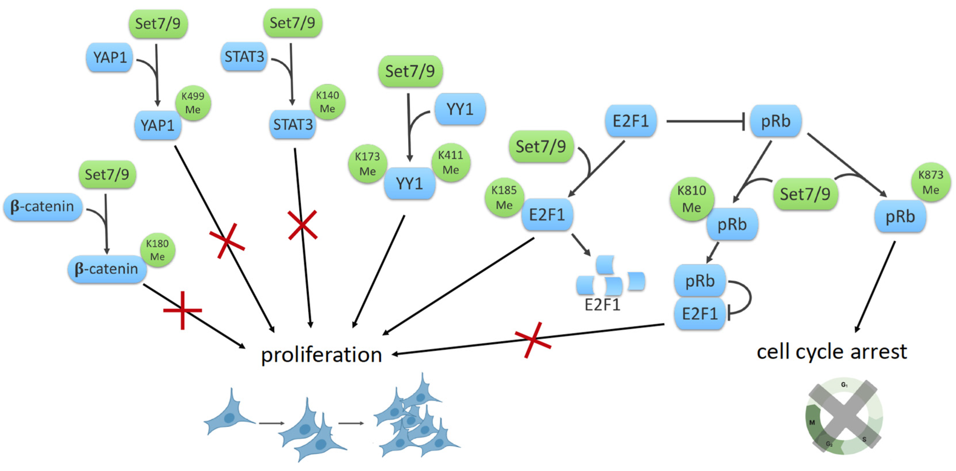
8. SET7/9 and Cell Proliferation

8.1. SET7/9, β-Catenin and YAP1

β-catenin is a key mediator of the Wnt/β-catenin signaling pathway and plays an important role in cell fate determination, cell proliferation and tumorigenesis. SET7/9 was shown to monomethylate β-catenin at lysine residue 180 in vivo and in vitro, thereby providing a novel mechanism by which the Wnt/β-catenin signaling pathway is regulated in response to oxidative stress. The binding of Wnt to its receptor LRP5/6 induces dissociation of β-catenin and its negative regulator glycogen synthase kinase (GSK)-3β, resulting in the stabilization of the β-catenin protein, its translocation to the nucleus and subsequent transactivation of the target genes [36]. It was demonstrated that methylation of β-catenin by SET7/9 facilitates its phosphorylation by (GSK)-3β and subsequent β-catenin degradation. Expression of the Wnt/β-catenin target genes such as c-Myc and CyclinD1 were significantly enhanced by either the depletion of SET7/9 or the mutation in the methylation site (K180R) of the β-catenin protein to promote the growth of cancer cells [36] (Figure 2).
On the other hand, SET7/9 was reported as a regulator of the methylation-dependent checkpoint in the Hippo/YAP1/TAZ pathway. SET7/9 monomethylates the YAP1 protein leading to its cytoplasmic retention [17]. YAP1 and TAZ are integral components of the β-catenin destruction complex while the β-catenin/TCF4 complex binds enhancer elements of the YAP gene to drive YAP expression in colorectal cancer cells [58,59]. Taken together, these facts indicate that SET7/9 may be considered as one of the key regulators of the Wnt/β-catenin and Hippo signaling pathways.

8.2. SET7/9 and STAT3

Another example of SET7/9 involvement in the regulation of the cell cycle is presented by the SET7/9-mediated methylation of the STAT3 transcription factor, which results in harnessing the activity of the latter. STAT3 forms dimers through reciprocal phosphor-tyrosine–SH2 interactions after phosphorylation on tyrosine and serine residues in response to different cytokines and growth factors. This phosphorylated form of STAT3 binds to and activates the promoters of its target genes. STAT3 can be methylated at K140 by SET7/9 and demethylated by LSD1. Methylation of K140 decreases the steady-state level of activated STAT3 and hence the expression of many STAT3 target genes [37] (Figure 2).

8.3. SET7/9 and YY1

YY1 (Yin Yang1) is a multifunctional zinc-finger transcription factor involved in a variety of biological processes such as DNA repair, apoptosis, cell proliferation, differentiation and development. SET7/9 methylates YY1 at K173 and K411 positions and enhances the DNA-binding activity of YY1 both in vitro and in cellulo at specific genomic loci in cultured cells. Functionally, SET7/9-mediated methylation of YY1 augments its transcriptional function and hence cell proliferation [42].

8.4. SET7/9 and E2F1/Rb1

The retinoblastoma protein (pRb) is a tumor suppressor protein playing an important role in regulating progression through the early stages of the cell cycle. pRb negatively regulates entry into the S-phase, thereby affecting the early cell cycle control [60]. The retinoblastoma protein interacts and blunts transcriptional activity of the E2F (E2 promoter-binding factor) family of transcription factors [61]. In addition to cell cycle control, pRb activity is associated with other types of cell fate, such as differentiation, senescence and apoptosis [62,63]. Munro et al. demonstrated that SET7/9 regulates the pRb tumor suppressor activity by methylating it at the K873 position [40]. SET7/9-mediated methylation of the C-terminal region of pRb facilitates the interaction between methylated pRb and the heterochromatin protein HP1, resulting in pRb-dependent transcriptional repression, cell cycle arrest, and differentiation [40].
It should be mentioned that the interplay between methylation and phosphorylation was observed for histone and non-histone proteins. In line with this, the phosphorylation of pRb required for the release of E2F1 and hence cell cycle progression was attenuated by the methylation of pRb at K810 by SET7/9 [41,62,63]. Apparently, SET7/9 locks pRb in a hypophosphorylated, growth-arresting state, thereby limiting the E2F target gene expression. Thus, cell cycle control could be regulated by the methylation/phosphorylation switch.
Moreover, the methylation of E2F1 by SET7/9 at lysine 185 inhibits acetylation and phosphorylation at the nearest positions, stimulating ubiquitination-depended degradation of the E2F1 protein [38]. At the same time, SET7/9 was shown as a critical co-activator of E2F1-dependent transcription under conditions of DNA damage [39]. SET7/9 affected the activity of E2F1 by indirect modulation of histone modifications in the promoters of E2F1-dependent genes, thereby promoting cell proliferation and repressing apoptosis. However, SET7/9 differentially affected E2F1 transcription targets: it promoted the expression of the CCNE1 gene, thereby facilitating cell proliferation, and it repressed the TP73 gene, hence preventing apoptosis [39] (Figure 3). Additionally, it was demonstrated that LSD1 removes the methyl mark required for the E2F1 stabilization and function in apoptosis [38]. Collectively, SET7/9 seems to be a critical element for the regulation of the cell cycle upon stress.

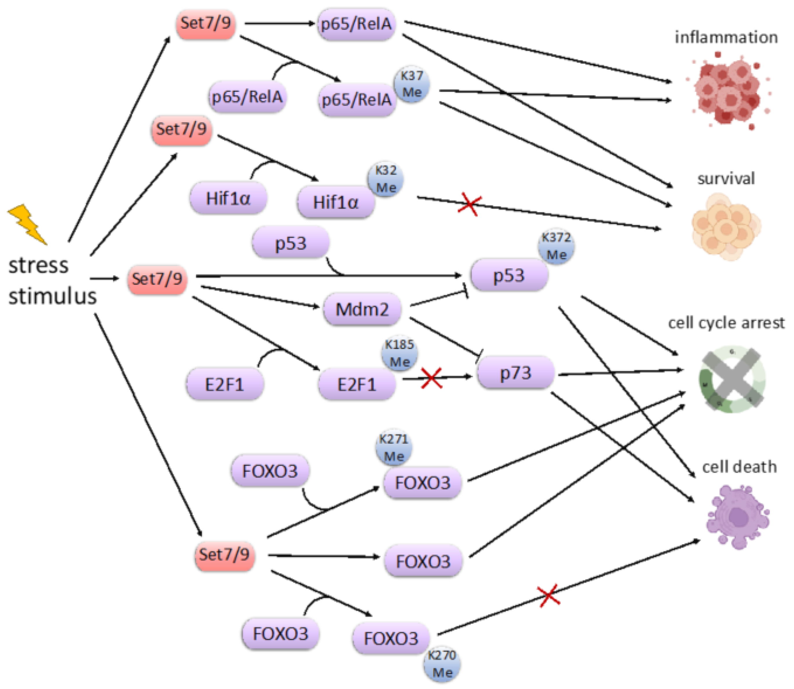
9. SET7/9 and Cell Stress Response

The tumor suppressor p53 was the first published non-histone methylation target for SET7/9. The p53 protein is the sequence-specific transcription factor that activates expression of its downstream transcription targets, whose products are involved in the regulation of cell cycle arrest and apoptosis [64]. The lysine-specific methylation of p53 at position K372 by SET7/9 is important for p53 transcriptional activation and stabilization mediated by its subsequent acetylation by p300/CBP [7]. Therefore, the cross talk between lysine methylation and acetylation is critical for p53 activation in response to DNA damage [18]. Importantly, both methylation and acetylation prevent p53 poly-ubiquitination mediated by an E3 ligase, Mdm2 [65]. The physical interaction between p53 and Mdm2 is critical for p53 ubiquitination and its subsequent degradation. Thus, small molecules that break this interaction were shown to stabilize p53 [66,67,68]. In line with this was the observation that SET7/9 physically interacts with Mdm2 and sequesters it away from p53 [69] (Figure 3). Both in vitro and in vivo experiments suggest that SET7/9 and Mdm2 have inverse expression. Accordingly, the unleashed expression of Mdm2 in cancer patients with diminished expression of SET7/9 correlated with poor survival outcomes [69].
It is worthy of note that SET7/9 was shown to be a new regulator of another p53-specific enzyme, Sirtulin 1 (SIRT1). The latter plays an important role during aging, metabolism and autophagy. SIRT1 interacts with SET7/9 mostly in response to DNA damage in human cells resulting in the dissociation of SIRT1 from p53 and the enhancement of p53 acetylation at K382. SET7/9 is able to both interact with and methylate SIRT1 at multiple sites [49]. The presence of SET7/9 attenuates the interaction between SIRT1 and p53, resulting in the transcriptional activation of p53 target genes and thereby inflicting cell cycle arrest and apoptosis [49].
The transcription factor FoxO3 of the Forkhead Box O (FoxO) family is involved in the regulation of the cellular response to ROS-induced DNA damage [70]. In response to oxidative stress, FoxO3 induces cell cycle arrest, apoptosis, autophagy, and hence can be considered as a tumor suppressor. It also affects metabolism and aging [46,70,71]. The phenotypic features of FoxO3-knockout mice support its involvement in the process of ageing and are exemplified by premature follicular activation, ovarian failure and early infertility [72].
FoxO3 inhibits transcription induced by ERα, thereby inhibiting the proliferation of breast cancer cells [73]. Accordingly, downregulation of the ERα activity is associated with poor prognosis in estrogen-dependent breast cancer and colorectal cancers [74]. SET7/9 methylates the FoxO3 protein at K271 [45]. Paradoxically, the methylation of FoxO3 destabilizes the protein but enhances its transcriptional activity towards the activation of pro-apoptotic genes. This paradoxical effect is similar to the one observed in the case of SET7/9-mediated methylation of E2F1. It should also be noted that SET7/9 apparently can methylate the additional FoxO3 lysine residue, K270 [46] (Figure 3). Surprisingly, this methylation has an opposite effect on the transcriptional activity of FoxO3, thereby preventing it from activation of the pro-apoptotic gene BIM and, hence, preventing cell death.
Hypoxia-inducible factor 1α (HIF-1α) is another target of SET7/9 methylation activity. HIF1a is a critical transcription factor for cellular hypoxic response. In response to oxygen deprivation, HIF1a is released from the ubiquitin-mediated degradation mediated by the von Hippel-Lindau disease tumor suppressor (VHL) E3 ligase [75]. SET7/9 methylates HIF-1α at K32, which competes with ubiquitination and its subsequent degradation. Thus, SET7/9 stabilizes the HIF-1α protein and stimulates the HIF-1α-dependent transcription of genes involved in the regulation of energy metabolism and angiogenesis to maintain tissue homeostasis [48]. However, several studies suggest that SET7/9-mediated methylation inhibits HIF1α transcriptional activity by preventing its DNA binding (Figure 3). Importantly, this effect was reversed by a SET7/9-specific inhibitor, (R)-PFI-2 [47,48]. Thus, additional experiments are required to elucidate the effect of SET7/9 on the function of HIF-1α.

10. Concluding Remarks

To assess the biological significance of SET7/9’s role in proliferation and stress response in vivo, it is important to develop the relevant tools. In this respect, small-molecule inhibitors are promising tools that allow for the probing functions of methyltransferases in diseases. In this respect, Barsyte-Lovejoy et al. designed and synthesized a novel inhibitor, (R)-PFI-2, against SET7/9 [76]. This inhibitor has demonstrated low toxicity even at high concentrations in human cells [77]. Mori et al. has developed an inhibitor which is an amine analogue of adenosylmethionine, bearing various alkylamino groups for increasing the inhibitory activity [78].
Berberine is a naturally occurring isoquinoline alkaloid which is commonly used in traditional Chinese medicine and exhibits anti-oxidant, anti-inflammatory, and anti-cancer activities. Berberin was shown to augment the activity of SET7/9 towards NFκB by sensitizing human cancer cells to ionizing radiation or chemotherapy. Berberine negatively regulates NFκB through SET7/9-mediated lysine methylation. Such methylation leads to a decrease in miR-21 levels and Bcl-2 levels [79].
Since there is a significant correlation between SET7/9 and different types of cancer, the wide application of small molecules as experimental tools should significantly facilitate the experimental work directed towards the elucidation of SET7/9 in tumorigenesis and other diseases.

Author Contributions

Conceptualization, N.A.B.; writing—original draft preparation, A.D., E.V., O.F., O.S. and N.A.B.; writing—review and editing, N.A.B.; visualization, A.D., E.V., O.F. and O.S. All authors have read and agreed to the published version of the manuscript.

Funding

This research was funded by RUSSIAN SCIENCE FOUNDATION (RSF), #19-75-10059.

Institutional Review Board Statement

Not applicable.

Informed Consent Statement

Not applicable.

Conflicts of Interest

The authors declare that they have no conflict of interest.

References

  1. Nishioka, K.; Chuikov, S.; Sarma, K.; Erdjument-Bromage, H.; Allis, C.D.; Tempst, P.; Reinberg, D. Set9, a Novel Histone H3 Methyltransferase that Facilitates Transcription by Precluding Histone Tail Modifications Required for Heterochromatin Formation. Genes Dev. 2002, 16, 479–489. [Google Scholar] [CrossRef] [PubMed] [Green Version]
  2. Wang, H.; Cao, R.; Xia, L.; Erdjument-Bromage, H.; Borchers, C.; Tempst, P.; Zhang, Y. Purification and Functional Characterization of a Histone H3-Lysine 4-Specific Methyltransferase. Mol. Cell 2001, 8, 1207–1217. [Google Scholar] [CrossRef]
  3. Dhayalan, A.; Kudithipudi, S.; Rathert, P.; Jeltsch, A. Specificity Analysis-Based Identification of New Methylation Targets of the SET7/9 Protein Lysine Methyltransferase. Chem. Biol. 2011, 18, 111–120. [Google Scholar] [CrossRef] [Green Version]
  4. Chakrabarti, S.K.; Francis, J.; Ziesmann, S.M.; Garmey, J.C.; Mirmira, R.G. Covalent Histone Modifications Underlie the Developmental Regulation of Insulin Gene Transcription in Pancreatic β Cells. J. Biol. Chem. 2003, 278, 23617–23623. [Google Scholar] [CrossRef] [PubMed] [Green Version]
  5. Evans-Molina, C.; Robbins, R.D.; Kono, T.; Tersey, S.A.; Vestermark, G.L.; Nunemaker, C.S.; Garmey, J.C.; Deering, T.G.; Keller, S.R.; Maier, B.; et al. Peroxisome Proliferator-Activated Receptor γ Activation Restores Islet Function in Diabetic Mice through Reduction of Endoplasmic Reticulum Stress and Maintenance of Euchromatin Structure. Mol. Cell. Biol. 2009, 29, 2053–2067. [Google Scholar] [CrossRef] [PubMed] [Green Version]
  6. Pradhan, S.; Chin, H.G.; Estève, P.-O.; Jacobsen, S.E. SET7/9 Mediated Methylation of Non-Histone Proteins in Mammalian Cells. Epigenetics 2009, 4, 383–387. [Google Scholar] [CrossRef]
  7. Chuikov, S.; Kurash, J.K.; Wilson, J.; Xiao, B.; Justin, N.; Ivanov, G.S.; McKinney, K.; Tempst, P.; Prives, C.; Gamblin, S.; et al. Regulation of p53 activity through lysine methylation. Nature 2004, 432, 353–360. [Google Scholar] [CrossRef]
  8. Couture, J.-F.; Collazo, E.; Hauk, G.; Trievel, R.C. Structural Basis for the Methylation Site Specificity of SET7/9. Nat. Struct. Mol. Biol. 2006, 13, 140–146. [Google Scholar] [CrossRef]
  9. Masatsugu, T.; Yamamoto, K. Multiple Lysine Methylation of PCAF by Set9 Methyltransferase. Biochem. Biophys. Res. Commun. 2009, 381, 22–26. [Google Scholar] [CrossRef]
  10. Vasileva, E.; Shuvalov, O.; Petukhov, A.; Fedorova, O.; Daks, A.; Nader, R.; Barlev, N. KMT Set7/9 Is a New Regulator of Sam68 STAR-Protein. Biochem. Biophys. Res. Commun. 2020, 525, 1018–1024. [Google Scholar] [CrossRef]
  11. Li, Y.; Reddy, M.A.; Miao, F.; Shanmugam, N.; Yee, J.-K.; Hawkins, D.; Ren, B.; Natarajan, R. Role of the Histone H3 Lysine 4 Methyltransferase, SET7/9, in the Regulation of NF-κB-Dependent Inflammatory Genes: Relevance to Diabetes and Inflammation. J. Biol. Chem. 2008, 283, 26771–26781. [Google Scholar] [CrossRef] [Green Version]
  12. Del Rizzo, P.A.; Trievel, R.C. Substrate and Product Specificities of SET Domain Methyltransferases. Epigenetics 2011, 6, 1059–1067. [Google Scholar] [CrossRef]
  13. Okabe, J.; Orlowski, C.; Balcerczyk, A.; Tikellis, C.; Thomas, M.; Cooper, M.E.; El-Osta, A. Distinguishing Hyperglycemic Changes by Set7 in Vascular Endothelial Cells. Circ. Res. 2012, 110, 1067–1076. [Google Scholar] [CrossRef] [PubMed] [Green Version]
  14. Batista, I.; Helguero, L.A. Biological Processes and Signal Transduction Pathways Regulated by the Protein Methyltransferase SETD7 and Their Significance in Cancer. Signal Transduct. Target. Ther. 2018, 3, 19. [Google Scholar] [CrossRef] [PubMed]
  15. Keating, S.T.; El-Osta, A. Transcriptional Regulation by the Set7 Lysine Methyltransferase. Epigenetics 2013, 8, 361–372. [Google Scholar] [CrossRef] [Green Version]
  16. Aksenova, V.; Turoverova, L.; Khotin, M.; Magnusson, K.-E.; Tulchinsky, E.; Melino, G.; Pinaev, G.P.; Barlev, N.; Tentler, D. Actin-Binding Protein Alpha-Actinin 4 (ACTN4) Is a Transcriptional Co-Activator of RelA/p65 Sub-unit of NF-kB. Oncotarget 2013, 4, 362. [Google Scholar] [CrossRef] [PubMed] [Green Version]
  17. Oudhoff, M.J.; Braam, M.J.; Freeman, S.A.; Wong, D.; Rattray, D.G.; Wang, J.; Antignano, F.; Snyder, K.; Refaeli, I.; Hughes, M.R.; et al. SETD7 Controls Intestinal Regeneration and Tumorigenesis by Regulating Wnt/β-Catenin and Hippo/YAP Signaling. Dev. Cell 2016, 37, 47–57. [Google Scholar] [CrossRef] [PubMed] [Green Version]
  18. Ivanov, G.S.; Ivanova, T.; Kurash, J.; Ivanov, A.; Chuikov, S.; Gizatullin, F.; Herrera-Medina, E.M.; Rauscher, F., III; Reinberg, D.; Barlev, N.A. Methylation-Acetylation Interplay Activates p53 in Response to DNA Damage. Mol. Cell. Biol. 2007, 27, 6756–6769. [Google Scholar] [CrossRef] [Green Version]
  19. Liu, H.; Li, Z.; Yang, Q.; Liu, W.; Wan, J.; Li, J.; Zhang, M. Substrate Docking–Mediated Specific and Efficient Lysine Methylation by the SET Domain–Containing Histone Methyltransferase SETD7. J. Biol. Chem. 2019, 294, 13355–13365. [Google Scholar] [CrossRef] [Green Version]
  20. El-Gebali, S.; Mistry, J.; Bateman, A.; Eddy, S.R.; Luciani, A.; Potter, S.C.; Qureshi, M.; Richardson, L.J.; Salazar, G.A.; Smart, A.; et al. The Pfam Protein Families Database in 2019. Nucleic Acids Res. 2019, 47, D427–D432. [Google Scholar] [CrossRef]
  21. Mecklenburg, K.L.; Freed, S.A.; Raval, M.; Quintero, O.A.; Yengo, C.M.; O’Tousa, J.E. Invertebrate and Vertebrate Class III Myosins Interact with MORN Repeat-Containing Adaptor Proteins. PLoS ONE 2015, 10, e0122502. [Google Scholar] [CrossRef] [PubMed] [Green Version]
  22. Hadano, S.; Kunita, R.; Otomo, A.; Suzuki-Utsunomiya, K.; Ikeda, J.-E. Molecular and Cellular Function of ALS2/Alsin: Implication of Membrane Dynamics in Neuronal Development and Degeneration. Neurochem. Int. 2007, 51, 74–84. [Google Scholar] [CrossRef] [PubMed]
  23. Landstrom, A.; Beavers, D.L.; Wehrens, X.H. The Junctophilin Family of Proteins: From Bench to Bedside. Trends Mol. Med. 2014, 20, 353–362. [Google Scholar] [CrossRef] [Green Version]
  24. Guo, A.; Wang, Y.; Chen, B.; Wang, Y.; Yuan, J.; Zhang, L.; Hall, D.; Wu, J.; Shi, Y.; Zhu, Q.; et al. E-C Coupling Structural Protein Junctophilin-2 Encodes a Stress-Adaptive Transcription Regulator. Science 2018, 362, eaan3303. [Google Scholar] [CrossRef]
  25. Sajko, S.; Grishkovskaya, I.; Kostan, J.; Graewert, M.; Setiawan, K.; Trübestein, L.; Niedermüller, K.; Gehin, C.; Sponga, A.; Puchinger, M. Structures of Three MORN Repeat Proteins and a Re-Evaluation of the Proposed Lipid-Binding Properties of MORN Repeats. PLoS ONE 2020, 15, e0242677. [Google Scholar] [CrossRef]
  26. Sehnal, D.; Bittrich, S.; Deshpande, M.; Svobodová, R.; Berka, K.; Bazgier, V.; Velankar, S.; Burley, S.K.; Koča, J.; Rose, A.S. Mol* Viewer: Modern Web App for 3D Visualization and Analysis of Large Biomolecular Structures. Nucleic Acids Res. 2021, 49, W431–W437. [Google Scholar] [CrossRef] [PubMed]
  27. Wilson, J.R.; Jing, C.; Walker, P.A.; Martin, S.R.; Howell, S.A.; Blackburn, G.M.; Gamblin, S.J.; Xiao, B. Crystal Structure and Functional Analysis of the Histone Methyltransferase SET7/9. Cell 2002, 111, 105–115. [Google Scholar] [CrossRef]
  28. Vasileva, E.; Barlev, N. The World of SET-Containing Lysine Methyltransferases. eLS 2017, 1–10. [Google Scholar] [CrossRef]
  29. Kouzarides, T. Chromatin Modifications and Their Function. Cell 2007, 128, 693–705. [Google Scholar] [CrossRef] [Green Version]
  30. Noma, K.-I.; Allis, C.D.; Grewal, S.I.S. Transitions in Distinct Histone H3 Methylation Patterns at the Heterochromatin Domain Boundaries. Science 2001, 293, 1150–1155. [Google Scholar] [CrossRef]
  31. Castaño, J.; Morera, C.; Sesé, B.; Boue, S.; Bonet-Costa, C.; Martí, M.; Roque, A.; Jordan, A.; Barrero, M.J. SETD7 Regulates the Differentiation of Human Embryonic Stem Cells. PLoS ONE 2016, 11, e0149502. [Google Scholar] [CrossRef] [PubMed]
  32. Wang, D.; Zhou, J.; Liu, X.; Lu, D.; Shen, C.; Du, Y.; Wei, F.-Z.; Song, B.; Lu, X.; Yu, Y. Methylation of SUV39H1 by SET7/9 Results in Heterochromatin Relaxation and Genome Instability. Proc. Natl. Acad. Sci. USA 2013, 110, 5516–5521. [Google Scholar] [CrossRef] [PubMed] [Green Version]
  33. Estève, P.-O.; Chin, H.G.; Benner, J.; Feehery, G.R.; Samaranayake, M.; Horwitz, G.A.; Jacobsen, S.E.; Pradhan, S. Regulation of DNMT1 Stability through SET7-Mediated Lysine Methylation in Mammalian Cells. Proc. Natl. Acad. Sci. USA 2009, 106, 5076–5081. [Google Scholar] [CrossRef] [PubMed] [Green Version]
  34. Wang, J.; Hevi, S.; Kurash, J.K.; Lei, H.; Gay, F.; Bajko, J.; Su, H.; Sun, W.; Chang, H.; Xu, G.; et al. The Lysine Demethylase LSD1 (KDM1) Is Required for Maintenance of Global DNA Methylation. Nat. Genet. 2008, 41, 125–129. [Google Scholar] [CrossRef] [PubMed]
  35. Kouskouti, A.; Scheer, E.; Staub, A.; Tora, L.; Talianidis, I. Gene-Specific Modulation of TAF10 Function by SET9-Mediated Methylation. Mol. Cell 2004, 14, 175–182. [Google Scholar] [CrossRef] [Green Version]
  36. Shen, C.; Wang, D.; Liu, X.; Gu, B.; Du, Y.; Wei, F.Z.; Cao, L.L.; Song, B.; Lu, X.; Yang, Q. SET7/9 Regulates Cancer Cell Proliferation by Influencing β-Catenin Stability. FASEB J. 2015, 29, 4313–4323. [Google Scholar] [CrossRef]
  37. Yang, J.; Huang, J.; Dasgupta, M.; Sears, N.; Miyagi, M.; Wang, B.; Chance, M.R.; Chen, X.; Du, Y.; Wang, Y.; et al. Reversible Methylation of Promoter-Bound STAT3 by Histone-Modifying Enzymes. Proc. Natl. Acad. Sci. USA 2010, 107, 21499–21504. [Google Scholar] [CrossRef] [Green Version]
  38. Kontaki, H.; Talianidis, I. Lysine Methylation Regulates E2F1-Induced Cell Death. Mol. Cell 2010, 39, 152–160. [Google Scholar] [CrossRef]
  39. Lezina, L.; Aksenova, V.; Ivanova, T.; Purmessur, N.; Antonov, A.; Tentler, D.; Fedorova, O.; Garabadgiu, A.; Talianidis, I.; Melino, G. KMTase Set7/9 Is a Critical Regulator of E2F1 Activity upon Genotoxic Stress. Cell Death Differ. 2014, 21, 1889–1899. [Google Scholar] [CrossRef] [PubMed]
  40. Munro, S.; Khaire, N.; Inche, A.; Carr, S.; La Thangue, N.B. Lysine Methylation Regulates the pRb Tumour Suppressor Protein. Oncogene 2010, 29, 2357–2367. [Google Scholar] [CrossRef] [Green Version]
  41. Carr, S.M.; Munro, S.; Kessler, B.; Oppermann, U.; La Thangue, N.B. Interplay between Lysine Methylation and Cdk Phosphorylation in Growth Control by the Retinoblastoma Protein. EMBO J. 2010, 30, 317–327. [Google Scholar] [CrossRef] [Green Version]
  42. Zhang, W.-J.; Wu, X.-N.; Shi, T.-T.; Xu, H.-T.; Yi, J.; Shen, H.-F.; Huang, M.-F.; Shu, X.-Y.; Wang, F.-F.; Peng, B.-L. Regulation of Transcription Factor Yin Yang 1 by SET7/9-Mediated Lysine Methylation. Sci. Rep. 2016, 6, 21718. [Google Scholar] [CrossRef] [Green Version]
  43. Ea, C.-K.; Baltimore, D. Regulation of NF-κB Activity through Lysine Monomethylation of p65. Proc. Natl. Acad. Sci. USA 2009, 106, 18972–18977. [Google Scholar] [CrossRef] [PubMed] [Green Version]
  44. Yang, X.-D.; Huang, B.; Li, M.; Lamb, A.; Kelleher, N.L.; Chen, L.-F. Negative Regulation of NF-κB Action by Set9-Mediated Lysine Methylation of the RelA Subunit. EMBO J. 2009, 28, 1055–1066. [Google Scholar] [CrossRef] [Green Version]
  45. Xie, Q.; Hao, Y.; Tao, L.; Peng, S.; Rao, C.; Chen, H.; You, H.; Dong, M.; Yuan, Z. Lysine Methylation of FOXO3 Regulates Oxidative Stress-Induced Neuronal Cell Death. EMBO Rep. 2012, 13, 371–377. [Google Scholar] [CrossRef] [PubMed] [Green Version]
  46. Calnan, D.R.; Webb, A.E.; White, J.L.; Stowe, T.R.; Goswami, T.; Shi, X.; Espejo, A.; Bedford, M.T.; Gozani, O.; Gygi, S.P.; et al. Methylation by Set9 Modulates FoxO3 Stability and Transcriptional Activity. Aging 2012, 4, 462–479. [Google Scholar] [CrossRef] [PubMed] [Green Version]
  47. Kim, Y.; Nam, H.J.; Lee, J.; Park, D.Y.; Kim, C.; Yu, Y.S.; Kim, D.; Park, S.W.; Bhin, J.; Hwang, D.; et al. Methylation-Dependent Regulation of HIF-1α Stability Restricts Retinal and Tumour Angiogenesis. Nat. Commun. 2016, 7, 10347. [Google Scholar] [CrossRef] [PubMed] [Green Version]
  48. Liu, X.; Chenxi, X.; Xu, C.; Leng, X.; Cao, H.; Ouyang, G.; Xiaoqian, L. Repression of Hypoxia-Inducible Factor α Signaling by Set7-Mediated Methylation. Nucleic Acids Res. 2015, 43, 5081–5098. [Google Scholar] [CrossRef] [PubMed] [Green Version]
  49. Liu, X.; Wang, D.; Zhao, Y.; Tu, B.; Zheng, Z.; Wang, L.; Wang, H.; Gu, W.; Roeder, R.G.; Zhu, W.-G. Methyltransferase Set7/9 Regulates p53 Activity by Interacting with Sirtuin 1 (SIRT1). Proc. Natl. Acad. Sci. USA 2011, 108, 1925–1930. [Google Scholar] [CrossRef] [Green Version]
  50. Kassner, I.; Barandun, M.; Fey, M.; Rosenthal, F.; Hottiger, M.O. Crosstalk between SET7/9-Dependent Methylation and ARTD1-Mediated ADP-Ribosylation of Histone H1. 4. Epigenet. Chromatin 2013, 6, 1. [Google Scholar] [CrossRef] [Green Version]
  51. Gaughan, L.; Stockley, J.; Wang, N.; McCracken, S.R.; Treumann, A.; Armstrong, K.; Shaheen, F.; Watt, K.; McEwan, I.J.; Wang, C. Regulation of the Androgen Receptor by SET9-Mediated Methylation. Nucleic Acids Res. 2011, 39, 1266–1279. [Google Scholar] [CrossRef] [PubMed] [Green Version]
  52. Subramanian, K.; Jia, D.; Kapoor-Vazirani, P.; Powell, D.R.; Collins, R.; Sharma, D.; Peng, J.; Cheng, X.; Vertino, P.M. Regulation of Estrogen Receptor α by the SET7 Lysine Methyltransferase. Mol. Cell 2008, 30, 336–347. [Google Scholar] [CrossRef] [PubMed] [Green Version]
  53. Heintzman, N.D.; Stuart, R.K.; Hon, G.; Fu, Y.; Ching, C.W.; Hawkins, R.D.; Barrera, L.O.; Van Calcar, S.; Qu, C.; Ching, K.A.; et al. Distinct and Predictive Chromatin Signatures of Transcriptional Promoters and Enhancers in the Human Genome. Nat. Genet. 2007, 39, 311–318. [Google Scholar] [CrossRef]
  54. Lehnertz, B.; Rogalski, J.C.; Schulze, F.M.; Yi, L.; Lin, S.; Kast, J.; Rossi, F.M. P53-Dependent Transcription and Tumor Suppression Are Not Affected in Set7/9-Deficient Mice. Mol. Cell 2011, 43, 673–680. [Google Scholar] [CrossRef] [Green Version]
  55. Sun, G.; Reddy, M.A.; Yuan, H.; Lanting, L.; Kato, M.; Natarajan, R. Epigenetic Histone Methylation Modulates Fibrotic Gene Expression. J. Am. Soc. Nephrol. 2010, 21, 2069–2080. [Google Scholar] [CrossRef]
  56. Nagashimada, M.; Ueda, T.; Ishita, Y.; Sakurai, H. TAF7 Is a Heat-Inducible Unstable Protein and Is Required for Sustained Expression of Heat Shock Protein Genes. FEBS J. 2018, 285, 3215–3224. [Google Scholar] [CrossRef] [PubMed]
  57. Estève, P.-O.; Chang, Y.; Samaranayake, M.; Upadhyay, A.K.; Horton, J.R.; Feehery, G.R.; Cheng, X.; Pradhan, S. A Methylation and Phosphorylation Switch between an Adjacent Lysine and Serine Determines Human DNMT1 Stability. Nat. Struct. Mol. Biol. 2010, 18, 42–48. [Google Scholar] [CrossRef] [Green Version]
  58. Konsavage, W.M.; Kyler, S.L.; Rennoll, S.A.; Jin, G.; Yochum, G.S. Wnt/β-Catenin Signaling Regulates Yes-associated Protein (YAP) Gene Expression in Colorectal Carcinoma Cells. J. Biol. Chem. 2012, 287, 11730–11739. [Google Scholar] [CrossRef] [Green Version]
  59. Azzolin, L.; Panciera, T.; Soligo, S.; Enzo, E.; Bicciato, S.; Dupont, S.; Bresolin, S.; Frasson, C.; Basso, G.; Guzzardo, V.; et al. YAP/TAZ Incorporation in the β-Catenin Destruction Complex Orchestrates the Wnt Response. Cell 2014, 158, 157–170. [Google Scholar] [CrossRef] [Green Version]
  60. Weinberg, R.A. The Retinoblastoma Protein and Cell Cycle Control. Cell 1995, 81, 323–330. [Google Scholar] [CrossRef] [Green Version]
  61. Stevens, C.; La Thangue, N.B. E2F and Cell Cycle Control: A Double-Edged Sword. Arch. Biochem. Biophys. 2003, 412, 157–169. [Google Scholar] [CrossRef]
  62. Classon, M.; Harlow, E. The Retinoblastoma Tumour Suppressor in Development and Cancer. Nat. Cancer 2002, 2, 910–917. [Google Scholar] [CrossRef]
  63. Giacinti, C.; Giordano, A. RB and Cell Cycle Progression. Oncogene 2006, 25, 5220–5227. [Google Scholar] [CrossRef] [PubMed] [Green Version]
  64. Kastan, M.B.; Onyekwere, O.; Sidransky, D.; Vogelstein, B.; Craig, R.W. Participation of p53 Protein in the Cellular Response to DNA Damage. Cancer Res. 1991, 51, 6304–6311. [Google Scholar] [CrossRef] [PubMed] [Green Version]
  65. Daks, A.; Melino, D.; Barlev, N.A. The Role of Different E3 Ubiquitin Ligases in Regulation of the P53 Tumor Suppressor Protein. Tsitologiia 2013, 55, 673–687. [Google Scholar]
  66. Fedorova, O.; Daks, A.; Petrova, V.; Petukhov, A.; Lezina, L.; Shuvalov, O.; Davidovich, P.; Kriger, D.; Lomert, E.; Tentler, D.; et al. Novel Isatin-Derived Molecules Activate p53 via Interference with Mdm2 to Promote Apoptosis. Cell Cycle 2018, 17, 1917–1930. [Google Scholar] [CrossRef] [PubMed] [Green Version]
  67. Vassilev, L.T.; Vu, B.T.; Graves, B.; Carvajal, D.; Podlaski, F.; Filipovic, Z.; Kong, N.; Kammlott, U.; Lukacs, C.; Klein, C.; et al. In Vivo Activation of the p53 Pathway by Small-Molecule Antagonists of MDM2. Science 2004, 303, 844–848. [Google Scholar] [CrossRef] [Green Version]
  68. Davidovich, P.B.; Aksenova, V.; Petrova, V.; Tentler, D.; Orlova, D.; Smirnov, S.V.; Gurzhiy, V.; Okorokov, A.; Garabadzhiu, A.V.; Melino, G.; et al. Discovery of Novel Isatin-Based p53 Inducers. ACS Med. Chem. Lett. 2015, 6, 856–860. [Google Scholar] [CrossRef] [Green Version]
  69. Lezina, L.; Aksenova, V.; Fedorova, O.; Malikova, D.; Shuvalov, O.; Antonov, A.V.; Tentler, D.; Garabadgiu, A.V.; Melino, G.; Barlev, N.A. KMT Set7/9 Affects Genotoxic Stress Response via the Mdm2 Axis. Oncotarget 2015, 6, 25843. [Google Scholar] [CrossRef] [Green Version]
  70. Greer, E.; Brunet, A. FOXO Transcription Factors at the Interface between Longevity and Tumor Suppression. Oncogene 2005, 24, 7410–7425. [Google Scholar] [CrossRef] [Green Version]
  71. Hwangbo, D.S.; Gersham, B.; Tu, M.-P.; Palmer, M.; Tatar, M. Drosophila dFOXO Controls Lifespan and Regulates Insulin Signalling in Brain and Fat Body. Nature 2004, 429, 562–566. [Google Scholar] [CrossRef] [PubMed]
  72. Castrillon, D.H.; Miao, L.; Kollipara, R.; Horner, J.W.; DePinho, R.A. Suppression of Ovarian Follicle Activation in Mice by the Transcription Factor Foxo3a. Science 2003, 301, 215–218. [Google Scholar] [CrossRef] [PubMed]
  73. Zou, Y.; Tsai, W.-B.; Cheng, C.-J.; Hsu, C.; Chung, Y.M.; Li, P.-C.; Lin, S.-H.; Hu, M.C.-T. Forkhead Box Transcription Factor FOXO3a Suppresses Estrogen-Dependent Breast Cancer Cell Proliferation and Tumorigenesis. Breast Cancer Res. 2008, 10, R21. [Google Scholar] [CrossRef] [PubMed] [Green Version]
  74. Bullock, M.D.; Bruce, A.; Sreekumar, R.; Curtis, N.; Cheung, T.; Reading, I.; Primrose, J.N.; Ottensmeier, C.; Packham, G.K.; Thomas, G.; et al. FOXO3 Expression during Colorectal Cancer Progression: Biomarker Potential Reflects a Tumour Suppressor Role. Br. J. Cancer 2013, 109, 387–394. [Google Scholar] [CrossRef]
  75. Maxwell, P.H.; Wiesener, M.S.; Chang, G.-W.; Clifford, S.C.; Vaux, E.C.; Cockman, M.E.; Wykoff, C.C.; Pugh, C.W.; Maher, E.R.; Ratcliffe, P.J. The Tumour Suppressor Protein VHL Targets Hypoxia-Inducible Factors for Oxygen-Dependent Proteolysis. Nature 1999, 399, 271–275. [Google Scholar] [CrossRef]
  76. Barsyte-Lovejoy, D.; Li, F.; Oudhoff, M.; Tatlock, J.H.; Dong, A.; Zeng, H.; Wu, H.; Freeman, S.A.; Schapira, M.; Senisterra, G.A.; et al. (R)-PFI-2 Is a Potent and Selective Inhibitor of SETD7 Methyltransferase Activity in Cells. Proc. Natl. Acad. Sci. USA 2014, 111, 12853–12858. [Google Scholar] [CrossRef] [Green Version]
  77. Daks, A.; Mamontova, V.; Fedorova, O.; Petukhov, A.; Shuvalov, O.; Parfenyev, S.; Netsvetay, S.; Venina, A.; Kizenko, A.; Imyanitov, E. Set7/9 Controls Proliferation and Genotoxic Drug Resistance of NSCLC Cells. Biochem. Biophys. Res. Commun. 2021, 572, 41–48. [Google Scholar] [CrossRef]
  78. Mori, S.; Iwase, K.; Iwanami, N.; Tanaka, Y.; Kagechika, H.; Hirano, T. Development of Novel Bisubstrate-Type Inhibitors of Histone Methyltransferase SET7/9. Bioorg. Med. Chem. 2010, 18, 8158–8166. [Google Scholar] [CrossRef]
  79. Hu, H.-Y.; Li, K.-P.; Wang, X.-J.; Liu, Y.; Lu, Z.-G.; Dong, R.-H.; Guo, H.-B.; Zhang, M.-X. Set9, NF-κB, and MicroRNA-21 Mediate Berberine-Induced Apoptosis of Human Multiple Myeloma Cells. Acta Pharmacol. Sin. 2012, 34, 157–166. [Google Scholar] [CrossRef] [Green Version]
Figure 1. The domain organization and 3D-structure (PDB ID 1N6C, [26]) of the SET7/9 protein. The amino-terminal domain includes three MORN repeats. The SET domain is located in the C-terminus. Several amino acid residues of the SET domain form the SAM-binding pocket. SAM—S-adenosyl-L-methionine.
Life 12 00362 g001
Figure 2. The scheme illustrating the participation of SET7/9 in regulation of proliferation.
Life 12 00362 g002
Figure 3. The scheme illustrating the key cellular stress effector pathways regulated by SET7/9. The functional outcomes are denoted on the right.
Life 12 00362 g003
Table 1. The substrates of SET7/9-dependent methylation playing roles in proliferation and cellular stress response.
SET7/9 Target ProteinMethylation SitesEffect of the ModificationReference
Histone H1K12, K14, K17, K20,
K21, K27, K111
Modulation of the affinity of histone H1 to chromatin during human pluripotent cells differentiation[31]
Histone H1.4K34, K127, K129, K130Prevention of acetylation at the same sites, heterochromatin formation[31]
Histone H2.AK5, K13, K15Unknown[3]
Histone H2.BK15Unknown[3]
Histone H3K4Activation of transcription[1]
Suv39H1K25, K123Heterohromatin relaxation, genome instability[32]
DNMT1K142Promotion of DNMT1 ubiquitination and proteasomal degradation [33]
K1094Decrease of the DNMT1 level [34]
TAF7K5Enhancement of TAF7 activity as co-factor of RNA polymerase II[8]
TAF10K189Enhancement of TAF10 activity as co-factor of RNA polymerase II, activation of transcription of TAF10 target genes[35]
YAP1K494Retention of YAP1 in the cytoplasm[17]
β-cateninK180Promotion of β-catenin ubiquitination by (GSK)-3b and its subsequent proteasomal degradation[36]
STAT3K140Dissociation of STAT3 from promoter elements, downregulation of STAT3-dependent genes expression[37]
E2F1K185Promotion of E2F1 ubiquitination and subsequent proteasomal degradation[38]
UnknownEnhancement of E2F transactivation of its target genes [39]
pRbK873Enhancement of pRB-dependent repression of transcription[40]
K810Promotion of p65/RelA ubiquitination and its subsequent proteasomal degradation [41]
YY1K173, K411Retention of YY1 in the cytoplasm[42]
p65/RelAK37Translocation to the nucleus and transactivation of target genes[43]
K314, K315Promotion of p65/RelA ubiquitination and subsequent proteasomal degradation [44]
FOXO3K270Downregulation of FOXO3-dependent transactivation of BIM [45]
K271Increase of the FOXO3 transactivation potential [46]
Hif1αK32Suppression of Hif1α transactivation of its target genes[47,48]
p53K372Stabilization, translocation to the nucleus and transactivation of target genes[7]
SIRT1K233, K235, K236, K238Enhancement of SIRT1-dependent p53 acetylation and activation[49]
Publisher’s Note: MDPI stays neutral with regard to jurisdictional claims in published maps and institutional affiliations.

