
Toxicity of Proton Therapy versus Photon Therapy on Salvage Re-Irradiation for Non-Small Cell Lung Cancer

 , , , , ,
Abstract
:1. Introduction
2. Materials and Methods
2.1. Patients
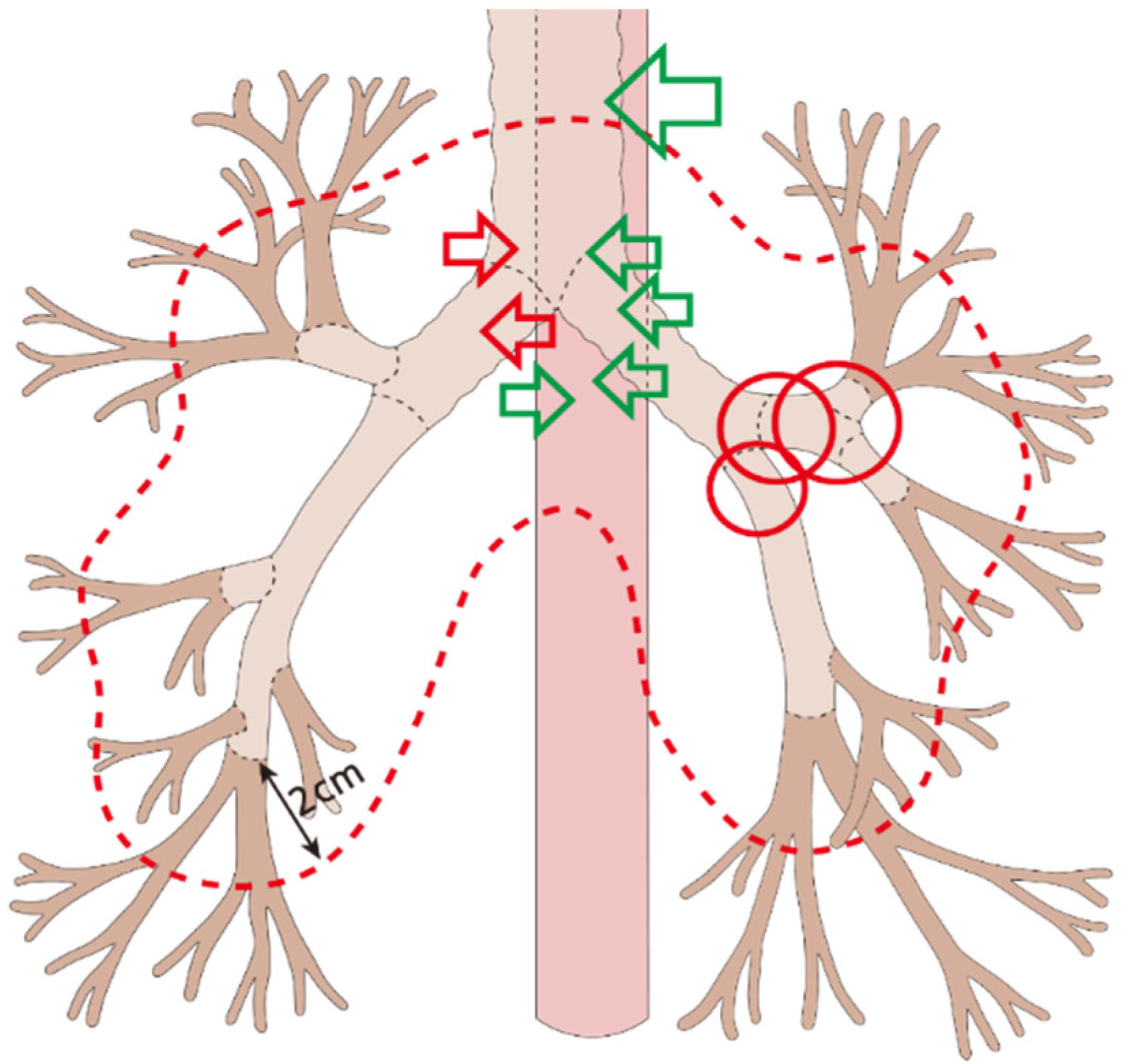
2.2. Re-Irradiation
2.3. Clinical Outcomes and Statistical Analysis
3. Results
3.1. Patients’ Characteristics
3.2. Clinical Outcomes
3.3. Radiation-related Complications
4. Discussion
5. Conclusions
Supplementary Materials
Author Contributions
Funding
Institutional Review Board Statement
Data Availability Statement
Conflicts of Interest
References
- Bray, F.; Ferlay, J.; Soerjomataram, I.; Siegel, R.L.; Torre, L.A.; Jemal, A. Global cancer statistics 2018: GLOBOCAN estimates of incidence and mortality worldwide for 36 cancers in 185 countries. CA Cancer J. Clin. 2018, 68, 394–424. [Google Scholar] [CrossRef] [Green Version]
- Travis, W.D.; Brambilla, E.; Nicholson, A.G.; Yatabe, Y.; Austin, J.H.M.; Beasley, M.B.; Chirieac, L.R.; Dacic, S.; Duhig, E.; Flieder, D.B.; et al. The 2015 World Health Organization Classification of Lung Tumors: Impact of Genetic, Clinical and Radiologic Advances Since the 2004 Classification. J. Thorac. Oncol. 2015, 10, 1243–1260. [Google Scholar] [CrossRef] [PubMed] [Green Version]
- Noh, J.M.; Ahn, Y.C.; Lee, H.; Pyo, H.; Kim, B.; Oh, D.; Park, H.; Lee, E.; Park, K.; Ahn, J.S.; et al. Definitive Bimodality Concurrent Chemoradiotherapy in Patients with Inoperable N2-positive Stage IIIA Non-small Cell Lung Cancer. Cancer Res. Treat. 2015, 47, 645–652. [Google Scholar] [CrossRef] [PubMed]
- Noh, J.M.; Kim, J.M.; Ahn, Y.C.; Pyo, H.; Kim, B.; Oh, D.; Ju, S.G.; Kim, J.S.; Shin, J.S.; Hong, C.S.; et al. Effect of Radiation Therapy Techniques on Outcome in N3-positive IIIB Non-small Cell Lung Cancer Treated with Concurrent Chemoradiotherapy. Cancer Res. Treat. 2016, 48, 106–114. [Google Scholar] [CrossRef] [PubMed]
- Shin, H.; Noh, J.M.; Pyo, H.; Ahn, Y.C.; Oh, D. Salvage proton beam therapy for locoregional recurrence of non-small cell lung cancer. Radiat. Oncol. J. 2021, 39, 24–32. [Google Scholar] [CrossRef]
- Ebara, T.; Tanio, N.; Etoh, T.; Shichi, I.; Honda, A.; Nakajima, N. Palliative re-irradiation for in-field recurrence after definitive radiotherapy in patients with primary lung cancer. Anticancer Res. 2007, 27, 531–534. [Google Scholar]
- Brooks, E.D.; Sun, B.; Feng, L.; Verma, V.; Zhao, L.; Gomez, D.R.; Liao, Z.; Jeter, M.; O’Reilly, M.; Welsh, J.W.; et al. Association of Long-term Outcomes and Survival With Multidisciplinary Salvage Treatment for Local and Regional Recurrence After Stereotactic Ablative Radiotherapy for Early-Stage Lung Cancer. JAMA Netw. Open 2018, 1, e181390. [Google Scholar] [CrossRef] [Green Version]
- McAvoy, S.; Ciura, K.; Wei, C.; Rineer, J.; Liao, Z.; Chang, J.Y.; Palmer, M.B.; Cox, J.D.; Komaki, R.; Gomez, D.R. Definitive reirradiation for locoregionally recurrent non-small cell lung cancer with proton beam therapy or intensity modulated radiation therapy: Predictors of high-grade toxicity and survival outcomes. Int. J. Radiat. Oncol. Biol. Phys. 2014, 90, 819–827. [Google Scholar] [CrossRef]
- Chao, H.H.; Berman, A.T.; Simone, C.B., 2nd; Ciunci, C.; Gabriel, P.; Lin, H.; Both, S.; Langer, C.; Lelionis, K.; Rengan, R.; et al. Multi-Institutional Prospective Study of Reirradiation with Proton Beam Radiotherapy for Locoregionally Recurrent Non-Small Cell Lung Cancer. J. Thorac. Oncol. 2017, 12, 281–292. [Google Scholar] [CrossRef] [Green Version]
- Cho, W.K.; Noh, J.M.; Ahn, Y.C.; Oh, D.; Pyo, H. Radiation Therapy Alone in cT1-3N0 Non-small Cell Lung Cancer Patients Who Are Unfit for Surgical Resection or Stereotactic Radiation Therapy: Comparison of Risk-Adaptive Dose Schedules. Cancer Res. Treat. 2016, 48, 1187–1195. [Google Scholar] [CrossRef] [Green Version]
- Lee, S.U.; Moon, S.H.; Cho, K.H.; Pyo, H.R.; Kim, J.Y.; Kim, D.Y.; Kim, T.H.; Suh, Y.G.; Kim, Y.J. Ablative dose proton beam therapy for stage I and recurrent non-small cell lung carcinomas: Ablative dose PBT for NSCLC. Strahlenther. Onkol. 2016, 192, 649–657. [Google Scholar] [CrossRef] [PubMed]
- Cox, J.D.; Stetz, J.; Pajak, T.F. Toxicity criteria of the Radiation Therapy Oncology Group (RTOG) and the European Organization for Research and Treatment of Cancer (EORTC). Int. J. Radiat. Oncol. Biol. Phys. 1995, 31, 1341–1346. [Google Scholar] [CrossRef]
- Ho, J.C.; Nguyen, Q.N.; Li, H.; Allen, P.K.; Zhang, X.; Liao, Z.; Zhu, X.R.; Gomez, D.; Lin, S.H.; Gillin, M.; et al. Reirradiation of thoracic cancers with intensity modulated proton therapy. Pract. Radiat. Oncol. 2018, 8, 58–65. [Google Scholar] [CrossRef] [PubMed]
- Vyfhuis, M.A.L.; Rice, S.; Remick, J.; Mossahebi, S.; Badiyan, S.; Mohindra, P.; Simone, C.B., 2nd. Reirradiation for locoregionally recurrent non-small cell lung cancer. J. Thorac. Dis. 2018, 10, S2522–S2536. [Google Scholar] [CrossRef]
- Chao, H.H.; Berman, A.T. Proton therapy for thoracic reirradiation of non-small cell lung cancer. Transl. Lung Cancer Res. 2018, 7, 153–159. [Google Scholar] [CrossRef] [PubMed]
- McAvoy, S.A.; Ciura, K.T.; Rineer, J.M.; Allen, P.K.; Liao, Z.; Chang, J.Y.; Palmer, M.B.; Cox, J.D.; Komaki, R.; Gomez, D.R. Feasibility of proton beam therapy for reirradiation of locoregionally recurrent non-small cell lung cancer. Radiother. Oncol. 2013, 109, 38–44. [Google Scholar] [CrossRef] [PubMed]
- Badiyan, S.N.; Rutenberg, M.S.; Hoppe, B.S.; Mohindra, P.; Larson, G.; Hartsell, W.F.; Tsai, H.; Zeng, J.; Rengan, R.; Glass, E.; et al. Clinical Outcomes of Patients With Recurrent Lung Cancer Reirradiated With Proton Therapy on the Proton Collaborative Group and University of Florida Proton Therapy Institute Prospective Registry Studies. Pract. Radiat. Oncol. 2019, 9, 280–288. [Google Scholar] [CrossRef]
- Maranzano, E.; Draghini, L.; Anselmo, P.; Casale, M.; Arcidiacono, F.; Chirico, L.; Italiani, M.; Trippa, F. Lung reirradiation with stereotactic body radiotherapy. J. Radiosurg. SBRT 2016, 4, 61–68. [Google Scholar]
- Ogawa, Y.; Shibamoto, Y.; Hashizume, C.; Kondo, T.; Iwata, H.; Tomita, N.; Ogino, H. Repeat stereotactic body radiotherapy (SBRT) for local recurrence of non-small cell lung cancer and lung metastasis after first SBRT. Radiat. Oncol. 2018, 13, 136. [Google Scholar] [CrossRef]
- Hong, J.H.; Kim, Y.-S.; Lee, S.-W.; Lee, S.J.; Kang, J.H.; Hong, S.H.; Hong, J.-Y.; Cheon, G. High-dose thoracic re-irradiation of lung cancer using highly conformal radiotherapy is effective with acceptable toxicity. Cancer Res. Treat. Off. J. Korean Cancer Assoc. 2019, 51, 1156. [Google Scholar] [CrossRef]
- Griffioen, G.H.; Dahele, M.; de Haan, P.F.; van de Ven, P.M.; Slotman, B.J.; Senan, S. High-dose, conventionally fractionated thoracic reirradiation for lung tumors. Lung Cancer 2014, 83, 356–362. [Google Scholar] [CrossRef] [PubMed]
- Trovo, M.; Minatel, E.; Durofil, E.; Polesel, J.; Avanzo, M.; Baresic, T.; Bearz, A.; Del Conte, A.; Franchin, G.; Gobitti, C. Stereotactic body radiation therapy for re-irradiation of persistent or recurrent non-small cell lung cancer. Int. J. Radiat. Oncol. Biol. Phys. 2014, 88, 1114–1119. [Google Scholar] [CrossRef] [PubMed]
- Kim, H.; Pyo, H.; Noh, J.M.; Lee, W.; Park, B.; Park, H.Y.; Yoo, H. Preliminary result of definitive radiotherapy in patients with non-small cell lung cancer who have underlying idiopathic pulmonary fibrosis: Comparison between X-ray and proton therapy. Radiat. Oncol. 2019, 14, 19. [Google Scholar] [CrossRef]
- Paganetti, H.; Niemierko, A.; Ancukiewicz, M.; Gerweck, L.E.; Goitein, M.; Loeffler, J.S.; Suit, H.D. Relative biological effectiveness (RBE) values for proton beam therapy. Int. J. Radiat. Oncol. Biol. Phys. 2002, 53, 407–421. [Google Scholar] [CrossRef]


| Variables | Total (N = 63) | XRT (n = 41) | PBT (n = 22) | p-Value |
|---|---|---|---|---|
| Age, years | ||||
| Median (range) | 63 (44–84) | 62 (44–80) | 66 (44–84) | 0.163 |
| Sex | ||||
| Male | 56 (88.9%) | 35 (85.4%) | 21 (95.5%) | 0.405 |
| Female | 7 (11.1%) | 6 (14.6%) | 1 (4.5%) | |
| Smoking | ||||
| Non-smoker | 11 (17.5%) | 8 (19.5%) | 3 (13.6%) | 0.733 |
| Current or ex-moker | 52 (82.5%) | 33 (80.5%) | 19 (86.4%) | |
| Histology | ||||
| SQ | 41 (65.1%) | 27 (65.9%) | 14 (63.6%) | 0.519 |
| AD | 20 (31.7%) | 12 (29.3%) | 8 (36.4%) | |
| Others or NOS | 2 (3.2%) | 2 (4.9%) | - | |
| CCI | ||||
| Median (range) | 4 (1–7) | 3 (1–6) | 4 (1–7) | 0.287 |
| Underlying COPD | ||||
| No | 51 (81.0%) | 35 (85.4%) | 16 (72.7%) | 0.314 |
| Yes | 12 (19.0%) | 6 (14.6%) | 6 (27.3%) | |
| Re-RT interval, months | ||||
| Median (range) | 15.2 (3.2–44.6) | 15.8 (3.2–44.6) | 14.0 (5.8–28.9) | 0.096 |
| ECOG at re-RT | ||||
| 0–1 | 60 (95.2%) | 41 (100%) | 19 (86.4%) | 0.039 |
| 2 | 3 (4.8%) | - | 3 (13.6%) | |
| FEV1, L | ||||
| Median (range) | 2.2 (1.2–3.8) | 2.2 (1.2–3.8) | 2.3 (1.5–2.9) | 0.567 |
| DLCO, % | ||||
| Median (range) | 72 (25–118) | 72 (25–118) | 67 (25–118) | 0.666 |
| Clinical stage at re-RT | ||||
| I-II | 30 (47.6%) | 18 (43.9%) | 12 (54.5%) | 0.428 |
| III | 33 (52.4%) | 23 (56.1%) | 10 (45.5%) | |
| Recurrence at re-RT | ||||
| 1st | 56 (88.9%) | 37 (90.2%) | 19 (86.4%) | 0.687 |
| 2nd or more | 7 (11.1%) | 4 (9.3%) | 3 (13.6%) | |
| CCRT | ||||
| No | 42 (66.7%) | 26 (63.4%) | 16 (72.7%) | 0.578 |
| Yes | 21 (33.3%) | 15 (36.6%) | 6 (27.3%) | |
| Re-RT technique | ||||
| XRT-3D | 23 (36.5%) | 23 (56.1%) | - | - |
| XRT-IMRT | 13 (20.6%) | 13 (31.7%) | - | |
| XRT-SBRT | 5 (7.9%) | 5 (12.2%) | - | |
| PBT-3DPT | 3 (4.8%) | - | 3 (13.6%) | |
| PBT-IMPT | 19 (30.2%) | - | 19 (86.4%) |
| XRT (n = 41) | PBT (n = 22) | p-Value | |||||
|---|---|---|---|---|---|---|---|
| Peripheral | Central | Ultracentral | Peripheral | Central | Ultracentral | ||
| Trachea | 22 (53.7%) | 3 (7.3%) | 16 (39.0%) | 15 (68.2%) | 3 (13.6%) | 4 (18.2%) | 0.215 |
| Proximal bronchi | 22 (53.7%) | 7 (17.1%) | 12 (29.3%) | 12 (54.5%) | 2 (9.1%) | 8 (36.4%) | 0.648 |
| Esophagus | 26 (63.4%) | 9 (22.0%) | 6 (14.6%) | 13 (59.1%) | 4 (18.2%) | 5 (22.7%) | 0.712 |
| Lung | Esophagus | |||||
|---|---|---|---|---|---|---|
| Variables | Grades 0–2 | Grades 3–5 | p-Value | Grades 0–2 | Grades 3–5 | p-Value |
| Target—trachea location | 0.534 | 1 | ||||
| Non-ultracentral | 37 (86.0%) | 6 (14.0%) | 40 (93.0%) | 3 (7.0%) | ||
| Ultracentral | 19 (95.0%) | 1 (5.0%) | 18 (90.0%) | 2 (10.0%) | ||
| Target—bronchus location | 0.271 | 0.055 | ||||
| Non-ultracentral | 40 (93.0%) | 3 (7.0%) | 42 (97.7%) | 1 (2.3%) | ||
| Ultracentral | 16 (80.0%) | 4 (20.0%) | 16 (80.0%) | 4 (20.0%) | ||
| Target—esophagus location | 1 | <0.001 | ||||
| Non-ultracentral | 46 (88.5%) | 6 (11.5%) | 52 (100%) | - | ||
| Ultracentral | 10 (90.9%) | 1 (9.1%) | 6 (54.5%) | 5 (45.5%) | ||
| CCRT at re-RT | 0.119 | 0.07 | ||||
| No | 35 (83.3%) | 7 (16.7%) | 41 (97.6%) | 1 (2.4%) | ||
| Yes | 21 (100%) | - | 17 (81.0%) | 4 (19.0%) | ||
| Re-RT technique | 0.427 | 0.194 | ||||
| PBT | 21 (95.5%) | 1 (4.5%) | 21 (95.5%) | 1 (4.5%) | 0.81 | |
| XRT | 35 (85.4%) | 6 (14.6%) | 37 (90.2%) | 4 (9.8%) | ||
| rCTV, cc | 81.4 ± 158.6 | 97.0 ± 92.3 | 0.801 | 87.3 ± 157.7 | 34.6 ± 41.4 | 0.074 |
| BED10 at re-RT, Gy | 85.3 ± 16.3 | 100.3 ± 34.1 | 0.292 | 87.6 ± 19.9 | 79.9 ± 2.3 | 0.009 |
| Re-RT fractions | 22.0 ± 9.5 | 14.0 ± 7.2 | 0.002 | 20.3 ± 9.4 | 30.8 ± 6.1 | 0.017 |
| Re-RT fraction size, Gy | 3.8 ± 3.0 | 6.7 ± 5.7 | 0.234 | 4.3 ± 3.5 | 2.2 ± 4.5 | <0.001 |
| Cumulative BED10, Gy | 164.4 ± 31.8 | 175.4 ± 28.6 | 0.387 | 167.6 ± 31.9 | 143.3 ± 11.7 | 0.098 |
| Lung, Dmean, Gy | 4.5 ± 2.9 | 6.4 ± 4.1 | 0.115 | |||
| Lung, V20, % | 7.5 ± 5.4 | 10.4 ± 8.1 | 0.22 | |||
| Lung, V5, % | 19.0 ± 13.0 | 24.7 ± 14.1 | 0.283 | |||
| Lung, cumulative Dmean, Gy | 14.1 ± 6.2 | 15.5 ± 4.8 | 0.682 | |||
| Lung, cumulative V20, % | 22.8 ± 12.2 | 24.8 ± 9.5 | 0.635 | |||
| Lung, cumulative V5, % | 46.6 ± 19.3 | 50.3 ± 18.3 | 0.865 | |||
| Esophagus, Dmax, Gy | 41.7 ± 23.8 | 67.2 ± 2.5 | <0.001 | |||
| Esophagus, Dmean, Gy | 8.6 ± 8.1 | 19.0 ± 7.4 | 0.008 | |||
| Esophagus, V50, % | 4.8 ± 9.7 | 21.8 ± 10.7 | <0.001 | |||
| Esophagus, cumulative Dmax, Gy | 83.6 ± 33.3 | 123.5 ± 10.4 | <0.001 | |||
| Esophagus, cumulative Dmean, Gy | 25.0 ± 14.4 | 42.2 ± 11.3 | 0.012 | |||
| Esophagus, cumulative V50, % | 22.3 ± 19.4 | 39.4 ± 9.2 | 0.057 |
Publisher’s Note: MDPI stays neutral with regard to jurisdictional claims in published maps and institutional affiliations. |
© 2022 by the authors. Licensee MDPI, Basel, Switzerland. This article is an open access article distributed under the terms and conditions of the Creative Commons Attribution (CC BY) license (https://creativecommons.org/licenses/by/4.0/).
Share and Cite
Yang, K.; Suh, Y.-G.; Shin, H.; Pyo, H.; Moon, S.H.; Ahn, Y.C.; Oh, D.; Chung, E.; Jo, K.; Noh, J.M. Toxicity of Proton Therapy versus Photon Therapy on Salvage Re-Irradiation for Non-Small Cell Lung Cancer. Life 2022, 12, 292. https://doi.org/10.3390/life12020292
Yang K, Suh Y-G, Shin H, Pyo H, Moon SH, Ahn YC, Oh D, Chung E, Jo K, Noh JM. Toxicity of Proton Therapy versus Photon Therapy on Salvage Re-Irradiation for Non-Small Cell Lung Cancer. Life. 2022; 12(2):292. https://doi.org/10.3390/life12020292
Chicago/Turabian StyleYang, Kyungmi, Yang-Gun Suh, Hyunju Shin, Hongryull Pyo, Sung Ho Moon, Yong Chan Ahn, Dongryul Oh, Eunah Chung, Kwanghyun Jo, and Jae Myoung Noh. 2022. "Toxicity of Proton Therapy versus Photon Therapy on Salvage Re-Irradiation for Non-Small Cell Lung Cancer" Life 12, no. 2: 292. https://doi.org/10.3390/life12020292