Article

Prokaryotic Diversity of Ruminal Content and Its Relationship with Methane Emissions in Cattle from Kazakhstan

1
LLP ‘Kazakh Research Institute for Livestock and Fodder Production’, St. Zhandosova 51, Almaty 050035, Kazakhstan
2
LLP ‘Scientific Production Center of Microbiology and Virology’, Bogenbay Batyr Str. 105, Almaty 050010, Kazakhstan
3
Istituto Zooprofilattico Sperimentale del Piemonte, Ligura e Valle d’Aosta, Via Bologna 148, 10154 Turin, Italy
*
Author to whom correspondence should be addressed.
Life 2022, 12(11), 1911; https://doi.org/10.3390/life12111911
Submission received: 18 August 2022 / Revised: 11 November 2022 / Accepted: 15 November 2022 / Published: 17 November 2022
(This article belongs to the Special Issue Evolutionary and Conservation Genetics: 2nd Edition)

Abstract

:
In this study, we analyzed the microbial composition of the rumen contents of cattle from Kazakhstan. Specifically, samples of the liquid and solid fractions of the rumen were collected to determine the quantitative and qualitative composition of methanogenic archaea. Cattle were six steers receiving hay-concentrate feeding. Methane emission was determined by repeated measurements for each animal. Rumen samples were then taken from fistulas and analyzed using 16S metabarcoding via Next-Generation Sequencing (NGS). The difference between the rumen fractions was investigated, resulting in differential distribution of the families Streptococccaceae, Lactobacillaceae, Desulfobulbaceae, and Succinivibrionaceae, which were more abundant in the liquid fraction, while Thalassospiraceae showed a higher presence in the solid fraction. These differences can be explained by the fact that fibrolytic bacteria are associated with the solid fraction compared to the liquid. A relationship between methane emission and methanogenic microbiota was also observed. Steers producing more methane showed microbiota richer in methanogens; specifically, most Mathanobacteriaceae resided in the liquid fraction and solid fraction of animals 1 and 6, respectively. The same animals carried most of the Methanobrevibacter and Methanosphaera genera. On the contrary, animals 2, 3, and 5 hosted a lower amount of methanogens, which also agreed with the data on methane emissions. In conclusion, this study demonstrated a relationship between methane emission and the content of methanogenic archaea in different rumen fractions collected from cattle in Kazakhstan. As a result of the studies, it was found that the solid fraction of the rumen contained more genera of methanogens than the liquid fraction of the rumen. These results prove that taking rumen contents through a fistula is more useful than taking it through a probe. The presented data may be of interest to scientists from all over the world engaged in similar research in a comparative aspect.

1. Introduction

The rumen contains one of the most complex microbial communities—prokaryotes, eukaryotes, and viruses. Prokaryotes include bacteria, primarily anaerobic bacteria, and archaea, including methanogens [1]. They have a role in digesting fibrous feed and providing nutrients to the animal host. There is an environmental cost, however, in which ruminants, via their ruminal microbiota, produce substantial amounts of greenhouse gas (GHG), i.e., methane [2,3,4]. The rumen is an anaerobic and methanogenic fermentation chamber that contains microorganisms that can utilize and increase the productivity of cellulolytic feeds (e.g., straw, hay, silage, and grass). Understanding the rumen microbiota and their connection to the ruminant itself is important for producing quality products, increasing profitability, and reducing environmental impacts [5]. Moreover, methane cannot be absorbed by the host, as it represents excess energy [6] and is therefore emitted to the environment. The methane production in the rumen is 2–12% of the total energy consumed, which could be used instead to increase the body weight of animals [7].
There are three intersecting microenvironments found in the rumen, which contain bacteria, archaea, protozoa, and fungi: the liquid phase, making up 25% of the microbial mass; the solid phase, making up 70% of the microbial mass; and the rumen epithelial cells and protozoa, containing 5% of the microbial mass [8]. In terms of cell numbers, bacteria are the most abundant, representing over 95% of microorganisms within the rumen ecosystem [9], and were first described using classical microbiology methods [10]. When cattle are fed a high amount of grains, the relative abundances of the members of the phyla Bacteroidetes and Fibrobacter in the microbial communities in the digestive tract decline, allowing Firmicutes and other opportunistic phyla, such as Gram-negative Proteobacteria, to proliferate faster, resulting in an increase in the proportions of Firmicutes and Proteobacteria [11].
Rumen archaea are strictly anaerobic and are the only known microorganisms present in the rumen capable of producing methane [12]. Such archaea are referred to as methanogens. Archaea are found in the rumen in the range of 106 to 108 cells per ml, accounting for less than 4% of the microbial community [13]. Wallace R.J. et al. [14] identified the Megasphaera genus as being significantly enriched in the rumen of low- methane-emitting cows.
The fiber-adherent microbiota is characterized by a higher proportion of taxa associated with fiber degradation, such as Ruminococcaceae and Fibrobacter, whereas the liquid phase is characterized by enrichment in members of the Prevotellaceae family, capable of utilizing a broad range of soluble substrates [15,16]. Studies by Brulc et al. [9] were performed on the rumen of three steers, revealing that one steer had a remarkably different microbiota composition compared with the other two steers analyzed, despite similar housing and dietary conditions and without any obvious differences in the animals’ physiology. Other authors had similar conclusions [17,18]. This observation is not limited to bacteria, and similar inter variation within the same host species can be seen in the other microbial domains inhabiting the rumen [19,20,21].
This study aimed to characterize the microbiota of liquid and solid fractions of the rumen in cattle from Kazakhstan, as well as investigate the association of identified bacteria and methanogens with methane production.

2. Materials and Methods

2.1. Animals

The experiment followed the rules for the treatment of animals (order of the Minister of Agriculture of the Republic of Kazakhstan dated 30 December 2014, no. 16-02/701) and was approved by the Bioethics Commission of the Kazakh Research Institute for livestock and Fodder Production. The research was carried out in the experimental farm of the ‘Kurazek’, Institute Zhambyl district, Almaty region. The animals enrolled in the study were 6 steers belonging to different breeds (i.e., Kazakh white-headed, Alatau, Simmental, and Holstein breeds) with a live weight of 350–400 kg and an average daily increase in live weight of 1200–1500 g. They were healthy animals of the same age (15 months old).

2.2. Average Daily Rations

The animals were fed alfalfa hay, straw, and concentrates. The average daily nutritional value of the diet was 11.9 EFU. The experiment duration was 52 days; then, sampling was carried out. The chemical analysis of feed was carried out in accordance with state standard 32040-2021. To determine the chemical composition of feed, a modern Infrasac device from FOSS (Denmark) was used.
The average daily ration was developed and calculated based on data on the amount of given feed; their palatability; and the actual consumption of feed, nutrients, and biologically active substances. The average daily ration characterizes the background and type of feeding for the period of experience. The average daily rations of animals’ actual consumed feed for the study period are given in Table 1.

2.3. Methane Measurement

During the experiment, the methane concentration in the exhaled air was measured for each animal four times a day for three days. Measurements of methane were carried out by the gas analyzer ‘Signal-44′ (TU 4215-002-80703968-07, according to the operating manual of the device). To ensure accurate analysis, the animal’s head was covered with plastic wrap (Figure 1). The operation of the gas analyzer was based on measuring the electrical signal coming from the gas-sensitive sensor (thermal catalytic sensor), which is proportional to the concentration of the measured substance within the measuring range. Methane measurement was carried out based on the Sniffer Method 22 with modifications.

2.4. DNA Extraction, PCR Amplification, and 16S Metabarcoding

The rumen contents were mixed manually, and then an aliquot was collected (Figure 2, Table 2). The liquid fraction was separated from the solid content by filtration through sterile cheesecloth. All the samples were transported on dry ice to the LLP ‘Scientific Production Center of Microbiology and Virology’ in Almaty for DNA extraction, amplification, and analysis. Samples from the solid and liquid phases were handled separately.
Two hundred mg of ruminal samples was taken for DNA isolation. DNA from the samples was isolated using the PureLinkTM Microbiome DNA Purification Kit (Invitrogen, Carlsbad, CA, USA) according to the manufacturer’s protocol. In addition to the samples, a positive control consisting of a standard microbial community (MOCK, Zymo Research) and a negative extraction control was set up. The DNA concentration was measured by a Qubit® 2.0 fluorimeter using a Qubit™ dsDNA HS Assay Kit (Life Technologies, Eugene, Oregon, USA). The genetic libraries were prepared for sequencing following the 16S Metagenomic Sequencing Library Preparation guide (part no. 15044223 rev. A). As an extraction control, a lysis buffer was used from a DNA isolation kit without any biological material. Molecularly pure water was used to control the libraries and was added to the amplification mixture. The amplification product was not observed in the controls. The libraries pooled with PhiX were sequenced on an Illumina MiSeq device using a 600-cycle MiSeq® Reagent Kit v3 (Illumina Inc., San Diego, CA, USA), following the manufacturer’s recommendations.

2.5. Bioinformatics and Data Analysis

The 16S metabarcoding data obtained from the rumen samples were analyzed using the Data QC workflow. The raw fastq data were analyzed with the ‘Data QC and OTU Clustering’ and ‘Estimate Alpha and Beta Diversity’ workflow tools of the Microbial Genomics Module in the CLC Genomic Workbench software (Qiagen). The paired-end reads were joined and trimmed for low quality score (Qscore < 0.05), nucleotide ambiguity (max of 2 nucleotides allowed), adapter sequences, and length. Duplicate sequences were merged and aligned against the SILVA database (v. 132) at 97% identity threshold. Chimeric reads were removed, and taxonomy was assigned, creating an OTU table. The profiles of the negative control and the mock communities were analyzed to check for correct procedures and cross-contamination, then removed. Rarefaction curves did not reach a plateau in all study samples, so they were compared at a sequencing depth of 50,000 reads. The alpha diversity (diversity within the groups) was estimated using the total number, Chao-1 bias-corrected, Simpson’s index, and Shannon entropy. The Bray–Curtis method, Jaccard, Unweighted UniFrac and Weighted UniFrac, and Principal Coordinates Analysis (PcoA) were used to estimate the beta diversity (diversity between groups). Statistical support was calculated using the Kruskal–Wallis test for alpha diversity and the PERMANOVA test for beta diversity (p-value ≤ 0.05). The OTU table showing the abundance of the genera in the analyzed samples was used to perform a generalized linear model test of differential abundance to compare microbial abundance of solid and liquid fractions. The ‘Differential Abundance Analysis’ tool in CLC Genomic Workbench performs a TMM normalization to make samples comparable, adjusting library sizes. The Wald test was used to determine significance between group pairs. Differential abundance analyses compared genera and families by liquid/solid fraction.

3. Results

3.1. Dynamics of Live Weight of Animals

Roughage and concentrates in the structure of the diet comprised 57.43% and 42.57%, respectively. Succulent foods were excluded from the diet. The eatability of hay was 86.8%, that of straw was 82.1%, and concentrates were consumed completely. On average, the animals consumed 12.6 kg of dry matter per day. The concentration EFU in 1 kg of dry matter was 0.94 MJ; the crude protein content was 156.36 g. Thus, the rations almost completely provided the animals’ need for basic nutrients, which positively affected their productivity (Table 3).
The data given in Table 3 shows that the average daily gain in live weight averaged 1476.0 ± 115.36 g, and the feed cost per 1 kg of gain was 8.1 ± 0.74 ECU.

3.2. Methane Emissions from Animals

The largest amount of methane in the exhaled air was found in animals 1 and 6. The averaged data on methane emission are shown in Table 4. Methane yield, in our case, is the concentration coefficient.

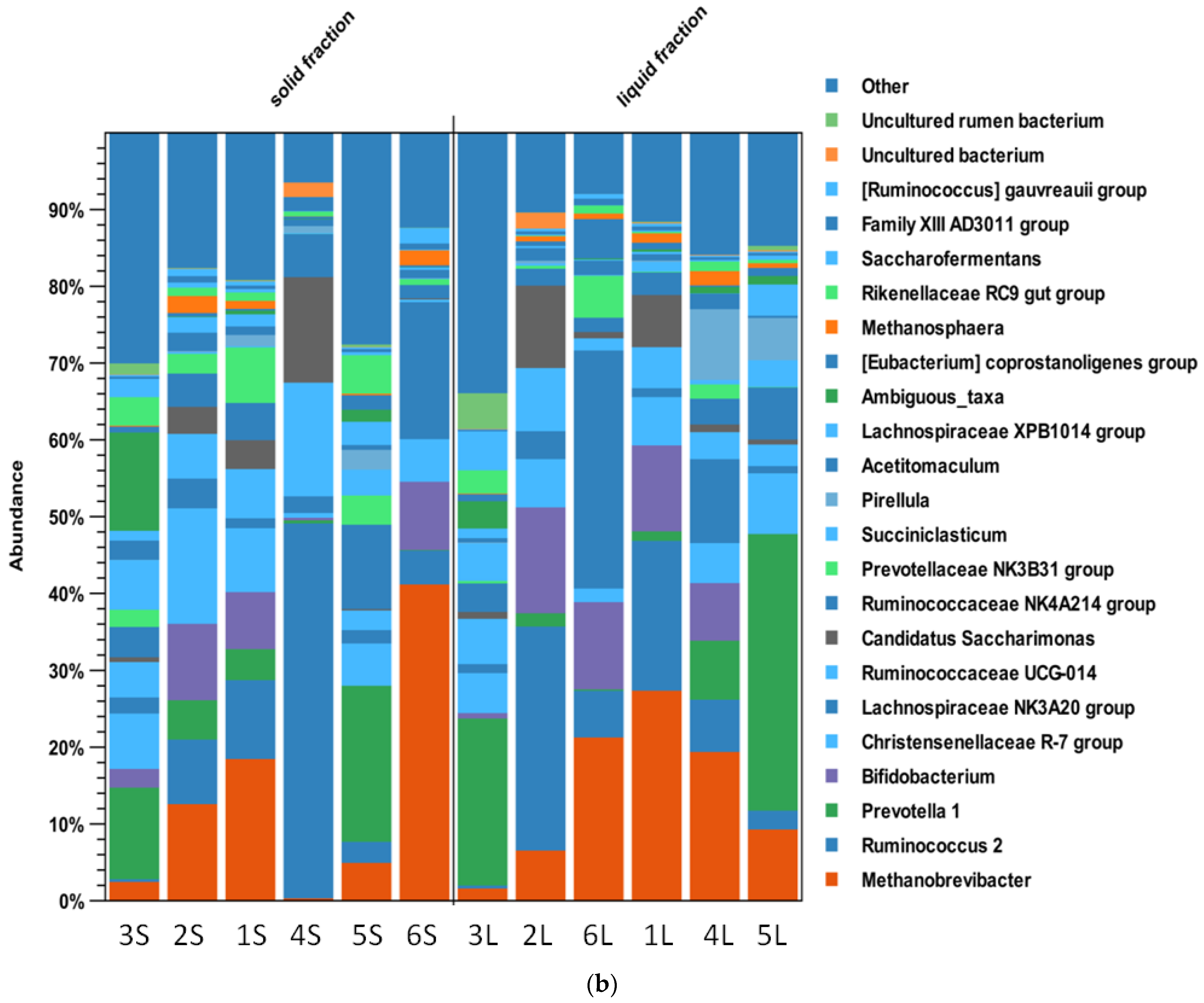
3.3. Rumen Microbiota

The high throughput sequencing of the V4 region of the 16S rRNA gene produced 3,963,970 reads; 3,959,335 paired reads were obtained for OTU clustering after quality filtering. The 392,411 unique nonchimeric sequences were assigned to 2,447 OTUs. Data from the mock community analysis matched the expected results, while the negative controls showed a low number of reads assigned to OTUs absent or with negligible abundance in the samples. Histograms comparing fractions (Figure 3a,b) showed differences at the family and genus level, not always significant after statistical analysis (as shown later in the text).
The alpha diversity was insignificant for all the compared samples with regard to fraction comparison. All the p-values were >0.05 for each of the different estimations. For example, the alpha-diversity results estimated by the total number comparing ruminal fractions are reported in Figure 4.
No differences were identified by considering the fraction by beta diversity analysis. As an example, in Figure 5, the results of the Bray–Curtis estimation are shown.
However, the differential abundance analysis showed significant differences for specific taxonomic groups comparing the liquid and solid fractions (Table 5).
At the family level, Streptococcaceae, Lactobacillaceae, Desulfobulbaceae, and Succinivibrionaceae were more abundant in the liquid fraction, while Thalassospiraceae showed a higher presence in the solid fraction. At the genus level, Ruminobacter, Howardella, Streptococcus, Lactobacillus, Ruminococcaceae UCG-013, Eubacterium Сoprostanoligenes, Succinivibrionaceae, Desulfobulbus, and Psychrobacter showed a higher abundance in the liquid fraction, while Thalassospira was more abundant in the solid fraction.
In the liquid fraction, the largest relative number of representatives of the families Ruminococcaceae (41%), Bifidobacteriaceae (14%), and Saccharimonadaceae (11%) was in 2L. The families Prevotellaceae (39%) and Christensenellaceae (8%) prevailed in another animal (5L). The family Lachnospiraceae (35%) was the most abundant in 1L. In 4L, the Pirellulaceae family was prevalent (11%). Among methanogens, a large relative number of members of the Methanobacteriaceae family (29%) was detected in 1L.
In the solid fraction, the largest relative number of representatives of the families Ruminococcaceae (70%) and Saccharimonadaceae (14%) was in 4S. In 3S, a large proportion of the families Muribaculaceae (16%) and Acidaminococcaceae (6%) was observed. The family Lachnospiraceae (24%) was also the most represented in 6S. The families Christensenellaceae (15%) and Bifidobacteriaceae (10%) prevailed in 2S. Additionally, in another animal (5S), the Prevotellaceae family prevailed (27%). Among methanogens, a large relative number of members of the family Methanobacteriaceae (44%) was found in 6S.
At the genus level, the largest relative number of representatives of the genera Candidatus Saccharimonas (41%), Ruminococcus 2 (29%), Bifidobacterium (14%), and Ruminococcus (15%) was in the liquid fraction collected from animal 2. The genus Prevotella (36%) prevailed in 5L. The family Lachnospiraceae NK3A20 (31%) was the most abundant in 6L. In 4L, the genus Pirellula prevailed (9%). Among methanogens, a large relative number of members of the genus Methanobrevibacter (27%) was in 1L and the genus Methanoshaera (2%) in 4L.
In the solid fraction of the rumen, the largest relative number of representatives of the genera Ruminococcus (64%) and Candidatus Saccharimonas (14%) was in animal 4S. In another animal (3L), a large number of reads assigned to an unidentified Muribaculaceae bacterium (13%) and the genus Succiniclasticum (6%) were observed. The family Lachnospiraceae (18%) and the genus Bifidobacterium (9%) were the most abundant in 6S. The family Ruminococcaceae (33%), Pirellulaceae (20%), Christensenellaceae (15%), Lachnospiraceae (15%), and Bifidobacteriaceae (10%) prevailed in 2S. Additionally, in 5S, the genus Prevotella 1 prevailed (8%). Animal 1 was dominated by the family Christensenellaceae R-7 group (8%) and the Prevotellaceae NK3B31 group (7%) in both liquid and solid fractions.
Among methanogens, a large relative number of members of the Methanobrevibacter genus was found in animals 1 and 6 (41%) and Methanoshaera in animals 2 and 6 (2%). The genus Prevotella1 showed a lower abundance in animal 6 compared with other animals. Animal 1 showed significant differences in Eubacterium ventriousum (Lachnospiraceae family), which was almost absent in all the other animals.

4. Discussion

In this study, the microbiota of liquid and solid fractions of the rumen in cattle from Kazakhstan was characterized, and the association of identified bacteria and methanogens with methane production was determined.
In previous studies, we determined how the microbiota of cattle affects methane in different regions of Kazakhstan. For this purpose, fecal samples were taken rectally from 37 cattle heads from 4 regions of Kazakhstan (Western, Southern, Northern, and Southeast Kazakhstan). As a result of the studies, it was found that methanogenic bacteria were less present in animals from the Western region grazing in the pasture compared with animals from other regions kept in the stall [22].
Diet is a major factor impacting the microbiota composition of the gastrointestinal tract. As a whole, since the feeding was the same for all animals (i.e., hay-concentrated feeding), the rumen microbiome was dominated by the families Lachnospiraceae, Bifidobacteriaceae, Prevotellaceae, and Succinivibrionaceae.
Members of the Lachnospiraceae family are Gram-positive, anaerobic, rod-shaped bacteria that predominantly ferment pectin [23]. Li and Guan [24] found that Lachnospiraceae are more abundant in the rumen of steers with the lowest average daily gain (ADG) and highest residual feed intake (RFI); however, these observations are not consistent with the findings of Myer et al. [25], who found that Lachnospiraceae are more abundant among steers with the highest ADG. Many Lachnospiraceae produce butyrate, which is a gut nutrient, and VFAs are produced from carbohydrate digestion [26].
Gagen et al. [27] found that acetogens exist in the Lachnospiraceae and Ruminococcaceae families. Acetogens can serve as a hydrogen scavenger and can increase if methane production is reduced. The presence of Lachnospiraceae and Ruminococcaceae indicates a more complete fermentation and an increase in digestible nutrients available to the animal [28]. The genus Ruminococcus plays an important role in the decomposition of cellulose [14].
The family Succinivibrionaceae (phylum Proteobacteria) was abundant in the liquid fraction of the rumen and predominated in grain-feeding animals [29]. The family Succinivibrionaceae are propionate producers and promote succinate. The family Succinivibrionaceae is known to inhibit methanogenesis [30].
Prevotella is usually found in large numbers and has many functions, such as breaking down polysaccharides and proteins. Classical studies showed that metabolic differences are widespread among the population, as they are often found in the rumen with different diets, and have also found that this genus grows freely at a pH of only 5.1 [23]. Delgado et al. [31] found a positive correlation between nutritional efficiency and the genus Prevotella. However, other authors note that the number of Prevotella in the rumen is greater in steers with the lowest ADG [26]. Previous analysis of the rumen microbiota showed that some OTUs belonging to the genus Prevotella were stimulated by the inhibition of methanogenesis. The Prevotella species includes propionate production by randomization upon inhibition of methanogenesis [32].
The genera Lactobacillus and Streptococcus showed a higher abundance in the liquid fraction of the rumen. These genera are recognized producers of lactic acid (lactate) and have higher relative abundance in grain-fed cattle with an acidic rumen environment (acidosis) [29]. Moreover, Streptococci produce toxic compounds by fermenting proteins [33] The genus Eubacterium ventriousum of the family Lachnospiraceae was only found in animal 1 and was nearly absent in all other animals. Eubacterium and Bifidobacterium are beneficial bacteria due to their ability to synthesize vitamins, aid digestion, stimulate immune function, and suppress pathogenic microbes.
Thus, the solid and liquid fractions of the rumen are different since fibrolytic bacteria are associated with the solid fraction compared with the liquid one. Therefore, for a complete picture of the diversity of the rumen, it is necessary to study both fractions.
To assess any relationship between GHG emission and microbiota composition, the first step was to measure methane emission in the studied animals. The existing methods for measuring methane are numerous, but they also have drawbacks and are unreliable. The need to improve and search for new methods for measuring methane in animals is reported by many authors [34,35,36]. To overcome this issue, multiple measurements were taken for each animal.
Among hydrogenotrophic methanogens, a relatively large number of members of the genus Methanobrevibacter (27%), the most common genus of methane producers in the rumen, was found in the liquid fraction of animal 1 and the solid fractions of animals 6 and 1 (41%); the genus Methanoshaera was detected in 4L (2%) and 6S (2%). Methanobrevibacter spp. is an important methanogen, accounting for 75–78% of methanogenic archaea in the rumen. Interestingly, Methanosphaera spp. is a methylotrophic methanogen that produces methane through a primary ethanol-dependent, hydrogen-independent methanogenesis pathway [36]. Family Methanobacteriaceae (15%) was observed in sample 2S.
Animal 1L had more abundant Methanobacteriaceae in the liquid fraction of the rumen, while animal 6S had more abundant Methanobacteriaceae in the solid fraction. The family Succinivibrionaceae suppressed methanogenesis and dominated in greater numbers in 3L. The genus Prevotella prevailed in sample 5L. The genus Prevotella1 showed a lower abundance in animal 6, which is consistent with the findings that the genus Prevotella inhibits methanogenesis.
Animals 3 and 4 had large numbers of members of the Succinovibrionaceae family in their rumen, which alters acetate and hydrogen production, resulting in less methanogenesis and, therefore, reduced methane emissions [14]. The family Succinovibrionaceae uses hydrogen to produce succinate, which is then rapidly converted to propionate and competes with the most common hydrogenotrophic methanogenesis.
In this study, we studied how the microbiota collected from the ruminal content of cattle affects methane. The animals involved in the experiment belonged to the Kazakh Research Institute for Livestock and Fodder Production. Due to the technical difficulty and high material costs of fistula installations, only six animals participated in the experiment.

5. Conclusions

The study results showed a differential distribution of the rumen microbiota, where Streptococccaceae, Lactobacillaceae, Desulfobulbaceae, and Succinivibrionaceae were more abundant in the liquid fraction, while Thalassospiraceae was more abundant in the solid fraction. These differences can be explained by the fact that fibrolytic bacteria are associated with the solid fraction compared with the liquid. Moreover, a relationship between methane emission and methanogenic microbiota was demonstrated. Steers producing more methane showed a microbiota richer in methanogens, especially Methanobrevibacter and Methanosphaera genera. As a result of the studies, it was found that the solid fraction of the rumen contained more genera of methanogens than the liquid fraction. These results prove that taking rumen contents through a fistula is more useful than taking it through a probe. This research is novel to Kazakhstan and adds to the growing global databases that have been developed by other countries under programs such as the global rumen census. The information developed from the project can also help the government to develop a national GHG emission policy.

Author Contributions

A.D., S.D., A.A., L.M., Z.Y. contributed to the experimental work. A.A., C.B., S.P. performed data analysis. A.D., S.D., A.A., S.P. contributed to the writing of the final manuscript. All authors have read and agreed to the published version of the manuscript.

Funding

This research was funded by the Science Committee of the Ministry of Education and Science of the Republic of Kazakhstan (grant no. AP09259133).

Institutional Review Board Statement

This experiment followed the rules for the treatment of animals (order of the Minister of Agriculture of the Republic of Kazakhstan dated 30 December 2014, no. 16-02/701) and was performed in accordance with the Bioethics Commission approved by Kazakh Research Institute for livestock and Fodder Production.

Informed Consent Statement

Not applicable.

Data Availability Statement

The data presented in this study are publicly available at the NCBI Sequence Read Archive (Bioproject ID: PRJNA854754).

Conflicts of Interest

The authors declare that the research was conducted in the absence of any commercial or financial relationships that could be construed as a potential conflict of interest.

References

  1. Kim, M.; Park, T.; Yu, Z. Metagenomic investigation of gastrointestinal microbiome in cattle. Asian Australas. J. Anim. Sci. 2017, 30, 1515–1528. [Google Scholar] [CrossRef] [PubMed] [Green Version]
  2. Wallace, R.J.; Sasson, G.; Garnsworthy, P.C.; Tapio, I.; Gregson, E.; Bani, P.; Huhtanen, P.; Bayat, A.R.; Strozzi, F.; Biscarini, F.; et al. A heritable subset of the core rumen microbiome dictates dairy cow productivity and emissions. Sci. Adv. 2019, 5, eaav8391. [Google Scholar] [CrossRef] [PubMed] [Green Version]
  3. Steinfeld, H.; Gerber, P.; Wassenaar, T.; Caste, V.; Rosales, M.; de Haan, C. Livestock’s Long Shadow; FAO: Rome, Italy, 2006. [Google Scholar]
  4. Fouts, D.E.; Szpakowski, S.; Purushe, J.; Torralba, M.; Waterman, R.C.; MacNeil, M.D.; Alexander, L.J.; Nelson, K.E. Next Generation Sequencing to Define Prokaryotic and Fungal Diversity in the Bovine Rumen. PLoS ONE 2012, 7, e48289. [Google Scholar] [CrossRef] [PubMed]
  5. Matthews, C.; Crispie, F.; Lewis, E.; Reid, M.; O’Toole, P.W.; Cotter, P.D. The rumen microbiome: A crucial consideration when optimising milk and meat production and nitrogen utilisation efficiency. Gut Microbes 2019, 10, 115–132. [Google Scholar] [CrossRef] [PubMed]
  6. Johnson, K.A.; Johnson, D.E. Methane emissions from cattle. J. Anim. Sci. 1995, 73, 2483–2492. [Google Scholar] [CrossRef]
  7. Johnson, D.E.; Ward, G.M. Estimates of animal methane emissions. Environ. Monit. Assess. 1996, 42, 133–141. [Google Scholar] [CrossRef]
  8. Ishler, V.; Heinrichs, A.J.; Varga, G. From Feed to Milk: Understanding Rumen Function; Pennsylvania State University Extension Circular: University Park, PA, USA, 1996; Volume 422. [Google Scholar]
  9. Brulc, J.M.; Antonopoulos, D.A.; Miller, M.E.B.; Wilson, M.K.; Yannarell, A.C.; Dinsdale, E.A.; Edwards, R.E.; Frank, E.D.; Emerson, J.B.; Wacklin, P.; et al. Gene-centric metagenomics of the fiber-adherent bovine rumen microbiome reveals forage specific glycoside hydrolases. Proc. Natl. Acad. Sci. USA 2009, 106, 1948–1953. [Google Scholar] [CrossRef] [Green Version]
  10. Hungate, R.E.; Bryant, M.P.; Mah, R.A. The rumen bacteria and protozoa. Annu. Rev. Microbiol. 1964, 18, 131–166. [Google Scholar] [CrossRef]
  11. Khafipour, E.; Li, S.; Plaizier, J.C.; Krause, D.O. Rumen Microbiome Composition Determined Using Two Nutritional Models of Subacute Ruminal Acidosis. Appl. Environ. Microbiol. 2009, 75, 7115–7124. [Google Scholar] [CrossRef] [Green Version]
  12. Hook, S.E.; Wright, A.-D.G.; McBride, B.W. Methanogens: Methane producers of the rumen and mitigation strategies. Archaea 2010, 2010, 945785. [Google Scholar] [CrossRef]
  13. Lin, C.; Raskin, L.; Stahl, D.A. Microbial community structure in gastrointestinal tracts of domestic animals: Comparative analyses using rRNA-targeted oligonucleotide probes. FEMS Microbiol. Ecol. 1997, 22, 281–294. [Google Scholar] [CrossRef]
  14. Wallace, R.J.; Rooke, J.A.; McKain, N.; Duthie, C.-A.; Hyslop, J.J.; Ross, D.W.; Waterhouse, A.; Watson, M.; Roehe, R. The rumen microbial metagenome associated with high methane production in cattle. BMC Genom. 2015, 16, 839. [Google Scholar] [CrossRef] [PubMed] [Green Version]
  15. Deusch, S.; Camarinha-Silva, A.; Conrad, J.; Beifuss, U.; Rodehutscord, M.; Seifert, J. A structural and functional elucidation of the rumen microbiome influenced by various diets and microenvironments. Front. Microbiol. 2017, 8, 1605. [Google Scholar] [CrossRef] [Green Version]
  16. Vaidya, J.D.; van den Bogert, B.; Edwards, J.E.; Boekhorst, J.; van Gastelen, S.; Saccenti, E.; Plugge, C.M.; Smidt, H. The effect of DNA extraction methods on observed microbial communities from fibrous and liquid rumen fractions of dairy cows. Front. Microbiol. 2018, 9, 92. [Google Scholar] [CrossRef] [PubMed]
  17. Welkie, D.G.; Stevenson, D.M.; Weimer, P.J. ARISA analysis of ruminal bacterial community dynamics in lactating dairy cows during the feeding cycle. Anaerobe 2009, 16, 94–100. [Google Scholar] [CrossRef]
  18. Jami, E.; Mizrahi, I. Composition and similarity of bovine rumen microbiota across individual animals. PLoS ONE 2012, 7, e33306. [Google Scholar] [CrossRef] [PubMed] [Green Version]
  19. Zhou, M.; Hernandez-Sanabria, E.; Guan, L.L. Characterization of variation in rumen methanogenic communities under different dietary and host feed efficiency conditions, as determined by PCR-denaturing gradient gel electro-phoresis analysis. Appl. Environ. Microbiol. 2010, 76, 3776–3786. [Google Scholar] [CrossRef] [Green Version]
  20. Henderson, G.; Cox, F.; Ganesh, S.; Jonker, A.; Young, W. Global Rumen Census Collaborators and Janssen PH Rumen microbial community composition varies with diet and host, but a core microbiome is found across a wide geographical range. Sci. Rep. 2015, 5, 14567. [Google Scholar] [CrossRef] [Green Version]
  21. Mizrahi Jami, E. Review: The compositional variation of the rumen microbiome and its effect on host performance and methane emission. Animal 2018, 12, 220–232. [Google Scholar] [CrossRef] [PubMed] [Green Version]
  22. Daugaliyeva, A.; Daugaliyeva, S.; Ashanin, A.; Kanatbayev, S.; Beltramo, C.; Peletto, S. Study of cattle microbiota in different regions of Kazakhstan using 16S metabarcoding analysis. Sci. Rep. 2022, 12, 16410. [Google Scholar] [CrossRef]
  23. Bach, A.; López-García, A.; González-Recio, O.; Elcoso, G.; Fàbregas, F.; Chaucheyras-Durand, F.; Castex, M. Changes in the rumen and colon microbiota and effects of live yeast dietary supplementation during the transition from the dry period to lactation of dairy cows. J. Dairy Sci. 2019, 102, 6180–6198. [Google Scholar] [CrossRef] [PubMed] [Green Version]
  24. Li, F.; Guan, L.L. Metatranscriptomic profiling reveals linkages between the active rumen microbiome and feed efficiency in beef cattle. Appl. Environ. Microbiol. 2017, 83, e00061-e17. [Google Scholar] [CrossRef] [PubMed] [Green Version]
  25. Myer, P.R.; Smith, T.P.; Wells, J.E.; Kuehn, L.A.; Freetly, H.C. Rumen microbiome from steers differing in feed efficiency. PLoS ONE 2015, 10, e0129174. [Google Scholar] [CrossRef] [PubMed] [Green Version]
  26. Freetly, H.C.; Dickey, A.; Lindholm-Perry, A.K.; Thallman, R.M.; Keele, J.W.; Foote, A.P.; Wells, J.E. Digestive tract microbiota of beef cattle that differed in feed efficiency. J. Anim. Sci. 2020, 98. [Google Scholar] [CrossRef] [Green Version]
  27. Gagen, E.J.; Padmanabha, J.; Denman, S.E.; McSweeney, C.S. Hydrogenotrophic culture enrichment reveals rumen Lachnospiraceae and Ruminococcaceae acetogens and hydrogen-responsive Bacteroidetes from pasturefed cattle. FEMS Microbiol. Lett. 2015, 362, fnv104. [Google Scholar] [CrossRef] [Green Version]
  28. Khafipour, E.; Li, S.; Tun, H.M.; Derakhshani, H.; Moossavi, S.; Plaizier, J.C. Effects of grain feeding on microbiota in the digestive tract of cattle. Anim. Front. 2016, 6, 13–19. [Google Scholar] [CrossRef]
  29. Liu, J.; Liu, F.; Cai, W.; Jia, C.; Bai, Y.; He, Y.; Zhu, W.; Li, R.W.; Song, J. Diet-induced Changes in Bacterial Communities in the Jejunum and Their Associations with Bile Acids in Angus Beef Cattle. Anim. Microbiome 2020, 2, 33. [Google Scholar] [CrossRef] [PubMed]
  30. Kelly, W.J.; Leahy, S.C.; Kamke, J.; Soni, P.; Koike, S.; Mackie, R.; Seshadri, R.; Cook, G.M.; Morales, S.E.; Greening, C.; et al. Occurrence and expression of genes encoding methyl-compound production in rumen bacteria. Anim. Microbiome. 2019, 1, 15. [Google Scholar] [CrossRef] [PubMed]
  31. Delgado, B.; Bach, A.; Guasch, I.; González, C.; Elcoso, G.; Pryce, J.E.; Gonzalez-Recio, O. Whole rumen metagenome sequencing allows classifying and predicting feed efficiency and intake levels in cattle. Sci. Rep. 2019, 9, 11. [Google Scholar] [CrossRef] [Green Version]
  32. Martinez-Fernandez, G.; Denman, S.E.; Yang, C.; Cheung, J.; Mitsumori, M.; McSweeney, C.S. Methane Inhibition Alters the Microbial Community, Hydrogen Flow, and Fermentation Response in the Rumen of Cattle. Front. Microbiol. 2016, 7, 1122. [Google Scholar] [CrossRef]
  33. Zhang, G.; Wang, Y.; Luo, H.; Qiu, W.; Zhang, H.; Hu, L.; Wang, Y.; Dong, G.; Guo, G. The Association Between Inflammaging and Age-Related Changes in the Ruminal and Fecal Microbiota Among Lactating Holstein Cows. Front. Microbiol. 2019, 10, 1803. [Google Scholar] [CrossRef] [PubMed] [Green Version]
  34. Raghavendra, B.; Osamu, E.; Mitsunori, K. Measurement of Methane Production from Ruminants. Asian Aust. J. Anim. Sci. 2007, 20, 1305–1318. [Google Scholar]
  35. Brouček, J. Methods of methane measurement in ruminants. Slovak J. Anim. Sci. 2014, 47, 51–60. [Google Scholar]
  36. Garnsworthy, P.C.; Difford, G.F.; Bell, M.J.; Bayat, A.R.; Huhtanen, P.; Kuhla, B.; Lassen, J.; Peiren, N.; Pszczola, M.; Sorg, D.; et al. Comparison of Methods to Measure Methane for Use in Genetic Evaluation of Dairy Cattle. Animals 2019, 9, 837. [Google Scholar] [CrossRef]
Figure 1. Measurement of methane emission using the gas analyzer ‘Signal-44′.
Life 12 01911 g001
Figure 2. Sampling from the rumen through the cannulated hole.
Life 12 01911 g002
Figure 3. (a). Histogram at the family level of the microbiota of each sample, compared by rumen fraction. (b). Histogram at the genus level of the microbiota of each sample, compared by rumen fraction.
Life 12 01911 g003aLife 12 01911 g003b
Figure 4. Alpha-diversity evaluated by comparing the fraction (green = liquid fraction; red = solid fraction).
Life 12 01911 g004
Figure 5. Scatter plot of the Beta diversity comparing rumen fractions (green—liquid fraction; red—solid fraction).
Life 12 01911 g005
Table 1. Average daily feeding rations of experimental steers’ actual consumed feed (per 1 head).
NameQuantity
Alfalfa hay, kg7.81
Straw, kg1.64
Concentrates, kg5
The diet contains
Energy feed units, Mj11.9
Exchange energy, Mj119.4
Dry matter, kg12.6
Crude protein, g1970.1
Digestible protein, g1350.8
Split protein, g1518.7
Non-degradable protein, g451.4
Crude fiber, g2701.6
Crude fat, g271.4
Sugar, g236.1
Starch, g2887.1
Calcium, g140.2
Phosphorus, g33.7
Carotene, mg390.9
Iron, mg2360.8
Zinc, g360.8
Manganese, g500.0
Copper, mg107.3
Cobalt, mg2.9
Iodine, mg4.3
Table 2. List of the analyzed samples.
Sample NameIDFractionAnimal Breed
1LSimJLiquid Simmental
1SSimTvSolidSimmental
2LAlJLiquid Alatau (local breed)
2SAl TBSolid Alatau (local breed)
3L8 BelKJLiquid Kazakh white-headed
3S7 KBKTvSolid Kazakh white-headed
4LT-1 JLiquid Kazakh white-headed
4ST1-TvSolid Kazakh white-headed
5LT2-JLiquid Alatau (local breed)
5ST2-TvSolid Alatau (local breed)
6LGKrJLiquidHolstein-Frisian
6SGKrTvSolid Holstein-Frisian
Table 3. Dynamics of live weight of experimental steers.
IndicatorsResult
Average live weight of steers when setting up for experiment, kg394 ± 16.4
Live weight of steers at the end of the experiment, kg470 ± 24.6
Absolute increase in live weight, kg76.8 ± 8.2
Average daily live weight gain, g1476 ± 115.4
Total feed costs, energy feed unit619 ± 35.7
Feed costs per 1 kg of live weight gain, EFU8.1 ±0.74
Table 4. Concentration of methane in exhaled gases of steers (average for the group, %).
Gas
(Volumetric
Share, %)
Steers
123456
Methane (CH4) 4.023.923.053.633.214.4
Carbon dioxide (CO2)3.623.112.923.313.914.54
Table 5. Results of differential abundance analysis for liquid vs. solid fractions.
Liquid Fraction vs. Solid Fraction
FamilyLog₂ Fold ChangeFold Changep-Value
Streptococcaceae3.148.790.005
Lactobacillaceae6.3581.330.010
Desulfobulbaceae2.485.570.013
Succinivibrionaceae3.6412.470.021
Thalassospiraceae−4.23−18.720.035
GenusLog₂ Fold ChangeFold Changep-Value
Ruminobacter5.1334.950.002
Howardella3.239.410.005
Streptococcus3.028.120.006
Lactobacillus6.71104.970.007
Ruminococcaceae UCG-0132.686.400.016
[Eubacterium] coprostanoligenes group2.024.060.026
Thalassospira−4.40−21.140.028
Succinivibrionaceae UCG-0023.319.910.033
Desulfobulbus2.395.220.036
Psychrobacter4.7927.700.040
Publisher’s Note: MDPI stays neutral with regard to jurisdictional claims in published maps and institutional affiliations.

