

Spectral Analysis and Antiulcer Potential of Lactuca sativa through the Amelioration of Proinflammatory Cytokines and Apoptosis Markers
 , , ,
, , ,  , and
, and
Abstract
:1. Introduction
2. Materials and Methods
2.1. Collection and Processing
2.2. Materials
2.3. Extraction Procedure
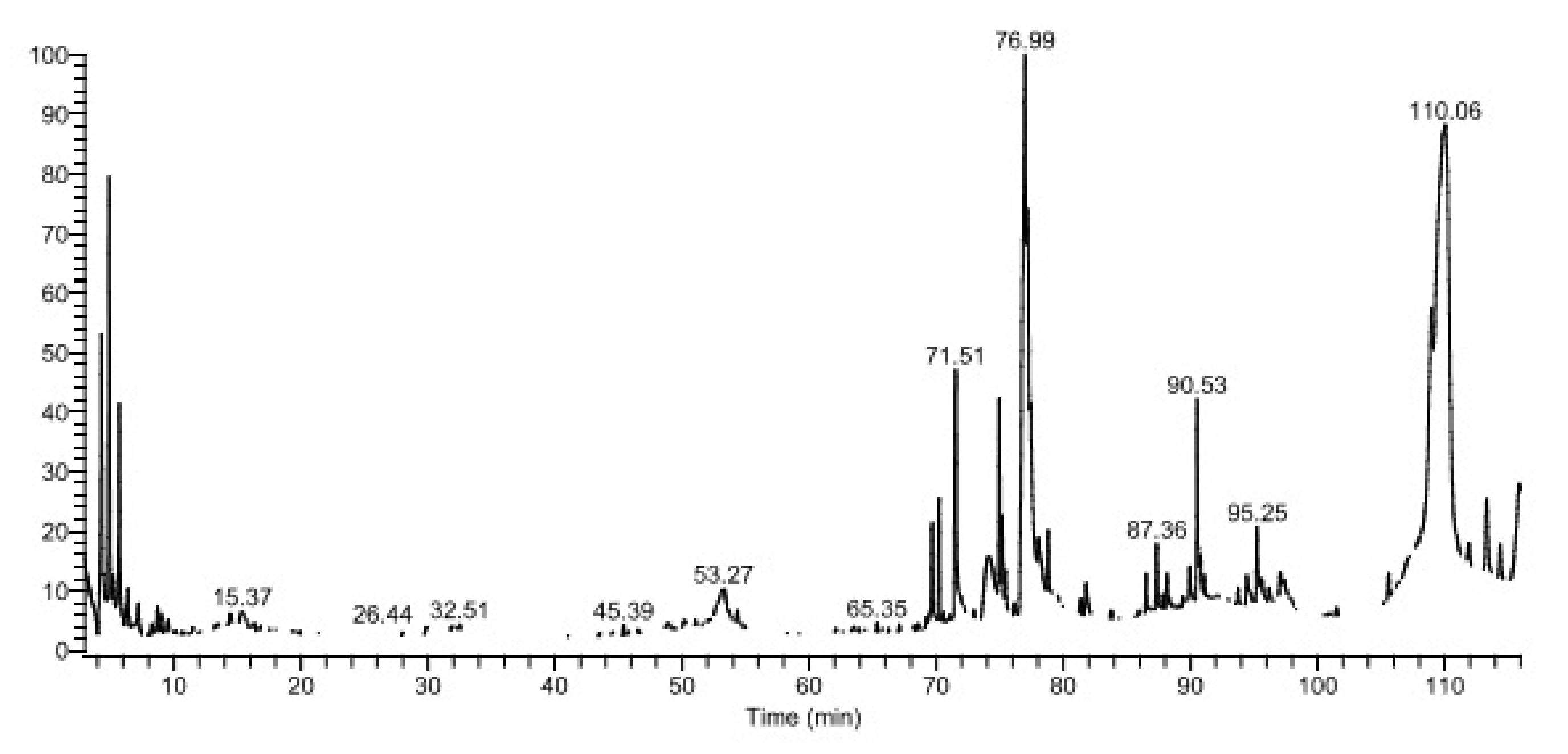
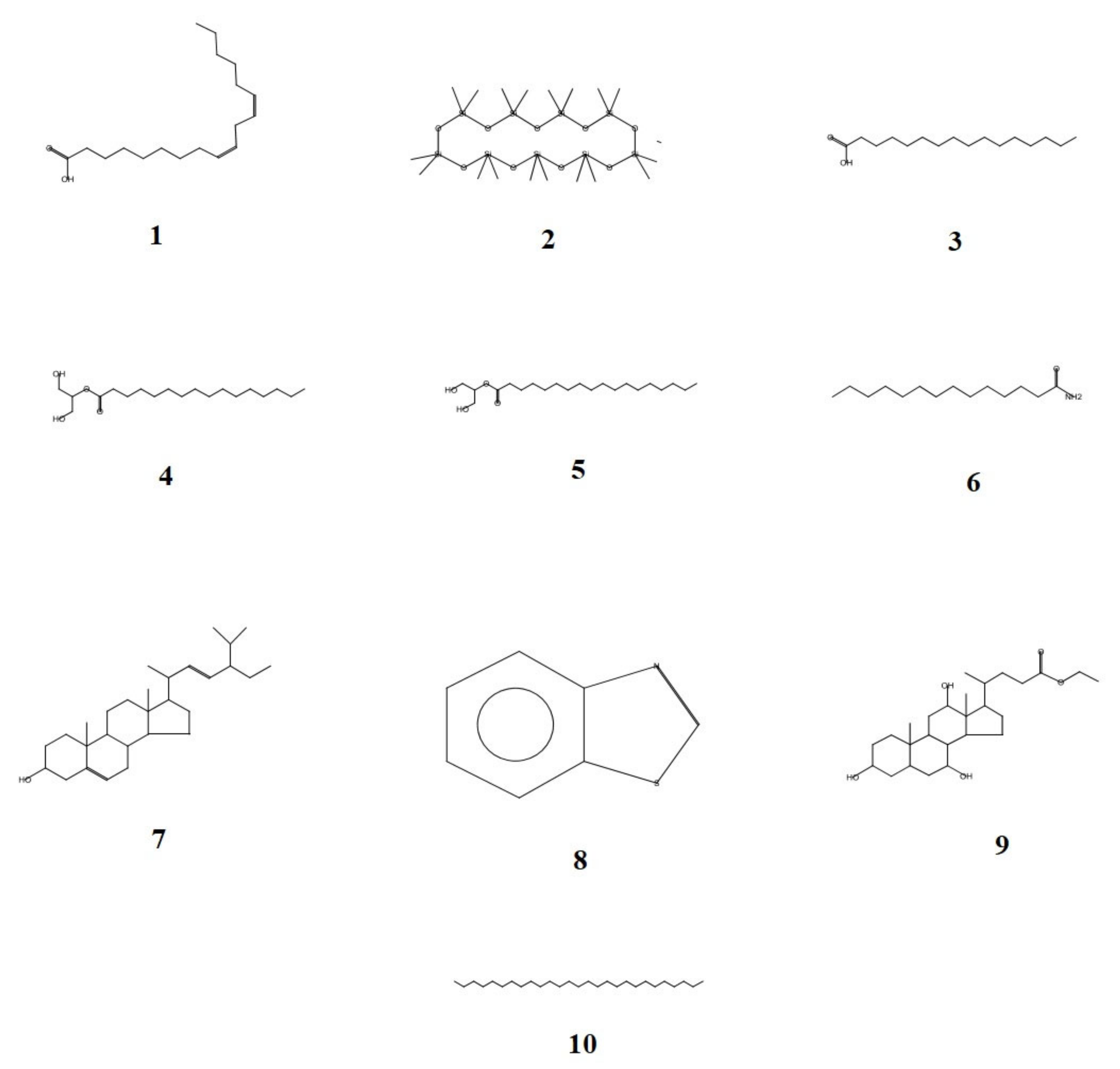
2.4. GC-MS Analysis
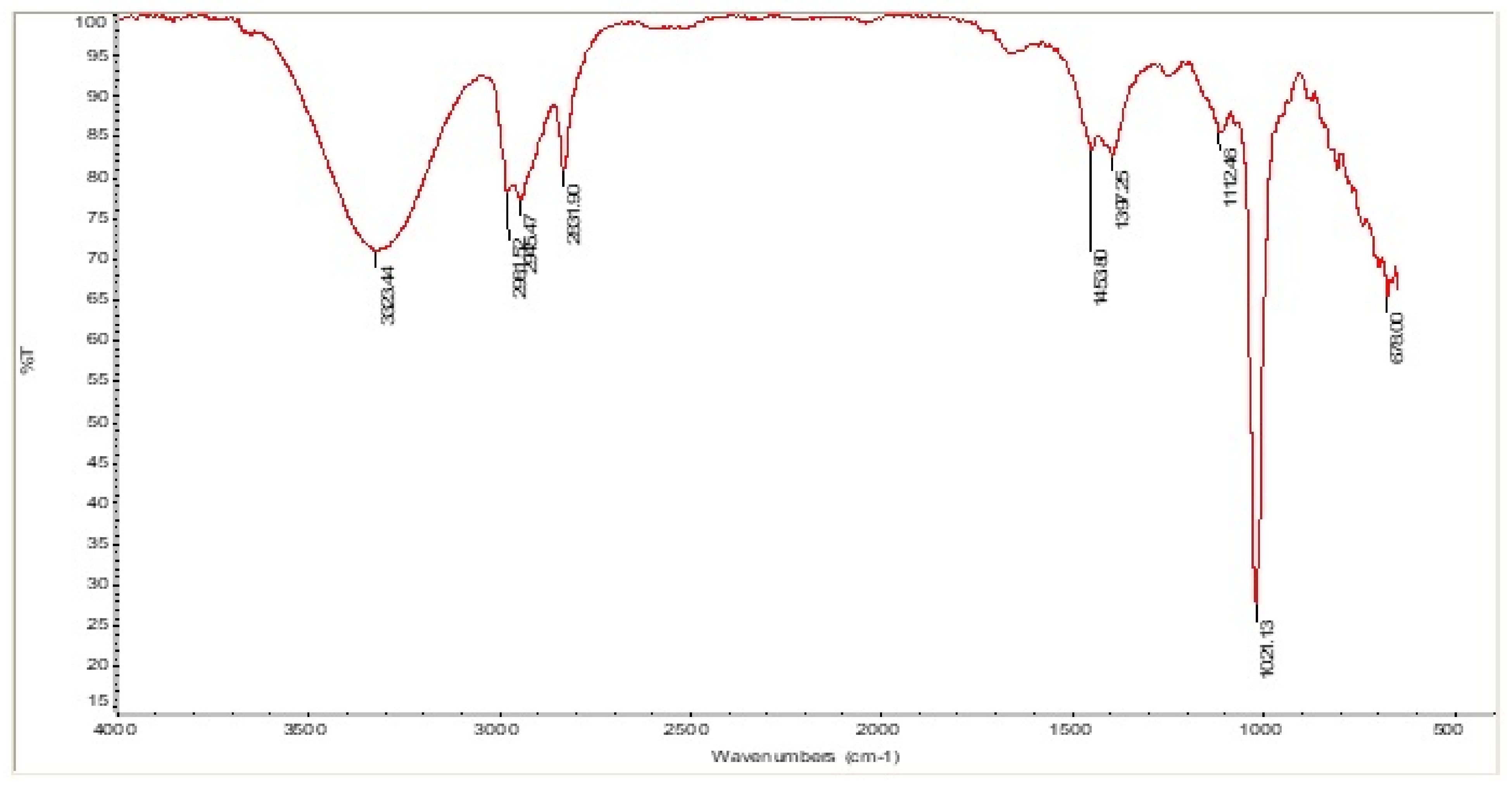
2.5. FT-IR Spectroscopy
2.6. In Vivo Study
2.6.1. Experimental Animals
2.6.2. Gastric Ulcer Model
2.6.3. Determination of Ulcer Index and % Inhibition of Ulcer [13]
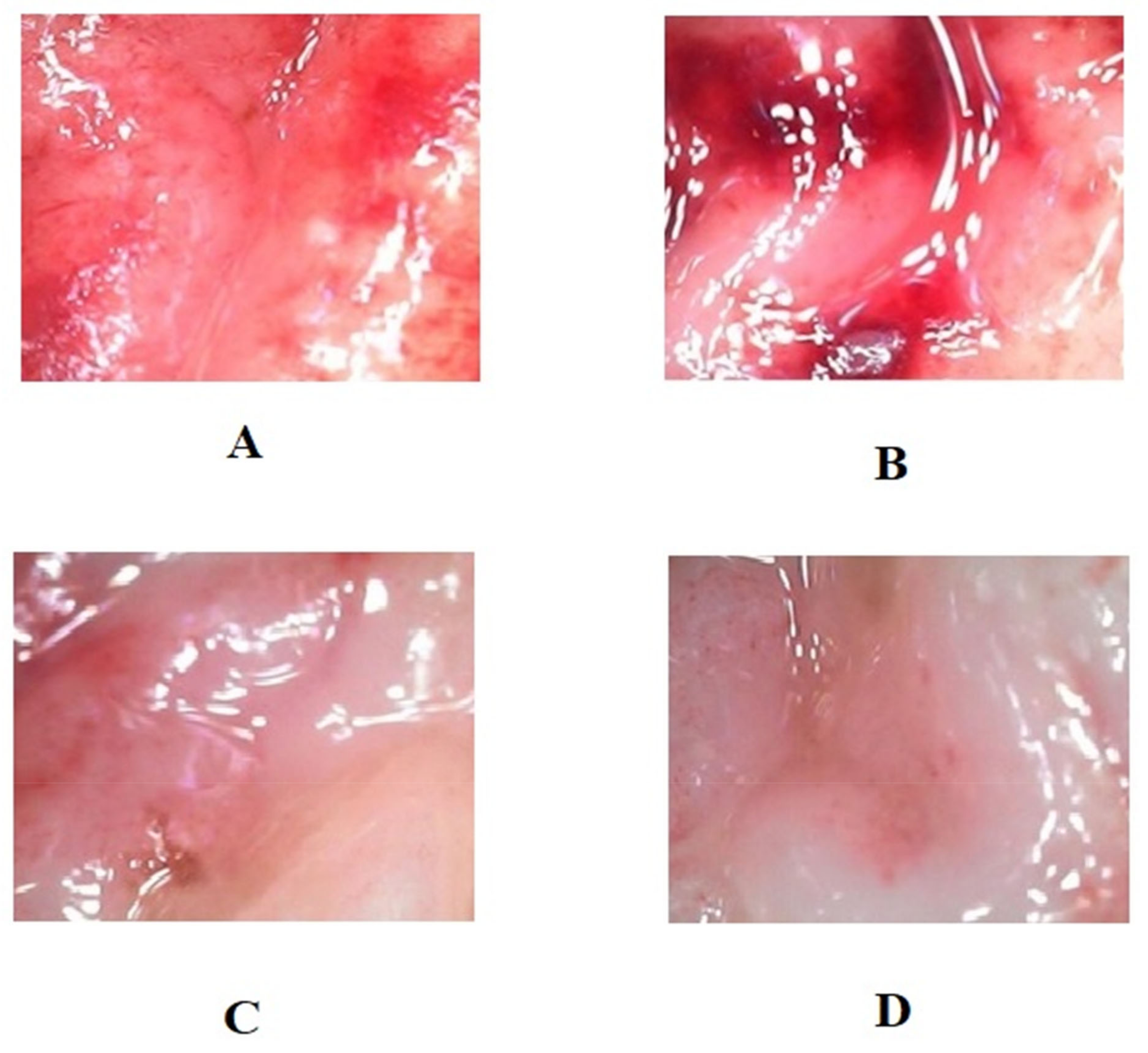
2.6.4. Macroscopic and Biochemical Gastric Assessments
2.6.5. Collection of Serum
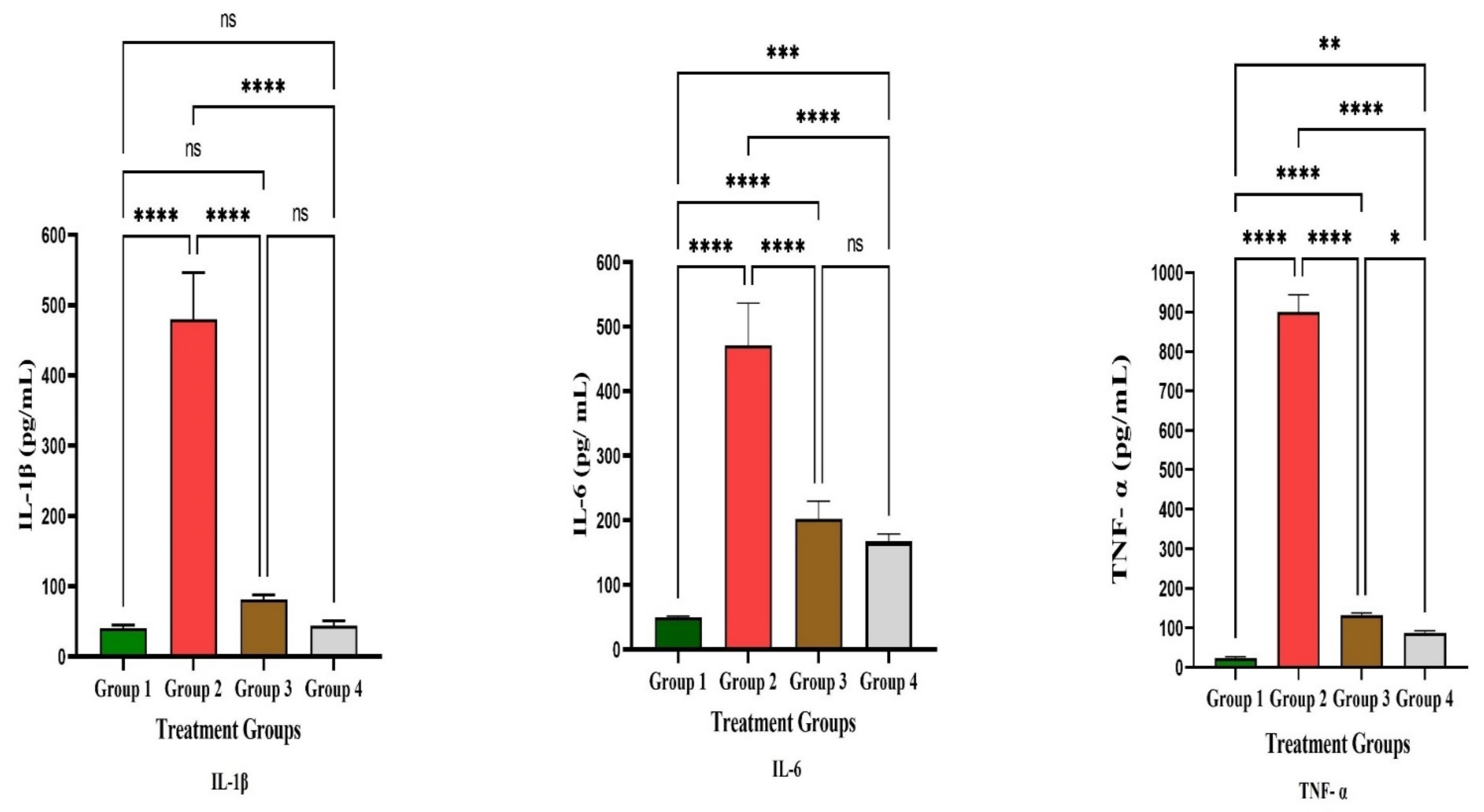
Serum IL-1β
Serum IL-6
Serum TNF-α
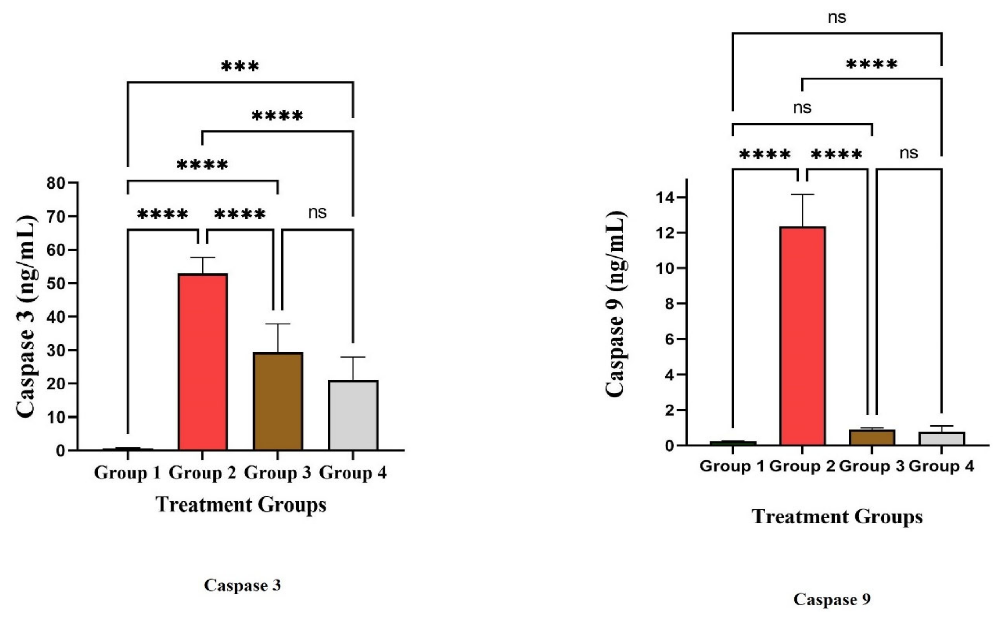
Caspase-3 and -9
2.7. Statistical Analysis
3. Results and Discussion
4. Conclusions
Author Contributions
Funding
Institutional Review Board Statement
Informed Consent Statement
Data Availability Statement
Acknowledgments
Conflicts of Interest
References
- Wallace, C.T.; Bailey, R.L.; Blumberg, J.B.; Freeman, B.B.; Oliver Chen, C.Y.; Crowe-White, K.M.; Drewnowski, A.; Hooshmand, S.; Johnson, E.; Lewis, R.; et al. Fruits, vegetables, and health: A comprehensive narrative, umbrella review of the science and recommendations for enhanced public policy to improve intake. Crit. Rev. Food Sci. Nutr. 2020, 60, 2174–2211. [Google Scholar] [CrossRef] [PubMed] [Green Version]
- Noumedem, J.A.K.; Djeussi, D.E.; Hritcu, L.; Mihasan, M.; Kuete, V. Chapter 20—Lactuca sativa. In Medicinal Spices and Vegetables from Africa; Victor, K., Ed.; Academic Press: Cambridge, MA, USA, 2017; pp. 437–449. [Google Scholar] [CrossRef]
- Kim, H.D.; Hong, K.B.; Dong, O.N.; Suh, H.J. Sleep-inducing effect of lettuce (Lactuca sativa) varieties on pentobarbital-induced sleep. Food Sci. Biotechnol. 2017, 26, 807–814. [Google Scholar] [CrossRef] [PubMed]
- Gopal, S.S.; Lakshmi, M.J.; Sharavana, G.; Sathaiah, G.; Sreerama, Y.N.; Baskaran, V. Lactucaxanthin—A potential anti-diabetic carotenoid from lettuce (Lactuca sativa) inhibits α-amylase and α-glucosidase activity in vitro and in diabetic rats. Food Funct. 2017, 8, 1124–1131. [Google Scholar] [CrossRef] [PubMed]
- Naseem, S.; Ismail, H. In vitro and in vivo evaluations of antioxidative, anti-Alzheimer, antidiabetic and anticancer potentials of hydroponically and soil grown Lactuca sativa. BMC Complement. Med. Ther. 2022, 22, 30. [Google Scholar] [CrossRef]
- Cheng, D.M.; Pogrebnyak, N.; Kuhn, P.; Krueger, C.G.; Johnson, W.D.; Raskin, I. Development and phytochemical characterization of high polyphenol red lettuce with anti-diabetic properties. PLoS ONE 2014, 9, e91571. [Google Scholar] [CrossRef] [Green Version]
- Hassan, I.A.; Basahi, J.M.; Ismail, I.M. Gas exchange, chlorophyll fluorescence and antioxidants as bioindicators of airborne heavy metal pollution in Jeddah, Saudi Arabia. Curr. World Environ. 2013, 8. [Google Scholar] [CrossRef]
- Sanpinit, S.; Chonsut, P.; Punsawad, C.; Wetchakul, P. Gastroprotective and Antioxidative Effects of the Traditional Thai Polyherbal Formula Phy-Blica-D against Ethanol-Induced Gastric Ulcers in Rats. Nutrients 2022, 14, 172. [Google Scholar] [CrossRef]
- Rahamat, U.S.; Sivakumar, S.M.; Raghad, H.A.; Rawan, H.A.; Nouf, F.A.; Khadijah, M.W.; Fayha, N.A.; Alshammari, M.H.; Fai, M.A.; ur Rehman, Z.; et al. Spectral characterization of the bioactive principles and antibacterial properties of a cold methanolic extract of Olea europaea from the Hail region of Saudi Arabia, Arab. J. Chem. 2022, 15, 104006. [Google Scholar] [CrossRef]
- Rahamat, U.S.; Sivakumar, S.M.; Bader, H.; Ahmed, A.; Afnan, A.A.; Amr, S.A.; Amr, S.A.L.; Marwa, H.A.; Humera, B.; Mohd, A.H.; et al. Bioactive principles, anti-diabetic, and anti-ulcer activities of Ducrosia anethifolia Boiss leaves from the Hail region, Saudi Arabia. Arab. J. Chem. 2022, 15, 104308. [Google Scholar] [CrossRef]
- Saad, S.A.; Sivakumar, S.M.; Muhammad, H.S.; Mohammed, A.B.; Osama, A.M.; Saeed, A.; Hafiz, A.M.; Santhosh, J.M.; ur Rehman, Z.; Alam, M.d.S.; et al. Potential bioactive secondary metabolites of Actinomycetes sp. isolated from rocky soils of the heritage village Rijal Alma, Saudi Arabia. Arab. J. Chem. 2022, 15, 103793. [Google Scholar] [CrossRef]
- Al-Wajeeh, N.S.; Hajerezaie, M.; Noor, S.M.; Halabi, M.F.; Al-Henhena, N.; Azizan, A.H.S.; Kamran, S.; Hassandarvish, A.N.S.; Ali, H.M. The gastro protective effects of cibotium barometz hair on ethanol-induced gastric ulcer in Sprague-Dawley rats. BMC Vet. Res. 2016, 13, 27. [Google Scholar] [CrossRef] [PubMed] [Green Version]
- Sabiu, S.; Garuba, T.; Sunmonu, T.; Emmanuel, A.; Abdulhakeem, S.; Ismaila, N.; Abdulazeez, B. Indomethacin-induced gastric ulceration in rats: Protective roles of Spondias mombin and Ficus exasperate. Toxicol. Rep. 2015, 2, 261–267. [Google Scholar] [CrossRef] [PubMed] [Green Version]
- Njar, V.C.; Adesanwo, J.K.; Raji, Y. Methyl angolensate: The antiulcer agent of the stem bark of Entandrophragma angolense. Planta Med. 1995, 61, 91–92. [Google Scholar] [CrossRef] [PubMed]
- Tan, P.V.; Nyasse, B.; Dimo, T.; Mezui, C. Gastric cytoprotective anti-ulcer effects of the leaf methanol extract of Ocimum suave (Lamiaceae) in rats. J. Ethnopharmacol. 2002, 82, 69–74. [Google Scholar] [CrossRef]
- Unissa, R.; Moni, S.S.; Banu, H.; Alrahef, S.S.; Alrahef, S.S.; AlenezI, T.K.; Abdallah, M.H.; Abu Lila, A.S.; EL-Horany, H.; Abouzied, A.S.; et al. Anti-ulcer properties, cytokines, and apoptosis regulatory effects of Olea europaea leaves from Hail Province, Saudi Arabia. Not. Bot. Horti Agrobot. Cluj-Napoca 2022, 50, 12891. [Google Scholar] [CrossRef]
- Ülger, T.G.; Ayşe, N.S.; Onur, C.; Funda, P.C. Chapter 2—Role of Vegetables in Human Nutrition and Disease Prevention. In Vegetables—Importance of Quality Vegetables to Human Health; Intech Open: London, UK, 2018; Available online: https://www.intechopen.com/chapters/61691 (accessed on 5 August 2021).
- Hanin, N.M.; Abdurazag, A.A. Lactuca sativa stems as the source of bioactive compounds as well as the leaves. J. Pharm. Pharmacol. 2020, 8, 143–150. [Google Scholar] [CrossRef]
- Emil, N.S.; Sara, V.; Marcello, I.; Stefania, G. Chemical characterization by GC/MS analysis of Lactuca tatarica (L.) C.A.Mey. aerial parts and seeds. Nat. Prod. Res. 2021, 1–5. [Google Scholar] [CrossRef]
- Chen, Z.Y.; Chan, P.T.; Kwan, K.Y.; Zhang, A. Reassessment of the antioxidant activity of conjugated linoleic acids. J. Amer. Oil. Chem. Soc. 1997, 74, 749–753. [Google Scholar] [CrossRef]
- Fagali, N.; Catalá, A. Antioxidant activity of conjugated linoleic acid isomers, linoleic acid and its methyl ester determined by photoemission and DPPH techniques. Biophys. Chem. 2008, 137, 56–62. [Google Scholar] [CrossRef]
- Den Hartigh, L.J. Conjugated linoleic acid effects on cancer, obesity, and atherosclerosis: A review of pre-clinical and human trials with current perspectives. Nutrients 2019, 11, 370. [Google Scholar] [CrossRef] [Green Version]
- Sina, N.; Dagfinn, A.; Joseph, B.; Sara, M.; Masoomeh, A.; Omid, S. Dietary intake and biomarkers of alpha linolenic acid and risk of all cause, cardiovascular, and cancer mortality: Systematic review and dose-response meta-analysis of cohort studies. BMJ 2011, 375, 2213. [Google Scholar] [CrossRef]
- Watanabe, Y.; Tatsuno, I. Omega-3 polyunsaturated fatty acids for cardiovascular diseases: Present, past and future. Expert Rev. Clin. Pharmacol. 2021, 10, 865–873. [Google Scholar] [CrossRef] [PubMed]
- Guasch-Ferré, M.; Li, Y.; Walter, C.W.; Qi, S.; Laura, S.; Salas-Salvadó, J.; Martínez-González, M.A.; Meir, J.S.; Hu, F.B. Consumption of olive oil and risk of total and cause-specific mortality among U.S. Adults. J. Am. Coll. Cardiol. 2022, 79, 101–112. [Google Scholar] [CrossRef] [PubMed]
- Yang, Q.; Cao, W.; Zhou, X.; Cao, W.; Xie, Y.; Wang, S. Anti-thrombotic effects of α-linolenic acid isolated from Zanthoxylum bungeanum maxim seeds. BMC Complement Altern. Med. 2014, 14, 348. [Google Scholar] [CrossRef] [Green Version]
- Brzosko, S.; De Curtis, A.; Murzilli, S.; de Gaetano, G.; Donati, M.B.; Iacoviello, L. Effect of extra virgin olive oil on experimental thrombosis and primary hemostasis in rats. Nutr. Metab. Cardiovasc. Dis. 2002, 12, 337–342. [Google Scholar]
- Ni Luh, S. Identification of the Substance Bioactive Leaf Extract Piper caninum Potential as Botanical Pesticides. Int. J. Pure App. Biosci. 2016, 4, 26–32. [Google Scholar]
- Aparna, V.; Dileep, K.V.; Mandal, P.K.; Karthe, P.; Sadasivan, C.; Haridas, M. Anti-inflammatory property of n-hexadecanoic acid: Structural evidence and kinetic assessment. Chem. Biol. Drug Des. 2012, 80, 434–439. [Google Scholar] [CrossRef] [PubMed]
- Bakar, K.; Mohamad, H.; Latip, J.; Tan, H.S.; Herng, G.M. Fatty acids compositions of Sargassum granuliferum and Dictyota dichotoma and their anti-fouling activities. J. Sustain. Sci. Manag. 2017, 12, 8–16. [Google Scholar]
- Bubenchikov, R.A.; Korableva, T.V.; Pozdnyakova, T.A.; Kuleshova, E.S. The study of the Fatty acid composition of compass Lettuce (Lactuca serriola L.). Res. J. Pharm. Technol. 2020, 13, 6105–6108. [Google Scholar] [CrossRef]
- Zhu, S.; Jiao, W.; Xu, X.; Hou, L.; Li, H.; Shao, J.; Zhang, X.; Wang, R.; Kong, D. Palmitic acid inhibits prostate cancer cell proliferation and metastasis by suppressing the PI3K/Akt pathway. Life Sci. 2021, 286, 120046. [Google Scholar] [CrossRef]
- Giancarlo, C.V.; Carlimar, O.M.; Solymar, M.; Christian, M.G.; René García, D.V.; Néstor, M.C.; Sanabria-Ríos, D.J. Antibacterial fatty acids: An update of possible mechanisms of action and implications in the development of the next-generation of antibacterial agents. Prog. Lipid Res. 2021, 82, 101093. [Google Scholar] [CrossRef]
- Havlicekova, Z.; Jesenak, M.; Banovcin, P.; Milan, K. Beta-palmitate—A natural component of human milk in supplemental milk formulas. Nutr. J. 2015, 15, 28. [Google Scholar] [CrossRef] [PubMed]
- Litmanovitz, I.; Bar-Yoseph, F.; Lifshitz, Y.; Davidson, K.; Eliakim, A.; Regev, H.; Nemet, D. Reduced crying in term infants fed high beta-palmitate formula: A double-blind randomized clinical trial. BMC Pediatr. 2014, 14, 152. [Google Scholar] [CrossRef] [Green Version]
- Glycerol Monostearate. Available online: https://www.chefsteps.com/ingredients/glycerol-monostearate (accessed on 5 August 2021).
- Gabay, O.; Sanchez, C.; Salvat, C.; Chevy, F.; Breton, M.; Nourissat, G.; Wolf, C.J.; Berenbaum, F. Stigmasterol: A phytosterol with potential anti-osteoarthritic properties. Osteoarthr. Cartil. 2010, 18, 106–116. [Google Scholar] [CrossRef] [PubMed]
- Tovey, F.I.; Capanoglu, D.; Langley, G.J.; Herniman, J.M.; Bor, S.; Ozutemiz, O.; Hobsley, M.; Bardhan, K.D.; Linclau, B. Dietary phytosterols protective against peptic ulceration. Gastroenterol. Res. 2011, 4, 149–156. [Google Scholar] [CrossRef] [Green Version]
- Shokraei, S.; Khandouzi, N.; Sina, Z.; Nasrollahzadeh, J. The acute effect of incorporating lettuce or watercress into a moderately high-fat meal on postprandial lipid, glycemic response, and plasma inflammatory cytokines in healthy young men: A randomized crossover trial. Lipids Health Dis. 2021, 20, 66. [Google Scholar] [CrossRef]


| S. No | Compound Name | Molecular Formula | Molecular Weight | Retention Time (Min) | Probability Index | Percent Area of Curve |
|---|---|---|---|---|---|---|
| 1 | 9,12-Octadecadienoic acid (Z,Z)- | C18H32O2 | 280 | 76.9 | 55.92 | 16.48 |
| 2 | Cyclononasiloxane, octadecamethyl- | C18H54O9Si9 | 666 | 110.08 | 22.90 | 23.76 |
| 3 | n-Hexadecanoic acid | C16H32O2 | 256 | 71.51 | 72.04 | 3.76 |
| 4 | Hexadecanoic acid, 2- hydroxy-1- (hydroxymethyl)ethyl | C19H38O4 | 330 | 90.53 | 61.55 | 2.69 |
| 5 | Octadecanoic acid, 2- hydroxy-1- (hydroxymethyl)ethyl ester | C21H42O4 | 358 | 95.25 | 63.34 | 1.37 |
| 6 | Octadecanamide | C18H37NO | 283 | 87.36 | 87 | 0.78 |
| 7 | Stigmasterol | C29H48O | 412 | 111.90 | 71.04 | 0.38 |
| 8 | Benzothiazole | C7H5NS | 135 | 34.31 | 8.06 | 0.2 |
| 9 | Ethyl iso-allocholate | C26H44O5 | 436 | 76.13 | 7.51 | 0.15 |
| 10 | Octacosane | C28H58 | 394 | 84.39 | 5.01 | 0.3 |
| Groups | Treatment | Ulcer Area (mm2) | % of Inhibition | Mucus Weight | pH |
|---|---|---|---|---|---|
| 1 | Normal control | 0.00 | 0.00 | 2.8 ± 0.11 | 3.61 ± 0.09 |
| 2 | Ulcer Control | 611 ± 32 | NA | 0.95 ± 0.2 | 3.61 ± 0.21 |
| 3 | Omeprazole | 90 ± 3.5 | 85 ± 1.9 | 1.45 ± 0.3 | 6.54 ± 0.09 |
| 4 | LPL | 90.5 ± 10.58 | 85.18 ± 2.71 | 3.14 ± 0.28 | 7.01 ± 0.34 |
Publisher’s Note: MDPI stays neutral with regard to jurisdictional claims in published maps and institutional affiliations. |
© 2022 by the authors. Licensee MDPI, Basel, Switzerland. This article is an open access article distributed under the terms and conditions of the Creative Commons Attribution (CC BY) license (https://creativecommons.org/licenses/by/4.0/).
Share and Cite
Syed, R.U.; Moni, S.S.; Lila, A.S.A.; Abdallah, M.H.; Abouzied, A.S.; Banu, H.; Alreshidi, K.S.M.; Alrashidi, B.M.W.; Hadi, M.A.; El-Horany, H.; et al. Spectral Analysis and Antiulcer Potential of Lactuca sativa through the Amelioration of Proinflammatory Cytokines and Apoptosis Markers. Life 2022, 12, 1641. https://doi.org/10.3390/life12101641
Syed RU, Moni SS, Lila ASA, Abdallah MH, Abouzied AS, Banu H, Alreshidi KSM, Alrashidi BMW, Hadi MA, El-Horany H, et al. Spectral Analysis and Antiulcer Potential of Lactuca sativa through the Amelioration of Proinflammatory Cytokines and Apoptosis Markers. Life. 2022; 12(10):1641. https://doi.org/10.3390/life12101641
Chicago/Turabian StyleSyed, Rahamat Unissa, Sivakumar S. Moni, Amr S. Abu Lila, Marwa H. Abdallah, Amr S. Abouzied, Humera Banu, Khetam Saad Mutni Alreshidi, Badriah Mansour Wadid Alrashidi, Mohd Abdul Hadi, Hemat El-Horany, and et al. 2022. "Spectral Analysis and Antiulcer Potential of Lactuca sativa through the Amelioration of Proinflammatory Cytokines and Apoptosis Markers" Life 12, no. 10: 1641. https://doi.org/10.3390/life12101641