Communication

Autologous Matrix Induced Chondrogenesis (AMIC) Compared to Microfractures for Chondral Defects of the Talar Shoulder: A Five-Year Follow-Up Prospective Cohort Study

1
Department of Orthopedics and Trauma Surgery, University Clinic Aachen, RWTH Aachen University Clinic, 52064 Aachen, Germany
2
School of Pharmacy and Bioengineering, Keele University School of Medicine, Staffordshire ST4 7QB, UK
3
Barts and the London School of Medicine and Dentistry, London E1 2AD, UK
4
Centre for Sports and Exercise Medicine, Queen Mary University of London, Mile End Hospital, London E1 4DG, UK
5
Department of Orthopedics, Klinikum Wels-Grieskirchen, A-4600 Wels, Austria
6
Department of Medicine, Surgery and Dentistry, University of Salerno, 84081 Baronissi, Italy
*
Author to whom correspondence should be addressed.
Life 2021, 11(3), 244; https://doi.org/10.3390/life11030244
Submission received: 2 February 2021 / Revised: 9 March 2021 / Accepted: 15 March 2021 / Published: 16 March 2021
(This article belongs to the Special Issue Focal Chondral Defects)

Abstract

:
Introduction: Many procedures are available to manage cartilage defects of the talus, including microfracturing (MFx) and Autologous Matrix Induced Chondrogenesis (AMIC). Whether AMIC or MFx are equivalent for borderline sized defects of the talar shoulder is unclear. Thus, the present study compared the efficacy of primary isolated AMIC versus MFx for borderline sized focal unipolar chondral defects of the talar shoulder at midterm follow-up. Methods: Patients undergoing primary isolated AMIC or MFx for focal unipolar borderline sized chondral defects of the talar shoulder were recruited prospectively. For those patients who underwent AMIC, a type I/III collagen resorbable membrane was used. The outcomes of interest were: Visual Analogic Scale (VAS), Tegner Activity Scale, American Orthopedic Foot and Ankle Score (AOFAS). The Magnetic Resonance Observation of Cartilage Repair Tissue (MOCART) was assessed by a blinded radiologist, who had not been involved in the clinical management of the patients. Data concerning complication rate and additional procedures were also collected. Results: The mean follow-up was 43.5 months. The mean age of the 70 patients at operation was 32.0 years, with a mean defect size of 2.7 cm2. The mean length of hospitalization was shorter in the MFx cohort (p = 0.01). No difference was found between the two cohorts in terms of length of prior surgery symptoms and follow-up, mean age and BMI, sex and side, and defect size. At a mean follow-up of 43.5 months, the AOFAS (p = 0.03), VAS (p = 0.003), and Tegner (p = 0.01) scores were greater in the AMIC group. No difference was found in the MOCART score (p = 0.08). The AMIC group evidenced lower rates of reoperation (p = 0.008) and failure (p = 0.003). Conclusion: At midterm follow-up, AMIC provides better results compared to MFx.

1. Introduction

Focal chondral defects of the talar shoulder are common [1,2]. Given the limited self-healing capability of cartilage, chondral defects are debilitating, often requiring surgical management [3,4,5]. Isolated microfractures (MFx) are recommended for defects smaller than 2.5 cm2 [6,7,8,9,10]. For bigger defects, several different surgical techniques have been described [11,12,13,14]. Osteochondral allo- or autograft transplantation (OAT) has been extensively performed as management of such chondral defects [15,16,17]. While autografts require a harvest site, allografts are expensive and have greater risk of failure [18,19]. Autologous chondrocyte implantation (ACI) has been widely used to address talar chondral defects [20,21]. ACI is a two sessions surgery which requires the harvest of cells and external chondrocytes expansion [22,23]. Autologous Matrix-Induced Chondrogenesis (AMIC) has been recently introduced [2,24]. AMIC does not require a harvest site, cell expansion, and is performed in a single surgical session [25,26]. AMIC is combined with MFx covering the lesions with a resorbable membrane to stabilize the resulting blood clot [27,28]. Thus, AMIC exploits the regenerative potential of bone marrow-derived mesenchymal stem cells arising from the subchondral bone [25,29].
Whether AMIC performed better than MFx for borderline sized defects (2.2 to 2.8 cm2) of the talar shoulder is unclear. The present study compared the efficacy of primary isolated AMIC versus MFx for borderline sized focal unipolar chondral defects of the talar shoulder at midterm follow-up. We hypothesized that AMIC provides better outcomes compared to MFx.

2. Material and Methods

2.1. Patients Recruitment

The present study was performed according to Strengthening the Reporting of Observational Studies in Epidemiology: the STROBE Statement [30]. In our setting, for patients with defect sized 2 to 3 cm2, both AMIC or isolated MFx were routinely performed. From 2012, patients undergoing primary isolated AMIC or MFx for focal unipolar borderline sized chondral defects of the talar shoulder (Figure 1), were recruited and followed-up prospectively. The inclusion criteria were: (1) symptomatic chondral defect of the talar shoulder, (2) single focal defect sized 2 to 3 cm2, (3) MRI evidence, (4) patients able to understand the nature of the treatment and the study. The exclusion criteria were: (1) kissing lesions, (2) bilateral lesions, (3) multifocal lesions, (4) previous ankle surgeries, (5) any bone disease, (6) any skeletal malformation, (7) any other relevant pathology that could have influenced the study. Suitable patients were informed about the pros and cons of both techniques, and were left free to decide their own procedure. The present study was approved and registered by the ethic committee of the RWTH University of Aachen (project ID EK 438-20), and conducted according to the principles expressed in the Declaration of Helsinki. All patients were able to understand the nature of their treatment and provided written consent to use their clinical and imaging data for research purposes.

2.2. Surgical Technique

All the surgeries were performed by three surgeons (BR, MT, AD) in a highly standardized fashion. All the surgeons were well behind their learning curve and had no preference on the surgical procedure. Briefly, the ankle was plantar flexed and a 2 mm K-wire was drilled in the distal tibia and another one in the talus. A Hintermann spreader (Integra LifeSciences, Plainsboro, NJ, USA) was used for joint distraction. Lesions were arthroscopically approached through standard anterolateral and anteromedial portals, according to the defect location shown on MRI. After identification of the defect, debridement and curettage of the non-viable border of the chondral tissue surrounding the lesion was performed until viable shoulder cartilage was reached. At this stage, the two surgical techniques take two different turns. In the MFx group, microfractures of 4 mm depth were arthroscopically performed into the defect. In the AMIC group, an arthroscopically-assisted mini-arthrotomy approach was used. A malleolar osteotomy was performed if the defect was not accessible by simple mini-arthrotomy. Microfractures of 4 mm depth were performed into the defect using a 1.2- or 1.4-mm Kirschner wire under constant irrigation with normosaline. If subchondral bone was not viable, this was debrided and substituted with autologous cancellous bone graft harvested from the osteotomy site or from the ipsilateral iliac crest. An aluminum template was trimmed according to the defect. A resorbable porcine type I/III collagen was used in all patients (Chondro-Gide®, Geistlich Pharma AG, Wolhusen, Switzerland). The membrane was trimmed according to the aluminum template to be slightly undersized in relation to the defect to avoid displacement, and hydrated in a saline solution. The membrane was placed into the lesion and secured with fibrin glue. The stability of the membrane was checked by flexing and extending the ankle. When an osteotomy was performed, it was fixed with two malleolar screws inserted through the predrilled holes and the wound sutured in a standard fashion. The rehabilitation protocol was performed according to that previously published [25].

2.3. Outcomes of Interest

Prior to surgery, the following data were recorded: age, gender, side, area of defect, additional autologous spongiosa transplantation, BMI (Kg/m2), score, symptoms duration, prior surgery, and length of the hospital stay. At the last follow-up, patients underwent an MRI and subsequently were invited to answer the following questionnaires: Visual Analogic Scale (VAS), Tegner Activity Scale, American Orthopedic Foot and Ankle Score (AOFAS). The Magnetic Resonance Observation of Cartilage Repair Tissue (MOCART) was assessed by a blinded radiologist, who had not been involved in the clinical management of the patients. Data concerning complications (failure, revision, arthroplasty, delamination, hypertrophy) and additional procedures were also collected. Failure was defined as persistent pain that affected negatively the quality of life and limited participation in recreational activities. For those patients who underwent a malleolar osteotomy, the occurrence of screw removal was not considered as revision surgery.
The primary outcome of interest was to compare the outcomes at the last follow-up between the AMIC and MFx group. The secondary outcome of interests was to compare within AMIC the location of the lesion (medial vs. lateral), the effect of the bone grafting (bone graft vs. no bone graft), and the approach (distraction vs. osteotomy).

2.4. Statistical Analysis

All statistical analyses were performed using the software IBM SPSS version 25. Continuous data were analyzed using the mean difference (MD), while for dichotomic data, the odd ratio (OR) effect measures. The confidence interval was set at 95% in all the comparisons. The T-test and x2 tests were performed, respectively, with values of p < 0.05 considered statistically significant. The confidence interval (CI) was set at 95% in all comparisons.

3. Results

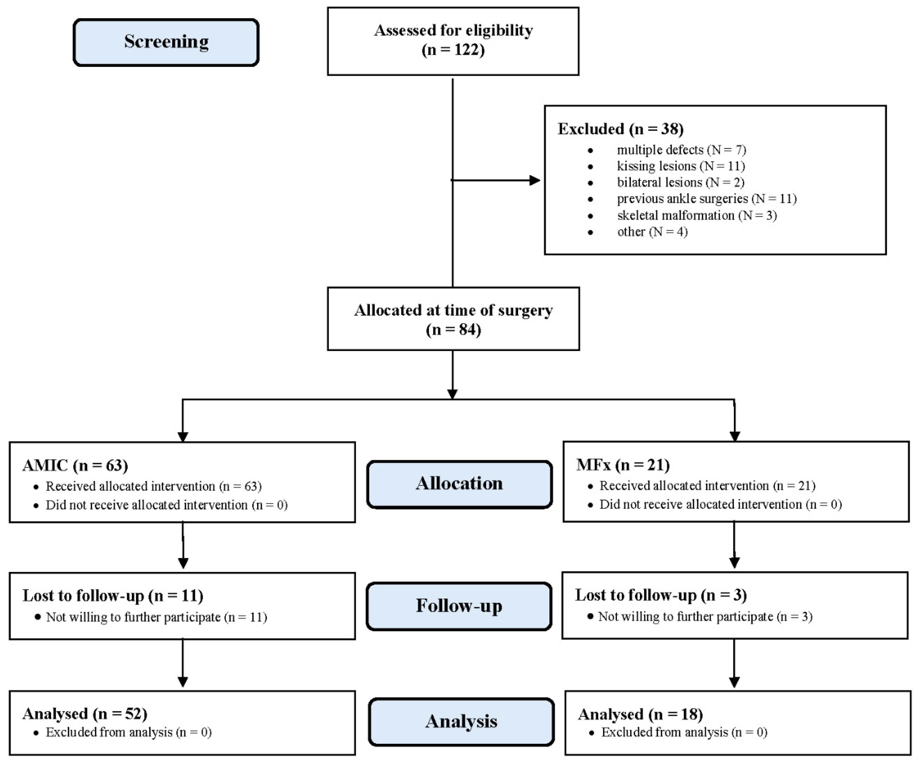
3.1. Recruitment Procedure

A total of 122 patients were initially evaluated. Of them, 38 were not eligible: multiple defects (N = 7), kissing lesions (N = 11), bilateral lesions (N = 2), previous ankle surgeries (N = 11), skeletal malformation (N = 3), other (N = 4). This left 84 patients: 63 AMIC and 21 MFx. A further 14 patients were lost to follow-up: 11 patients in the AMIC group, and 3 in the MFx group did not wish to further participate in the study for geographical reasons, but they declared themselves satisfied on telephone interviews. Eventually, 70 patients took part in the present study: 52 in the AMIC group and 18 in the MFx (Figure 2).

3.2. Patients Demographics

The mean follow-up was 43.5 months. The mean age of the 70 patients on admission was 32.0 years, with a mean defect size of 2.7 cm2, 44% (31 of 70 patients) were women, and in 53% (37 of 70) of patients the right side was involved. The mean symptoms duration before surgery was 45.8 months. The mean length of hospitalization was shorter in the MFx cohort (p = 0.01). No other difference was found between the two cohorts in terms of length of prior surgery symptoms and follow-up, mean age and BMI, sex and side, and defect size (Table 1).

3.3. Outcomes of Interest

At a mean follow-up of 43.1 months, the AOFAS (p = 0.03), VAS (p = 0.003), and Tegner (p = 0.01) scores were greater in the AMIC group. No difference was found in the MOCART score (p = 0.08) (Table 2).

3.4. Complications

The AMIC group experienced lower rates of reoperation (p = 0.008) and failure (p = 0.003). No hypertrophy or delamination were observed during follow-up (Table 3). Five patients (10%) underwent revision surgery for persistent pain within four years postoperatively in the AMIC group, seven (39%) in the MFx group. No complications related to the bone harvest site were experienced by any patient.

3.5. Subgroup Analysis

Within AMIC, no difference was evidenced by the subgroup analysis medial vs. lateral lesions (p = 0.08), bone grafting vs. no-bone grafting (p = 0.09), and between joint distractors vs. malleolar osteotomy (p = 0.07).

4. Discussion

According to the main findings of the present study, AMIC performed better than MFx at midterm follow-up. AOFAS and VAS both scored better in the AMIC group, along with a greater sporting activity, according to the Tegner score. The rate of reoperation and failure was also lower in the AMIC group at midterm follow-up. No complications related to the harvest site were reported. Within the AMIC group, no difference was evidenced by the subgroup analyses medial vs. lateral lesions, bone grafting vs. no-bone grafting, and joint distractors vs. malleolar osteotomy.
Symptomatic chondral defects negatively affect the activities of daily life and sporting activity level [25,31]. The low metabolic activity of hyaline cartilage, along with its alymphatic and hypocellular structure, are some of the reasons behind its poor regenerative capabilities [3,4,5]. Cartilage healing typically does not restore the original tissue, and fibrosis and residual chondral defects are common [32,33]. This impairs the cartilage biomechanical proprieties and predisposes to chronic pain [34,35]. AMIC has been successfully used to address chondral defects of the talus, with a growing trend of clinical studies in the current literature [36,37,38].
Only the study by Becher et al. [39] compared primary AMIC (N = 16) versus MFx (N = 16) for talar defects in a retrospective fashion; both techniques produced similar outcomes at five years follow-up. However, their patients had a mean defect smaller than 2 cm2 [39]. Chung et al. [40] compared AMIC versus MFx in 64 patients for knee chondral defects. They included patients with a mean defect size of 1.3 cm2 in the AMIC group and 1.5 cm2 in the MFx group, evidencing similarity between the two groups at two years follow-up. Both these studies were performed in a cohort of patients with small defects. Indeed, for small defects, MFx is still the most appropriate procedure [3,4,5,9]. Furthermore, the relatively short length of the follow-up may also affect the results. In the present study, we included only patients with borderline-sized defect (mean 2.7 cm2). We were unable to identify further studies that compare AMIC vs. MFx for talus chondral defects of this size. A similar study on chondral defects of the knee was performed by Volz et al. [41]. They compared AMIC versus MFx at five years in 47 patients with a mean defect size of 3.6 cm2. Similarly, they found a significant greater value of the Cincinnati score and lower pain level in the AMIC cohort.
We used plantar flexion and Hintermann spreader to distract the tibiotalar joint as standard, whereas, for lesions placed dorsally, a malleolar osteotomy was performed. Malleolar osteotomy leads to possible bony complications, intraoperative cartilage damage, loss of osteotomy reduction, delayed union or nonunion, persistent pain and/or swelling at the osteotomy site, painful hardware requiring surgical revision [42,43]. The osteotomy might damage the articular facet, and, given the poor regenerative capabilities of hyaline cartilage, this may lead to pain and early osteoarthrosis of the tibiotalar joint. Plantar flexion and Hintermann spreader to distract the tibiotalar allows patients to faster full bearing and recovery, but it may predispose to soft tissue damage, especially the neurovascular structures [44]. Indeed, in ankle arthroscopy series with mechanical distraction, the average complication rate ranges between 8 and 17% [45,46,47,48,49,50], while in a series of 1305 consecutive procedures with only plantar flexion the rate of complication was 3.4% [51]. In a cadaveric study, de Leeuw et al. [52] demonstrated greater distance between the anterior distal tibia and the overlying anterior neurovascular bundle with the ankle in a dorsiflexed position compared to the distracted ankle position. Whether plantar flexion and Hintermann spreader perform better than malleolar osteotomy is still controversial, and future studies are required.
The relatively small number of patients included in the present investigation represents the most important limitation of the present study, and may affect the ability to identify uncommon complications. The unblinded design, along with the lack of randomization, are two important limitations of the study. Further larger randomized controlled trials are required. We emphasize, however, that randomization and blinding in surgery may not be easily acceptable to patients and surgeons alike. The MOCART score was used to assess the degree of cartilage regeneration [53], but MRI is not reliable in predicting clinical outcome after cartilage repair is uncertain [54,55,56]. During the time elapsed between the onset of symptoms and the index surgery, all the patients underwent conservative management. However, given the heterogeneous nature and/or the lack of documentation of these treatments, it was not possible to analyze them separately. Similarly, most of the 15 patients who underwent revision surgeries were not aware of the treatment and/or data were lacking; thus, no further analyses were possible.

5. Conclusions

The present study confirmed our hypothesis that AMIC demonstrated superiority over MFx in focal osteochondral lesions of the talus between 2 and 3 cm2. At midterm follow-up, AOFAS and VAS scores were both better in the AMIC group, along with a greater sporting activity, according to the Tegner score. The rate of reoperation and failure was also lower in the AMIC group.

Author Contributions

Conceptualization, F.M.; methodology, F.M.; software, F.M.; Validation, N.M., M.T., and B.R.; formal analysis, F.M.; resources, F.M.; data curation, F.M.; writing—original draft preparation, F.M.; writing—review & editing, N.M., B.R. and M.T.; visualization, F.M., N.M., B.R., M.T., A.D., J.E. and H.S.; supervision, M.T.; project administration, J.E. All authors have read and agreed to the published version of the manuscript.

Funding

This research was supported by research grants from the German Federal Ministry of Education and Research (BMBF, workHEALTH, FZK 01EC1905A). The sponsor had no role in the study design or the writing of the manuscript, or the decision to submit the manuscript for publication.

Institutional Review Board Statement

The present study was approved and registered by the ethic committee of the RWTH University of Aachen (project ID EK 438-20).

Informed Consent Statement

All patients provided written consent to use their clinical and imaging data for research purposes.

Data Availability Statement

Data is contained within the article.

Acknowledgments

We would like to thank Nadja Sippel for her contribution in this study.

Conflicts of Interest

The authors declare that they have no conflict of interest.

References

  1. Årøen, A.; Løken, S.B.; Heir, S.; Alvik, E.; Ekeland, A.; Granlund, O.G.; Engebretsen, L. Articular Cartilage Lesions in 993 Consecutive Knee Arthroscopies. Am. J. Sports Med. 2004, 32, 211–215. [Google Scholar] [CrossRef] [Green Version]
  2. Migliorini, F.; Berton, A.; Salvatore, G.; Candela, V.; Khan, W.; Longo, U.G.; Denaro, V. Autologous Chondrocyte Implan-tation and Mesenchymal Stem Cells for the Treatments of Chondral Defects of the Knee—A Systematic Review. Curr. Stem Cell Res. Ther. 2020, 15, 547–556. [Google Scholar] [CrossRef]
  3. Kreuz, P.; Steinwachs, M.; Erggelet, C.; Krause, S.; Konrad, G.; Uhl, M.; Südkamp, N. Results after microfracture of full-thickness chondral defects in different compartments in the knee. Osteoarthr. Cartil. 2006, 14, 1119–1125. [Google Scholar] [CrossRef] [Green Version]
  4. Scillia, A.J.; Aune, K.T.; Andrachuk, J.S.; Cain, E.L.; Dugas, J.R.; Fleisig, G.S.; Andrews, J.R. Return to Play After Chondroplasty of the Knee in National Football League Athletes. Am. J. Sports Med. 2015, 43, 663–668. [Google Scholar] [CrossRef] [PubMed]
  5. Castrodad, I.M.D.; Mease, S.J.; Werheim, E.; McInerney, V.K.; Scillia, A.J. Arthroscopic Chondral Defect Repair with Extracellular Matrix Scaffold and Bone Marrow Aspirate Concentrate. Arthrosc. Tech. 2020, 9, e1241–e1247. [Google Scholar] [CrossRef]
  6. Gudas, R.; Kalesinskas, R.J.; Kimtys, V.; Stankevičius, E.; Toliušis, V.; Bernotavičius, G.; Smailys, A. A Prospective Randomized Clinical Study of Mosaic Osteochondral Autologous Transplantation Versus Microfracture for the Treatment of Osteochondral Defects in the Knee Joint in Young Athletes. Arthrosc. J. Arthrosc. Relat. Surg. 2005, 21, 1066–1075. [Google Scholar] [CrossRef]
  7. Richter, D.L.; Schenck, J.R.C., Jr.; Wascher, D.C.; Treme, G. Knee Articular Cartilage Repair and Restoration Techniques: A Review of the Literature. Sports Health 2016, 8, 153–160. [Google Scholar] [CrossRef] [Green Version]
  8. Bertho, P.; Pauvert, A.; Pouderoux, T.; Robert, H. Treatment of large deep osteochondritis lesions of the knee by autologous matrix-induced chondrogenesis (AMIC): Preliminary results in 13 patients. Orthop. Traumatol. Surg. Res. 2018, 104, 695–700. [Google Scholar] [CrossRef]
  9. Smith, G.D.; Knutsen, G.; Richardson, J.B. A clinical review of cartilage repair techniques. J. Bone Jt. Surgery. Br. Vol. 2005, 87, 445–449. [Google Scholar] [CrossRef]
  10. Steinwachs, M.; Guggi, T.; Kreuz, P. Marrow stimulation techniques. Injury 2008, 39 (Suppl. 1), 26–31. [Google Scholar] [CrossRef]
  11. Aae, T.F.; Randsborg, P.-H.; Lurås, H.; Årøen, A.; Lian, Ø.B. Microfracture is more cost-effective than autologous chondrocyte implantation: A review of level 1 and level 2 studies with 5 year follow-up. Knee Surg. Sports Traumatol. Arthrosc. 2017, 26, 1044–1052. [Google Scholar] [CrossRef] [Green Version]
  12. El-Rashidy, H.; Villacis, D.; Omar, I.; Kelikian, A.S. Fresh Osteochondral Allograft for the Treatment of Cartilage Defects of the Talus: A Retrospective Review. J. Bone Jt. Surg. Am. Vol. 2011, 93, 1634–1640. [Google Scholar] [CrossRef]
  13. Behrens, P.; Bitter, T.; Kurz, B.; Russlies, M. Matrix-associated autologous chondrocyte transplantation/implantation (MACT/MACI)—5-year follow-up. Knee 2006, 13, 194–202. [Google Scholar] [CrossRef] [PubMed]
  14. Migliorini, F.; Eschweiler, J.; Maffulli, N.; Schenker, H.; Baroncini, A.; Tingart, M.; Rath, B. Autologous Matrix-Induced Chondrogenesis (AMIC) and Microfractures for Focal Chondral Defects of the Knee: A Medium-Term Comparative Study. Life 2021, 11, 183. [Google Scholar] [CrossRef]
  15. Sabaghzadeh, A.; Mirzaee, F.; Rad, H.S.; Bahramian, F.; Alidousti, A.; Aslani, H. Osteochondral autograft transfer (mosaicplasty) for treatment of patients with osteochondral lesions of talus. Chin. J. Traumatol. 2020, 23, 60–62. [Google Scholar] [CrossRef] [PubMed]
  16. Park, C.H.; Song, K.-S.; Kim, J.R.; Lee, S.-W. Retrospective evaluation of outcomes of bone peg fixation for osteochondral lesion of the talus. Bone Jt. J. 2020, 102-B, 1349–1353. [Google Scholar] [CrossRef]
  17. Weigelt, L.; Hartmann, R.; Pfirrmann, C.; Espinosa, N.; Wirth, S.H. Autologous Matrix-Induced Chondrogenesis for Osteochondral Lesions of the Talus: A Clinical and Radiological 2- to 8-Year Follow-up Study. Am. J. Sports Med. 2019, 47, 1679–1686. [Google Scholar] [CrossRef] [PubMed]
  18. Shimozono, Y.; Hurley, E.T.; Nguyen, J.T.; Deyer, T.W.; Kennedy, J.G. Allograft Compared with Autograft in Osteochondral Transplantation for the Treatment of Osteochondral Lesions of the Talus. J. Bone Jt. Surg. Am. Vol. 2018, 100, 1838–1844. [Google Scholar] [CrossRef]
  19. Ahmad, J.; Jones, K. Comparison of Osteochondral Autografts and Allografts for Treatment of Recurrent or Large Talar Osteochondral Lesions. Foot Ankle Int. 2015, 37, 40–50. [Google Scholar] [CrossRef] [PubMed]
  20. Kreulen, C.; Giza, E.; Walton, J.; Sullivan, M. Seven-Year Follow-up of Matrix-Induced Autologous Implantation in Talus Articular Defects. Foot Ankle Spéc. 2018, 11, 133–137. [Google Scholar] [CrossRef]
  21. Schneider, T.E.; Karaikudi, S. Matrix-Induced Autologous Chondrocyte Implantation (MACI) Grafting for Osteochondral Lesions of the Talus. Foot Ankle Int. 2009, 30, 810–814. [Google Scholar] [CrossRef]
  22. Pagliazzi, G.; Vannini, F.; Battaglia, M.; Ramponi, L.; Buda, R. Autologous Chondrocyte Implantation for Talar Osteochondral Lesions: Comparison Between 5-Year Follow-Up Magnetic Resonance Imaging Findings and 7-Year Follow-Up Clinical Results. J. Foot Ankle Surg. 2018, 57, 221–225. [Google Scholar] [CrossRef]
  23. Dixon, S.; Harvey, L.; Baddour, E.; Janes, G.; Hardisty, G. Functional Outcome of Matrix-Associated Autologous Chondrocyte Implantation in the Ankle. Foot Ankle Int. 2011, 32, 368–374. [Google Scholar] [CrossRef] [PubMed]
  24. Behrens, P. Matrixgekoppelte Mikrofrakturierung. Arthroskopie 2005, 18, 193–197. [Google Scholar] [CrossRef]
  25. Götze, C.; Nieder, C.; Felder, H.; Migliorini, F. AMIC for Focal Osteochondral Defect of the Talar Shoulder. Life 2020, 10, 328. [Google Scholar] [CrossRef] [PubMed]
  26. De Girolamo, L.; Schönhuber, H.; Viganò, M.; Bait, C.; Quaglia, A.; Thiebat, G.; Volpi, P. Autologous Matrix-Induced Chondrogenesis (AMIC) and AMIC Enhanced by Autologous Concentrated Bone Marrow Aspirate (BMAC) Allow for Stable Clinical and Functional Improvements at up to 9 Years Follow-Up: Results from a Randomized Controlled Study. J. Clin. Med. 2019, 8, 392. [Google Scholar] [CrossRef] [PubMed] [Green Version]
  27. Panni, A.S.; Del Regno, C.; Mazzitelli, G.; D’Apolito, R.; Corona, K.; Vasso, M. Good clinical results with autologous matrix-induced chondrogenesis (Amic) technique in large knee chondral defects. Knee Surg. Sports Traumatol. Arthrosc. 2017, 26, 1130–1136. [Google Scholar] [CrossRef]
  28. Schagemann, J.; Behrens, P.; Paech, A.; Riepenhof, H.; Kienast, B.; Mittelstädt, H.; Gille, J. Mid-term outcome of arthroscopic AMIC for the treatment of articular cartilage defects in the knee joint is equivalent to mini-open procedures. Arch. Orthop. Trauma Surg. 2018, 138, 819–825. [Google Scholar] [CrossRef]
  29. Gille, J.; Behrens, P.; Volpi, P.; De Girolamo, L.; Reiss, E.; Zoch, W.; Anders, S. Outcome of Autologous Matrix Induced Chondrogenesis (AMIC) in cartilage knee surgery: Data of the AMIC Registry. Arch. Orthop. Trauma Surg. 2012, 133, 87–93. [Google Scholar] [CrossRef] [Green Version]
  30. Von Elm, E.; Altman, D.G.; Egger, M.; Pocock, S.J.; Gøtzsche, P.C.; Vandenbroucke, J.P. The strengthening the reporting of observational studies in epidemiology (STROBE) statement: Guidelines for reporting observational studies. J. Clin. Epidemiol. 2008, 61, 344–349. [Google Scholar] [CrossRef] [Green Version]
  31. Heir, S.; Nerhus, T.K.; Røtterud, J.H.; Løken, S.; Ekeland, A.; Engebretsen, L.; Årøen, A. Focal Cartilage Defects in the Knee Impair Quality of Life as Much as Severe Osteoarthritis: A Comparison of Knee Injury and Osteoarthritis Outcome Score in 4 Patient Categories Scheduled for Knee Surgery. Am. J. Sports Med. 2010, 38, 231–237. [Google Scholar] [CrossRef] [PubMed] [Green Version]
  32. Atala, A.; Irvine, D.J.; Moses, M.; Shaunak, S. Wound Healing Versus Regeneration: Role of the Tissue Environment in Regenerative Medicine. MRS Bull. 2010, 35, 597–606. [Google Scholar] [CrossRef] [PubMed] [Green Version]
  33. Buckwalter, J.A. Articular Cartilage Injuries. Clin. Orthop. Relat. Res. 2002, 402, 21–37. [Google Scholar] [CrossRef]
  34. Irwin, R.M.; Gao, T.; Boys, A.J.; Ortved, K.; Cohen, I.; Bonassar, L.J. Microscale strain mapping demonstrates the importance of interface slope in the mechanics of cartilage repair. J. Biomech. 2021, 114, 110159. [Google Scholar] [CrossRef] [PubMed]
  35. Trengove, M.A.; Di Bella, C.; O’Connor, A.J. The Challenge of Cartilage Integration: Understanding a Major Barrier to Chondral Repair. Tissue Eng. Part B Rev. 2021. [Google Scholar] [CrossRef]
  36. Wiewiorski, M.; Barg, A.; Valderrabano, V. Autologous Matrix-induced Chondrogenesis in Osteochondral Lesions of the Talus. Foot Ankle Clin. 2013, 18, 151–158. [Google Scholar] [CrossRef]
  37. Galla, M.; Duensing, I.; Kahn, T.L.; Barg, A. Open reconstruction with autologous spongiosa grafts and matrix-induced chondrogenesis for osteochondral lesions of the talus can be performed without medial malleolar osteotomy. Knee Surg. Sports Traumatol. Arthrosc. 2018, 27, 2789–2795. [Google Scholar] [CrossRef]
  38. Valderrabano, V.; Miska, M.; Leumann, A.; Wiewiorski, M. Reconstruction of Osteochondral Lesions of the Talus with Autologous Spongiosa Grafts and Autologous Matrix-Induced Chondrogenesis. Am. J. Sports Med. 2013, 41, 519–527. [Google Scholar] [CrossRef]
  39. Becher, C.; Malahias, M.A.; Ali, M.M.; Maffulli, N.; Thermann, H. Arthroscopic microfracture vs. arthroscopic autologous matrix-induced chondrogenesis for the treatment of articular cartilage defects of the talus. Knee Surg. Sports Traumatol. Arthrosc. 2019, 27, 2731–2736. [Google Scholar] [CrossRef]
  40. Chung, J.Y.; Lee, D.-H.; Kim, T.H.; Kwack, K.-S.; Yoon, K.H.; Min, B.-H. Cartilage extra-cellular matrix biomembrane for the enhancement of microfractured defects. Knee Surg. Sports Traumatol. Arthrosc. 2014, 22, 1249–1259. [Google Scholar] [CrossRef]
  41. Volz, M.; Schaumburger, J.; Frick, H.; Grifka, J.; Anders, S. A randomized controlled trial demonstrating sustained benefit of Autologous Matrix-Induced Chondrogenesis over microfracture at five years. Int. Orthop. 2017, 41, 797–804. [Google Scholar] [CrossRef] [PubMed] [Green Version]
  42. Lee, K.T.; Kim, J.S.; Young, K.W.; Lee, Y.K.; Park, Y.U.; Kim, Y.H.; Cho, H.K. The use of fibrin matrix-mixed gel-type autologous chondrocyte implantation in the treatment for osteochondral lesions of the talus. Knee Surg. Sports Traumatol. Arthrosc. 2012, 21, 1251–1260. [Google Scholar] [CrossRef] [PubMed] [Green Version]
  43. Leumann, A.; Horisberger, M.; Buettner, O.; Mueller-Gerbl, M.; Valderrabano, V. Medial malleolar osteotomy for the treatment of talar osteochondral lesions: Anatomical and morbidity considerations. Knee Surg. Sports Traumatol. Arthrosc. 2015, 24, 2133–2139. [Google Scholar] [CrossRef]
  44. Zengerink, M.; Van Dijk, C.N. Complications in ankle arthroscopy. Knee Surg. Sports Traumatol. Arthrosc. 2012, 20, 1420–1431. [Google Scholar] [CrossRef] [PubMed] [Green Version]
  45. Amendola, A.; Petrik, J.; Webster-Bogaert, S. Ankle arthroscopy: Outcome in 79 consecutive patients. Arthrosc. J. Arthrosc. Relat. Surg. 1996, 12, 565–573. [Google Scholar] [CrossRef]
  46. Barber, F.A.; Click, J.; Britt, B.T. Complications of Ankle Arthroscopy. Foot Ankle 1990, 10, 263–266. [Google Scholar] [CrossRef]
  47. Bonnin, M.; Bouysset, M. Arthroscopy of the ankle: Analysis of results and indications on a series of 75 cases. Foot Ankle Int. 1999, 20, 744–751. [Google Scholar] [CrossRef]
  48. Cutsuries, A.M.; Saltrick, K.R.; Wagner, J.; Catanzariti, A.R. Arthroscopic arthroplasty of the ankle joint. Clin. Podiatr. Med. Surg. 1994, 11, 449–467. [Google Scholar]
  49. Ferkel, R.D.; Small, H.N.; Gittins, J.E. Complications in Foot and Ankle Arthroscopy. Clin. Orthop. Relat. Res. 2001, 391, 89–104. [Google Scholar] [CrossRef] [PubMed]
  50. Frey, C.; Feder, K.S.; DiGiovanni, C. Arthroscopic Evaluation of the Subtalar Joint: Does Sinus Tarsi Syndrome Exist? Foot Ankle Int. 1999, 20, 185–191. [Google Scholar] [CrossRef] [PubMed]
  51. Van Dijk, N.C.; Van Bergen, C.J.A. Advancements in Ankle Arthroscopy. J. Am. Acad. Orthop. Surg. 2008, 16, 635–646. [Google Scholar] [CrossRef] [PubMed]
  52. De Leeuw, P.A.J.; Golanó, P.; Clavero, J.A.; Van Dijk, C.N. Anterior ankle arthroscopy, distraction or dorsiflexion? Knee Surg. Sports Traumatol. Arthrosc. 2010, 18, 594–600. [Google Scholar] [CrossRef] [PubMed] [Green Version]
  53. Albano, D.; Martinelli, N.; Bianchi, A.; Giacalone, A.; Sconfienza, L.M. Evaluation of reproducibility of the MOCART score in patients with osteochondral lesions of the talus repaired using the autologous matrix-induced chondrogenesis technique. La Radiol. Med. 2017, 122, 909–917. [Google Scholar] [CrossRef] [PubMed]
  54. Blackman, A.J.; Smith, M.V.; Flanigan, D.C.; Matava, M.J.; Wright, R.W.; Brophy, R.H. Correlation between Magnetic Reso-Nance Imaging and Clinical Outcomes after Cartilage Repair surgery in the Knee: A Systematic Review and Meta-Analysis. Am. J. Sports Med. 2013, 41, 1426–1434. [Google Scholar] [CrossRef] [PubMed]
  55. Shive, M.S.; Stanish, W.D.; McCormack, R.; Forriol, F.; Mohtadi, N.; Pelet, S.; Desnoyers, J.; Méthot, S.; Vehik, K.; Restrepo, A. BST-CarGel® Treatment Maintains Cartilage Repair Superiority over Microfracture at 5 Years in a Multicenter Randomized Controlled Trial. Cartilage 2014, 6, 62–72. [Google Scholar] [CrossRef] [PubMed]
  56. Albano, D.; Martinelli, N.; Bianchi, A.; Messina, C.; Malerba, F.; Sconfienza, L.M. Clinical and imaging outcome of osteochondral lesions of the talus treated using autologous matrix-induced chondrogenesis technique with a biomimetic scaffold. BMC Musculoskelet. Disord. 2017, 18, 306. [Google Scholar] [CrossRef]
Figure 1. MRI evidencing a focal defect of the medial talar shoulder.
Life 11 00244 g001
Figure 2. Diagram of the recruitment process (AMIC: Autologous Matrix Induced Chondrogenesis; MFx: Microfracures). Compared to Microfractures.
Life 11 00244 g002
Table 1. Demographic data of the patients (n.s.: not significant; AMIC: Autologous Matrix Induced Chondrogenesis; MFx: Microfracures; BMI: Body Mass Index).
EndpointAMIC (n = 52)MFx (n = 18)p
Follow-up (months)44.2 ± 19.941.5 ± 18.1n.s.
Mean age 31.5 ± 2.133.3 ± 6.2n.s.
Sex (female)44% (23 of 52)44% (8 of 18)n.s.
Right ankle52% (27 of 52)56% (10 of 18)n.s.
Articular side (talus)
medial60% (31 of 52)72% (13 of 18)n.s.
lateral40% (21 of 52)18% (5 of 18)n.s.
Cancellous bone grafting (n)39% (20 of 52)
from osteotomy site14% (7 of 52)
from iliac crest25% (13 of 52)
Approach
Malleolar osteotomy (n)44% (23 of 52)
Distraction (n)56% (29 of 52)
Symptom duration (months)48.1 ± 80.739.3 ± 50.41n.s.
Length of stay (days)3.5 ± 1.61.9 ± 2.00.01
Area of defect (cm2)2.8 ± 1.52.4 ± 0.4n.s.
BMI (kg/m2) 27.1 ± 6.426.9 ± 3.8n.s.
Table 2. Results of scores (AMIC: Autologous Matrix Induced Chondrogenesis; MFx: Microfracures; VAS: Visual Analogue Scale; AOFAS: American Orthopedic Foot and Ankle Score; MOCART: Magnetic Resonance Observation of Cartilage Repair Tissue).
EndpointAMIC (n = 52)MFx (n = 18)MD95% CIp
MOCART80.0 ± 25.466.8 ± 33.113.2−1.822 to 28.2220.08
AOFAS83.8 ± 12.475.0 ± 19.38.80.921 to 16.6790.03
VAS (0–10)1.9 ± 0.83.3 ± 3.11.4−2.326 to −0.4740.003
Tegner4.3 ± 1.53.1 ± 2.11.20.288 to 2.1120.01
Table 3. Complications.
EndpointAMIC (n = 52)MFx (n = 18)OR95% CIp
Reoperation10% (5 of 52)39% (7 of 18)0.170.0446 to 0.62710.008
Failures13% (7 of 52)50% (9 of 18)0.160.0459 to 0.52680.003
Publisher’s Note: MDPI stays neutral with regard to jurisdictional claims in published maps and institutional affiliations.

