Article

Whole Genome Sequencing and Comparative Genome Analysis of the Halotolerant Deep Sea Black Yeast Hortaea werneckii

1
Department of Chemical, Biological, Pharmaceutical and Environmental Sciences, University of Messina, 98166 Messina, Italy
2
Department of Clinical and Experimental Medicine, University Hospital of Messina, 98125 Messina, Italy
*
Author to whom correspondence should be addressed.
Life 2020, 10(10), 229; https://doi.org/10.3390/life10100229
Submission received: 14 September 2020 / Revised: 25 September 2020 / Accepted: 30 September 2020 / Published: 2 October 2020
(This article belongs to the Special Issue Advances in Fungal -Omics)

Abstract

:
Hortaea werneckii, an extreme halotolerant black yeast in the order of Capnodiales, was recently isolated from different stations and depths in the Mediterranean Sea, where it was shown to be the dominant fungal species. In order to explore the genome characteristics of these Mediterranean isolates, we carried out a de-novo sequencing of the genome of one strain isolated at a depth of 3400 m (MC873) and a re-sequencing of one strain taken from a depth of 2500 m (MC848), whose genome was previously sequenced but was highly fragmented. A comparative phylogenomic analysis with other published H. werneckii genomes was also carried out to investigate the evolution of the strains from the deep sea in this environment. A high level of genome completeness was obtained for both genomes, for which genome duplication and an extensive level of heterozygosity (~4.6%) were observed, supporting the recent hypothesis that a genome duplication caused by intraspecific hybridization occurred in most H. werneckii strains. Phylogenetic analyses showed environmental and/or geographical specificity, suggesting a possible evolutionary adaptation of marine H. werneckii strains to the deep sea environment. We release high-quality genome assemblies from marine H. werneckii strains, which provides additional data for further genomics analysis, including niche adaptation, fitness and evolution studies.

1. Introduction

Hortaea werneckii belongs to the so-called black yeast, a polyphyletic group of pleomorphic and melanised fungi that present in many cases a polyextremotolerant lifestyle [1,2]. Up to date, this species was mainly studied for its remarkable halo-tolerance, being the only known fungus able to grow in a wide range of salinity, from 0 M NaCl to saturation at 5.1 M NaCl [3,4]. For this reason, the fungus is used as a model organism for understanding the osmoadaptation strategies and the molecular mechanisms involved in the tolerance to high salinities of eukaryotic cells [5,6,7,8].
H. werneckii is assigned to the division of Ascomycota in the family Teratosphaeriaceae (Pezizomycotina, Dothideomycetes, Capnodiales) and the species was previously classified as Exophiala werneckii, Cladosporium werneckii or Phaeoannellomyces werneckii [9], until the genus Hortaea was established [10]. The genus Hortaea currently also includes the species Hortaea thailandica, isolated from Syzygium siamense leaf spots in Thailand [11]. The identification at species level is complicated by the fact that some housekeeping genes commonly used for taxonomic purposes, such as β-tubulin, mini-chromosome maintenance protein and translation elongation factor 1, produce ambiguous results in some H. werneckii strains [12,13]. So, the sequencing of ribosomal genes together with morphology, biochemical and physiological characteristics is considered the main useful criteria to identify this species [12].
Before 2000, H. werneckii was studied mainly for medical interest, as the etiology agent of the human disease “tinea nigra”, a non-invasive skin infection of hands and feet with global distribution but with a higher incidence in tropical and subtropical climates [14,15,16,17]. The physiological preferences of the species, such as an ability to grow better at higher salt conditions at 37 °C, should facilitate skin infection by the fungus [13]. However, the pathogenetic mechanisms are still not clear.
H. werneckii is a cosmopolitan fungal species, well adapted to live in hypersaline environments worldwide, such as brine in eutrophic solar salterns, which are considered its primary ecological niche [18]. In fact, it has been shown to be the dominant fungal species in these habitats at salinities above 20% NaCl, and thus its presence was reported in different salty environments such as seawater, beach soils, saltern microbial mats, immersed wood, fish, corals and salt marsh plants [7,18,19,20].
Although H. werneckii is not listed as a marine species [21], it is stated that it also occurs in marine habitats, and also in the depths of the oceans [22,23,24].
Recently, H. werneckii was also isolated from Mediterranean seawater up to the depth of 3400 m [13,25], where it represented the dominant fungal species. These Mediterranean strains were proven to be highly similar in terms of physiological and genetic characteristics, and significantly deviated from other strains from different sources, being also less halophilic [13]. These findings were explained by the fact that these strains may be less often related to the hypersaline environment of coastal saltpans, suggesting a possible marine origin or evolution in these habitats.
To date, the genomes of 12 H. werneckii strains isolated from diverse sources (3 from hypersaline water, 1 from deep-sea water, 1 from rocks, 3 from spider webs, 1 from coast soil and 3 clinical strains) and different countries have been sequenced [26].
The first whole genome sequencing of an H. werneckii strain (EXF-2000) isolated from the hypersaline water of salt-pans in Slovenia showed that, differently from related species within the Dothideomycetes, the fungus has a diploid genome, characterized by high levels of heterozygosity between the two sub-genomes [27,28]. However, when the genomes of a further 11 H. werneckii strains were sequenced, 2 haploid strains (EXF-562 and EXF-2788, isolated from sea coast soil in Namibia and from salterns in Slovenia, respectively) were detected, while the remaining 9 strains, including the strain isolated from the depths of the Mediterranean Sea, were diploid and highly heterozygous, as with the reference genome [26]. Although at least one mating locus was identified in all sequenced strains, the species undergoes clonal reproduction, and the duplication of the genomes seems to be due to intraspecific hybridization events between ancestors [26].
The aim of this study was to sequence the whole genomes of two H. werneckii strains isolated from Mediterranean Sea water sampled at 3400 m and 2500 m depths. A de-novo whole genome assembly was carried out for the strain MC873 (3400 m depth), while a re-sequencing of the strain MC848 (2500 m depth) was performed in order to get a better assembly for this strain, as its current draft genome sequence is highly fragmented (5734 contigs; GenBank assembly accession: GCA_003704595.1).
A comparative phylogenomic analysis with other published H. werneckii genomes, from different environmental and clinical sources, was also carried out to investigate the evolutionary history and the possible marine origin of the strains from the deep sea.

2. Materials and Methods

2.1. Fungal Isolates and Genomic DNA Isolation

The genomes of two H. werneckii strains, MC848 and MC873, isolated from the stations of Vector and Geostar in the Mediterranean Sea at 2500 and 3400 m depths, respectively [13,25], were examined in this study. Both strains are maintained in the collection of the Department of Chemical, Biological, Pharmaceutical and Environmental Sciences of University of Messina, Italy. Information relative to the Hortaea werneckii strains included in this study for comparative genome analysis is reported in Table 1.
For whole-genome sequencing, genomic DNA was extracted from the two marine H. werneckii strains after growth on MEA medium for 7 days, using a mechanical glass-beads disruption method followed by traditional phenol\chloroform extraction and ethanol precipitation. Briefly, fungal cells were transferred to a 2 mL eppendorf tube, containing 500 μL of lysis buffer (1% SDS; 0.1 M Tris, pH 8.0; 50 mM EDTA, pH 8.0), 25 μL of NaCl 5 M and about 400 μL of acid washed glass beads (Ø 0.50 mm). The mixture was vortexed for 2 min and centrifuged for 3 min at 14,000 rpm, then the supernatant was collected in a new tube. A volume of 500 μL of lysis buffer was added to the pellet, then vortexed and centrifuged for 3 min at 14,000 rpm. The supernatant was recovered and added to the one previously collected. After addition of 400 μL phenol, the solution was mixed and centrifuged for 5 min at 12,000 rpm and the supernatant was transferred to a new tube with an equal volume of phenol-chloroform (4:1), then the mixture was mixed and centrifuged for 5 min at 12,000 rpm. The supernatant was collected, and the DNA was precipitated with a volume of 1 mL of absolute ethanol and incubated at −80 °C for 1 h, then centrifuged for 10 min at 12,000 rpm. Subsequently the pellet was washed with cold 70% ethanol, dried and dissolved in 50 μL of ultrapure water. RNase treatment was carried out by adding 2 μL of RNase (20 μg/mL) and incubating for 45 min at 37 °C. Finally, the samples were stored at −20 °C. DNA integrity and purity were evaluated spectrophotometrically and by 1.2% agarose gel electrophoresis. High quality DNA (A260/280 ≥ 1.8) was used for library construction.

2.2. Library Preparation, Sequencing and Genome Assembly

The genomes of the H. werneckii MC873 and MC848 strains were sequenced using Illumina NextSeq 500 technology (Illumina, Italy). A total of two sequencing libraries, with insert sizes of approximately 350 bp and 550 bp, respectively, were prepared for each strain and sequenced using the paired-end (2 × 150 bp) strategy. After sequencing, raw reads were preprocessed using the fastpv.0.20.0 software (options: sliding window of 5 bp; average base quality-score of 25; minimum read length of 35 bp) [29] to remove adapters and/or other Illumina technical sequences, including low-quality reads/bases. The final data set, used for genome assembling, contained a total of 70,946,302 (MC873 strain) and 97,637,450 (MC848 strain) quality-controlled reads. Using these clean reads, a de-novo genome assembly was performed using SPAdesv.3.13.0 assembler [30] and the resulting contigs were further processed with SSPACE-Standard v.3.0 software [31] to obtain scaffold sequences. The GapFiller v.1-10 program [32] was then used to close the gaps within pre-assembled scaffolds by reducing the number of undetermined bases in the genome.

2.3. Evaluation of the Quality and Completeness of the Genome Assembly and Determination of the Level of Heterozygosity

The quality of genome assemblies was assessed using the QUAST v.5.0.2 program [33] whereas their completeness was quantitatively evaluated with Benchmarking Universal Single-Copy Orthologs (BUSCO) v.3.1.0 [34] by searching for universal single-copy orthologs in different lineage-specific datasets (eukaryota_odb9, fungi_odb9 and ascomycota_odb9) [34]. Finally, the KAT tool v. 2.4.2 [35] and GenomeScope [36] were employed to perform the K-mer frequency (k = 27) analysis and to estimate the level of heterozygosity and duplication of each genome.

2.4. Gene Prediction, Functional Genome Annotation and Phylogenomic Analysis
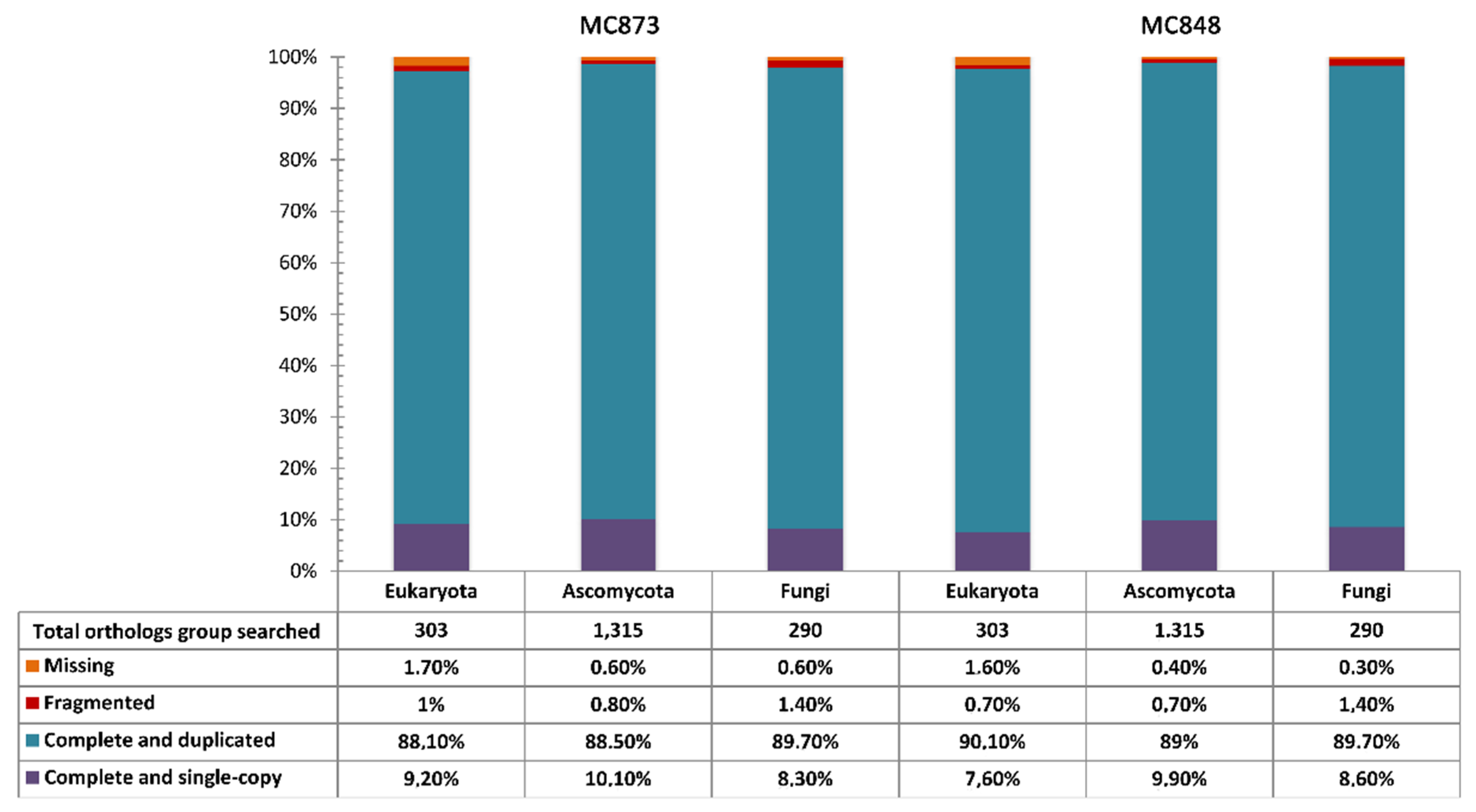
Gene models were predicted separately for each H. werneckii genome using the MAKER pipeline (v.3.00.0) [37] combined with the ab-initio gene predictors SNAP (v.2.39) [38] and AUGUSTUS (v. 3.3.3) [39], and further integrated with a full set of reference proteins and expressed sequence tags (ESTs) from Capnodiales (NCBI: txid134362) downloaded from the NCBI Protein and Nucleotide databases, respectively. However, before using these datasets, sequences sharing more than 90% similarity were removed by CD-HIT software [40] in order to reduce the redundancy.
Automatic functional annotation of predicted proteins was performed using PANNZER2 program [41] and resulting proteomes were compared by OrthoFinder (v.2.3.11) (https://github.com/davidemms/OrthoFinder) in order to evaluate the content of orthologous genes and the abundance of distinct orthogroups among the two sequenced strains. An orthogroup was defined as a set of genes derived from a single gene in the last common ancestor of different species. Orthogroups, containing a different number of genes, were subsequently submitted to ShinyGO v0.61 (http://bioinformatics.sdstate.edu/go) to determine if any particular metabolic pathway was enriched in a strain-specific manner.
Finally, the two genome assemblies were also screened for transposable elements (TE) and repetitive DNA sequence content by using Tandem Repeat Finder v.4.09 [42] and RepeatMasker v.4.0.9 (www.repeatmasker.org) softwares. tRNAs were also predicted using the tRNAscan-SE program v.1.3.1 [43].
Phylogenomic analysis was conducted using our two sequenced genomes and additional publicly available H. werneckii genome assemblies retrieved from the NCBI BioProject database (accession number: PRJNA428320) and GenBank (assembly accession: GCA_002127715.1) (Table 1). Phylogenetic distance was estimated by the Mash 2.2 toolkit [44] (sketch size, s  =  1000; k-mer size, k  =  21) while cluster analysis was performed with the pvclustR package [45], using the UPGMA algorithm with a bootstrap analysis of 1000 replicates. The resulting tree was visualized and edited using iTOL (https://itol.embl.de).

3. Results

Basic statistics of the H. werneckii genome assemblies generated in this study are shown in Table 2. The MC848 and MC873 genomes were sequenced with an average coverage of ~276× and ~207×, respectively, and their predicted size was quite similar, consisting of approximately ~51 Mbp each, with a total GC content of 53.4% (Table 2). The raw reads have been deposited in the Sequence Reads Archive (SRA) database and are available under BioProject identifier (ID) PRJNA641248 (https://www.ncbi.nlm.nih.gov/Traces/study/?acc=PRJNA641248).
The draft genome sequences contained a total of 1218 (MC848 strain) and 925 (MC873 strain) assembled contigs (minimum length: ≥200 bp), and have been submitted to the GenBank database under the following accession numbers: JACSRB000000000 (MC848) and JACSRC000000000 (MC873). The comparison of k-mer frequencies to the final assemblies revealed that most of the 27-mers in the reads were represented once in the respective assemblies, consistent with a high-quality genome assembly (estimated assembly completeness: >99.8%) of a predominantly heterozygous microorganism (Figure 1a). This genome-level variability was also confirmed by GenomeScope analysis (k-mer: 27), which showed an extensive level of heterozygosity (average ~4.6%) for both H. werneckii genomes.
To examine the phylogenetic relationships among our H. werneckii strains and other previously sequenced strains, we generated a Mash-based tree using the whole genomes currently available in GenBank (Figure 1b).
Phylogenomic analysis showed the existence of two main clusters (Figure 1b). The strains from the Mediterranean Sea clustered together, and they were very close to one clinical strain from Brazil (EXF-171) and two environmental isolates—one from salterns of Spain (EXF-120) and one haploid strain from Namibia (EXF-562). The second cluster grouped eight strains: four from the Atacama desert in Chile (EXF-6651, EXF-6654, EXF-6669 and EXF-6656), two clinical strains from Portugal (EXF-151) and from Italy (EXF-2682), and two strains from salterns of Slovenia (respectively, EXF-2000 and the haploid strain EXF-2788).
For both MC848 and MC873 genomes, we observed evidence of genome duplication by k-mer frequency analysis (Figure 1a), which was further corroborated byBUSCOanalysis (Figure 2).
Notably, for both H. werneckii genomes, we detected a high level of complete and duplicated BUSCO genes (>88%; Figure 2), which is consistent with the level of genome duplication observed. Overall, BUSCO results also confirmed a high level of genome completeness, with over 97% of eukaryotic and fungal genes found to be complete in our assemblies (Figure 2).
Totals of 15,542 and 15,565 genes were predicted in the H. werneckii MC873 and MC848 genomes, respectively. There was only a slight difference in protein-coding gene content between the two sequenced genomes (Table 2). Further small variations were also observed for the transposon content (23 extra TE found only in the MC848 strain), especially for the LTR-retrotransposon gipsy and non-LTR LINE-like retrotransposons, including some DNA TE that were exclusively detected in the MC848 strain (Table 3).
Regarding functional analysis of the gene set annotated in this study, we observed small differences among the two Hortaea genomes. In fact, over 99% of these genes were assigned to 10,911 unique orthogroups, of which 6052 (55.5%) contained single-copy orthologs, whereas the remaining 4859 (44.5%) included at least 2 genes from a single strain (Supplementary Material Table S1). Interestingly, 1047 orthogroups (1047/10,911;9.6%) showed strain-level differences in the number of genes included in each group. More specifically, of the 1047 orthogroups, 528 (50.4%) were enriched in the MC848 strain and 497 (47.5%) were enriched in the MC873 strain, while the remaining 22 (2.1%) orthogroups included strain-specific genes. In particular, the MC848 strain displayed 10 specific orthogroups containing a total of 20 genes, while the remaining 12 MC873-specific orthogroups included a total of 24 genes (Supplementary Material Table S1). However, most of the strain-specific genes encoded proteins with unknown or still uncharacterized functions (Supplementary Material Table S1).

4. Discussion

The recent finding that H. werneckii has a ubiquitous distribution in the seawater of the Mediterranean Sea, being the dominant fungal species up to a depth of 3400 m [13,25], has led us to deepen the genomic characterization of these strains to better understand their origin and evolution.
The presence of the fungus was demonstrated also in other deep-sea environments, such as deep-sea hydrothermal ecosystems in the Pacific Ocean [22,24] and sediments at 5000 m depths in the Central Indian Basin [23]. However, the species is not currently considered a marine fungus [21].
Comparative analyses of Mediterranean isolates with other H. werneckii strains recovered from different sources evidenced the peculiar genetic and physiological characteristics of the seawater strains compared to the others [13], but these were not sufficient to assign them to another taxonomic group [12].
In this study we carried out a de-novo sequencing of the genome of one strain isolated at a 3,400 m depth (MC873), and a re-sequencing of the genome derived from a 2500 m depth (MC848). The latter strain MC848 was previously sequenced and assembled in 5734 contigs, which were highly fragmented and presumably had an extensive number of errors, especially in terms of total gene number predicted [46].
In this study, a high-quality genome assembly (>99.8%) was obtained for both MC848 and MC873 genomes, which proved to be diploid. In fact, k-mer frequency analysis (Figure 1a) and BUSCO analysis (Figure 2) showed the existence of genome duplication in both genomes. In addition, a high level of heterozygosity (~4.6%) was also detected for both H. werneckii genomes. This is a typical hallmark of diploid species and/or eukaryotic lineages that have undergone whole-genome duplication (WGD) by occasional intraspecific hybridization events between two haploid progenitors. This finding was suggested also by Gostinčar et al. [26], who, after analyzing 12 genomes of H. werneckii, found that only 2 (EXF-562 and EXF-2788) were haploid, while the remaining strains were diploid and highly heterozygous. Our results support this hypothesis.
Phylogenetic analyses carried out in this study showed the existence of two separate clusters. Interestingly, our strains, recovered from the deep sea at 3400 and 2500 m of depth, but in different stations of the Mediterranean Sea, clustered together, suggesting a degree of environmental specificity for these genotypes (Figure 1b). The placement of our strains in a single well-supported group, together with the previously sequenced EXF-10513 strain (BioSample accession n°: SAMN08295408) (Figure 1b), is consistent with our expectation, as this latter strain and MC848 are the same strain, although strain codes may vary in the publications [26]. However, from the re-sequencing of the genome of this strain (MC848=EXF-10513), we obtained a better assembly with fewer contigs (1218 vs. 5734 contigs), and consequently an improved genome annotation according to other studies [46]. In fact, we detected a lower number of predicted genes (15,410 vs. 17,094) and exons (32,716 vs. 37,837) [26] as expected for less fragmented genome assemblies [46].
Environmental and/or geographical specificity was also observed for other H. werneckii genomes in the phylogenetic tree, such as EXF-2000 and EXF-2788 strains, isolated from hypersaline water, or EXF-6651 and EXF-6669 strains, recovered from spider webs in the Atacama desert, Chile [26]. These findings may suggest an evolutionary adaptation of H. werneckii strains to such environments.
The sizes of the MC848 and MC873 genomes sequenced in this study were 51,030,830 bp and 50,705,820 bp, respectively, confirming that the DNA content of marine strains may be slightly larger than that of other H. werneckii genomes sequenced so far, whose sizes range from 25.2 Mbp to 49.9 Mbp [26].
Genome sizes show dramatic variation among the Dothideomycetes [47], and after Pseudocercospora (Mycosphaerella) fijiensis (genome size ~74.14 Mbp) [47], H. werneckii possesses the largest genome among members of the order of Capnodiales [28] (www.zbi.ee/fungal-genomesize). However, additional large genomes are frequently described in other dothideomycetous black fungi, such as Friedmanniomyces endolithicus (genome size 46.75 Mbp), isolated from the extreme environment of the Antarctic [48].
Large scale genome duplication in fungi seems to be associated with selective advantage in terms of stressful environmental conditions. In Rhizopus oryzae and Phycomyces blakeesleeanus species, WGD contributes to pathogenicity and the expansion of signal transduction and light sensing [49,50], while increases in genome size were also observed in experimental evolution studies in Saccharomyces cerevisiae, when the yeast is exposed to stressful concentrations of salt and UV radiations [51,52]. In fact, genome duplication provides the raw material for further evolution processes. In the case of H. werneckii, the large genome also contains expanded gene families encoding metal cation transporters [27] that are supposed to confer a selective advantage in hypersaline environments [28].
The results obtained in this study support the recent hypothesis that most H. werneckii strains are likely derivatives from intraspecific hybridization and, due the phylogenomic differences observed between strains from different sources, we could suppose that marine strains are evolving in this environment where they are well adapted.
The release of high-quality genome assemblies from marine H. werneckii strains provides additional data enabling further genomics analysis, including niche adaptation, fitness and evolution studies for investigating the diversification of Hortaea species and their specific associations with stressful and harsh environments.

Supplementary Materials

The following are available online at https://www.mdpi.com/2075-1729/10/10/229/s1, Table S1: Overall statistics of all orthogroups detected in the two Hortaea werneckii strains.

Author Contributions

Conceptualization, O.R., C.U. and F.D.L.; methodology, O.R., D.G. and F.D.L.; investigation, D.G., L.G. and A.M.; resources, O.R. and F.D.L.; writing of the original draft preparation and editing, A.M., O.R. and F.D.L.; supervision, F.D.L. All authors have read and agreed to the published version of the manuscript.

Funding

This research was supported in part by FFABR_2017 (Italian fund of basic research activities) to F.D.L. and by the University of Messina CT_ROMEO_GOM - grant agreement n° 36036 to O.R.

Conflicts of Interest

The authors declare no conflict of interest.

Data Availability

The Illumina raw reads have also been submitted into the Sequence Read Archive (SRA) database and are associated with the BioProject ID: PRJNA641248. The draft whole-genome sequences of strains MC848 and MC873 have also been deposited at DDBJ/ENA/GenBank under accession numbers JACSRB000000000 andJACSRC000000000, respectively. The versions described in this article are the first versions JACSRB000000000 and JACSRC000000000.

References

  1. Gostinčar, C.; Muggia, L.; Grube, M. Polyextremotolerant black fungi: Oligotrophism, adaptive potential, and a link to lichen symbioses. Front. Microbiol. 2012, 3, 390. [Google Scholar] [CrossRef] [Green Version]
  2. Moreno, L.F.; Vicente, V.A.; de Hoog, S. Black yeasts in the omics era: Achievements and challenges. Med. Mycol. 2018, 56, S32–S41. [Google Scholar] [CrossRef]
  3. Butinar, L.; Sonjak, S.; Zalar, P.; Plemenitaš, A.; Gunde-Cimerman, N. Melanized halophilic fungi are eukaryotic members of microbial communities in hypersaline waters of solar salterns. Bot. Mar. 2005, 48, 73–79. [Google Scholar] [CrossRef]
  4. Gunde-Cimerman, N.; Zalar, P.; de Hoog, G.S.; Plemenitaš, A. Hypersaline waters in salterns- Natural ecological niches for halophilic black yeasts. FEMS Microbiol. Ecol. 2000, 32, 235–240. [Google Scholar] [CrossRef]
  5. Petrovič, U.; Gunde-Cimerman, N.; Plemenitaš, A. Cellular responses to environmental salinity in the halophilic black yeast Hortaea werneckii. Mol. Microbiol. 2002, 45, 665–672. [Google Scholar] [CrossRef] [Green Version]
  6. Kogej, T.; Stein, M.; Volkmann, M.; Gorbushina, A.A.; Galinski, E.A.; Gunde-Cimerman, N. Osmotic adaptation of the halophilic fungus Hortaea werneckii: Role of osmolytes and melanization. Microbiology 2007, 153, 4261–4273. [Google Scholar] [CrossRef] [Green Version]
  7. Plemenitaš, A.; Lenassi, M.; Konte, T.; Kejžar, A.; Zajc, J.; Gostinčar, C.; Gunde-Cimerman, N. Adaptation to high concentrations in halotolerant/halophilic fungi: A molecular prospective. Front. Microbiol. 2014, 5, 199. [Google Scholar] [CrossRef] [Green Version]
  8. Gunde-Cimerman, N.; Plemenitaš, A.; Oren, A. Strategies of adaptation of microorganisms of the three domains of life to high salt concentrations. FEMS Microbiol. Rev. 2018, 42, 353–375. [Google Scholar] [CrossRef]
  9. Gunde-Cimerman, N.; Plemenitaš, A. Ecology and molecular adaptations of the halophilic black yeast Hortaeawerneckii. Rev. Environ. Sci. Biotechnol. 2006, 5, 323–331. [Google Scholar] [CrossRef]
  10. Nishimura, K.; Miyaji, M. Hortaea, a new genus to accommodate Cladosporium werneckii. Jpn. J. Med. Mycol. 1984, 25, 139–146. [Google Scholar] [CrossRef]
  11. Crous, P.W.; Schoch, C.L.; Hyde, K.D.; Wood, A.R.; Gueidan, C.; de Hoog, G.S.; Groenewald, J.Z. Phylogenetic lineages in the Capnodiales. Stud. Mycol. 2009, 64, 17–47. [Google Scholar] [CrossRef]
  12. Zalar, P.; Zupančič, J.; Gostinčar, C.; Zajc, J.; de Hoog, G.S.; De Leo, F.; Azua-Bustos, A.; Gunde-Cimerman, N. The extremely halotolerant black yeast Hortaea werneckii–a model for intraspecific hybridization in clonal fungi. IMA Fungus 2019, 10, 10. [Google Scholar] [CrossRef] [Green Version]
  13. Marchetta, A.; van den Ende, G.B.; Al-Hatmi, A.M.S.; Hagen, F.; Zalar, P.; Sudhadham, M.; Gunde-Cimerman, N.; Urzì, C.; de Hoog, S.; De Leo, F. Global molecular diversity of the halotolerant fungus Hortaea werneckii. Life 2018, 8, 31. [Google Scholar] [CrossRef] [Green Version]
  14. Göttlich, E.; de Hoog, G.S.; Yoshida, S.; Takeo, K.; Nishimura, K.; Miyaji, M. Cell-surface hydrophobicity and lipolysis as essential factors in human tinea nigra. Mycoses 1995, 38, 489–494. [Google Scholar] [CrossRef]
  15. Perez, C.; Colella, M.T.; Olaizola, C.; de Capriles, C.H.; Magaldi, S.; Mata-Essayag, S. Tineanigra: Report of twelvecases in Venezuela. Mycopathologia 2005, 160, 235–238, ds11046–s005. [Google Scholar] [CrossRef]
  16. Bonifaz, A.; Badali, H.; de Hoog, G.S.; Cruz, M.; Araiza, J.; Cruz, M.A.; Fierro, L.; Ponce, R.M. Tinea nigra by Hortaea werneckii, a report of 22 cases from Mexico. Stud. Mycol. 2008, 61, 77–82. [Google Scholar] [CrossRef]
  17. Giordano, M.C.; De la Fuente, A.; Lorca, M.B.; Kramer, D. Tinea nigra: Report of three pediatrics cases. Rev. Chil. Pediatr. 2018, 89, 506–510. [Google Scholar] [CrossRef]
  18. Gunde-Cimerman, N.; Zalar, P. Extremely halotolerant and halophilic fungi inhabit brine in solar salterns around the globe. Food Technol. Biotech. 2014, 52, 170–179. [Google Scholar]
  19. Xu, W.; Pang, K.-L.; Luo, Z.-H. High fungal diversity and abundance recovered in the deep-sea sediments of the Pacific Ocean. Microb. Ecol. 2014, 68, 688–698. [Google Scholar] [CrossRef]
  20. Formoso, A.; Heidrich, D.; Felix, C.R.; Tenório, A.C.; Leite, B.R.; Pagani, D.M.; Ortiz-Monsalve, S.; Ramírez-Castrillón, M.; FontesLandell, M.; Scroferneker, M.L.; et al. Enzymatic activity and susceptibility to antifungal agents of brazilian environmental isolates of Hortaeawerneckii. Mycopathologia 2015, 180, 345–352, ds11046–s015. [Google Scholar] [CrossRef]
  21. Jones, E.B.G.; Suetrong, S.; Bahkali, A.H.; Abdel-Wahab, M.A.; Boekhout, T.; Pang, K.-L. Classification of marine Ascomycota, Basidiomycota, Blastocladiomycota and Chytridiomycota. Fungal Divers. 2015, 3, 1–72. [Google Scholar] [CrossRef]
  22. Le Calvez, T.; Burgaud, G.; Mahé, S.; Barbier, G.; Vandenkoornhuyse, P. Fungal diversity in deep-sea hydrothermal ecosystems. Appl. Environ. Microbiol. 2009, 75, 6415–6421. [Google Scholar] [CrossRef] [PubMed] [Green Version]
  23. Singh, P.; Raghukumar, C.; Meena, R.M.; Verma, P.; Shouche, Y. Fungal diversity in deep-sea sediments revealed by culture-dependent and culture-independent approaches. Fungal Ecol. 2012, 5, 543–555. [Google Scholar] [CrossRef]
  24. Pang, K.-L.; Guo, S.-Y.; Chen, I.-A.; Burgaud, G.; Luo, Z.-H.; Dahms, H.U.; Hwang, J.-S.; Lin, Y.-L.; Huang, J.-S.; Ho, T.-W.; et al. Insights into fungal diversity of a shallow-water hydrothermal vent field at Kueishan Island, Taiwan by culture-based and metabarcoding analyses. PLoS ONE 2019, 14, e0226616. [Google Scholar] [CrossRef] [Green Version]
  25. De Leo, F.; Lo Giudice, A.; Alaimo, C.; De Carlo, G.; Rappazzo, A.C.; Graziano, M.; De Domenico, E.; Urzì, C. Occurrence of the blackyeast Hortaeawerneckii in the Mediterranean Sea. Extremophiles 2019, 23, 9–17. [Google Scholar] [CrossRef]
  26. Gostinčar, C.; Stajich, J.E.; Zupančič, J.; Zalar, P.; Gunde-Cimerman, N. Genomic evidence for intraspecific hybridization in a clonal and extremely halotolerant yeast. BMC Genom. 2018, 19, 364. [Google Scholar] [CrossRef]
  27. Lenassi, M.; Gostinčar, C.; Jackman, S.; Turk, M.; Sadowski, I.; Nislow, C.; Jones, S.; Birol, I.; Gunde-Cimerman, N.; Plemenitaš, A. Whole genome duplication and enrichment of metal cation transporters revealed by de-novo genome sequencing of extremely halotolerant black yeast Hortaeawerneckii. PLoS ONE 2013, 8, e71328. [Google Scholar] [CrossRef] [Green Version]
  28. Sinha, S.; Flibotte, S.; Neira, M.; Formby, S.; Plemenitaš, A.; Gunde-Cimerman, N.; Lenassi, M.; Gostinčar, C.; Stajich, J.E.; Nislow, C. Insight into the recent genome duplication of the halophilic yeast Hortaea werneckii: Combining an improved genome with gene expression and chromatin structure. G3 2017, 7, 2015–2022. [Google Scholar] [CrossRef] [Green Version]
  29. Chen, S.; Zhou, Y.; Chen, Y.; Gu, J. fastp: An ultra-fast all-in-one FASTQ preprocessor. Bioinformatics 2018, 34, i884–i890. [Google Scholar] [CrossRef]
  30. Bankevich, A.; Nurk, S.; Antipov, D.; Gurevich, A.A.; Dvorkin, M.; Kulikov, A.S.; Lesin, V.M.; Nikolenko, S.I.; Pham, S.; Prjibelski, A.D.; et al. SPAdes: A new genome assembly algorithm and its applications to single-cell sequencing. J. Comput. Biol. 2012, 19, 4554–4577. [Google Scholar] [CrossRef] [Green Version]
  31. Boetzer, M.; Henkel, C.V.; Jansen, H.J.; Butler, D.; Pirovano, W. Scaffolding pre-assembled contigs using SSPACE. Bioinformatics 2011, 27, 578–579. [Google Scholar] [CrossRef] [Green Version]
  32. Boetzer, M.; Pirovano, W. Toward almost closed genomes with GapFiller. Genome Biol. 2012, 13, R56. [Google Scholar] [CrossRef] [PubMed] [Green Version]
  33. Gurevich, A.; Saveliev, V.; Vyahhi, N.; Tesler, G. QUAST: Quality assessment tool for genome assemblies. Bioinformatics 2013, 29, 1072–1075. [Google Scholar] [CrossRef] [PubMed]
  34. Seppey, M.; Manni, M.; Zdobnov, E.M. BUSCO: Assessing Genome Assembly and Annotation Completeness. In Gene Prediction. Methods in Molecular Biology; Kollmar, M., Ed.; Humana: New York, NY, USA, 2019; Volume 1962, pp. 2272–2345. [Google Scholar] [CrossRef]
  35. Mapleson, D.; Garcia Accinelli, G.; Kettleborough, G.; Wright, J.; Clavijo, B.J. KAT: A K-mer analysis toolkit to quality control NGS datasets and genome assemblies. Bioinformatics 2017, 33, 574–576. [Google Scholar] [CrossRef] [PubMed]
  36. Vurture, G.W.; Sedlazeck, F.J.; Nattestad, M.; Underwood, C.J.; Fang, H.; Gurtowski, J.; Schatz, M.C. GenomeScope: Fast reference-free genome profiling from short reads. Bioinformatics 2017, 33, 2202–2204. [Google Scholar] [CrossRef] [Green Version]
  37. Campbell, M.S.; Holt, C.; Moore, B.; Yandell, M. Genome Annotation and Curation Using MAKER and MAKER-P. Curr. Protoc. Bioinform. 2014, 48, 4–11. [Google Scholar] [CrossRef] [Green Version]
  38. Korf, I. Gene finding in novel genomes. BMC Bioinform. 2004, 5, 59. [Google Scholar] [CrossRef] [Green Version]
  39. Stanke, M.; Morgenstern, B. AUGUSTUS: A web server for gene prediction in eukaryotes that allows user-defined constraints. Nucleic Acids Res. 2005, 33, W465–W467. [Google Scholar] [CrossRef] [Green Version]
  40. Fu, L.; Niu, B.; Zhu, Z.; Wu, S.; Li, W. CD-HIT: Accelerated for clustering the next-generation sequencing data. Bioinformatics 2012, 28, 3150–3152. [Google Scholar] [CrossRef]
  41. Törönen, P.; Medlar, A.; Holm, L. PANNZER2: A rapid functional annotation web server. Nucleic Acids Res. 2018, 46, W84–W88. [Google Scholar] [CrossRef]
  42. Benson, G. Tandem repeats finder: A program to analyze DNA sequences. Nucleic Acids Res. 1999, 27, 573–580. [Google Scholar] [CrossRef] [PubMed] [Green Version]
  43. Chan, P.P.; Lowe, T.M. tRNAscan-SE: Searching for tRNA Genes in Genomic Sequences. In Gene Prediction. Methods in Molecular Biology; Kollmar, M., Ed.; Humana: New York, NY, USA, 2019; Volume 1962, pp. 11–14. [Google Scholar] [CrossRef]
  44. Ondov, B.D.; Treangen, T.J.; Melsted, P.; Mallonee, A.B.; Bergman, N.H.; Koren, S.; Phillippy, A.M. Mash: Fast genome and metagenome distance estimation using MinHash. Genome Biol. 2016, 17, 132. [Google Scholar] [CrossRef] [PubMed] [Green Version]
  45. Suzuki, R.; Shimodaira, H. Pvclust: An R package for assessing the uncertainty in hierarchical clustering. Bioinformatics 2006, 22, 15401–15542. [Google Scholar] [CrossRef] [PubMed]
  46. Denton, J.F.; Lugo-Martinez, J.; Tucker, A.E.; Schrider, D.R.; Warren, W.C.; Hahn, M.W. Extensive error in the number of genes inferred from draft genome assemblies. Ploscomput. Biol. 2014, 10, e1003998. [Google Scholar] [CrossRef] [Green Version]
  47. Ohm, R.A.; Feau, N.; Henrissat, B.; Schoch, C.L.; Horwitz, B.A.; Barry, K.W.; Condon, B.J.; Copeland, A.C.; Dhillon, B.; Glaser, F.; et al. Diverse lifestyles and strategies of plant pathogenesis encoded in the genomes of eighteen Dothideomycetes fungi. PLoS Pathog. 2012, 8, e1003037. [Google Scholar] [CrossRef] [Green Version]
  48. Coleine, C.; Masonjones, S.; Sterflinger, K.; Onofri, S.; Selbmann, L.; Stajich, J.E. Peculiar genomic traits in the stress-adapted cryptoendolithic Antarctic fungus Friedmanniomyces Endolithicus. Fungal Biol. 2020, 124, 458–467. [Google Scholar] [CrossRef]
  49. Ma, L.-J.; Ibrahim, A.S.; Skory, C.; Grabherr, M.G.; Burger, G.; Butler, M.; Elias, M.; Idnurm, A.; Lang, B.F.; Sone, T.; et al. Genomic analysis of the basal lineage fungus Rhizopus oryzae reveals a whole-genome duplication. PLoS Genet. 2009, 5, e1000549. [Google Scholar] [CrossRef]
  50. Corrochano, L.M.; Kuo, A.; Marcet-Houben, M.; Polaino, S.; Salamov, A.; Villalobos-Escobedo, J.M.; Grimwood, J.; Álvarez, M.I.; Avalos, J.; Bauer, D.; et al. Expansion of Signal Transduction Pathways in Fungi by Extensive Genome Duplication. Curr. Biol. 2016, 26, 1577–1584. [Google Scholar] [CrossRef] [Green Version]
  51. Lidzbarsky, G.A.; Shkolnik, T.; Nevo, E. Adaptive response to DNA-damaging agents in natural Saccharomyces cerevisiae populations from Evolution Canyon, Mt. Carmel, Israel. PLoS ONE 2009, 4, e5914. [Google Scholar] [CrossRef]
  52. Dhar, R.; Sägesser, R.; Weikert, C.; Yuan, J.; Wagner, A. Adaptation of Saccharomyces cerevisiae to saline stress through laboratory evolution. J. Evol. Biol. 2011, 24, 1135–1153. [Google Scholar] [CrossRef] [Green Version]
Figure 1. Genome assembly analysis and phylogenomic relationships of H. werneckii strains. (a) k-mer frequency analysis generated by the KAT tool; plots show KAT spectra of the 27-mer multiplicity of the read set (x axis) against their respective assembly (y axis) for MC873 and MC848 strains. Read content in black represents the 27-mers present in the reads but absent in the assemblies and suggests a fairly complete genome assembly. Red indicates 27-mers are present once in the assembly while purple 27-mers are present twice, indicating that genome duplication events likely occurred; (b) Mash-based phylogenetic tree generated using our two H. werneckii genome assemblies along with whole-genomes currently available in GenBank database. The code of each strain used is shown to the right of each branch tip and its GenBank accession number. Numbers at the nodes represent percent bootstrap support values based on 1000 replicates.
Life 10 00229 g001
Figure 2. (BUSCO) results showing the number of complete (single and duplicated), missing and fragmented orthologs obtained by searching the Eukaryota, Ascomycota and fungal lineage datasets.
Life 10 00229 g002
Table 1. Strains of Hortaea werneckii included in this study.
StrainOther CollectionSourceCountryReference
EXF-2000CBS 100457Hypersaline water of salt-pansSlovenia[28]
EXF-2788 Hypersaline water of salt-pansSlovenia[26]
EXF-120 Hypersaline water of salt-pansSpain[26]
EXF-562 Soil on the sea costNamibia[26]
EXF-171CBS 111.31Human, KeratomycosisBrazil[26]
EXF-2682CBS 126.35Human, Trichomycosis nigraItaly[26]
EXF-151CBS 107.67Human, Tinea nigraPortugal[26]
EXF-6651 Spider web, Atacama desertChile[26]
EXF-6654 Spider web, Atacama desertChile[26]
EXF-6669 Spider web, Atacama desertChile[26]
EXF-6656 Rock in a cave, Atacama desertChile[26]
EXF-10513MC848
V2500 b
Seawater of Mediterranean Sea; 2500 m depth, Vector stationItaly[26]; This study
MC873Geo f 100 cSeawater, Mediterranean Sea; 3400 m depth, Geostar stationItalyThis study
Table 2. Overall assembly statistics and gene content of the two H. werneckii genomes sequenced in this study.
H. werneckii Genome FeaturesMC873 StrainMC848 Strain
Total number of sequenced bases10,481,489,01314,036,248,558
Total number of reads with Q-score ≥2570,784,39798,305,008
Total number of mapped reads (%)70,711,190 (99.9%)95,135,691 (96.8%)
Unmapped reads73,207 (0.1%)3,169,317 (3.2%)
Number of total contigs9251218
Number of contigs >1 Kbp761612
Largest contig (bp)604,832576,474
Genome size (bp)50,705,82051,030,830
Average coverage depth~207×~276×
GC content (%)53.453.4
Total number of predicted genes15,54215,565
Protein-coding genes15,39715,410
tRNAs126132
snRNAs55
Total gene length (bp)26,021,39426,184,209
Longest gene (bp)15,05219,877
Number of exons32,68132,716
Longest exon (bp)12,96612,966
Exon average length (bp)~742~746
Gene average length (bp)~1690~1699
Tandem Repeat Number13,18813,315
Simple repeats11,18311,282
Low-complexity repeats20052033
Table 3. Classes of transposable elements (TE) and their relative numbers detected in the two sequenced marine H. werneckii genomes.
TE ClassMC848MC873TE ClassMC848MC873
DNA (notclassified)23LINE I-Jockey11
DNA CMC-EnSpm23LINE L143
DNA Dada108LINE L1-Tx143
DNA Ginger-122LINE L210
DNA hAT12LINE RTE10
DNA hAT-Ac1817LINE RTE-BovB35
DNA hAT-Charlie22LTR (notclassified)33
DNA Kolobok-T210LTR Copia81
DNA Merlin10LTR ERV176
DNA-Maverick01LTR ERVK68
DNA Mule-MuDR11LTR Gypsy165151
DNA P11LTR Ngaro1212
DNA TcMar-ISRm1111LTR Pao1817
DNA-TcMar-Tc101RC Helitron12
LINE CR110SINE B211
LINE I10SINE-tRNA-RTE01
Total279256

