Parafiniukite, Ca2Mn3(PO4)3Cl, a New Member of the Apatite Supergroup from the Szklary Pegmatite, Lower Silesia, Poland: Description and Crystal Structure

 , , ,
, , ,
Abstract
:1. Introduction
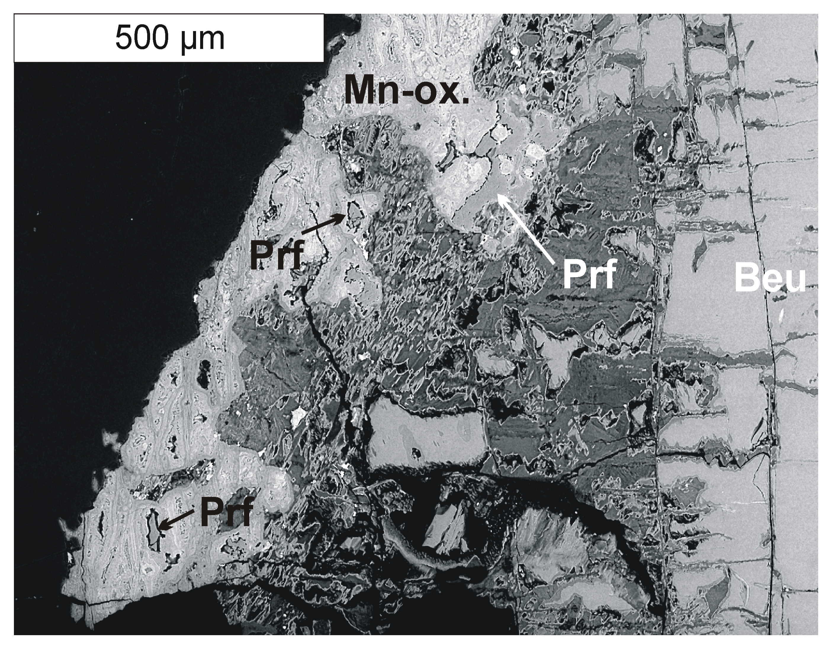
2. Occurrence
3. Experimental Data
3.1. Mineral Description and Physical Properties
3.2. Chemical Data
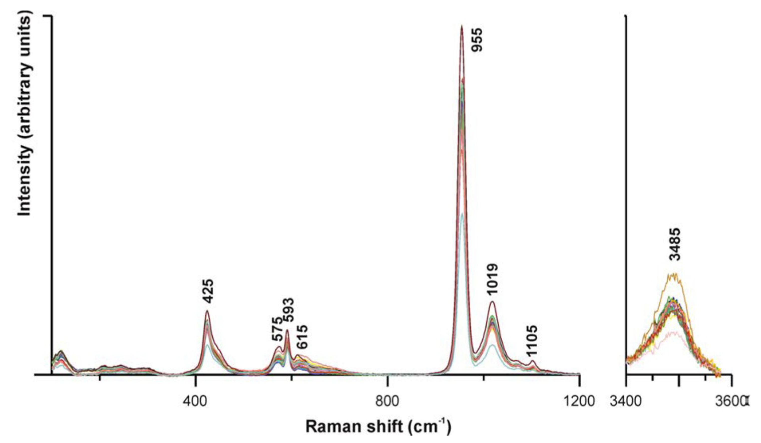
3.3. Micro-Raman Spectroscopy
3.4. Crystallography
4. Discussion
4.1. Crystal Structure Description
4.2. Crystallization of Parafiniukite
Supplementary Materials
Author Contributions
Funding
Conflicts of Interest
References
- Pieczka, A. Beusite and unusual Mn-rich apatite from the Szklary granitic pegmatite, Lower Silesia, Poland. Can. Mineral. 2007, 45, 901–914. [Google Scholar] [CrossRef]
- Szuszkiewicz, A.; Pieczka, A.; Gołębiowska, B.; Dumańska-Słowik, M.; Marszałek, M.; Szełęg, E. Chemical composition of Mn- and Cl-rich apatites from the Szklary pegmatite, Central Sudetes, SW Poland: Taxonomic and genetic implications. Minerals 2018, 8, 350. [Google Scholar] [CrossRef]
- Černý, P.; Ercit, T.S. The classification of granitic pegmatites revisited. Can. Mineral. 2005, 43, 2005–2026. [Google Scholar] [CrossRef]
- Pieczka, A.; Szuszkiewicz, A.; Szełęg, E.; Janeczek, J.; Nejbert, K. Granitic pegmatites of the Polish part of the Sudetes (NE Bohemian massif, SW Poland). In Fieldtrip Guidebook, Proceedings of the 7th International Symposium on Granitic Pegmatites, Książ, Poland, 17–19 June 2015; PEG2015: Książ, Poland, 2015; pp. 73–103. [Google Scholar]
- Oliver, G.J.H.; Corfu, F.; Krogh, T.E. U–Pb ages from SW Poland: Evidence for a Caledonian suture zone between Baltica and Gondwana. J. Geol. Soc. Lond. 1993, 150, 355–369. [Google Scholar] [CrossRef]
- Van Breemen, O.; Bowes, D.R.; Aftalion, M.; Żelaźniewicz, A. Devonian tectonothermal activity in the Sowie Góry gneissic block, Sudetes, southwestern Poland: Evidence from Rb-Sr and U-Pb isotopic studies. J. Pol. Geol. Soc. 1988, 58, 3–10. [Google Scholar]
- Timmermann, H.; Parrish, R.R.; Noble, S.R.; Kryza, R. New U–Pb monazite and zircon data from the Sudetes Mountains in SW Poland: Evidence for a single-cycle Variscan orogeny. J. Geol. Soc. Lond. 2000, 157, 265–268. [Google Scholar] [CrossRef]
- Turniak, K.; Pieczka, A.; Kennedy, A.K.; Szełęg, E.; Ilnicki, S.; Nejbert, K.; Szuszkiewicz, A. Crystallisation age of the Julianna pegmatite system (Góry Sowie Block, NE margin of the Bohemian massif): Evidence from U-Th-Pb SHRIMP monazite and CHIME uraninite studies. In Book of Abstracts, Proceedings of the 7th International Symposium on Granitic Pegmatites, Książ, Poland, 17–19 June 2015; PEG2015: Książ, Poland, 2015; pp. 111–112. [Google Scholar]
- Pieczka, A. A rare mineral-bearing pegmatite from the Szklary serpentynite massif, the Fore-Sudetic Block, SW Poland. Geol. Stud. 2000, 33, 23–31. [Google Scholar]
- Pieczka, A. Primary Nb-Ta minerals in the Szklary pegmatite, Poland: New insights into controls of crystal chemistry and crystallization sequences. Am. Mineral. 2010, 95, 1478–1492. [Google Scholar] [CrossRef]
- Pieczka, A.; Grew, E.S.; Groat, L.A.; Evans, R.J. Holtite and dumortierite from the Szklary pegmatite, Lower Silesia, Poland. Mineral. Mag. 2011, 75, 303–315. [Google Scholar] [CrossRef]
- Pieczka, A.; Evans, R.J.; Grew, E.S.; Groat, L.A.; Ma, C.; Rossman, G.R. The dumortierite supergroup. II. Three new minerals from the Szklary pegmatite, SW Poland: Nioboholtite, (Nb0.6□0.4)Al6BSi3O18, titanoholtite, (Ti0.75□0.25)Al6BSi3O18, and szklaryite, □Al6BAs3+3O15. Mineral. Mag. 2013, 77, 2841–2856. [Google Scholar] [CrossRef]
- Tait, K.; Ball, N.A.; Hawthorne, F.C. Pieczkaite, ideally Mn5(PO4)3Cl, a new apatite-supergroup mineral from Cross Lake, Manitoba, Canada: Description and crystal structure. Am. Mineral. 2015, 100, 1047–1052. [Google Scholar] [CrossRef]
- Mandarino, J.A. The Gladstone-Dale relationship. Part III. Some general applications. Can. Mineral. 1979, 17, 71–76. [Google Scholar]
- Mandarino, J.A. The Gladstone-Dale relationship. Part IV. The compatibility concept and its application. Can. Mineral. 1981, 19, 441–450. [Google Scholar]
- Pouchou, J.-L.; Pichoir, F. Quantitative analysis of homogeneous or stratified microvolumes applying the model “PAP”. In Electron Probe Quantitation; Heinrich, K.F.J., Newbury, D.E., Eds.; Plenum Press: New York, NY, USA, 1991; pp. 31–75. [Google Scholar]
- Bruker AXS Inc. APEX 3. In Bruker Advanced X-ray Solution; Bruker AXS Inc.: Madison, WI, USA, 2004. [Google Scholar]
- Biagioni, C.; Bosi, F.; Hålenius, U.; Pasero, M. The crystal structure of turneaureite, Ca5(AsO4)3Cl, the arsenate analog of chlorapatite, and its relationships with the arsenate apatites johnbaumite and svabite. Am. Mineral. 2017, 102, 1981–1986. [Google Scholar] [CrossRef]
- Sheldrick, G.M. Crystal structure refinement with SHELXL. Acta Crystallogr. 2015, C71, 3–8. [Google Scholar]
- Wilson, A.J.C. Volume C: Mathematical, Physical and Chemical Tables. In International Tables for Crystallography; Kluwer Academic: Dordrecth, The Netherlands, 1992. [Google Scholar]
- Brese, N.E.; O’Keeffe, M. Bond-valence parameters for solids. Acta Crystallogr. 1991, B47, 192–197. [Google Scholar] [CrossRef]
- Kraus, W.; Nolze, G. POWDER CELL—A program for the representation and manipulation of crystal structures and calculation of the resulting X-ray powder patterns. J. Appl. Crystallogr. 1996, 29, 301–303. [Google Scholar] [CrossRef]
- Hughes, J.M.; Rakovan, J. The crystal structure of apatite, Ca5(PO4)3(F,OH,Cl). In Phosphates: Geochemical, Geobiological and Materials Importance; Kohn, M.L., Rakovan, J., Hughes, J.M., Eds.; Mineralogical Society of America: Chantilly, VA, USA, 2002. [Google Scholar]
- Shannon, R.D. Revised effective ionic radii and systematic studies of interatomic distances in halides and chalcogenides. Acta Crystallogr. 1976, A32, 751–767. [Google Scholar] [CrossRef]
- Hughes, J.M.; Cameron, M.; Crowley, K.D. Structural variations in natural F, OH, and Cl apatites. Am. Mineral. 1989, 74, 870–876. [Google Scholar]
- Hughes, J.M.; Cameron, M.; Crowley, K.D. Crystal structures of natural ternary apatites: Solid solution in the Ca5(PO4)3X (X = F, OH, Cl) system. Am. Mineral. 1990, 75, 295–304. [Google Scholar]
- Suitch, P.R.; Lacout, J.L.; Hewat, A.W.; Young, R.A. The structural location and role of Mn2+ partially substituted for Ca2+ in fluorapatite. Acta Crystallogr. 1985, B41, 173–179. [Google Scholar] [CrossRef]
- Hughes, J.M.; Ertl, A.; Bernhardt, H.J.; Rossman, G.R.; Rakovan, J. Mn-rich fluorapatite from Austria: Crystal structure, chemical analysis and spectroscopic investigations. Am. Mineral. 2004, 89, 629–632. [Google Scholar] [CrossRef]
- Strunz, H.; Nickel, E.H. Strunz Mineralogical Tables, 9th ed.; Schweizerbart’sche Verlagsbuchhandlung: Stuttgart, Germany, 2001. [Google Scholar]
- Gaines, R.V.; Skinner, H.C.; Foord, E.E.; Mason, B.; Rosenzweig, A. Dana’s New Mineralogy, 9th ed.; John Wiley & Sons, Inc.: Hoboken, NJ, USA, 1997. [Google Scholar]
- Pasero, M.; Kampf, A.R.; Ferraris, C.; Pekov, I.V.; Rakovan, J.; White, T.J. Nomenclature of the apatite supergroup minerals. Eur. J. Mineral. 2010, 22, 163–179. [Google Scholar] [CrossRef]
- Černý, P.; Fryer, B.J.; Chapman, R. Apatite from granitic pegmatite exocontact in Moldanubian serpentinites. J. Czech Geol. Soc. 2001, 46, 15–20. [Google Scholar]
- Deschamps, F.; Godard, M.; Guillot, S.; Hattori, K. Geochemistry of subduction zone serpentynites: A review. Lithos 2013, 178, 96–127. [Google Scholar] [CrossRef]


| Constituent | Mean (n = 10) | Range | e.s.d. |
|---|---|---|---|
| P2O5 | 39.20 | 38.98–39.44 | 0.14 |
| MgO | 0.19 | 0.12–0.27 | 0.05 |
| CaO | 24.14 | 23.66–24.64 | 0.39 |
| MnO | 31.19 | 30.04–31.78 | 0.62 |
| FeO | 2.95 | 2.72–3.15 | 0.16 |
| Na2O | 0.05 | 0.01–0.07 | 0.02 |
| F | 0.39 | 0.29–0.46 | 0.05 |
| Cl | 3.13 | 3.00–3.29 | 0.09 |
| H2O(calc) | 0.68 | 0.61–0.71 | 0.03 |
| O=(F + Cl) | −0.87 | ||
| Total | 101.05 |
| Crystal Data | |
| Crystal size (mm) | 0.07 × 0.04 × 0.03 |
| Cell setting, space group | Hexagonal, P63/m |
| a (Å) | 9.4900(6) |
| c (Å) | 6.4777(5) |
| V (Å3) | 505.22(5) |
| Z | 2 |
| Data Collection and Refinement | |
| Radiation, wavelength (Å) | Mo Kα, λ = 0.71073 |
| Temperature (K) | 293 |
| 2θmax (°) | 54.89 |
| Measured reflections | 6047 |
| Unique reflections | 422 |
| Reflections with Fo > 4σ(Fo) | 320 |
| Rint | 0.1008 |
| Rσ | 0.0422 |
| Range of h, k, l | −12 ≤ h ≤ 12, −12 ≤ k ≤ 12, −8 ≤ l ≤ 8 |
| R [Fo > 4σ(Fo)] | 0.0463 |
| R (all data) | 0.0676 |
| wR (on Fo2) | 0.0933 |
| Goof | 1.163 |
| Number of least-squares parameters | 41 |
| Maximum and minimum residual peak (e Å−3) | 0.77 (at 0.98 Å from Xb) −0.80 (at 0.76 Å from T) |
| Site | Wyckoff | x/a | y/b | z/c | Ueq |
|---|---|---|---|---|---|
| M(1) | 4f | 2/3 | 1/3 | 0.0054(3) | 0.0082(5) |
| M(2) | 6h | 0.0177(2) | 0.2645(2) | ¼ | 0.0206(5) |
| T | 6h | 0.3752(2) | 0.4034(2) | ¼ | 0.0103(6) |
| O(1) | 6h | 0.5008(7) | 0.3465(6) | ¼ | 0.0119(13) |
| O(2) | 6h | 0.4621(7) | 0.5920(7) | ¼ | 0.0247(18) |
| O(3) | 12i | 0.2629(5) | 0.3424(5) | 0.4403(7) | 0.0199(12) |
| Xa | 2a | 0 | 0 | ¼ | 0.0132(17) * |
| Xb | 4e | 0 | 0 | 0.186(4) | 0.0132(17) * |
| M(1) | –O(1) | 2.280(4) × 3 | M(2) | –O(3) | 2.204(5) × 2 | T | –O(1) | 1.535(6) |
| –O(2) | 2.366(4) × 3 | –O(2) | 2.259(6) | –O(3) | 1.540(5) × 2 | |||
| –O(3) | 2.835(4) × 3 | –O(3) | 2.401(4) × 2 | –O(2) | 1.552(6) | |||
| –Xa | 2.431(2) | |||||||
| –Xb | 2.466(4) | |||||||
| –O(1) | 3.070(6) |
| Site | Refined Site Scattering | Proposed Site Population | Calculated Site Scattering |
|---|---|---|---|
| M(1) | 43.4 | Ca1.25Mna0.74Na0.01 | 43.6 |
| M(2) | 69.0 | Mna1.90Ca1.06Mg0.04 | 69.2 |
| Site | M(1) | M(2) | T | ΣAnions |
|---|---|---|---|---|
| O(1) | 2×→0.37↓×3 | 0.04 | 1.21 | 1.99 |
| O(2) | 2×→0.29↓×3 | 0.34 | 1.15 | 2.07 |
| O(3) | 0.08↓×3 | 0.40↓×2 0.23↓×2 | 1.19↓×2 | 1.90 |
| Xa | 3×→0.26 | 0.78 | ||
| Xb | 3×→0.08 | 0.24 | ||
| ΣCations | 2.22 | 1.98 | 4.74 |
| Icalc | dcalc | h k l | Icalc | dcalc | h k l |
|---|---|---|---|---|---|
| 13 | 8.22 | 1 0 0 | 10 | 2.150 | 3 1 1 |
| 7 | 5.09 | 1 0 1 | 6 | 1.965 | 1 1 3 |
| 6 | 4.109 | 2 0 0 | 31 | 1.914 | 2 2 2 |
| 16 | 3.470 | 2 0 1 | 8 | 1.885 | 3 2 0 |
| 39 | 3.239 | 0 0 2 | 22 | 1.864 | 1 3 2 |
| 14 | 3.106 | 1 2 0 | 6 | 1.864 | 3 1 2 |
| 16 | 3.013 | 1 0 2 | 15 | 1.810 | 3 2 1 |
| 55 | 2.801 | 2 1 1 | 13 | 1.793 | 1 4 0 |
| 76 | 2.801 | 1 2 1 | 17 | 1.773 | 1 2 3 |
| 100 | 2.740 | 3 0 0 | 16 | 1.773 | 2 1 3 |
| 50 | 2.675 | 1 1 2 | 17 | 1.735 | 4 0 2 |
| 69 | 2.544 | 2 0 2 | 14 | 1.629 | 2 3 2 |
| 7 | 2.523 | 3 0 1 | 17 | 1.619 | 0 0 4 |
| 13 | 2.279 | 3 1 0 | 12 | 1.466 | 5 0 2 |
| 10 | 2.242 | 1 2 2 | 7 | 1.439 | 1 5 1 |
© 2018 by the authors. Licensee MDPI, Basel, Switzerland. This article is an open access article distributed under the terms and conditions of the Creative Commons Attribution (CC BY) license (http://creativecommons.org/licenses/by/4.0/).
Share and Cite
Pieczka, A.; Biagioni, C.; Gołębiowska, B.; Jeleń, P.; Pasero, M.; Sitarz, M. Parafiniukite, Ca2Mn3(PO4)3Cl, a New Member of the Apatite Supergroup from the Szklary Pegmatite, Lower Silesia, Poland: Description and Crystal Structure. Minerals 2018, 8, 485. https://doi.org/10.3390/min8110485
Pieczka A, Biagioni C, Gołębiowska B, Jeleń P, Pasero M, Sitarz M. Parafiniukite, Ca2Mn3(PO4)3Cl, a New Member of the Apatite Supergroup from the Szklary Pegmatite, Lower Silesia, Poland: Description and Crystal Structure. Minerals. 2018; 8(11):485. https://doi.org/10.3390/min8110485
Chicago/Turabian StylePieczka, Adam, Cristian Biagioni, Bożena Gołębiowska, Piotr Jeleń, Marco Pasero, and Maciej Sitarz. 2018. "Parafiniukite, Ca2Mn3(PO4)3Cl, a New Member of the Apatite Supergroup from the Szklary Pegmatite, Lower Silesia, Poland: Description and Crystal Structure" Minerals 8, no. 11: 485. https://doi.org/10.3390/min8110485