Article

Isolation and Characterization of A Novel Iron–Sulfur Oxidizing Bacterium Acidithiobacillus Ferrooxidans YQ-N3 and its Applicability in Coal Biodesulfurization

1
School of Environment Science and Spatial Informatics, China University of Mining and Technology, Xuzhou 221116, China
2
Engineering Research Center of Ministry of Education for Mine Ecological Restoration, China University of Mining and Technology, Xuzhou 221116, China
*
Author to whom correspondence should be addressed.
Minerals 2023, 13(1), 95; https://doi.org/10.3390/min13010095
Submission received: 20 November 2022 / Revised: 3 January 2023 / Accepted: 5 January 2023 / Published: 7 January 2023
(This article belongs to the Topic Green Mining)

Abstract

:
Acidithiobacillus ferrooxidans is a chemotrophic, aerobic, acidophilic, and Gram-negative bacterium that plays a key role in iron and sulfur cycling and has a wide range of applications in the industrial field. A novel A. ferrooxidans strain, hereinafter referred to as strain “YQ-N3”, was isolated from sediments of a river polluted by acid mine drainage (AMD) of an abandoned mine in Shanxi, China. The whole genome sequencing results revealed that A. ferrooxidans YQ-N3 has a 3,217,720 bp genome, which is comprised of one circular chromosome and five circular plasmids (Plasmid A, Plasmid B, Plasmid C, Plasmid D, Plasmid E). Plasmid E, a new plasmid, had not been annotated in the reference database. A. ferrooxidans YQ-N3 had a close evolutionary relationship with A. ferrooxidans ATCC23270 and A. ferridurans JCM18981 and exhibited higher similarity in its genomic structure with A. ferrooxidans ATCC23270. Multiple genes related to environmental resistance and iron and sulfur metabolism were predicted from its genome. A. ferrooxidans YQ-N3 can remarkably increase the oxidation rate of Fe2+ and S0 and enhance the hydrophilicity of S0, which was supported by functional gene analysis and laboratory experiments. The biological desulfurization experiment demonstrated that A. ferrooxidans YQ-N3 can reduce the sulfur content in coal by removing pyrite sulfur and organic sulfur.

1. Introduction

Acidithiobacillus ferrooxidans (A. ferrooxidans) is an aerobic, acidophilic, Gram-negative, chemotrophic prokaryote [1]. This bacterium is widely present in acidic mine soils, puddles, and environments containing iron or sulfur deposits. Its major energy sources are iron, sulfur, and sulfide minerals, and its metabolites include sulfate and Fe3+ [2]. Therefore, this bacterium plays a key role in the natural biogeochemical cycles of Fe and S. A. ferrooxidans participates in the oxidation of metal sulfide minerals in mining areas via the oxidation of Fe2+ and reduced sulfur compounds, resulting in acid mine drainage (AMD) in mining areas. Due to its low pH, high sulfate, and heavy metal contents, AMD can severely impact the surrounding soil and groundwater [3]. In fact, the United States Environmental Protection Agency (EPA) has reported that the environmental risks posed by AMD are second only to global warming and ozone depletion [4]. However, despite considerable efforts to identify the generation mechanisms of AMD and potential removal strategies, little progress has been made toward the development of effective AMD mitigation methods.
Due to its capacity to oxidize Fe2+ and reduce sulfur compounds on an industrial scale, A. ferrooxidans has been widely applied in bioleaching, biological desulfurization, biosynthesis, and biochemical production [5]. For instance, Lorenzo-Tallafigo et al. developed a new process for the recovery of lead, silver, and gold from polymetallic sulfide ores using A. ferrooxidans and demonstrated that the process was cleaner than using traditional hydrometallurgical methods (e.g., hot brine leaching) [6]. The process included a bio-oxidation stage, where sulfides were oxidized in the presence of extremophiles, followed by pickling and citric acid leaching, after which lead was successfully recovered. Nie et al. isolated A. ferrooxidans Z1 from printed circuit board waste and confirmed that A. ferrooxidans Z1 was able to extract 96% of copper from waste printed circuit boards at an initial Fe2+ concentration of 12 g L−1 after seven days [7]. A. ferrooxidans can also be used to produce a variety of biomaterials, including schwertmannite, jarosite, iron–sulfur clusters (ISC), and magnetosomes [8]. Additionally, the biological desulfurization of coal would greatly contribute to its clean utilization. Rout et al. reported that A. ferrooxidans cells catalyzed the removal of sulfur in both organic and inorganic forms in coal samples, and approximately 79% of the total sulfur was removed from coal samples during a microbiological desulfurization process in a 500 mL flask within 14 days [9]. Currently, most studies on A. ferrooxidans have focused on model strains (e.g., ATCC23270), whereas newly discovered A. ferrooxidans strains have not been studied in depth. Therefore, examining novel A. ferrooxidans strains may provide new insights into the involvement of A. ferrooxidans in biogeochemical cycles and its applicability in the biological desulfurization of coal.
In this study, a novel A. ferrooxidans strain was isolated from sediments of a river polluted by the AMD of an abandoned mine in Shanxi, China. The whole genome of the newly isolated strain was sequenced, the oxidation features of Fe2+, S0, and FeS2 were studied, and the performance of coal desulfurization was also tested. Additionally, the genomic characteristics, the capacity to oxidize iron and sulfur, and the environmental adaptability of the newly discovered strain were analyzed from a comparative and functional genomics perspective. Therefore, this study provides a theoretical basis for studying the formation and processing of AMD, as well as for the development and optimization of industrial applications using A. ferrooxidans YQ-N3.

2. Materials and Methods

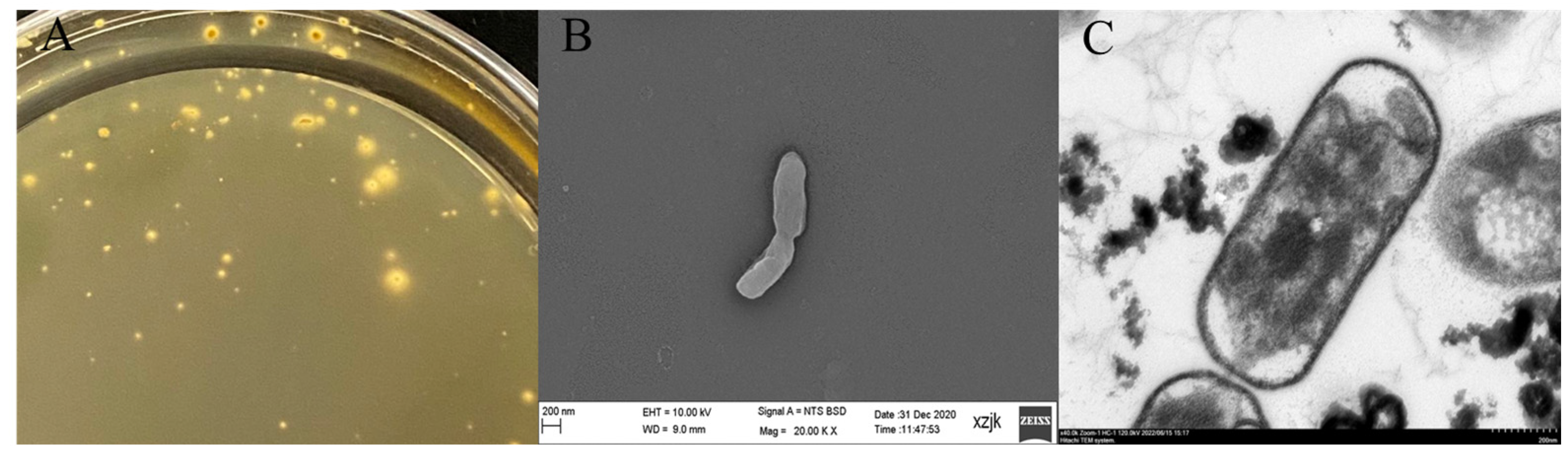
2.1. Isolation, Purification, and Identification

Due to the growth characteristics of A. ferrooxidans, Leathen or 9K media are often used during the isolation and purification of this bacterium [10]. In this study, 9K medium was selected for the isolation and purification of A. ferrooxidans. The 9K liquid medium is composed of solutions A and B. Solution A is composed of 3.00 g/L (NH4)2∙SO4, 0.10 g/L KCl, 0.655 g/L K2HPO4·3H2O, 0.50 g/L MgSO3·7H2O, and 0.01 g/L Ca(NO3)2·4H2O. After the preparation of solution A, the pH of the solution was adjusted to 1.8 with 1:1 concentrated sulfuric acid and sterilized in an autoclave at 121 °C for 20 min. Solution B contains FeSO4·7H2O and was sterilized using a microporous membrane (d = 0.22 μm). After cooling down to room temperature, Solution A was evenly mixed with Solution B before use. Furthermore, the 9K solid medium contained Solution A, Solution B, and Solution C. Solutions A and B were prepared as described above, whereas solution C was a 7.5 g/L agarose solution. After solution C was heated and dissolved, it was sterilized in an autoclave at 121 °C for 20 min. Once solution A was cooled to approximately 60 °C, it was mixed with Solution B. When the pH was adjusted to 2.0, Solution C was quickly added to the mixture to make a solid medium [11].
The strains used in this study were isolated from sediments of a river polluted by AMD from an abandoned mine in Shanxi, China. Following aseptic procedures, the sediment samples were collected and stored at 4 °C and then transported to the laboratory. Next, 10 g of sediment sample was added to 100 mL of sterile water and the mixture was incubated at 30 °C and 180 r/min for three days. The samples were then filtered to obtain a bacterial solution, which was stored at 4 °C. The bacteria-containing solution was inoculated into an Erlenmeyer flask containing 9K liquid medium at a 10% inoculum volume. Next, the flask was covered with a perforated sealing film (polypropylene, Beckman) and transferred to a constant temperature incubator at 30 °C and 180 r/min for continuous enrichment culture. Once the bacteria had multiplied until the medium turned reddish–brown, the culture was inoculated into a new 9K liquid medium at a 10% inoculum volume rate, and isolation and purification were performed after 5–6 consecutive enrichments. The enrichment experiments were conducted in triplicate. An alternate solid–liquid culture method was used for the isolation and purification of A. ferrooxidans. The gradient dilution method was adopted during the isolation. Sterile sulfuric acid solution (pH = 1.8) was used to dilute the enriched bacterial liquid samples, and each sample was sequentially diluted from the initial concentration 10, 102, 103, 104, 105, and 106 times. Next, 0.2 mL of bacterial solutions at different concentrations was inoculated into the solidified 9K solid medium and evenly coated using a sterile coating rod. After coating, the Petri dish was placed upright in a constant temperature incubator at 30 °C for 10 h, after which the Petri dish was incubated upside down to prevent contamination caused by the backflow of water to the 9K solid medium. Isolation and purification were performed when the liquid medium turned reddish–brown. This process was repeated until pure strains were obtained.
A small amount of bacterial solution was taken after isolation and purification, after which a 1–1.5 cm diameter bacterial smear was evenly spread on a glass slide with a sterile inoculation loop, and ammonium oxalate crystal violet staining solution was added in a dropwise fashion to the smeared area, which was then stained for 1 min and washed with water. After blotting with absorbent paper, anhydrous ethanol was also added in a dropwise fashion to cover the entire smear area and then washed with water for 30 s. After blotting with absorbent paper, safranin dye solution was added in a dropwise fashion and stained for 1 min. The samples were then washed with water, blot dried with absorbent paper, and placed under a light microscope to observe bacterial morphology and Gram staining. Afterward, 100 mL of bacterial liquid cultured to the logarithmic phase was centrifuged at high speed at 4 °C, fixed with 2.5% glutaraldehyde overnight, and centrifuged to store the bacteria. The bacteria were subjected to gradient dehydration with 30%, 50%, 75%, 90%, and 100% ethanol (10 min per step). The samples were then freeze-dried at −20, −40, −60, and −80 °C and kept at each temperature for 12 h. After sample loading and gold spraying, the samples were examined via scanning electron microscopy (Regulus 8100, Hitachi High-Tech, Tokyo, Japan). Next, 20 μL of resuspended samples were placed dropwise on 200-mesh grids and incubated at room temperature for 10 min. Then, the cell morphology was examined using a transmission electron microscope (HT7700, Hitachi High-Tech, Tokyo, Japan).
Bacterial cultures in the logarithmic phase were frozen and centrifuged to obtain the bacterial cells for DNA purification and taxonomic classification based on 16S rRNA gene sequences. The extracted sample DNA was amplified by 16S rRNA, and PCR amplification was performed using a PCR instrument (2720 PCR instrument) with the 27F (5’- AGA GTT TGA TCC TGG CTC AG-3’) and 1492R (5’-GGT TAC CTT GTT ACG ACT G-3’) primer pair. The PCR amplification reaction system (30 μL) included 2 X EasyTaq SuperMix (15.0 μL), 27F (1μL), 1492R (1μL), DNA (1–2 μL), and ddH2O (up to 30 μL). The amplification consisted of a pre-denaturation step at 94 °C for 5 min, 25 cycles (denaturation at 94 °C for 30 s, annealing at 57 °C for 30 s, extension at 72 °C for 90 s), extension at 72 °C for 7 min, and storage at 4 °C. The amplicons were sequenced with a 3730 sequencer for the first generation of double-ended sequencing to obtain an ABI sequencing peak map file. After assembly, the sequence was compared with the 16s sub-library to identify closely related species. Upon comparing the 16S rRNA sequences in the NCBI database, the 19 strains that were closest to the species level were selected and the NJ (Neighbor-Joining) method was used to construct a phylogenetic tree using the MEGA 6.0 software to visualize the evolutionary relationship of the sample and the near-source species.
The bacteria obtained in the experiment were identified as pure A. ferrooxidans, after which their growth conditions were studied, including the optimum growth pH, temperature, and inoculum size. The cells were cultured in growth media at different pH conditions (pH 1.2, 1.8, 2.4, 3.0, 3.6, and 4.2) to explore the optimal pH conditions for bacterial growth. The cells were cultured at 15, 20, 25, 30, 35, and 40 °C to study the optimal growth temperature. Finally, the cultures were inoculated with 5%, 10%, 15%, and 20% of the inoculum to explore the optimal inoculum amount. A blood cell counting plate was used to observe and calculate the bacterial concentration.

2.2. Whole genome Sequencing

The bacteria collected above were sent to Beijing BMK Biological Co., Ltd., and whole genome sequencing was carried out using PacBio sequencing technology. The experimental process was performed according to the standard protocol provided by PacBio [12], including sample quality detection, library construction, library quality detection, and library sequencing. Low-quality and short fragments with a length of less than 2000 bp were removed from the reads obtained after sequencing, and clean reads were obtained for genome assembly and functional annotation. To fully display the features of the genome, a genome circle diagram of a single sample was generated using the Circos software (Version 0.69-6, http://www.circos.ca (accessed on 4 January 2023)) and a variety of information was displayed in the diagram to provide a comprehensive and intuitive understanding of the characteristics of the bacterial genome. Chromosome genes were predicted using Glimmer and the plasma genome was predicted using GeneMarkS [13,14]. The predicted genome was compared with the COG and KEGG databases based on protein sequence, and functional annotation was performed to obtain the corresponding gene function information. Upon sequencing the whole genome of A. ferrooxidans YQ-N3, short-length circular consensus sequencing (ccs) reads were filtered out from the raw data as a quality control measure. The filtered ccs reads were assembled de novo, and the assembled genome was corrected for errors. Once the assembly was completed, genome analysis and functional annotation were performed. Mobile genetic elements (MGEs) can help bacteria overcome challenges or occupy dominant niches. Therefore, studying the MGEs in the A. ferrooxidans YQ-N3 genome could provide insights into their ability to adapt to environmental changes. In this study, we analyzed three types of MGEs: genome islands, prophages, and CRISPR (clustered regularly interspersed short palindromic repeats)-Cas. Gene island prediction of the A. ferrooxidans YQ-N3 genome was performed using Islander (Version 1.2, http://www.pathogenomics.sfu.ca/islandviewer/ (accessed on 4 January 2023)) [15]. Prophage prediction was performed using Phage_Finder (phage-finder.sourceforge.net) [16]. CRISPR-Cas prediction was performed using Minced [17].

2.3. Comparative Genomic Analysis

To further examine the genomic characteristics of A. ferrooxidans YQ-N3, the genomes of five strains closest to it at the species level based on 16S rRNA sequences were selected for comparative analysis. The genome sequences of the selected strains (A. ferrooxidans ATCC23270, A. ferrooxidans BY0502, A. ferridurans JCM18981, A. thiooxidans ATCC 19377, and A. ferrivorans SS3) were obtained from the GenBank database. Glimmer 3.0 (http://ccb.jhu.edu/software/glimmer/index.shtml (accessed on 4 January 2023)) and GeneMarkS (Predictive Plasmid Genome) software (Version 4.3, http://topaz.gatech.edu/GeneMark (accessed on 4 January 2023)) were used to predict the coding sequence (CDS) of genome. The core gene and pan-genome in the predicted sequence were analyzed using BPGA (Bacterial Pan-genome Analysis tool ver. 1.2, https://iicb.res.in/bpga/index.html (accessed on 4 January 2023)). The empirical power law equation and exponential equation were used to calculate the extrapolation of the pan-genome and core genome curves, respectively [18,19]. ANI analysis of the selected genome was conducted by using the pyani software following the ANIm method. AAI analysis of the selected genome was performed using the CompareM software (https://github.com/dparks1134/CompareM (accessed on 4 January 2023)). Genome alignments between A. ferrooxidans YQ-N3 and A. ferrooxidans ATCC23270, A. ferrooxidans BY0502, A. ferridurans JCM18981, A. thiooxidans ATCC 19377, and A. ferrivorans SS3 were conducted using the MUMmer software (ver. 3.23, https://sourceforge.net/projects/mummer/files/latest/download (accessed on 4 January 2023)) related to the ucmer comparison method. Based on protein sequence alignments, the coding genes predicted on the A. ferrooxidans YQ-N3 and the genome of the other five studied strains were compared with the COG (clusters of orthologous groups of proteins) databases for functional annotation, and the corresponding functional annotations were obtained.

2.4. Oxidation Characteristics of Fe2+, S0, and FeS2

As the dominant microbe in metallic sulfide mining areas, A. ferrooxidans plays a vital role in Fe and S biogeochemical cycling [20]. Therefore, studies on the Fe2+, S0, and FeS2 oxidation capacities of novel A. ferrooxidans strains are essential, specifically due to their theoretical and practical values.
This study characterized the A. ferrooxidans strain YQ-N3 isolated in the previous stage, and its ability to oxidize FeSO4·7H2O, S0, and FeS2 as energy sources was discussed. The preserved YQ-N3 strain was transferred to 9K medium containing FeSO4·7H2O, S0, and FeS2 with 10% of the inoculum, and was then placed in a constant temperature culture shaker at 30 °C and 180 r/min for multiple activations. The bacterial density was determined using a hemocytometer, after which acclimated log-phase bacteria were collected. The domesticated strains were inoculated into 9K medium containing FeSO4·7H2O, S0, and FeS2 at a 10% inoculum volume proportion, and the bacterial concentration and Fe2+, Fe3+, SO42−, pH, and Eh in the culture system were monitored thereafter. The specific experimental design is shown in Table 1. Three parallel groups and one blank control were set for each group of experiments.
At the end of the experiment, mineral samples generated via the oxidation of Fe2+ and FeS2 before and after the reaction were collected. The samples were then ground and screened for mineral composition analysis. The phases and components of the collected samples were determined via X-ray diffraction (XRD) using an X-ray diffractometer (Bruker D8 ADVANCE, Karlsruhe, Germany) with Cu Kα radiation operating at 40 kV and 30 mA. The samples were scanned at a 0.02 °/s rate to record the patterns within a 2θ range of 3–105°; the mineral composition of the sample was then analyzed.

2.5. Coal Biodesulfurization Experiment

To explore the applicability of A. ferrooxidans YQ-N3 in coal desulfurization, three coal samples with different sulfur content from different mining areas in China were selected to conduct biological desulfurization experiments. The sulfur contents of the coal samples are shown in Table 2. The coal desulfurization experiments were conducted using a sequential batch experiment approach. Each coal sample was set with six groups of experiments, which included three replicates and one blank. Coal samples were taken at 5, 10, 15, 20, 25, and 30 days, respectively, to detect the sulfur contents. The experiment was carried out in 250 mL conical flasks in a constant temperature culture shaker at 30 °C and 180 r/min. All conical flasks contained 90 mL 9K liquid medium (free Fe2+), 5 g of coal sample powder (200 mesh), and 10 mL of A. ferrooxidans YQ-N3 bacterial solution (2.0 × 10⁷ cells/mL). The initial pH of all growth media was adjusted to 2. The total sulfur and sulfur contents of coal samples were analyzed according to the national standards (GB/T214-1996 and GB/T215-1996) of China.

3. Results and Discussion

3.1. Growth Characteristics of A. ferrooxidans YQ-N3

In this study, sediment was collected from a river polluted by AMD of an abandoned mine in Shanxi, China. The sediment samples were treated with sterile water and inoculated in 9K-Fe2+ liquid medium for enrichment culture. After cultivation, all three replicate cultures exhibited the same characteristics. The color of the medium gradually changed from light blue–green to turbid, after which it became clear red–brown after continued cultivation. After culturing on 9K-Fe2+ solid medium for 30 days via the dilution coating method, colonies appeared on the plate, as illustrated in Figure 1A. The colonies were round, with prominent centers, neat edges, and yellow surroundings. After repeated solid–liquid alternating culture, the bacteria were collected, and DNA was extracted for 16S rRNA gene amplification and sequencing. After gene amplification, there was a single clear target band, which has 99.93% similarity with Acidithiobacillus ferrooxidans (NR_074193.1) according to the comparison between the sequencing results and BLAST. Therefore, the bacterial strain was named A. ferrooxidans YQ-N3. The collected bacteria were diluted several times using normal saline and then observed via Gram staining, thus confirming that the bacteria were Gram-negative. Optical microscope observations showed that the bacteria were rod-shaped (both as single cells and aggregates) and were able to swim rapidly. SEM and TEM analyses indicated that the bacteria had a short rod shape with blunt rounded ends, as shown in Figure 1B,C, with a length of approximately 0.8–1.2 μm and a width of 0.2–0.5 μm.
Upon comparing the 16S rRNA sequences in the GenBank database, the 18 strains that were closest at the species level were selected, and the NJ method was used to construct the phylogenetic tree of A. ferrooxidans YQ-N3 using the MEGA 6.0 software, as shown in Figure 2. The phylogenetic tree analyses demonstrated that A. ferrooxidans YQ-N3 was distinct from Pseudomonas, Oceanicoccus, Luteimonas, and Xanthomonas, but appeared to be related to A. thiooxidans, A. ferridurans, and A. ferrivorans. Particularly, the 16S rDNA sequence of A. ferrooxidans YQ-N3 was more than 99.93% similar to that of A. ferrooxidans ATCC23270, and it was therefore concluded that the isolated strains were A. ferrooxidans.
After screening and obtaining pure A. ferrooxidans YQ-N3 cultures, the optimum growth conditions were explored. Our findings demonstrated that A. ferrooxidans YQ-N3 could grow at a pH range of 1.6–2.4 and a temperature range of 20–35 °C, and were therefore consistent with previous studies. The strain achieved optimal growth at an initial pH of 1.8, a temperature of 30 °C, and an inoculum volume of 10%. Under these conditions, A. ferrooxidans YQ-N3 could reach a density of up to 2.3 × 10⁸ cells/mL after 16 h.

3.2. Genome Overview of A. ferrooxidans YQ-N3

Whole genome analysis indicated that the genome size of A. ferrooxidans YQ-N3 was 3,217,720 bp, including one circular chromosome and five circular plasmids (Plasmid A, Plasmid B, Plasmid C, Plasmid D, and Plasmid E). The sequence length of the circular chromosome was 3,043,496 bp and the GC content was 58.64%. To fully demonstrate the genome features of A. ferrooxidans YQ-N3, a genome circle plot was drawn using the Circos software (Figure 3). A total of 3200 coding sequences (CDs), 6 rRNAs, 46 tRNAs, and 18 sRNAs were predicted. The annotation results of plasmids are shown in Table 3. The sequence length of the circular Plasmid A was 79,659 bp and the GC content was 61.64%. The length of Plasmid B was 34,460 bp and the GC content was 60.54%. The sequence length of Plasmid C was 29,178 bp and the GC content was 62.67%. The sequence length of Plasmid D was 23,017 bp and the GC content was 60.69%. The sequence length of Plasmid E was 7910 bp and the GC content was 52.40%. Among them, the Plasmid E sequence had not been annotated in the reference database. The complete genome sequences for the main chromosome and Plasmids A-E of A. ferrooxidans YQ-N3 were submitted to the GenBank database under accession numbers CP084172.1, CP084173.1, CP084174.1, CP084175.1, CP084176.1, and CP084177.1, respectively. A. ferrooxidans can grow in a variety of extreme environments and has great potential applications in industries such as bioleaching and biological desulfurization [21,22,23]. However, there are 26 published genomes of A. ferrooxidans strains, and therefore the whole genome sequencing and analysis of A. ferrooxidans YQ-N3 could provide key insights into its genetic properties and potential applicability. To understand the differences between the genomes of A. ferrooxidans YQ-N3 and other A. ferrooxidans strains, the genome information of A. ferrooxidans YQ-N3 and A. ferrooxidans that has been published in GenBank was selected for tabulation analysis, as shown in Supplementary Materials Table S1. The genome size distribution of A. ferrooxidans was 2.82631–4.18422 Mb, the GC content was 56.8%–58.9%, and the CDS (coding sequence) distribution was 2705–3998. The genome size, GC%, and number of CDSs of A. ferrooxidans YQ-N3 are similar to those of other A. ferrooxidans strains. However, with the exception of A. ferrooxidans YQ-N3, the assembled A. ferrooxidans genome in the published database included no more than five plasmids.

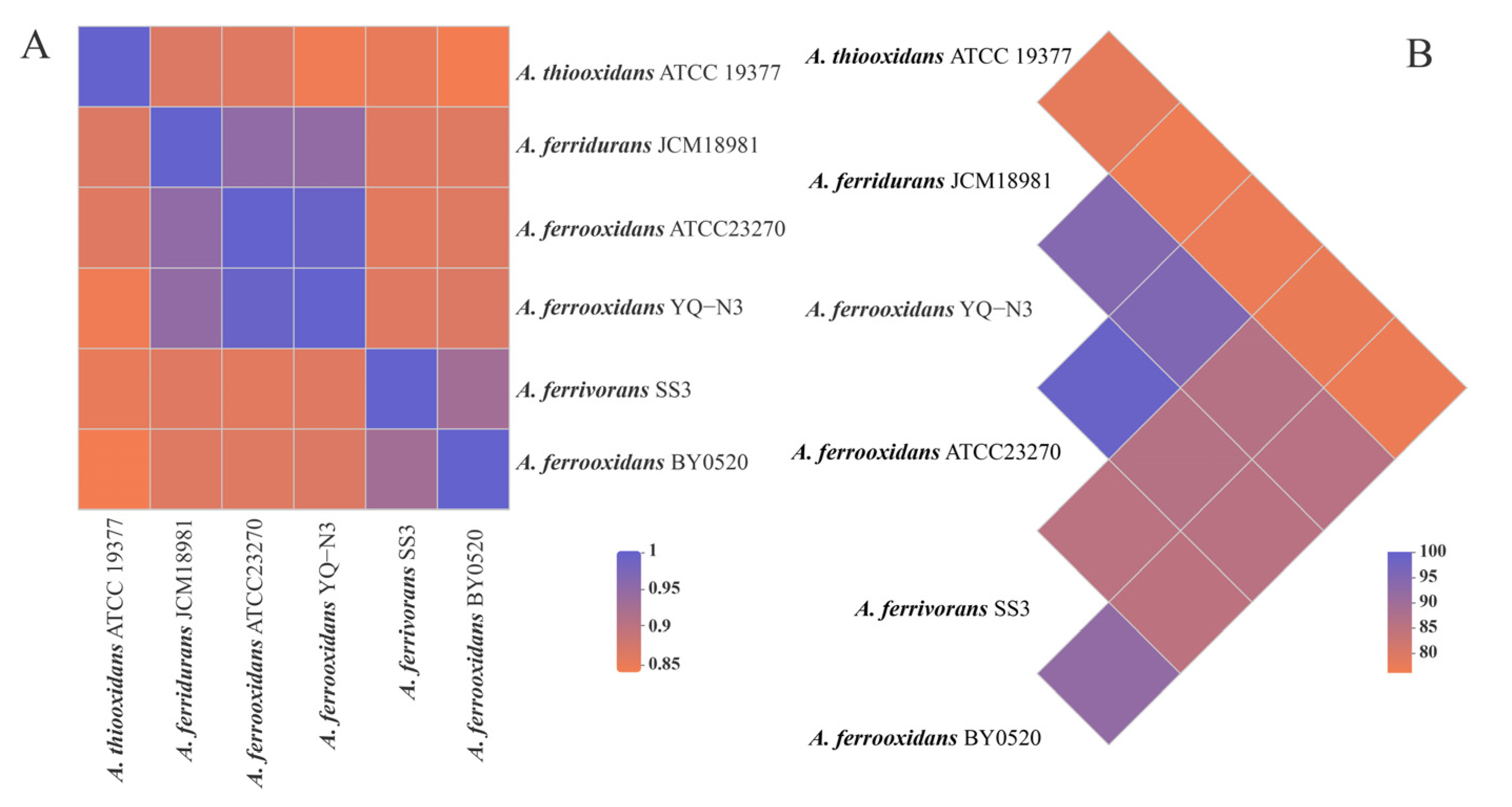
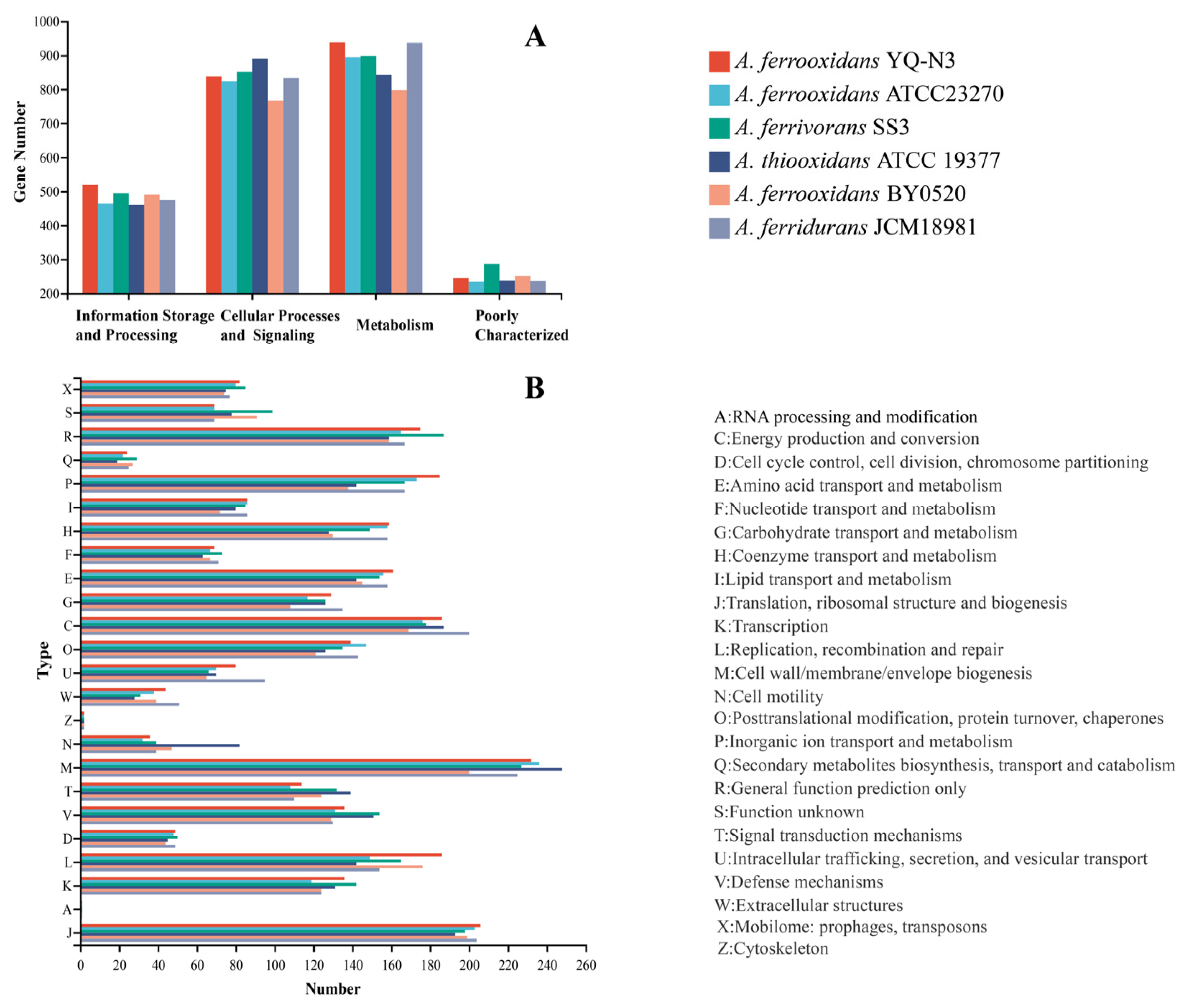
3.3. Comparative Genomic Analysis

A summary of the features of six selected genomes is listed in Table 4. These strains were collected from different environments around the world but mainly from acid mine drainage. These genomes vary in size by approximately 0.44 Mb (ranging from 2.98 to 3.42 Mb) with coding sequence (CDS) numbers ranging from 2927 to 3498 and the G+C contents of the genomes ranging from 53% to 58.8%. Figure 4 shows the results of ANI and AAI analysis among strains. The squares with different colors show the similarity of nucleotides and amino acids among stains. It can be seen that A. ferrooxidans YQ-N3 is closely related to A. ferrooxidans ATCC23270 and A. ferridurans JCM18981. The ANI value and AAI value between A. ferrooxidans YQ-N3 and A. ferrooxidans ATCC23270 exceeded 95%, indicating that A. ferrooxidans YQ-N3 and A. ferrooxidans ATCC23270 are the same species. The results of the whole genome alignment between A. ferrooxidans YQ-N3 and the selected genomes are shown in Figure 5. Our findings indicated that the genomic arrangement of A. ferrooxidans YQ-N3 has the best co-linearity with A. ferrooxidans ATCC23270, and has better co-linearity with A. ferridurans JCM18981 than with A. ferrooxidans BY0502. This result demonstrates that A. ferrooxidans YQ-N3 and A. ferrooxidans ATCC23270 have closer evolutionary distances and highlights the abundant evolutionary progress of A. ferrooxidans.
The bacterial pan-genome (BGPA) was used to identify orthologous groups among the studied genomes to reveal the unique genomic characteristics of the studied strains. The results are shown in Figure 6. The pan-genome contains 6062 genes, including 1400 core genomes and 2918 unique genomes. Figure 6B shows the curve of the relationship between the pan-genome size, core genome size, and genome number. According to the formula of the relationship curve between the pan-genome size and genome number (f(n) = 2239.763n0.497 + 580.101), we can conclude that the pan-genome in this study is open-type (0 < 0.497 < 1) [24], which means that as the number of sequenced genomes continues to increase, the pan-genome size will increase indefinitely. The core genome is composed of genes that exist in all strains and is generally related to the basic biological functions and main phenotypic characteristics of bacteria. The unique genome is composed of genes only existing in some strains and genes unique to a strain, which reflects the diversity of strains and is generally related to some special biological metabolic pathways and environmental adaptation, such as antibiotic production, tolerance, and virulence. The rich specific genome content in this study indicates that the studied strains may undergo horizontal gene transfer (HGT) to improve their own survival competitiveness and increase microbial diversity [25].
Figure 7 shows the COG annotation category and function statistics of the studied strains. These COGs were divided into four categories and 24 functional classes. The proportion of genes related to replication, recombination, and repair (L), cell wall/membrane/envelope biogenesis (M), energy production and conversion (C), and inorganic ion transport and metabolism (P) was slightly higher than that of genes with other functions, accounting for 5.55%–7.18%, 8.16%–9.71%, 6.66%–7.58%, and 5.55%–6.89% of all annotated genes, respectively. Additionally, defense mechanisms accounted for 4.92%–5.91% of all annotated genes, suggesting that these strains have a strong ability to self-repair and resist harsh environments [1]. A. ferrooxidans YQ-N3 has a total of 2571 genes annotated in COG, accounting for approximately 80.34% of the total number of genes. Compared to other strains, A. ferrooxidans YQ-N3 had the most genes associated with functions supporting P, L, and J (translation, ribosomal structure, and biogenesis). Moreover, the distribution of functional genes in the A. ferrooxidans YQ-N3, A. ferrooxidans ATCC23270, and A. ferrooxidans BY0502 genomes showed a similar pattern.
A. ferrooxidans is widely found in metal mines, high-sulfate coal mines, and other extreme environments due to its high environmental resistance, as well as its tolerance to metal ions and high acidity [26]. The characterization of genes associated with environmental resistance in A. ferrooxidans YQ-N3 would provide insights into the mechanisms that mediate its adaptability, which also lays the foundation for future research on its survival state and industrial application value in extreme environments. The results of COG annotation showed that the YQ-N3-gene-1491 and YQ-N3-gene-2286 located in the chromosome were annotated as ENOG410ZI6F and their COG description was “heavy metal transport detoxification protein”, which further suggested that this strain was resistant to heavy metals. Some metal resistance genes have also been found, such as cusA, cusB, cusR, and cusS. Previous studies have demonstrated that the cus proton pump system is widely distributed in Gram-negative bacteria and is associated with copper and silver resistance in A. ferrooxidans [27]. In fact, cusA, cusB, and cusF possess metal-binding sites, and Cu2+ is directly transferred from protein to protein in the cus system during ion transport, thereby reducing the potential toxicity of free Cu2+ to cells [28,29]. Nan et al. linked the adaptability of A. ferrooxidans to chemotactic movement and quorum sensing (QS), both of which allow this bacterium to grow, develop, and reproduce in extreme environments [30].

3.4. MEGs

More than 20% of genes in microbial genomes may be obtained through horizontal gene transfer (HGT), and these genes are usually referred to as mobile genetic elements (MGEs) [31,32]. MGEs refer to some exogenous gene fragments by way of the HGT that can be incorporated into bacterial genomes to adapt to environmental changes or improve the likelihood of survival. These fragments generally contain genes that encode the enzymes of other proteins with specific functions to help bacteria overcome adverse conditions or take advantage of resources that otherwise could not be exploited. The gene islands often carry functional genes, contain integrase and plasmid conjugation-related factors [33]. The prophage sequence often contained some functional genes, such as antibiotic resistance genes and virulence genes, among others, all of which enhanced the adaptability of this bacterium to its environment [34]. The CRISPR/Cas system is a prokaryotic immune system designed to resist the invasion of exogenous genetic material [35]. Integrators can enhance the adaptability of bacteria by capturing exogenous genes. Some studies have indicated that HGT is the main mechanism for acidophilic bacteria to adapt to heavy metal-rich environments [36,37,38]. The research on MGEs is helpful to further explore the whole genome characteristics of species. In this study, we analyzed the MGEs of A. ferrooxidans YQ-N3 from phages, genomic islands (GIs), and integrons. The predicted results indicated that the chromosome of A. ferrooxidans YQ-N3 contained 10 gene islands and Plasmid A contained one gene island. In turn, these gene islands contained a total of 181 CDs. The linear map of genome islands is shown in Figure 8. The description of genes 1255, 1259, 1624, 1270, and 1274 are “DEAD/DEAH box helicase”, “hypothetical protein”, “phage anti-repressor Ant”, “M48 family metalloprotease”, and “MULTISPECIES: restriction endonuclease”, respectively. Interestingly, our analyses indicated that a prophage genome had integrated into the chromosome of A. ferrooxidans YQ-N3. The sequence length of the prophage was 17,089 bp and had 19 CDs, in addition to having a 58.64% GC content. The chromosome of A. ferrooxidans YQ-N3 was predicted to contain four CRISPRs with average repeat lengths of 27, 28, 23, and 27 bp, respectively. The average lengths of the spacer sequences were 27, 32, 43, and 45 bp, respectively. An integron was also found on a chromosome of A. ferrooxidans YQ-N3. Its structure is shown in Figure 9; its length was 123 bp, including 9 CDs. Therefore, from the perspective of the whole genome, A. ferrooxidans YQ-N3 is likely resistant to heavy metals and is highly adaptable, making it a promising candidate for various industrial applications such as biological desulfurization, bioleaching, metal processing, and others.

3.5. Genes Associated with Iron and Sulfur Metabolism

To further understand the whole genome characteristics of A. ferrooxidans YQ-N3, its genes related to iron and sulfur metabolism were deeply explored from a functional gene perspective. According to the KEGG annotation results of A. ferrooxidans YQ-N3, the genes related to iron and sulfur metabolism are summarized in Table S2. Our findings demonstrated that this strain possesses multiple genes related to Fe2+ oxidation metabolism and sulfur metabolism. For example, cyc2, cyc1, coxB, coxA, and coxC, which encode the Rus operon components, were identified in A. ferrooxidans YQ-N3. Moreover, hdrB, hdrA, hdrC, tusA, and dsrE, which encode the HdrABC (isodisulfide reductase complex), were also detected. A. ferrooxidans is a chemoautotrophic bacterium with a complex energy metabolism pathway, which obtains energy through the oxidation of Fe2+ and reduction of sulfur compounds, and generates energy to sustain its growth by fixing carbon and nitrogen in the air [39,40]. The oxidative metabolism of ferrous and sulfur compounds by A. ferrooxidans makes this bacterium especially well suited to industrial applications [41]. Previous studies have demonstrated that Fe2+ oxidation by A. ferrooxidans in the electron transport chain has two patterns, including the downhill potential gradient and the uphill potential gradient [42]. Most electrons generated via Fe2+ oxidation pass along the downhill potential gradient. Specifically, electrons generated by Fe2+ oxidation enter the cytoplasm from the outer cell membrane, then pass to the cytochrome protein cyc2, after which they are received by rus, a ceruloplasmin, and passed to cyc2, a cytochrome protein. Afterward, they are received by cytochrome oxidase aa3 and then passed to O2, where they finally combine with protons to generate H2O [43]. In this process, the oxidation process of the rus operon plays a dominant role, and the rus operon is composed of multiple genes including cyc2, coxA, coxB, coxC, and rus [44]. Furthermore, studies on the regulation of the expression of key rus operon genes in model microorganisms show that the expression of the rus operon is induced by Fe2+ [45,46]. The sqr gene, which encodes the sulfide quinone reductase enzyme, has been previously identified in various A. ferrooxidans strains [47]. doxDA encoding thiosulfate quinone reductase was also identified in ATCC23270 and CCM4253. When elemental sulfur was used as an energy source, the transcription level of the heterodisulfide reductase (HdrABC) complex encoded by ten genes including hdrB, hdrA, hdrC2, tusA, and hdrB2 was significantly upregulated, indicating that this complex was related to sulfur metabolism in A. ferrooxidans [48]. The query of functional genes confirmed that A. ferrooxidans YQ-N3 can oxidize Fe2+ and sulfur, but its metabolic pathways require being further explored.

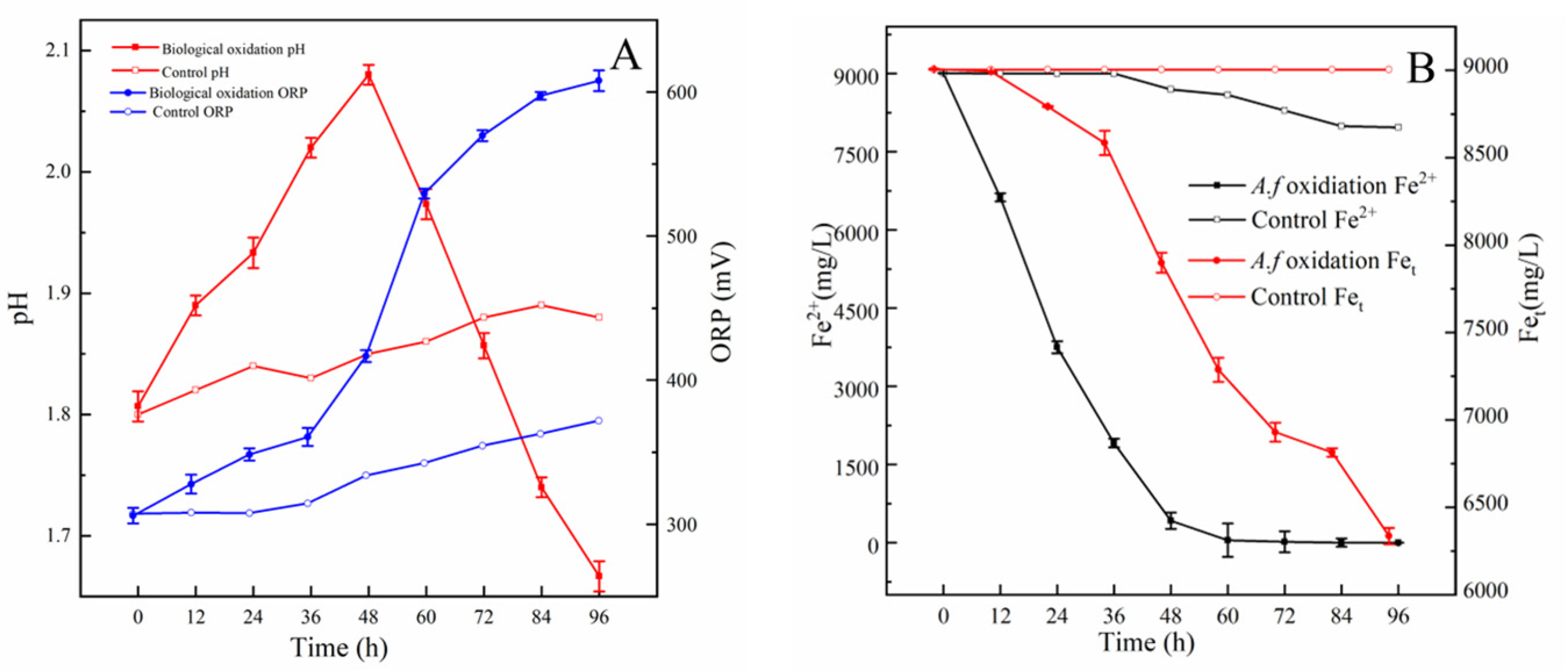
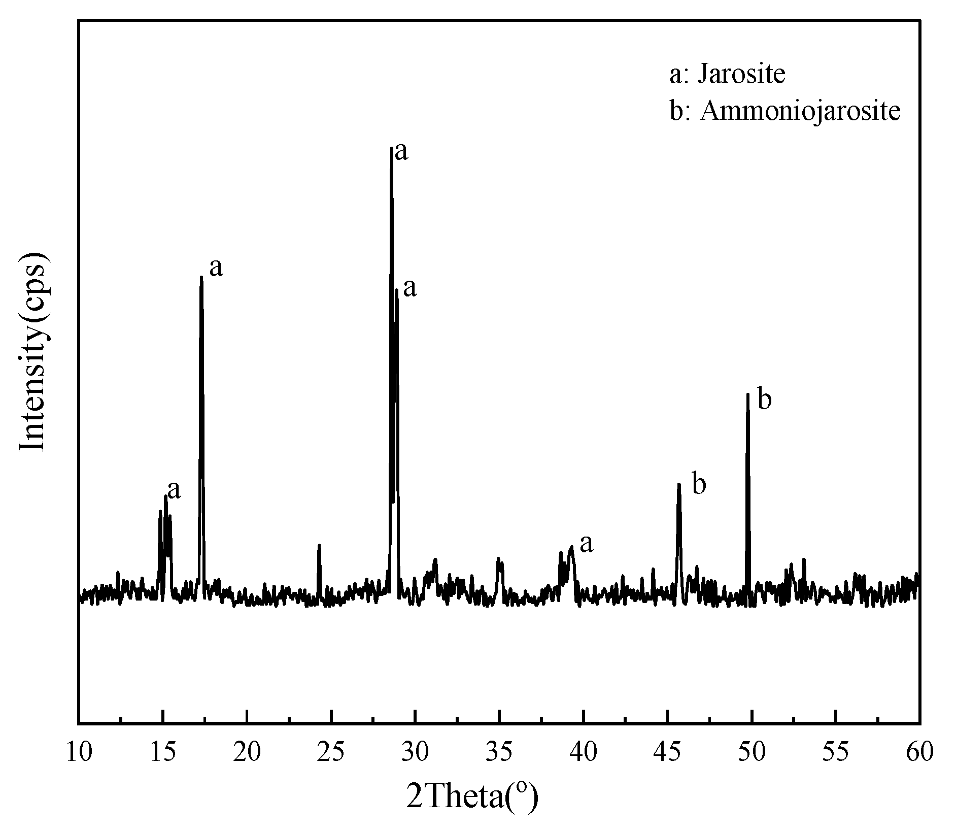
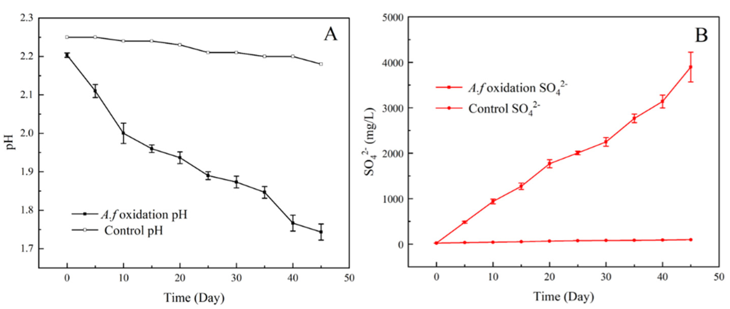
3.6. Oxidation of Fe2+, S0, and Pyrite

During Fe2+ oxidation by A. ferrooxidans YQ-N3, the color of the medium gradually shifted from clear light green to yellow–green at first. After 16 h, the medium completely changed to reddish–brown. Then, a yellow precipitate gradually appeared in the reaction system. The blank control group did not exhibit any obvious color change. Figure 10 shows the trends of pH, ORP, Fe2+ content, and total iron content of the medium during Fe2+ oxidation. According to Figure 10A, the pH in the experiment involving A. ferrooxidans YQ-N3 increased from 1.8 to 2.16 after 48 h, and then rapidly decreased to 1.7. ORP also increased throughout the experiment but this trend decelerated slightly after 48 h. In the blank control group, pH and ORP showed a slight upward trend throughout the experiment. This can be attributed to H+ consumption by the oxidation of Fe2+ into Fe3+ [49]. Fe3+, SO42−, and other cations (e.g., K+, Na+, NH4+) begin to react as the Fe3+ content increases, which results in the production of H+ and the minerals jarosite (KFe3(SO4)2(OH)6) and ammoniojarosite ((NH4)2Fe6(SO4)4(OH)12) (Figure 11), which is consistent with previous studies [50]. Therefore, the pH rapidly decreased and the ORP increase rate slowed down in the later stage of the experimental group [51]. Figure 10B shows that the Fe2+ content in the experiment involving A. ferrooxidans YQ-N3 continued to decrease rapidly and the Fe2+ content reached 0 mg/L when the reaction reached 60 h. The total iron concentration decreased by approximately 2700 mg/L during the whole reaction period, whereas only 14.5% Fe2+ was oxidized and the total iron concentration in the blank control group remained unchanged for 96 h. This can be attributed to the fact that A. ferrooxidans YQ-N3 accelerates the oxidation of Fe2+, and the Fe3+ generated by the reaction leads to a decrease in the total iron content in the liquid phase. Fe2+ is naturally oxidized and generates free Fe3+ in the blank control group during the reaction, and therefore the total iron content remained unchanged in the system.
During the oxidation of S0 by A. ferrooxidans YQ-N3, the medium gradually changed from clear and colorless to light yellow, the particle size of sulfur powder gradually became smaller, and its hydrophobicity gradually weakened. However, no obvious change was observed during the reaction of the blank control group. This phenomenon was mainly because A. ferrooxidans adsorbed on the surface of S when oxidizing it and secreted hydrophilic organic substances that covered the surface of S to enhance its hydrophilicity [52,53]. During the oxidation of S0 by A. ferrooxidans YQ-N3, the pH value of the medium decreased from 2.2 to 1.74, whereas the SO42− concentration of the medium increased to 3896.66 mg/L after 45 d (Figure 12). This can be attributed to the release of H+ through the oxidation of S0 to SO42−.
Figure 13 illustrates the trends of pH, ORP, SO42−, Fe2+, and Fet during pyrite oxidation by A. ferrooxidans YQ-N3. In the experimental group, the pH value decreased from 1.9 to 1.2 after 45 d, the ORP increased from 353 to 632 mV after 45 d, the SO42− concentration increased to 5433.8 mg/L after 45 d, the Fe2+ content decreased drastically from 43.6 mg/L to a negligible level after 23 d, and the Fet content increased to 3853 mg/L after 45 d. Characterization by XRD revealed that pyrite oxidized by A. ferrooxidans YQ-N3 contains jarosite and FeOOH, in addition to quartz impurities (Figure 14). In the blank control group, the pH value decreased slightly, the ORP decreased and then slightly increased, the SO42− concentration increased to 563 mg/L after 45 d, the Fe2+ content increased to 178.2 mg/L after 45 d, and the Fet concentration increased to 197.7 mg/L after 45 d. This could be because the H+ released by the oxidation of S0 into SO42− exceeded the levels consumed by Fe2+ oxidation. A. ferrooxidans YQ-N3 can accelerate the oxidation of Fe2+ to Fe3+ and pyrite oxidation can be carried out continuously and rapidly. The dissolved Fe mainly exists in the form of Fe3+ in this system, and therefore ORP continues to increase.
The oxidation of different energy substances (Fe2+, S0, and pyrite) by A. ferrooxidans YQ-N3 was investigated via the shake bottle experiment. The results indicate that A. ferrooxidans YQ-N3 can accelerate the oxidation of Fe2+ and that it contributes to the formation of minerals such as jarosite and ammoniojarosite while also accelerating the oxidation of S0 and enhancing its hydrophilicity. A. ferrooxidans YQ-N3 can also achieve rapid and continuous oxidation of pyrite by accelerating Fe2+ oxidation, thereby increasing the ORP. As a result, the pH value decreased, the SO42− concentration increased, and minerals (e.g., jarosite and FeOOH) were generated, all of which are hallmarks of the AMD generation process [54]. These findings suggest that A. ferrooxidans YQ-N3 can accelerate the production of AMD.

3.7. Desulfurization of Coal

The direct combustion of coal can lead to a variety of environmental pollution problems. For example, the main reason for the formation of acid rain is the SO2 gas released by the direct combustion of coal [55]. Therefore, it is particularly important to develop efficient desulfurization technology. The microbial desulfurization of coal not only promotes environmental protection but is also highly efficient and consumes few resources [56]. A. ferrooxidans is a commonly used coal desulfurization bacteria. Therefore, it is of great significance to study the coal desulfurization ability of A. ferrooxidans YQ-N3. The performance of A. ferrooxidans YQ-N3 during the coal desulfurization is illustrated in Figure 15.
During the desulfurization process, the sulfur removal rate of A. ferrooxidans YQ-N3 on the three coal samples continued to increase, and the sulfur removal rate of coal sample II was higher than that of coal samples III and Ι. After 30 days of reaction, the total sulfur removal rates of the I, II, and III coal samples were 40.17%, 62.25%, and 58.40%, respectively.
Table 5 shows the coal desulfurization characteristics of strain A. ferrooxidans YQ-N3 and other A. ferrooxidans. Our findings indicated that the coal desulfurization ability of A. ferrooxidans YQ-N3 was at a medium level. This may be due to the fact that the strain only grew in the ferrous environment and was not domesticated in any environment before the coal desulfurization experiments. Domesticated strains in specific environments (e.g., medium containing pyrite) and the optimization of desulfurization experimental parameters may improve the desulfurization capacity of A. ferrooxidans YQ-N3 [57].
Additionally, coal chemical analysis of coal samples after the desulfurization reaction was carried out. The results showed that the total sulfur content of Ι after the reaction was 2.07%, including 0.43% pyrite sulfur, 0.36% organic sulfur, and 1.28% sulfate sulfur. The total sulfur content of II was 1.14%, including 0.32% pyrite sulfur, 0.29% organic sulfur, and 0.53% sulfate sulfur. Coal sample III contains 1.165 total sulfur, 0.26% pyrite sulfur, 0.42% organic sulfur, and 0.48% sulfate sulfur. It can be seen that the sulfur removal rate for coal samples with high pyrite content (II) is higher than that for coal samples with high organic sulfur content (III), and higher than that for coal samples with high sulfate sulfur content (Ι). Some studies showed that the desulfurization capacity of A. ferrooxidans is mainly due to the ability of this bacterium to oxidize pyrite in coal [8]. The mechanisms through which A. ferrooxidans oxidizes the pyrite in coal can be divided into two modes: direct action and indirect action. The direct action mechanism refers to that during the oxidation of pyrite. Particularly, the bacteria uses the adhesion of fixed organs, pili, or mineral surfaces to directly adsorb on the pyrite surface. Pyrite is directly decomposed into ferrous ions and sulfur through protein secretion or other metabolites (ferrous oxidase, cytochrome C oxidase, cytochrome oxidoreductase, etc.), and sulfur is further oxidized to SO42− by A. ferrooxidans [58,61]. Indirect action means that although A. ferrooxidans cells adsorb on the surface of minerals, they do not directly oxidize pyrite, but oxidize Fe2+ to Fe3+ in solution. Fe3+ has a strong oxidation ability to oxidize pyrite to release Fe2+ and generate H+ and SO42−. Fe2+ is further oxidized in solution to generate Fe3+. This process is repeated to continuously remove pyrite from coal [62]. The organic sulfur compounds of coal exist in the form of thiols, mercaptans, sulfide, and disulfide linkages, as well as complex thiophene moieties. Organic sulfur is considered more recalcitrant compared to its inorganic counterparts. Rout et al. reported that the microbial desulfurization process assisted by A. ferrooxidans successfully resulted in the removal of 84.75% of organic sulfur from coal samples. The authors also reported that A. ferrooxidans performed oxidative desulfurization of the model sulfur-containing organic compound dibenzothiophene to 2-hydroxy biphenyl through the formation of dioxy-dibenzothiophene [9,63]. Therefore, the results of this study have demonstrated that A. ferrooxidans YQ-N3 cells are capable of biological desulfurization. Therefore, these cells can be used to oxidize pyrite sulfur and organic sulfur in coal.

4. Conclusions

A novel A. ferrooxidans strain was isolated from sediment from a river polluted by the AMD of an abandoned mine in Shanxi, China. Whole genome sequencing analyses revealed that the genome of A. ferrooxidans YQ-N3 includes one circular chromosome and five circular plasmids, among which one plasmid had not been annotated in public databases. A. ferrooxidans YQ-N3 is closely related to A. ferrooxidans ATCC23270 and A. ferridurans JCM18981, and its distribution of functional genes is similar to that of A. ferrooxidans ATCC23270 and A. ferrooxidans BY0502. This newly identified strain possesses multiple genes related to iron and sulfur metabolism. However, additional studies are required to characterize its energy metabolism pathways. The results of functional genome analysis and laboratory experiments jointly demonstrated that A. ferrooxidans YQ-N3 can accelerate the oxidation of Fe2+, S0, and FeS2. Additionally, a 62.25% total sulfur removal rate was achieved in the biological coal desulfurization experiments conducted in this study. This study not only established a theoretical foundation for future research on the generation and treatment of AMD but also isolated a new bacterial strain that could be used for industrial desulfurization, biometallurgy, and the disposal of waste electronic products.

Supplementary Materials

The following supporting information can be downloaded at https://www.mdpi.com/article/10.3390/min13010095/s1, Table S1. General features and genomic comparison between A. ferrooxidans YQ-N3 and the other published genomes of A. ferrooxidans. Table S2. List of selected genes identified in the YQ-N3 strain via KEGG annotation, including genes for iron and sulfur metabolism.

Author Contributions

Conceptualization, Q.F.; investigation, W.L. and Z.L.; data analysis, W.L.; writing, W.L. All authors have read and agreed to the published version of the manuscript.

Funding

This research was supported by the National Natural Science Foundation of China (Grant No. 41977159; “Mechanism and kinetics of microbial acid inhibition in acid mine drainage of an abandoned coal mine”).

Data Availability Statement

Not applicable.

Acknowledgments

We thank Beijing BMK Biological Co., Ltd. for conducting DNA sequencing.

Conflicts of Interest

The authors declare no conflict of interest.

References

  1. Zhan, Y.; Yang, M.R.; Zhang, S.; Zhao, D.; Duan, J.G.; Wang, W.D.; Yan, L. Iron and sulfur oxidation pathways of Acidithiobacillus ferrooxidans. World J. Microbiol. Biotechnol. 2019, 35, 12. [Google Scholar] [CrossRef] [PubMed]
  2. Moinier, D.; Byrne, D.; Amouric, A.; Bonnefoy, V. The Global Redox Responding RegB/RegA Signal Transduction System Regulates the Genes Involved in Ferrous Iron and Inorganic Sulfur Compound Oxidation of the Acidophilic Acidithiobacillus ferrooxidans. Front. Microbiol. 2017, 8, 16. [Google Scholar] [CrossRef] [PubMed] [Green Version]
  3. Zhang, S.; Yan, L.; Xing, W.J.; Chen, P.; Zhang, Y.; Wang, W.D. Acidithiobacillus ferrooxidans and its potential application. Extremophiles 2018, 22, 563–579. [Google Scholar] [CrossRef]
  4. Iakovleva, E.; Mäkilä, E.; Salonen, J.; Sitarz, M.; Wang, S.B.; Sillanpää, M. Acid mine drainage (AMD) treatment: Neutralization and toxic elements removal with unmodified and modified limestone. Ecol. Eng. 2014, 81, 30–40. [Google Scholar] [CrossRef]
  5. Valdes, J.; Pedroso, I.; Quatrini, R.; Dodson, R.J.; Tettelin, H.; Blake, R.; Eisen, J.A.; Holmes, D.S. Acidithiobacillus ferrooxidans metabolism: From genome sequence to industrial applications. BMC Genom. 2008, 9, 24. [Google Scholar] [CrossRef] [Green Version]
  6. Lorenzo-Tallafigo, J.; Iglesias-González, N.; Mazuelos, A.; Romero, R.; Carranza, F. An alternative approach to recover lead, silver and gold from black gossan (polymetallic ore). Study of biological oxidation and lead recovery stages. J. Clean. Prod. 2019, 207, 510–521. [Google Scholar] [CrossRef]
  7. Nie, H.Y.; Yang, C.; Zhu, N.W.; Wu, P.X.; Zhang, T.; Zhang, Y.Q.; Xing, Y.J. Isolation of Acidithiobacillus ferrooxidans strain Z1 and its mechanism of bioleaching copper from waste printed circuit boards. J. Chem. Technol. Biotechnol. 2015, 90, 714–721. [Google Scholar] [CrossRef]
  8. Yang, M.R.; Zhan, Y.; Zhang, S.; Wang, W.D.; Yan, L. Biological materials formed by Acidithiobacillus ferrooxidans and their potential applications. 3 Biotech 2020, 10, 9. [Google Scholar] [CrossRef]
  9. Rout, P.G.; Mohanty, A.K.; Pradhan, N.; Biswal, S.K.; Behera, S.K. Study on the Reaction Mechanism of Oxidative Microbial Desulfurization of Organic Sulfur-Rich Coal. Geomicrobiol. J. 2022, 39, 210–218. [Google Scholar] [CrossRef]
  10. Lavalle, L.; Chiacchiarini, P.; Pogliani, C.; Donati, E. Isolation and characterization of acidophilic bacteria from Patagonia, Argentina. Process Biochem. 2005, 40, 1095–1099. [Google Scholar] [CrossRef]
  11. Kai, M.; Yano, T.; Fukumori, Y.; Yamanaka, T. Cytochrome oxidase of an acidophilic iron-oxidizing bacterium, Thiobacillus ferrooxidans, functions at pH 3.5. Biochem. Biophys. Res. Commun. 1989, 160, 839–843. [Google Scholar] [CrossRef] [PubMed]
  12. Rhoads, A.; Au, K.F. PacBio Sequencing and Its Applications. Genom. Proteom. Bioinform. 2015, 13, 278–289. [Google Scholar] [CrossRef] [PubMed] [Green Version]
  13. Delcher, A.L.; Bratke, K.A.; Powers, E.C.; Salzberg, S.L. Identifying bacterial genes and endosymbiont DNA with Glimmer. Bioinformatics 2007, 23, 673–679. [Google Scholar] [CrossRef] [Green Version]
  14. Besemer, J.; Borodovsky, M. GeneMark: Web software for gene finding in prokaryotes, eukaryotes and viruses. Nucleic Acids Res. 2005, 33, W451–W454. [Google Scholar] [CrossRef] [PubMed] [Green Version]
  15. Bertelli, C.; Laird, M.R.; Williams, K.P.; Lau, B.Y.; Hoad, G.; Winsor, G.L.; Brinkman, F.S.L.; Simon Fraser Univ Res Comp, G. IslandViewer 4: Expanded prediction of genomic islands for larger-scale datasets. Nucleic Acids Res. 2017, 45, W30–W35. [Google Scholar] [CrossRef] [PubMed]
  16. Fouts, D.E. Phage_Finder: Automated identification and classification of prophage regions in complete bacterial genome sequences. Nucleic Acids Res. 2006, 34, 5839–5851. [Google Scholar] [CrossRef] [PubMed]
  17. Bland, C.; Ramsey, T.L.; Sabree, F.; Lowe, M.; Brown, K.; Kyrpides, N.C.; Hugenholtz, P. CRISPR Recognition Tool (CRT): A tool for automatic detection of clustered regularly interspaced palindromic repeats. BMC Bioinform. 2007, 8, 8. [Google Scholar] [CrossRef] [Green Version]
  18. Cury, J.; Jove, T.; Touchon, M.; Neron, B.; Rocha, E.P.C. Identification and analysis of integrons and cassette arrays in bacterial genomes. Nucleic Acids Res. 2016, 44, 4539–4550. [Google Scholar] [CrossRef] [Green Version]
  19. Chen, J.J.; Liu, Y.L.; Diep, P.; Mahadevan, R. Genomic Analysis of a Newly Isolated Acidithiobacillus ferridurans JAGS Strain Reveals Its Adaptation to Acid Mine Drainage. Minerals 2021, 11, 14. [Google Scholar] [CrossRef]
  20. Akcil, A.; Ciftci, H.; Deveci, H. Role and contribution of pure and mixed cultures of mesophiles in bioleaching of a pyritic chalcopyrite concentrate. Miner. Eng. 2007, 20, 310–318. [Google Scholar] [CrossRef]
  21. Gonzalez-Toril, E.; Llobet-Brossa, E.; Casamayor, E.O.; Amann, R.; Amils, R. Microbial ecology of an extreme acidic environment, the Tinto river. Appl. Environ. Microbiol. 2003, 69, 6959. [Google Scholar] [CrossRef] [Green Version]
  22. Umanskii, A.B.; Klyushnikov, A.M. Bioleaching of low grade uranium ore containing pyrite using A. ferrooxidans and A. thiooxidans. J. Radioanal. Nucl. Chem. 2013, 295, 151–156. [Google Scholar] [CrossRef] [Green Version]
  23. Zhang, Y.; Zhang, S.; Zhao, D.; Ni, Y.Q.; Wang, W.D.; Yan, L. Complete Genome Sequence of Acidithiobacillus ferrooxidans YNTRS-40, a Strain of the Ferrous Iron- and Sulfur-Oxidizing Acidophile. Microorganisms 2020, 8, 10. [Google Scholar] [CrossRef] [Green Version]
  24. Hinger, I.; Ansorge, R.; Mussmann, M.; Romano, S. Phylogenomic Analyses of Members of the Widespread Marine Heterotrophic Genus Pseudovibrio Suggest Distinct Evolutionary Trajectories and a Novel Genus, Polycladidibacter gen. nov. Appl. Environ. Microbiol. 2020, 86, 17. [Google Scholar] [CrossRef]
  25. Hemme, C.L.; Green, S.J.; Rishishwar, L.; Prakash, O.; Pettenato, A.; Chakraborty, R.; Deutschbauer, A.M.; Van Nostrand, J.D.; Wu, L.Y.; He, Z.L.; et al. Lateral Gene Transfer in a Heavy Metal-Contaminated-Groundwater Microbial Community. mBio 2016, 7, 14. [Google Scholar] [CrossRef] [PubMed] [Green Version]
  26. Acosta, M.; Beard, S.; Ponce, J.; Vera, M.; Mobarec, J.C.; Jerez, C.A. Identification of putative sulfurtransferase genes in the extremophilic Acidithiobacillus ferrooxidans ATCC 23270 genome: Structural and functional characterization of the proteins. Omics 2005, 9, 13–29. [Google Scholar] [CrossRef] [PubMed]
  27. Mealman, T.D.; Blackburn, N.J.; McEvoy, M.M. Metal export by CusCFBA, the periplasmic Cu(I)/Ag(I) transport system of Escherichia coli. Curr. Top. Membr. 2012, 69, 163–196. [Google Scholar]
  28. Chacon, K.N.; Perkins, J.; Mathe, Z.; Alwan, K.; Ho, E.N.; Ucisik, M.N.; Merz, K.M.; Blackburn, N.J. Trapping intermediates in metal transfer reactions of the CusCBAF export pump of Escherichia coli. Commun. Biol. 2018, 1, 11. [Google Scholar] [CrossRef] [Green Version]
  29. Almarcegui, R.J.; Navarro, C.A.; Paradela, A.; Albar, J.P.; von Bernath, D.; Jerez, C.A. Response to copper of Acidithiobacillus ferrooxidans ATCC 23270 grown in elemental sulfur. Res. Microbiol. 2014, 165, 761–772. [Google Scholar] [CrossRef]
  30. Wenbin, N.; Dejuan, Z.; Feifan, L.; Lei, Y.; Peng, C.; Xiaoxuan, Y.; Hongyu, L.; Nan, W.B.; Dejuan, L.Z.; Feifan, Y.; et al. Quorum-sensing system in Acidithiobacillus ferrooxidans involved in its resistance to Cu2+. Lett. Appl. Microbiol. 2011, 53, 84–91. [Google Scholar] [CrossRef]
  31. Popa, O.; Hazkani-Covo, E.; Landan, G.; Martin, W.; Dagan, T. Directed networks reveal genomic barriers and DNA repair bypasses to lateral gene transfer among prokaryotes. Genome Res. 2011, 21, 599–609. [Google Scholar] [CrossRef] [PubMed]
  32. Polz, M.F.; Alm, E.J.; Hanage, W.P. Horizontal gene transfer and the evolution of bacterial and archaeal population structure. Trends Genet. 2013, 29, 170–175. [Google Scholar] [CrossRef] [Green Version]
  33. Coutinho, T.J.D.; Franco, G.R.; Lobo, F.P. Homology-Independent Metrics for Comparative Genomics. Comp. Struct. Biotechnol. J. 2015, 13, 352–357. [Google Scholar] [CrossRef] [PubMed] [Green Version]
  34. Dominguez-Mirazo, M.; Jin, R.; Weitz, J.S. Functional and comparative genomic analysis of integrated prophage-like sequences in “candidatus liberibacter asiaticus”. mSphere 2019, 4, 409–419. [Google Scholar] [CrossRef] [PubMed] [Green Version]
  35. Cady, K.C.; Bondy-Denomy, J.; Heussler, G.E.; Davidson, A.R.; O’Toole, G.A. The CRISPR/Cas Adaptive Immune System of Pseudomonas aeruginosa Mediates Resistance to Naturally Occurring and Engineered Phages. J. Bacteriol. 2012, 194, 5728–5738. [Google Scholar] [CrossRef] [PubMed] [Green Version]
  36. Dopson, M.; Holmes, D.S. Metal resistance in acidophilic microorganisms and its significance for biotechnologies. Appl. Microbiol. Biotechnol. 2014, 98, 8133–8144. [Google Scholar] [CrossRef] [PubMed]
  37. Nies, D.H. Efflux-mediated heavy metal resistance in prokaryotes. Fems Microbiol. Rev. 2003, 27, 313–339. [Google Scholar] [CrossRef]
  38. Hemme, C.L.; Deng, Y.; Gentry, T.J.; Fields, M.W.; Wu, L.Y.; Barua, S.; Barry, K.; Tringe, S.G.; Watson, D.B.; He, Z.L.; et al. Metagenomic insights into evolution of a heavy metal-contaminated groundwater microbial community. ISME J. 2010, 4, 660–672. [Google Scholar] [CrossRef] [Green Version]
  39. Hallberg, K.B.; Gonzalez-Toril, E.; Johnson, D.B. Acidithiobacillus ferrivorans, sp nov.; facultatively anaerobic, psychrotolerant iron-, and sulfur-oxidizing acidophiles isolated from metal mine-impacted environments. Extremophiles 2010, 14, 9–19. [Google Scholar] [CrossRef]
  40. Bonnefoy, V.; Holmes, D.S. Genomic insights into microbial iron oxidation and iron uptake strategies in extremely acidic environments. Environ. Microbiol. 2012, 14, 1597–1611. [Google Scholar] [CrossRef]
  41. Ponce, J.S.; Moinier, D.; Byrne, D.; Amouric, A.; Bonnefoy, V. Acidithiobacillus ferrooxidans oxidizes ferrous iron before sulfur likely through transcriptional regulation by the global redox responding RegBA signal transducing system. Hydrometallurgy 2012, 127, 187–194. [Google Scholar] [CrossRef]
  42. Johnson, D.B.; Hallberg, K.B. Carbon, Iron and Sulfur Metabolism in Acidophilic Micro-Organisms. In Advances in Microbial Physiology; Poole, R.K., Ed.; Academic Press Ltd-Elsevier Science Ltd: London, UK, 2009; Volume 54, pp. 201–255. [Google Scholar]
  43. Bird, L.J.; Bonnefoy, V.; Newman, D.K. Bioenergetic challenges of microbial iron metabolisms. Trends Microbiol. 2011, 19, 330–340. [Google Scholar] [CrossRef] [PubMed]
  44. Yarzabal, A.; Appia-Ayme, C.; Ratouchniak, J.; Bonnefoy, V. Regulation of the expression of the Acidithiobacillus ferrooxidans rus operon encoding two cytochromes c, a cytochrome oxidase and rusticyanin. Microbiology 2004, 150, 2113–2123. [Google Scholar] [CrossRef] [PubMed] [Green Version]
  45. Amouric, A.; Appia-Ayme, C.; Yarzabal, A.; Bonnefoy, V. Regulation of the iron and sulfur oxidation pathwayways in the acidophilic A. Ferrooxidans. Adv. Mater. Res. 2009, 71–73, 163–166. [Google Scholar] [CrossRef]
  46. Kucera, J.; Bouchal, P.; Lochman, J.; Potesil, D.; Janiczek, O.; Zdrahal, Z.; Mandl, M. Ferrous iron oxidation by sulfur-oxidizing Acidithiobacillus ferrooxidans and analysis of the process at the levels of transcription and protein synthesis. Antonie Van Leeuwenhoek 2013, 103, 905–919. [Google Scholar] [CrossRef]
  47. Wakai, S.; Tsujita, M.; Kikumoto, M.; Manchur, M.A.; Kanao, T.; Kamimura, K. Purification and characterization of sulfide: Quinone oxidoreductase from an acidophilic iron-oxidizing bacterium, acidithiobacillus ferrooxidans. Biosci. Biotechnol. Biochem. 2007, 71, 2735–2742. [Google Scholar] [CrossRef] [Green Version]
  48. Liu, Y.D.; Ji, J.J.; Yu, R.L.; Qiu, G.Z. Expression, Purification and Molecular Modeling of Another HdrC from Acidithiobacillus ferrooxidans Which Binds Only One 4Fe-4S Cluster. Curr. Microbiol. 2012, 65, 416–423. [Google Scholar] [CrossRef]
  49. Offeddu, F.G.; Cama, J.; Soler, J.M.; Dávila, G.; McDowell, A.; Craciunescu, T. Processes affecting the efficiency of limestone in passive treatments for AMD: Column experiments. J. Environ. Chem. Eng. 2015, 3, 304–316. [Google Scholar] [CrossRef] [Green Version]
  50. Nazari, B.; Jorjani, E.; Hani, H.; Manfi, Z.; Riahi, A. Formation of jarosite and its effect on important ions for A. ferrooxidans bacteria. Trans. Nonferrous Met. Soc. China 2014, 24, 1152–1160. [Google Scholar] [CrossRef]
  51. Li, W.B.; Feng, Q.Y.; Liang, H.Q.; Chen, D.; Li, X.D. Passive treatment test of acid mine drainage from an abandoned coal mine in Kaili Guizhou, China. Water Sci. Technol. 2021, 84, 1981–1996. [Google Scholar]
  52. Konishi, Y.; Asai, S.; Yoshida, N. Growth Kinetics of Thiobacillus thiooxidans on the Surface of Elemental Sulfur. Appl. Environ. Microbiol. 1995, 61, 3617–3622. [Google Scholar] [CrossRef] [Green Version]
  53. Knickerbocker, C.; Nordstrom, D.K.; Southam, G. The role of “blebbing” in overcoming the hydrophobic barrier during biooxidation of elemental sulfur by Thiobacillus thiooxidans. Chem. Geol. 2000, 169, 425–433. [Google Scholar] [CrossRef]
  54. Kuang, J.L.; Huang, L.N.; Chen, L.X.; Hua, Z.S.; Li, S.J.; Hu, M.; Li, J.T.; Shu, W.S. Contemporary environmental variation determines microbial diversity patterns in acid mine drainage. ISME J. 2013, 7, 1038–1050. [Google Scholar] [CrossRef] [PubMed] [Green Version]
  55. Cardona, I.C.; Marquez, M.A. Biodesulfurization of two Colombian coals with native microorganisms. Fuel Process. Technol. 2009, 90, 1099–1106. [Google Scholar] [CrossRef]
  56. Yang, X.; Wang, S.; Liu, Y.; Zhang, Y. Identification and characterization of Acidithiobacillus ferrooxidans YY2 and its application in the biodesulfurization of coal. Can. J. Microbiol. 2015, 61, 65–71. [Google Scholar] [CrossRef]
  57. Yang, X.P.; Wang, S.M.; Liu, Y.J.; Liang, Y. A Comparative Study of the Biodesulfurization Efficiency of Acidithiobacillus ferrooxidans LY01 Cells Domesticated with Ferrous Iron and Pyrite. Geomicrobiol. J. 2016, 33, 488–493. [Google Scholar] [CrossRef]
  58. Newman, D.K. Feasting on Minerals. Science 2010, 327, 793–794. [Google Scholar] [CrossRef]
  59. Liu, F.W.; Lei, Y.S.; Shi, J.; Zhou, L.X.; Wu, Z.H.; Dong, Y.; Bi, W.L. Effect of microbial nutrients supply on coal bio-desulfurization. J. Hazard. Mater. 2020, 384, 10. [Google Scholar] [CrossRef]
  60. Arslan, V. The application of combined lignite cleaning processes, bacterial leaching and flotation, for reducing higher ash and sulfur contents. Int. J. Coal Prep. Util. 2022, 42, 2114–2126. [Google Scholar] [CrossRef]
  61. Silva, R.A.; Park, J.; Ilyas, S.; Borja, D.; Zhao, H.B.; Urik, M.; Rastegar, S.O.; Kim, H. Biodegradation mechanism of arsenopyrite mine tailing with Acidithiobacillus ferrooxidans and influence of ferric supplements. Int. Biodeterior. Biodegrad. 2020, 153, 6. [Google Scholar] [CrossRef]
  62. Schippers, A. Sand, Bacterial leaching of metal sulfides proceeds by two indirect mechanisms via thiosulfate or via polysulfides and sulfur. Appl. Environ. Microbiol. 1999, 65, 319–321. [Google Scholar] [CrossRef] [PubMed] [Green Version]
  63. Chen, Z.H.; Huang, X.Y.; He, H.; Tang, J.L.; Tao, X.X.; Huang, H.Z.; Haider, R.; Ali, M.I.; Jamal, A.; Huang, Z.X. Bioleaching Coal Gangue with a Mixed Culture of Acidithiobacillus ferrooxidans and Acidithiobacillus thiooxidans. Minerals 2021, 11, 11. [Google Scholar] [CrossRef]
Figure 1. Phenotypic features of the new isolated A. ferrooxidans YQ-N3. ((A) is colonies on the plate; (B) is morphology of A. ferrooxidans YQ-N3 under SEM; (C) is morphology of A. ferrooxidans YQ-N3 under TEM.).
Minerals 13 00095 g001
Figure 2. Phylogenetic tree of A. ferrooxidans YQ-N3 sequences and other sequences obtained from the GenBank database.
Minerals 13 00095 g002
Figure 3. Circular genome map of A. ferrooxidans YQ-N3. From outside to center: genes on the direct strand, genes on the complementary strand, tRNAs (orange), rRNA (purple), CRISPR (blue), and genomic island (green). GC-skew and sequencing depths are also displayed. (A) Chromosome, (B) Plasmid A, (C) Plasmid B, (D) Plasmid C, (E) Plasmid D, and (F) Plasmid E.
Minerals 13 00095 g003
Figure 4. Strain ANI matrix thermogram (A) and strain AAI triangle thermogram (B).
Minerals 13 00095 g004
Figure 5. Whole genome alignment between A. ferrooxidans YQ-N3 and the selected genomes.
Minerals 13 00095 g005
Figure 6. Pan-genome analysis of strains in this study. (A) Venn diagram of the pan-genome; (B) mathematical modeling of the pan-genome and core genome of strains in this study.
Minerals 13 00095 g006
Figure 7. Comparison of COG categories (A) and COG functions (B) of strains in this study.
Minerals 13 00095 g007
Figure 8. Linear map of genome islands. (The arrows represent genes, the length of the arrow represents the gene length, the direction indicates that the gene is encoded by a sense chain or antisense chain, and the color represents its COG function classification. The arrow without color represents that the corresponding COG function is not annotated. COG function classifications of gene 1252, gene 1259, gene 1264, htpX and mrr are type L, type S, type K, type O and type V).
Minerals 13 00095 g008
Figure 9. Integron structure diagram (attC is the recombination site necessary for integration).
Minerals 13 00095 g009
Figure 10. Trends of physicochemical indexes during Fe2+ oxidation by A. ferrooxidans YQ-N3.((A) is the change of pH and ORP; (B) is the change of content Fe2+ and total Fe. The error bar represents the standard deviation of three parallel experiments).
Minerals 13 00095 g010
Figure 11. XRD pattern of minerals generated by Fe2+ oxidated by A. ferrooxidans YQ-N3.
Minerals 13 00095 g011
Figure 12. Trends of physicochemical indexes during S0 oxidation by A. ferrooxidans YQ-N3. ((A) is the change of pH; (B) is the change of content SO42-. The error bar represents the standard deviation of three parallel experiments).
Minerals 13 00095 g012
Figure 13. Trends of physicochemical indexes during FeS2 oxidation by A. ferrooxidans YQ-N3. ((A) is the change of pH and ORP; (B) is the change of content SO42-; (C) is the change of content Fe2+ and total Fe. The error bar represents the standard deviation of three parallel experiments).
Minerals 13 00095 g013
Figure 14. XRD pattern of residues after FeS2 was oxidated by A. ferrooxidans YQ-N3.
Minerals 13 00095 g014
Figure 15. Sulfur removal efficiency during desulfurization.
Minerals 13 00095 g015
Table 1. Study design for the assessment of Fe2+, S0, and FeS2 oxidation using A. ferrooxidans YQ-N3.
GroupEnergy SourceBacterial Inoculum
A.f Oxidation Fe2+FeSO4·7H2O (8.95 g)10%
Control 1FeSO4·7H2O (8.95 g)0%
A.f Oxidation S0S0 (0.5 g)10%
Control 2S0 (0.5 g)0%
A.f Oxidation FeS2FeS2 (5 g)10%
Control 3FeS2 (5 g)0%
All experiments were conducted with 200 mL of 9K medium without Fe.
Table 2. Sulfur contents of coal samples.
Coal SamplesTotal SSp, AdSo, AdSs, Ad
Ι3.46%1.02%0.81%1.63%
II3.02%1.27%0.83%0.92%
III2.77%0.83%1.42%0.52%
Sp, ad: sulfur in the form of pyrite; So, ad: sulfur in organic form; Ss, ad: sulfur in the form of sulfate.
Table 3. Table of Plasmids’ Annotation Details.
LocationSequence Length
(Bp)
GC Content (%)Accession NumberAccession Strain NameAccession Plasmid NameIdentity (%)
Plasmid E791052.4----
Plasmid D23,01760.69NC_015188.1Acidiphilium multivorum AIU301pACMV488.265
Plasmid C29,17862.67NC_015178.1Acidiphilium multivorum AIU301pACMV188.293
Plasmid B34,46060.54NC_009470.1Acidiphilium cryptum JF-5pACRY0496.491
Plasmid A79,65961.64NC_009469.1Acidiphilium cryptum JF-5pACRY0399.949
“-” indicates no data.
Table 4. General features of bacterial genomes used in this study.
StrainGeographic OriginGenome Size (Mb)GC%LevelCDSGenes
A. ferrooxidans YQ-N3Shanxi, China3.2258.7Complete31953252
A. ferrooxidans ATCC23270Bituminous coal
mine effluent
2.9858.8Complete29273087
A. ferrooxidans BY0502Gansu, China2.9856.8Contig30263186
A. ferridurans JCM18981Okayama, Japan2.9858.4Complete28023043
A. thiooxidans ATCC 19377-3.4253Contig34983584
A. ferrivorans SS3Norilsk, Russia3.2056.5Complete30893200
“-” indicates no data.
Table 5. Main characteristics of different desulfurization strains.
StrainGeographic OriginHighest Desulfurization Rate for Coal Desulfurization Reaction TimeReferences
A. ferrooxidans YY2Guizhou, China75%30 days[56]
A. ferrooxidans LY01Guizhou, China67.8%13 days[57]
A. ferrooxidansJohannesburg, South Africa79%14 days[58]
A. ferrooxidans
LX5
China31.6%32 days[59]
A. ferrooxidans DSM 583-30.84%12 h[60]
A. ferrooxidans YQ-N3Shanxi, China 62.25%30 daysThis study
“-” indicates no data.
Disclaimer/Publisher’s Note: The statements, opinions and data contained in all publications are solely those of the individual author(s) and contributor(s) and not of MDPI and/or the editor(s). MDPI and/or the editor(s) disclaim responsibility for any injury to people or property resulting from any ideas, methods, instructions or products referred to in the content.

