
New Data for the Internal Structure of Ultramafic Hosted Seafloor Massive Sulfides (SMS) Deposits: Case Study of the Semenov-5 Hydrothermal Field (13°31′ N, MAR)
 ,
,
Abstract
:1. Introduction
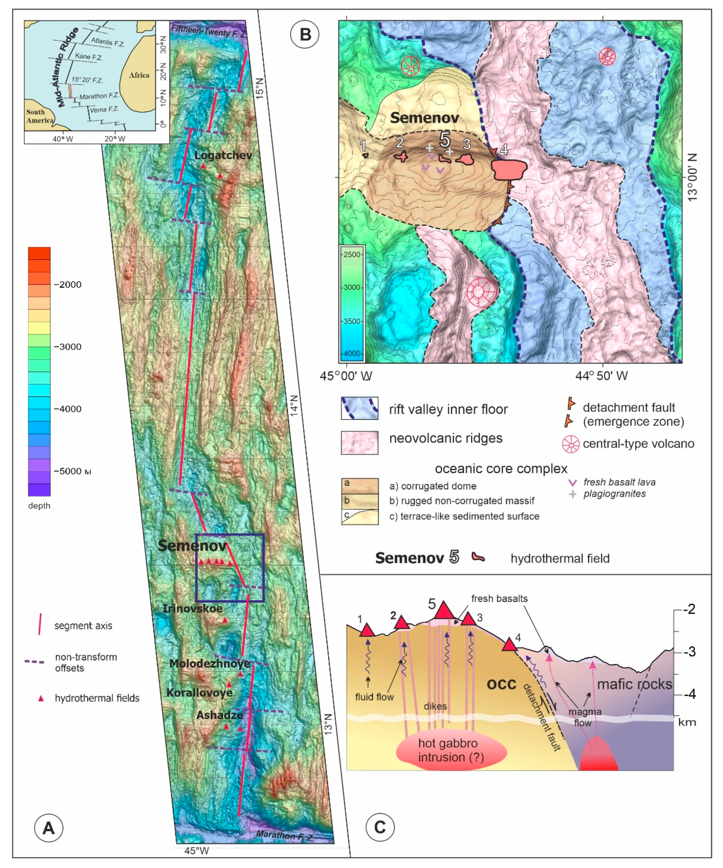
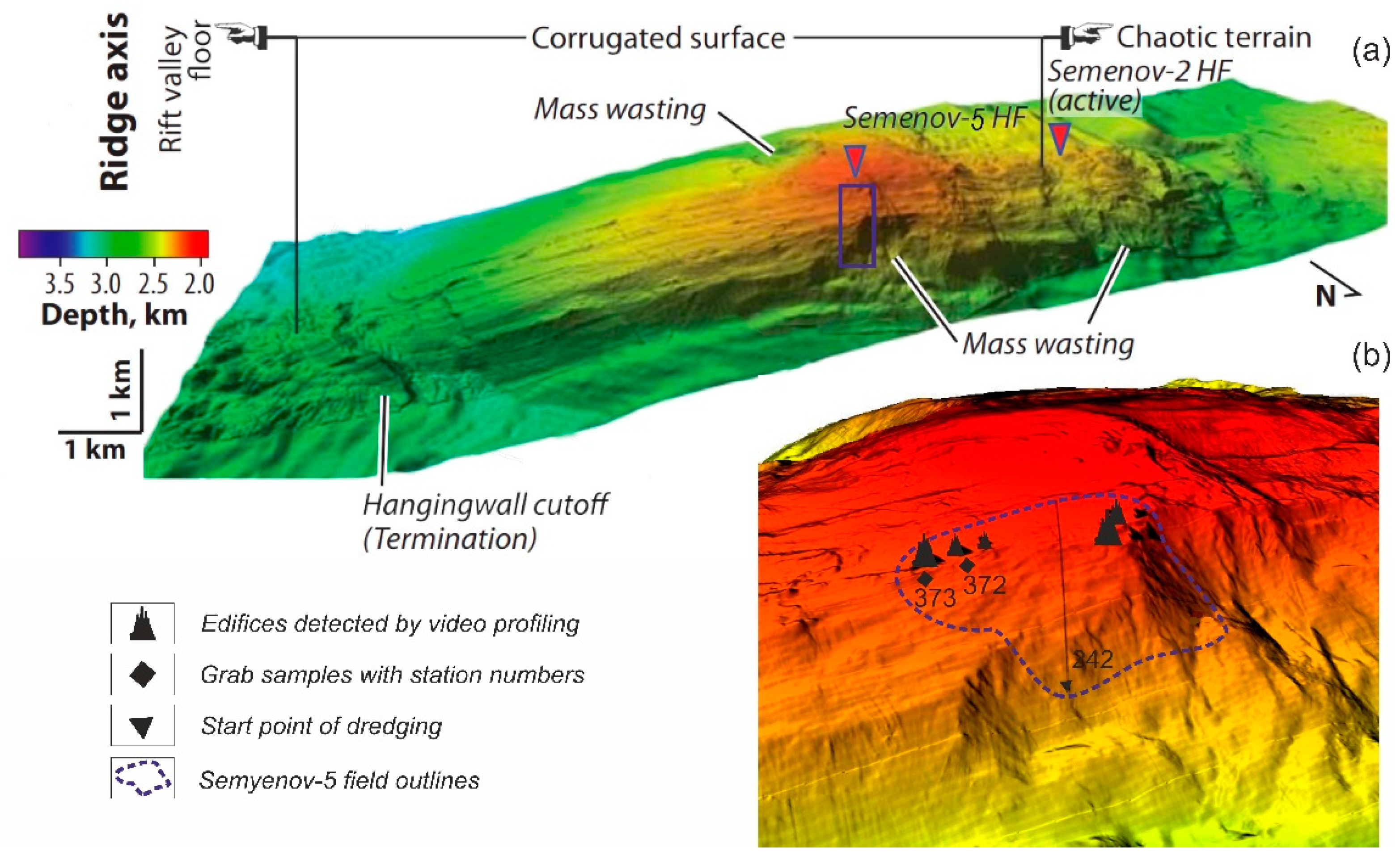
2. Previous Study and Geological Setting of the Semenov-5 Hydrothermal Field
3. Materials and Methods
4. Results
4.1. Mineralogy
4.1.1. St. 242
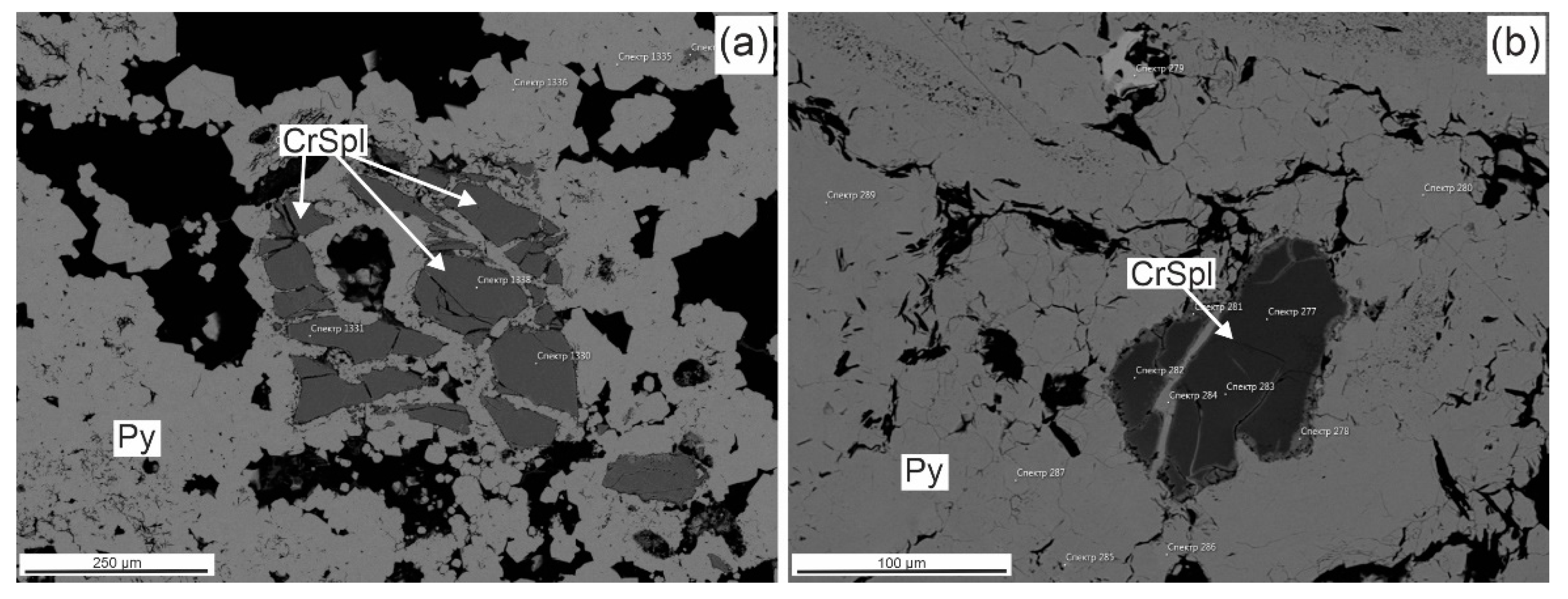
Cr-Spinels from St. 242
4.1.2. St. 372
4.1.3. St. 373
4.2. Bulk Geochemistry
4.2.1. St. 242
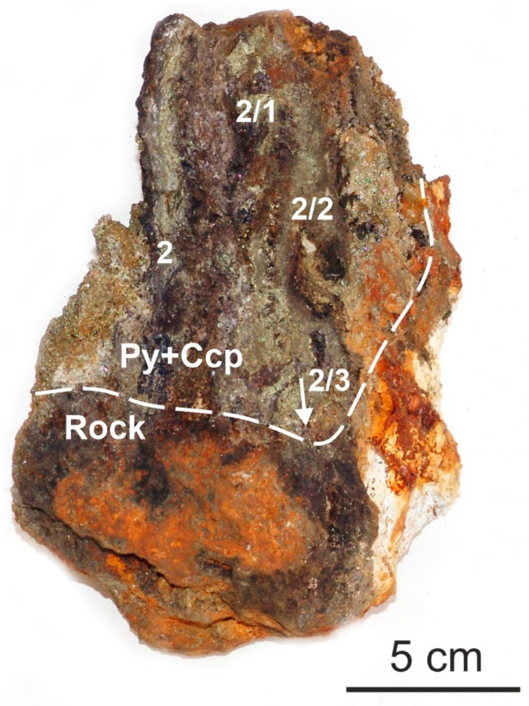
4.2.2. St. 372
4.2.3. St. 373
5. Discussion
6. Conclusions
- (a)
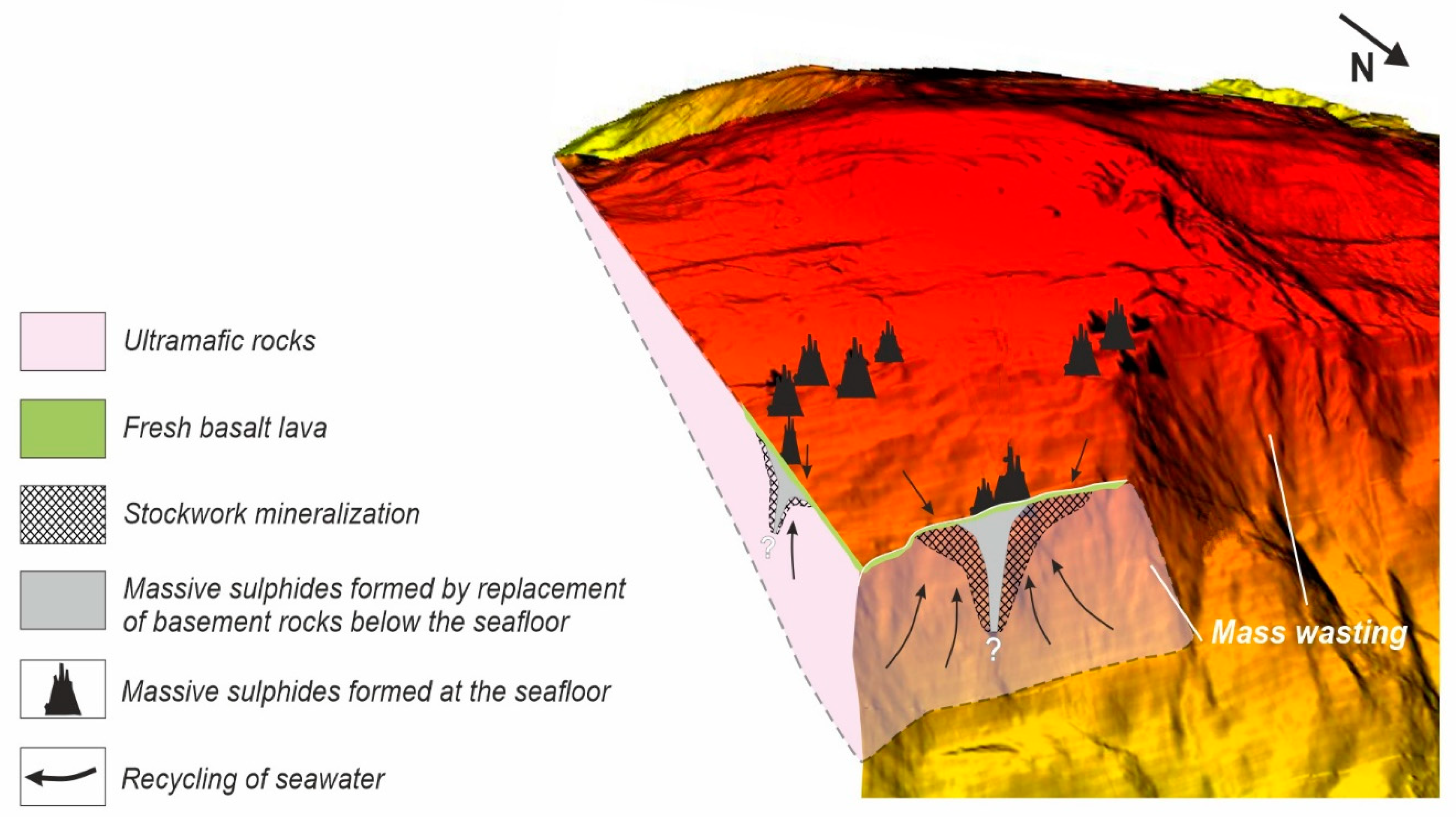
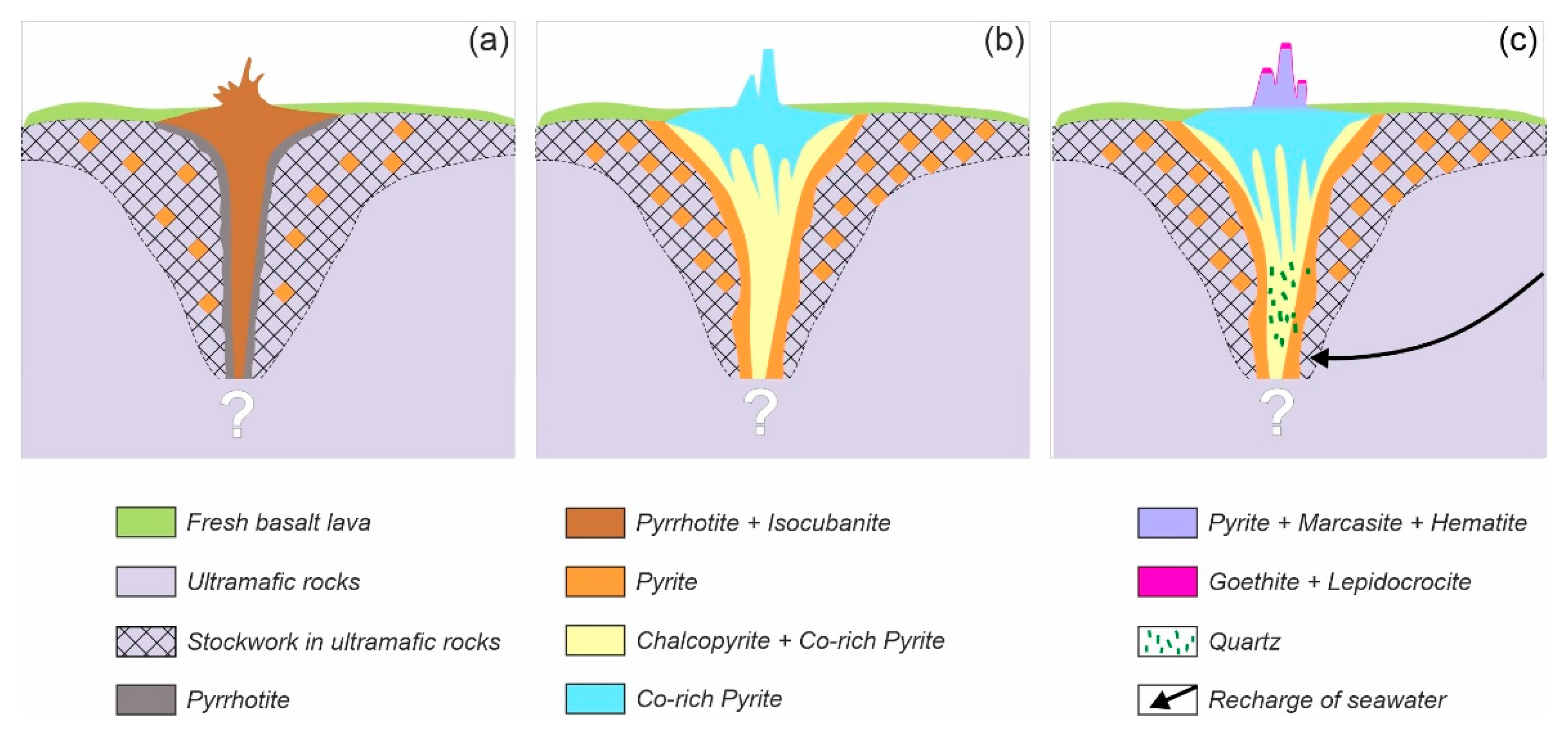
- the significant differences of composition between Semenov-5’s (1) sub-seafloor and (2) seafloor mineralization are most likely connected with altering the physical and chemical parameters of the hydrothermal system and a difference in the mode of formation: (1) metasomatically within hosted rocks and (2) from the discharged fluid on the seafloor surface;
- (b)
- sub-seafloor and seafloor massive sulfides have a common history of formation from the hydrothermal fluids which have been circulated within the ultramafic rocks and discharged on the surface;
- (c)
- distribution of fresh, thin basalt lava flowing within the studied area does not significantly affect seafloor massive sulfide composition.
Author Contributions
Funding
Data Availability Statement
Acknowledgments
Conflicts of Interest
References
- Pertsev, A.N.; Bortnikov, N.S.; Vlasov, E.A.; Beltenev, V.E.; Dobretsova, I.G.; Ageeva, O.A. Recent massive sulphide deposits of the Semenov ore district, Mid-Atlantic Ridge, 13°31° N: Associated rocks of the oceanic core complex and their hydrothermal alteration. Geol. Ore Depos. 2012, 54, 334–346. [Google Scholar] [CrossRef]
- Cherkashov, G.A. Morphology and Internal Structure of Hydrothermal Ore Bodies Formed in Various Geological Settings of the World Ocean. Okeangeology 2021, 61, 262–271. [Google Scholar]
- van Wijk, J.M. Public Report: Blue Mining, Breakthrough Solutions for Mineral Extraction and Processing in Extreme Environments. 2018. pp. 3–30. Available online: http://www.bluemining.eu/download/project_results/public_reports/Blue-mining-Public-Report-2018.pdf (accessed on 30 September 2018).
- Humphris, S.E.; Herzig, P.M.; Miller, D.J.; Alt, J.C.; Becker, K.; Brown, D.; Brügmann, G.; Chiba, H.; Fouquet, Y.; Gemmell, J.B.; et al. The internal structure of an active sea-floor massive sulphide deposit. Nature 1995, 377, 713–716. [Google Scholar] [CrossRef] [Green Version]
- Sawkins, F.J. Massive sulfide deposits in relation to geotectonics. Spec. Pap. Geol. Ass. Can. 1976, 14, 221–240. [Google Scholar]
- Zierenberg, R.A.; Fouquet, Y.; Miller, D.J.; Bahr, J.M.; Baker, P.A.; Bjerkgård, T.; Brunner, C.A.; Duckworth, R.C.; Gable, R.C.; Gieskes, J.; et al. The deep structure of a sea-floor hydrothermal deposit. Nature 1998, 392, 485–488. [Google Scholar] [CrossRef] [Green Version]
- Yamasaki, T. The Role of Bimodal Magmatism in Seafloor Massive Sulfide (SMS) Ore-forming Systems at the Middle Okinawa Trough. Japan. Ocean Sci. J. 2018, 53, 413–436. [Google Scholar] [CrossRef]
- Petersen, S.; Monecke, T.; Westhues, A.; Hannington, M.D.; Gemmell, J.B.; Sharpe, R.; Peters, M.; Strauss, H.; Lackschewitz, K.; Augustin, N.; et al. Drilling shallow water massive sulfides at the Palinuro Volcanic Complex, Aeolian Island Arc, Italy. Econ. Geol. 2014, 109, 2129–2157. [Google Scholar] [CrossRef]
- Lipton, I. Mineral Resource Estimate Solwara Project; Technical Report; Golder Associate: Singapore, 2012; 240p. [Google Scholar]
- Murton, B.J.; Lehrmann, B.; Dutrieux, A.M.; Martins, S.; de la Iglesia, A.G.; Stobbs, I.J.; Barriga, F.J.; Bialas, J.; Dannowski, A.; Vardy, M.E.; et al. Geological fate of seafloor massive sulphides at the TAG hydrothermal field (Mid-Atlantic Ridge). Ore Geol. Rev. 2019, 107, 903–925. [Google Scholar] [CrossRef]
- Beltenev, V.; Ivanov, V.; Rozhdestvenskaya, I.; Cherkashov, G.; Stepanova, T.; Shilov, V.; Pertsev, A.; Davydov, M.; Egorov, I.; Melekestseva, I.; et al. A new hydrothermal field at l3°30′ N on the Mid-Atlantic Ridge. InterRidge News 2007, 16, 9–10. [Google Scholar]
- Beltenev, V. Polar Marine Geosurvey Expedition (PMGE) report on regional works on a scale of 1:500,000–1:1,000,000 for deep-sea polymetallic sulphides in the axial zone of the Mid-Atlantic Ridge and searching works at Ashadze hydrothermal field: Polar Marine Geosurvey Expedition (PMGE), Lomonosov-St-Petersburg. Unpublished Report. 2007; 67p. (In Rusian) [Google Scholar]
- Beltenev, V. Polar Marine Geosurvey Expedition (PMGE) report on regional works for deep-sea polymetallic sulphides in the axial zone of the Mid-Atlantic Ridge: Polar Marine Geosurvey Expedition (PMGE), Lomonosov-St-Petersburg. Unpublished Report. 2009; 75p. (In Russian) [Google Scholar]
- Firstova, A.; Stepanova, T.; Sukhanova, A.; Cherkashov, G.; Poroshina, I. Au and Te Minerals in Seafloor Massive Sulphides from Semyenov-2 Hydrothermal Field, Mid-Atlantic Ridge. Minerals 2019, 9, 294. [Google Scholar] [CrossRef] [Green Version]
- Kuznetsov, V.; Maksimov, F.; Zheleznov, A.; Cherkashov, G.; Bel’tenev, V.; Lazareva, I. 230Th/U chronology of ore formation within the Semyenov hydrothermal district (13°31′ N) at the Mid-Atlantic Ridge. Geochronometria 2011, 38, 72–76. [Google Scholar] [CrossRef] [Green Version]
- Melekestseva, I.Y.; Tret’yakov, G.A.; Nimis, P.; Yuminov, A.M.; Maslennikov, V.V.; Maslennikova, S.P.; Kotlyarov, V.A.; Beltenev, V.E.; Danyushevsky, L.V.; Large, R. Barite-rich massive sulfides from the Semenov-1 hydrothermal field (Mid-Atlantic Ridge, 13°30.87′ N): Evidence for phase separation and magmatic input. Mar. Geol. 2014, 349, 37–54. [Google Scholar] [CrossRef]
- Beltenev, V.; Ivanov, V.; Rozhdestvenskaya, I.; Cherkashov, G.; Stepanova, T.; Shilov, V.; Davydov, M.; Laiba, A.; Kaylio, V.; Narkevsky, E.; et al. New data about hydrothermal fields on the Mid-Atlantic Ridge between 11°–14° N: 32nd cruise of R/V Professor Logatchev. InterRidge News 2009, 18, 14–18. [Google Scholar]
- Melekestseva, I.Y.; Maslennikov, V.V.; Tret’yakov, G.A.; Nimis, P.; Beltenev, V.E.; Rozhdestvenskaya, I.I.; Maslennikova, S.P.; Belogub, E.V.; Danyushevsky, L.; Large, R.; et al. Gold- and Silver-Rich Massive Sulfides from the Semenov-2 Hydrothermal Field, 13°31.13′ N, Mid-Atlantic Ridge: A Case of Magmatic Contribution? Econ. Geol. 2017, 112, 741–773. [Google Scholar] [CrossRef]
- Melekestseva, I.Y.; Kotlyarov, V.A.; Khvorov, P.V.; Ivanov, V.N.; Beltenev, V.E.; Dobretsova, I.G. Noble-metal mineralization in the Semenov-2 hydrothermal field (13°31′ N), Mid-Atlantic Ridge. Geol. Ore Depos. 2010, 52, 800–810. [Google Scholar] [CrossRef]
- Escartín, J.; Mevel, C.; Petersen, S.; Bonnemains, D.; Cannat, M.; Andreani, M.; Godard, M. Tectonic structure, evolution and the nature of oceanic core complexes and their detachment fault zones (13°20′ N and 13°30′ N, Mid Atlantic Ridge). Geochem. Geophys. Geosystems 2017, 18, 1451–1482. [Google Scholar] [CrossRef] [Green Version]
- Murton, B.J. Online Report of Project ULTRA. Available online: https://noc.ac.uk/projects/ultra (accessed on 1 April 2020).
- Smith, D.K.; Cann, J.R.; Escartín, J. Widespread active detachment faulting and core complex formation near 13° N on the Mid-Atlantic Ridge. Nature 2006, 442, 440–443. [Google Scholar] [CrossRef]
- Smith, D.K.; Escartin, J.; Schouten, H.; Cann, J.R. Fault rotation and core complex formation: Significant processes in seafloor formation at slow-spreading mid-ocean ridges (Mid-Atlantic Ridge, 13°–15° N). Geochem. Geophys. Geosystems 2008, 9. [Google Scholar] [CrossRef]
- MacLeod, C.J.; Searle, R.C.; Casey, J.F.; Mallows, C.; Unsworth, M.; Achenbach, K.; Harris, M. Life cycle of oceanic core complexes. Earth Planet. Sci. Lett. 2009, 287, 333–344. [Google Scholar] [CrossRef]
- Mallows, C.; Searle, R.C. A geophysical study of oceanic core complexes and surrounding terrain, Mid-Atlantic Ridge 13°–14° N. Geochem. Geophys. Geosystems 2012, 13. [Google Scholar] [CrossRef]
- Cann, J.R.; Blackman, D.K.; Smith, D.K.; McAllister, E.; Janssen, B.; Mello, S.; Avgerinos, E.; Pascoe, A.R.; Escartín, J. Corrugated slip surfaces formed at North Atlantic ridge-transform intersections. Nature 1997, 385, 329–332. [Google Scholar] [CrossRef]
- Tucholke, B.E.; Lin, J.; Kleinrock, M.C. Megamullions and mullion structure defining oceanic metamorphic core complexes on the Mid-Atlantic Ridge. J. Geophys. Res. Solid Earth 1998, 103, 9857–9866. [Google Scholar] [CrossRef] [Green Version]
- MacLeod, C.J.; Escartin, J.; Banerji, D.; Banks, G.J.; Gleeson, M.; Irving, D.H.B.; Smith, D.K. Direct geological evidence for oceanic detachment faulting: The Mid-Atlantic Ridge, 15°45′ N. Geology 2002, 30, 879–882. [Google Scholar] [CrossRef]
- Escartín, J.; Mevel, C.; MacLeod, C.J.; McCaig, A.M. Constraints on deformation conditions and the origin of oceanic detachments: The Mid-Atlantic Ridge core complex at 15°45′ N. Geochem. Geophys. Geosystems 2003, 4, 1067. [Google Scholar] [CrossRef]
- Ribeiro da Costa, I.; Barriga, F.J.A.S. Chromite Oxidation Patterns Associated to Serpentinization: Case Studies from the Mid-Atlantic Ridge, the Alter do Chão Massif (NE Alentejo, Portugal) and the Ronda Massif (Spain). Minerals 2022, 12, 1300. [Google Scholar] [CrossRef]
- Fouquet, Y.; Cambon, P.; Etoubleau, J.; Charlou, J.-L.; Ondréas, H.; Barriga, F.J.A.S.; Cherkashev, G.; Semkova, T.; Poroshina, I.; Bohn, M.; et al. Geodiversity of Hydrothermal Processes along the Mid-Atlantic Ridge and Ultramafic-Hosted Mineralization: A New Type of Oceanic Cu-Zn-Co-Au Volcanogenic Massive Sulfide Deposit; American Geophysical Union: Washington, DC, USA, 2010; pp. 321–368. [Google Scholar]
- Sukhanova, A.; Firstova, A. Uranium in seafloor massive sulfides at the Mid-Atlantic Ridge. In Proceedings of the Underwater Mining Conference, Sanya, China, 22–27 September 2019. [Google Scholar]
- Melekestseva, I.Y. Heterpgeneous Co-bearing massive sulfide deposits associated with ultramafites of paleo-island arc structure. Nauka 2007, 245. (In Russian) [Google Scholar]
- Malakhov, I.A.; Savokhin, I.V.; Burmako, P.L.; Kuznetsov, V.I. Influence of processes of metamorphism and metasomatism on the composition of chrome spinels in ultramafics and chromites of Urals. In Proceedings of the Ural State Mining and Geological Academy. Geol. Geophys. 2001, 13, 66–73. (In Russian) [Google Scholar]
- Maslennikov, V.V.; Maslennikova, S.P.; Large, R.R.; Danyushevsky, L.V.; Herrington, R.J.; Stanley, C.J. Tellurium-bearing minerals in zoned sulphide chimneys from Cu-Zn massive sulphide deposits of the Urals, Russia. Mineral. Petrol. 2013, 107, 67–99. [Google Scholar] [CrossRef]
- Mozgova, N.N.; Trubkin, N.V.; Borodaev, Y.S.; Cherkashov, G.A.; Stepanova, T.V.; Semkova, T.A.; Uspenskaya, T.Y. Mineralogy of massive sulphides from the Ashadze hydrothermal field, 13° N, Mid-Atlantic Ridge. Can. Miner. 2008, 46, 545–567. [Google Scholar] [CrossRef]


| Samples | 242-1 | 242-2 | 372 | 373 | ||||||||||||
|---|---|---|---|---|---|---|---|---|---|---|---|---|---|---|---|---|
| Zones | Py Layer | Contact of Sulfides with the Rock | Py-Chp Layer | Contact of Sulfides with the Rock | Py-Chp Layer | |||||||||||
| Minerals | 1 | 1/1 | 1/1a | 1/1b | 1/2 | 1/3 | 2/3 | 2 | 2/1 | 2/2 | 1 | 1 | 3 | 3/1 | 4 | |
| Pyrrhotite Fe1−xS | x | |||||||||||||||
| Pyrite FeS2 | +++ | +++ | +++ | +++ | +++ | +++ | +++ | +++ | +++ | +++ | +++ | +++ | +++ | +++ | +++ | |
| Marcasite FeS2 | x | x | x | + | +++ | ++ | +++ | +++ | ||||||||
| Chalcopyrite CuFeS2 | + | + | ++ | ++ | +++ | +++ | ++ | +++ | +++ | +++ | x | x | x | x | ||
| Isocubanite CuFe2S3 | ? | x | ||||||||||||||
| Secondary Cu sulfides (Bn,Cv) | x | x | x | x | x | x | ||||||||||
| Sphalerite (Zn,Fe)S | x | x | x | + | ||||||||||||
| Cr-spinels (Fe,Mg)(Cr,Al,Fe)2O4 | x | x | x | x | x | x | x | x | x | x | ||||||
| Rutile TiO2 | x | x | x | x | x | |||||||||||
| Quartz SiO2 | + | + | + | + | + | + | +++ | ++ | ++ | ++ | ||||||
| Baryte BaSO4 | x | + | + | + | ++ | |||||||||||
| Hematite Fe2O3 | x | ++ | + | ++ | ||||||||||||
| Goethite FeO(OH) | x | + | + | + | ++ | |||||||||||
| Lepidocrocite γ-Fe3+O(OH) | + | |||||||||||||||
| Mineral (Number of Analyses, n) | Element (wt %) | ||||||
|---|---|---|---|---|---|---|---|
| Fe | Cu | Zn | S | O | Si | Total | |
| Fe-sphalerite (39) | 10.27 | - | 56.12 | 33.40 | - | - | 99.79 |
| Chalcopyrite (47) | 31.37 | 33.68 | - | 34.49 | - | - | 99.54 |
| Secondary Cu sulfides (18) | 2.32 | 57.13 | - | 32.54 | 8.20 | - | 100.19 |
| Quartz (27) | - | - | - | - | 50.93 | 48.92 | 99.85 |
| Element (wt %) | ||||||||
|---|---|---|---|---|---|---|---|---|
| S | Fe | Co | Ni | Cu | Zn | Cr | Total | |
| Pyrites from pyrite layer, n = 198 | ||||||||
| Average | 52.78 | 47.02 | 0.43 | 0.47 | 0.35 | - | 0.32 | 100.05 |
| Min | 45.92 | 45.40 | 0.01 | 0.05 | 0.04 | - | 0.27 | 98.69 |
| Max | 54.97 | 54.08 | 1.08 | 1.11 | 1.16 | - | 0.37 | 101.77 |
| Frequency of occurrence in a layer | 100 vol.% | 100 vol.% | 15 vol.% | 14 vol.% | 13 vol.% | - | <1 vol.% | |
| Pyrites from chalcopyrite-pyrite layer, n = 297 | ||||||||
| Average | 52.27 | 46.17 | 0.78 | 0.56 | 0.71 | 0.37 | 1.45 | 100.31 |
| Min | 47.37 | 42.37 | 0.02 | 0.04 | 0.08 | 0.18 | 0.32 | 98.38 |
| Max | 54.04 | 50.36 | 2.97 | 2.01 | 4.80 | 0.56 | 4.06 | 101.80 |
| Frequency of occurrence in a layer | 100 vol.% | 100 vol.% | 62 vol.% | 61 vol.% | 63 vol.% | <1 vol.% | <1 vol.% | |
| Mineral (Number of Analyses, n) | Element (wt %) | |||||||||||||
|---|---|---|---|---|---|---|---|---|---|---|---|---|---|---|
| Fe | Cu | S | Ni | Co | O | Si | Mg | Al | Cr | Mn | Total | |||
| Pyrite (11) | 45.66 | - | 52.72 | 1.41 | 1.29 | - | - | - | - | - | - | 101.08 | ||
| Chalcopyrite (5) | 30.91 | 32.98 | 34.01 | 0.43 | 0.24 | 2.92 | - | - | - | - | - | 101.49 | ||
| Oxides (wt %) | ||||||||||||||
| FeO | NiO | CoO | SiO2 | MgO | Al2O2 | Cr2O3 | MnO | Total | ||||||
| Enstatite (5) | 3.33 | - | - | 66.45 | 31.25 | - | 0.22 | - | 101.25 | |||||
| Cr-spinels. Core (3) | 15.06 | - | - | - | 13.48 | 28.11 | 38.57 | 6.48 | 101.70 | |||||
| Cr-spinels. Rims (2) | 63.81 | 0.87 | 0.70 | 0.74 | 0.90 | 2.15 | 25.73 | 6.42 | 101.32 | |||||
| O | Mg | Al | Cr | Fe | Si | Ti | V | Mn | Cu | Zn | S | Frequency of Cr-Spinels Occurrence in | ||
|---|---|---|---|---|---|---|---|---|---|---|---|---|---|---|
| Py Layer | Cph-Py Layer | |||||||||||||
| Primary Cr-spinels from peridotites [12,13] | ||||||||||||||
| Average | 35.88 | 8.08 | 14.99 | 26.35 | 14.70 | - | - | - | - | - | - | - | ||
| Primary Cr-spinels from SMS: Fe = 10%–17%; Mg + Al > 20%; Cr/Fe ≥ 2; n = 59 | ||||||||||||||
| Average | 32.77 | 8.83 | 15.97 | 28.72 | 13.49 | - | - | - | - | - | - | - | 80% | 36% |
| Min | 31.10 | 6.37 | 13.49 | 23.48 | 11.09 | - | - | - | - | - | - | - | ||
| Max | 34.92 | 9.80 | 18.32 | 31.90 | 19.85 | - | - | - | - | - | - | - | ||
| Slightly hydrothermally altered Cr-spinels from SMS: Fe = 17%–26%; Mg + Al = 10%–20%; Cr/Fe = 1–2; n = 26 | ||||||||||||||
| Average | 30.55 | 5.10 | 9.69 | 32.66 | 21.31 | - | - | - | - | - | - | - | 7% | 24% |
| Min | 28.29 | 3.06 | 6.05 | 27.16 | 17.54 | - | - | - | - | - | - | - | ||
| Max | 33.74 | 6.81 | 14.05 | 39.74 | 27.00 | - | - | - | - | - | - | - | ||
| Strong hydrothermally altered Cr-spinels from SMS: Fe > 26%; Mg + Al < 10%; Cr/Fe < 1; n = 43 | ||||||||||||||
| Average | 27.35 | 1.77 | 3.36 | 28.97 | 33.50 | 1.80 | 0.50 | 0.45 | 1.65 | 2.19 | 1.53 | 1.20 | 13% | 40% |
| Min | 23.35 | 0.31 | 0.50 | 20.07 | 21.95 | 0.13 | 0.21 | 0.22 | 0.69 | 0.14 | 0.64 | 0.23 | ||
| Max | 31.85 | 4.64 | 8.15 | 36.81 | 48.12 | 9.11 | 1.19 | 0.85 | 3.77 | 7.94 | 2.85 | 5.19 | ||
| Frequency of trace elements occurrence, vol.% | 77 | 58 | 42 | 58 | 42 | 44 | 60 | |||||||
| Mineral (Number of Analyses, n) | Element (wt %) | |||||||
|---|---|---|---|---|---|---|---|---|
| Fe | Cu | Zn | S | O | Ba | Sr | Total | |
| Pyrite * (108) | 47.33 | - | - | 52.61 | - | - | - | 99.94 |
| Chalcopyrite (32) | 31.97 | 33.09 | - | 34.80 | - | - | - | 99.86 |
| Secondary Cu sulfides (24) | 6.73 | 59.87 | - | 31.23 | 4.59 | - | - | 100.42 |
| Fe-sphalerite (12) | 14.42 | - | 49.15 | 35.72 | 2.34 | - | - | 101.63 |
| Baryte (11) | 1.45 | - | - | 14.41 | 22.76 | 60.86 | 1.55 | 101.03 |
| Station | St. 242 | St. 372 | St. 373 | Avg | |||||||||||||||
|---|---|---|---|---|---|---|---|---|---|---|---|---|---|---|---|---|---|---|---|
| Sample | 1 | 1/1 | 1/1b | 1/2 | 1/3 | 1/3a | 2 | 2/1 | 2/2 | 2/3 | 1 | 1 | 2 | 3 | 3/1 | 4 | 5 | 6 | |
| Fe, wt % | 44.5 | 39.5 | - | 36.6 | 35.8 | - | 25.4 | 25.0 | 25.4 | 25.3 | 44.7 | 42.4 | - | 43.1 | 41.0 | - | - | - | 35.7 |
| S | 34.2 | 45.1 | 44.0 | 34.2 | 29.1 | 42.8 | 23.2 | 28.0 | 29.7 | 31.9 | 39.9 | 36.5 | 46.2 | 38.6 | 35.7 | 39.2 | 45.8 | 44.8 | 37.2 |
| Cu | 1.39 | 7.62 | 12.9 | 13.1 | 16.6 | 13.9 | 12.0 | 11.8 | 11.9 | 12.9 | 0.11 | 1.38 | 0.79 | 1.27 | 0.71 | 1.27 | 1.03 | 0.99 | 6.76 |
| Zn | 0.02 | 0.06 | 0.10 | 0.09 | 0.13 | 0.10 | 0.06 | 0.07 | 0.08 | 0.06 | 0.05 | 0.11 | 0.09 | 0.19 | 0.07 | 0.15 | 0.03 | 0.07 | 0.09 |
| Mg | 0.19 | 0.13 | - | 0.04 | 0.05 | - | 0.22 | 0.05 | 0.07 | 0.01 | 0.04 | 0.05 | - | 0.06 | 0.04 | - | - | - | 0.08 |
| Al | 0.01 | - | - | 0.01 | 0.03 | - | 0.01 | 0.01 | 0.01 | 0.01 | 0.02 | 0.06 | - | 0.06 | 0.03 | - | - | - | 0.02 |
| Si | 1.01 | 1.48 | - | 2.11 | 2.08 | - | 14.6 | 16.2 | 14.3 | 12.6 | 0.41 | 0.54 | - | 0.54 | 0.34 | - | - | - | 5.52 |
| Ca | 0.08 | 0.08 | - | 0.01 | 0.05 | - | 0.03 | 0.01 | 0.01 | 0.01 | 0.01 | 0.01 | - | 0.01 | 0.04 | - | - | - | 0.03 |
| Co, ppm | 1150 | 1220 | 1020 | 1230 | 1090 | 1210 | 859 | 1600 | 1540 | 1170 | 191 | 9.3 | 234 | 242 | 220 | 509 | 132 | 218 | 769 |
| Ni | 1510 | 1610 | 1330 | 1600 | 1450 | 1540 | 958 | 1390 | 1110 | 1000 | 6.4 | 4.0 | 36 | 44 | 22 | 45 | 22 | 26 | 761 |
| Bi | 0.8 | 1.4 | 1.4 | 1.1 | 1.0 | 0.7 | 0.3 | 0.5 | 0.6 | 0.4 | 1.9 | <0.1 | 0.4 | 3.3 | 2 | 5.6 | 2.9 | 5.7 | 1.8 |
| Se | 482 | 248 | 382 | 370 | 270 | 483 | 102 | 415 | 358 | 327 | 451 | 6.8 | 34 | 50 | 38 | 95 | 26 | 47 | 232 |
| Te | 12 | 9.7 | 8.4 | 7.5 | 4.5 | 11 | 2.7 | 6.8 | 6.3 | 6.2 | 34 | <0.2 | 0.4 | 1.9 | 3.8 | 2.7 | 1.5 | 0.9 | 7.1 |
| In | 11 | 4.9 | 8.2 | 10 | 11 | 10 | 2.9 | 8.4 | 8.4 | 9.1 | 0.2 | 0.2 | 0.7 | 1.7 | 38 | 1.5 | 1.4 | 1.2 | 7.2 |
| Au | 0.11 | 0.14 | 0.09 | 0.12 | 0.01 | 0.13 | 0.05 | 0.10 | 0.12 | 0.07 | 1.70 | 0.17 | 0.12 | 0.38 | 0.40 | 0.49 | 0.24 | 0.13 | 0.25 |
| Ag | 23 | 36 | 21 | 33 | 23 | 28 | 7.6 | 17 | 24 | 24 | 24 | 3.2 | 6.5 | 19.3 | 12 | 13 | 18 | 14 | 19 |
| Cd | 3.3 | 2.9 | 3.1 | 2.9 | 4.4 | 3.8 | 1.2 | 2.8 | 2.8 | 3.0 | 2.9 | 0.1 | 1.1 | 0.8 | 11 | 0.6 | 0.6 | 0.7 | 2.7 |
| Ge | 0.7 | 0.5 | 0.6 | 0.6 | 0.5 | 0.7 | 0.3 | 0.6 | 0.6 | 0.6 | 11 | 0.8 | 1.2 | 2.9 | 52 | 3.1 | 2.3 | 1.9 | 4.5 |
| Ga | 1.0 | 1.1 | 0.3 | 1.3 | 1.0 | 1.3 | 2.9 | 0.7 | 0.8 | 0.7 | 4.2 | 4.2 | 1.3 | 5.1 | 10 | 2.5 | 9.4 | 2.1 | 2.8 |
| Pb | 32 | 47 | 29 | 34 | 23 | 44 | 23 | 9.6 | 20 | 25 | 116 | 184 | 508 | 224 | 93 | 301 | 217 | 330 | 125 |
| Sb | 2.0 | 3.0 | 1.8 | 3.0 | 1.2 | 3.0 | 1.3 | 0.5 | 1.2 | 1.5 | 4.9 | 2.6 | 6.9 | 9.6 | 1.5 | 8.9 | 5.9 | 7.9 | 3.7 |
| As | 27 | 35 | 25 | 38 | 21 | 34 | 23 | 6.9 | 9.6 | 17 | 48 | 117 | 103 | 225 | 4.4 | 303 | 112 | 111 | 70 |
| Sn | 18 | 19 | 14 | 18 | 12 | 19 | 8.3 | 3.5 | 6.6 | 15 | 13 | 2.3 | 2.3 | 9.1 | 10 | 6.2 | 12 | 5.8 | 11 |
| Mo | 10 | 11 | 9.1 | 8.8 | 5.3 | 14 | 2.3 | 1.1 | 1.8 | 2.5 | 7.3 | 80 | 176 | 120 | 39 | 165 | 117 | 199 | 54 |
| V | 14 | 12 | 13 | 12 | 7.7 | 15 | 9.3 | 13 | 14 | 6.6 | 2.6 | 2.7 | 10 | 32 | - | 18 | 15 | 21 | 13 |
| Cr | 1510 | 980 | 1120 | 894 | 780 | 1220 | 538 | 1020 | 1090 | 584 | 4.6 | 7.9 | 23 | 11 | - | 4.8 | 12 | 19 | 578 |
| U | <0.1 | 0.1 | <0.1 | <0.1 | <0.1 | <0.1 | 0.13 | <0.1 | <0.1 | <0.1 | 1.1 | 3.1 | 14 | 25 | 37 | 13 | 11 | 15 | 12 |
| Ba | 28 | 9.1 | 11 | 9.9 | 61 | 15 | 236 | <3 | 41 | 16 | 1220 | 11900 | 7890 | 3560 | 20 | 35100 | 16100 | 6200 | 4848 |
| Elements | Host Rocks | SMS MAR * | ||||||||||||
|---|---|---|---|---|---|---|---|---|---|---|---|---|---|---|
| Mafic | Mafic and Ultramafic | Ultramafic | ||||||||||||
| Jubileynoye | Zenith-Victoria | Krasnov | Semenov-4 | Semenov-5 | Semenov-2 | Semenov-1 | Logatchev-1 | Irinovskoye | Ashadze-1 | Ashadze-2 | ||||
| St. 242 | St. 372 | St. 373 | ||||||||||||
| Fe, % | 31.5 | 39.1 | 39.8 | 41.2 | 32.1 | 44.7 | 42.7 | 13.9 | 29.3 | 19.4 | 18.0 | 28.3 | 32.0 | 32.4 |
| S | 35.7 | 44.5 | 45.3 | 47.7 | 31.3 | 39.9 | 36.9 | 14.8 | 40.0 | 23.3 | 26.1 | 28.9 | 32.3 | 36.4 |
| Cu | 4.44 | 3.02 | 1.84 | 0.93 | 11.0 | 0.11 | 1.12 | 30.1 | 4.6 | 34.1 | 21.8 | 10.2 | 18.2 | 9.56 |
| Zn | 0.45 | 1.41 | 0.63 | 0.09 | 0.07 | 0.05 | 0.12 | 3.46 | 0.16 | 2.33 | 5.16 | 17.94 | 0.86 | 4.39 |
| Si | 10.4 | 2.44 | 1.62 | 0.68 | 8.11 | 0.41 | 0.47 | 5.51 | 0.47 | 2.04 | 11.18 | 0.71 | 0.51 | 3.2 |
| Ca | 0.26 | 0.03 | 0.09 | 0.15 | 0.03 | 0.01 | 0.02 | 3.38 | 0.02 | 1.12 | 0.27 | 0.49 | 0.11 | 0.42 |
| Mg | 0.34 | 0.01 | 0.04 | 0.06 | 0.09 | 0.04 | 0.05 | 0.32 | 0.03 | 0.37 | 0.17 | 0.22 | 0.81 | 0.14 |
| Al | 0.11 | 0.12 | 0.13 | 0.14 | 0.03 | 0.08 | 0.05 | 0.22 | 0.08 | 0.21 | 0.29 | 0.16 | 0.19 | 0.16 |
| Ti, ppm | 298 | 269 | 591 | 372 | 465 | 240 | 240 | 966 | 270 | 493 | 432 | 460 | 559 | 450 |
| Co | 526 | 342 | 539 | 113 | 1267 | 191 | 223 | 92 | 48 | 467 | 150 | 2039 | 1223 | 619 |
| Ni | 6.1 | 5.1 | 6.1 | <10 | 1446 | 6.4 | 29.4 | 11 | <10 | 76 | 11 | 234 | 24 | 55 |
| Bi | 1.6 | 1.3 | 1.5 | 0.75 | 0.85 | 1.87 | 2.99 | 18 | 0.49 | 7.4 | 6.4 | 2.7 | 2.6 | 4.5 |
| Se | 355 | 11.3 | 71.4 | 101 | 383 | 451 | 43 | 409 | 130 | 276 | 365 | 229 | 89.5 | 280 |
| Pb | 61 | 161 | 47 | 81 | 28.3 | 116 | 294 | 246 | 41 | 145 | 201 | 274 | 14 | 157 |
| Ga | 15 | 12 | 7.7 | 3.2 | 0.97 | 4.2 | 4.1 | 36 | 9.2 | 7.5 | 5.1 | 7.4 | 7.0 | 19 |
| Ge | 2.1 | 14 | 3.3 | 3.6 | 0.62 | 11.5 | 2.0 | 345 | 24 | 9.1 | 35 | 9,3 | 1.7 | 36 |
| Sn | 5.8 | 2.5 | 24 | 24 | 13.4 | 12.8 | 6.3 | 108 | 26 | 132 | 44 | 368 | 40 | 65 |
| Cd | 8.3 | 22 | 26 | 0.9 | 3.1 | 2.9 | 0.67 | 113 | 3 | 77 | 129 | 287 | 13 | 110 |
| Sb | 6.1 | 34 | 9.1 | 3.8 | 1.9 | 4.9 | 6.9 | 17 | 10 | 152 | 90 | 51 | 10.9 | 33 |
| As | 19 | 188 | 54 | 140 | 24 | 47 | 161 | 106 | 6.6 | 386 | 356 | 84 | 215 | 318 |
| Mn | 388 | 54 | 29 | 15 | 44.2 | 8.0 | 18 | 86 | 215 | 245 | 164 | 473 | 108 | 159 |
| Ba | 1451 | 450 | 8623 | 6200 | 21.5 | 1220 | 13,458 | 1144 | 10,045 | 1900 | 218 | 690 | 300 | 2900 |
| Ag | 15 | 28 | 25 | 11 | 26 | 24 | 12.4 | 185 | 20 | 46 | 86 | 84 | 7.5 | 49 |
| Au | 0.5 | 0.3 | 0.8 | 0.4 | 0.1 | 1.7 | 0.2 | 25 | 5.2 | 14 | 5.2 | 3.3 | 11 | 3.2 |
| Cr | 88 | 30 | 38 | 47 | 984 | 4.6 | 12.8 | 36 | 2.5 | 38 | 129 | 24 | 14 | 38 |
| Mo | 147 | 126 | 139 | 88 | 6.7 | 7.3 | 143 | 167 | 145 | 163 | 25 | 43 | 80 | 97 |
| Te | 11 | 1.2 | 4.3 | 1.0 | 8.5 | 35 | 1.2 | 18.3 | 3.6 | 15 | 16 | 7.7 | 14 | 9.9 |
| In | 3.8 | 1.4 | 2.5 | 1.5 | 8.8 | 0.2 | 1.1 | 0.8 | 0.6 | 6.8 | 3.2 | 9.3 | 3.7 | 4.4 |
| V | 49 | 50 | 15 | 70 | 12 | 2.6 | 16 | 55 | 341 | 187 | 69 | 57 | 258 | 160 |
| n | 35 | 63 | 161 | 99 | 9 | 1 | 6 | 35 | 11 | 90 | 10 | 120 | 53 | 1052 |
Publisher’s Note: MDPI stays neutral with regard to jurisdictional claims in published maps and institutional affiliations. |
© 2022 by the authors. Licensee MDPI, Basel, Switzerland. This article is an open access article distributed under the terms and conditions of the Creative Commons Attribution (CC BY) license (https://creativecommons.org/licenses/by/4.0/).
Share and Cite
Firstova, A.; Cherkashov, G.; Stepanova, T.; Sukhanova, A.; Poroshina, I.; Bel’tenev, V. New Data for the Internal Structure of Ultramafic Hosted Seafloor Massive Sulfides (SMS) Deposits: Case Study of the Semenov-5 Hydrothermal Field (13°31′ N, MAR). Minerals 2022, 12, 1593. https://doi.org/10.3390/min12121593
Firstova A, Cherkashov G, Stepanova T, Sukhanova A, Poroshina I, Bel’tenev V. New Data for the Internal Structure of Ultramafic Hosted Seafloor Massive Sulfides (SMS) Deposits: Case Study of the Semenov-5 Hydrothermal Field (13°31′ N, MAR). Minerals. 2022; 12(12):1593. https://doi.org/10.3390/min12121593
Chicago/Turabian StyleFirstova, Anna, Georgy Cherkashov, Tamara Stepanova, Anna Sukhanova, Irina Poroshina, and Victor Bel’tenev. 2022. "New Data for the Internal Structure of Ultramafic Hosted Seafloor Massive Sulfides (SMS) Deposits: Case Study of the Semenov-5 Hydrothermal Field (13°31′ N, MAR)" Minerals 12, no. 12: 1593. https://doi.org/10.3390/min12121593