Article

Vascular Underpinnings of Cerebral Lateralisation in the Neonate

1
Melbourne School of Psychological Sciences, University of Melbourne, Redmond Barry Building 115, Parkville, VIC 3010, Australia
2
Florey Institute of Neurosciences and Mental Health, Royal Parade, Parkville, VIC 3052, Australia
3
Royal Women’s Hospital, Flemington Rd., Parkville, VIC 3052, Australia
4
Murdoch Children’s Research Institute, Royal Children’s Hospital, Flemington Rd., Parkville, VIC 3052, Australia
5
Monash Institute of Cognitive & Clinical Neurosciences, Monash University, Clayton, VIC 3800, Australia
6
Departments of Paediatrics and Obstetrics & Gynaecology, University of Melbourne, Parkville, VIC 3010, Australia
7
Department of Psychology & Neuroscience Institute, University of Cape Town, Private Bag, Rondebosch 7701, South Africa
*
Author to whom correspondence should be addressed.
Symmetry 2024, 16(2), 161; https://doi.org/10.3390/sym16020161
Submission received: 22 June 2023 / Revised: 16 October 2023 / Accepted: 8 January 2024 / Published: 30 January 2024
(This article belongs to the Special Issue Neuroscience, Neurophysiology and Asymmetry—Volume II)

Abstract

:
Traditionally, adult and neonatal cerebral perfusion have been presumed to be symmetrical. Contrary to this, our adult work shows that supra-aortic cerebral supply is systematically biased towards the left, in terms of both vessel geometry and blood flow volumes. Although this asymmetry is meaningfully related to hand preference, the developmental origins of this association remain unknown. Our detailed investigations of the cerebral vasculature confirm analogous asymmetries in term neonates. Specifically, we demonstrate that the structure and flow of neonatal middle cerebral vessels are consistently asymmetric and predominantly left-dominant. Building on our work from the same cohort, we now report further analyses of these new-found asymmetries. Namely, exploring for the first time, the relationship between arterial lateral biases and the neonatal head-turning response—a reliable early behavioural precursor of handedness that shows a systematic rightward bias in the population. Here, we demonstrate a contralateral relationship between vessel morphology and primitive expressions of lateralisation that predate the establishment of definitive handedness in the course of postnatal development. This relationship mimics patterns observed in adults and suggests that lateralising trends in angiogenesis may ultimately influence the emergence of human lateral preferences.

1. Introduction

The expression of behavioural lateralisation in humans sets us apart from other animals, with 85% to 95% of the adult population demonstrating consistent right-hand dominance. The discovery of arterial correlates associated with adult hand preference [1] in our recent work poses a fundamental question: does lateralised hemispheric perfusion predate neurobehavioural expressions of cerebral lateralisation such as language and manual dexterity?
Here, we demonstrate that the middle cerebral arterial structure and supply are systematically asymmetric in the neonatal brain. Moreover, the observed vessel asymmetry is meaningfully related to the supine head orientation bias, an involuntary neonatal behaviour that reliably predicts handedness. The typical arterio-postural relationship manifests with wider arterial diameters and increased blood flow volumes in the left hemisphere of neonates that favour a right-sided head posture. Deviations from this typical left-sided arterial dominance, as manifested in diameter and flow volumes, correlate with increasing heterogeneity in head-turning behaviour. This finding has substantial implications for understanding the origins of human cerebral lateralisation.
In 1865, Broca [2] proposed that language is lateralised to the cerebral hemisphere responsible for writing and manual dexterity (see also Dax [3]), thereby originating the classical doctrine of left-hemispheric cerebral ”dominance”. Prior investigations into the typical superiority of the right hand for writing and manual dexterity primarily focused on underlying anatomical asymmetries in the body periphery. For example, Hyrtl’s ”subclavian artery theory” postulated that preferential arterial perfusion of the right upper limb could explain this phenomenon [4,5]. Following Broca’s discovery, researchers shifted attention to investigating the hypothesis of asymmetric cerebral perfusion [6,7]. This proposition seemed entirely plausible at the time, considering the asymmetrical origin of the carotid vessels, where the right common carotid artery stems from the neck of the brachiocephalic trunk, while the left common carotid originates directly from the aortic arch [6,7]. The search for a vascular origin of cerebral lateralisation was initially abandoned following the discovery of the anterior communicating artery [8,9,10]. However, this retreat turned out to be premature, as subsequent research has demonstrated that the anterior communicating artery does very little to balance supply between the hemispheres (see [1] for a historical review).
The cerebral hemispheres have since been shown to be asymmetric not only in function but also in morphology and cerebral perfusion [11,12,13,14]. Our own investigation [1] further revealed that adult cerebrovascular supply (via the common and internal carotid arteries) is structurally and haemodynamically asymmetric at rest (n = 195; n = 228), with a systematic bias to the left cerebral hemisphere. The asymmetry is closely related to handedness, and it manifests in wider arterial calibres, increased blood flow velocities, and increased overall flow volumes directed to the dominant hemisphere. This discovery of arterial correlates of hand preference is consistent with the notion of a more nutrient-rich and better perfused left cerebral hemisphere [12,15,16,17], thereby challenging classical assumptions regarding vascular phenotypes [18]. However, whether or not the foundational basis for this asymmetry is detectable in neonates prior to the emergence of manual preferences and the onset of language development remains unresolved. The direction of causation therefore remains unknown.
Head turning has been well studied as a marker of neonatal lateral preferences and later handedness. Approximately 65 to 92% of newborns spontaneously and consistently prefer a right-sided head position when supine [19,20,21,22,23,24,25]. The left–right distribution of this behavioural asymmetry suggests that it is a precursor of adult manual specialisation [19,21,25], and there is evidence that it predicts initial hand preference in infancy (p < 0.05 at 16 weeks; p = 0.007 at 22 weeks), habitual reaching preference between 16 and 22 weeks [24], full reaching preference at four months [23], and later childhood handedness [19].
Given the compelling evidence for a vascular correlate of hemispheric lateralisation in adults [1], the work described here takes a vital step towards untangling the extent to which these factors contribute to the genesis and maintenance of behavioural and cerebral lateralisation. This study aimed to determine whether the geometry and local resting haemodynamics of the middle cerebral arterial trunk are asymmetric in healthy term neonates. A further aim of this work was to determine whether these putative lateralised trends in brain vasculature predict neonatal head turning (this will be referred to as the arterio-postural relationship). The middle cerebral artery was chosen since it is a major and accessible arterial conduit to the neocortex, and it perfuses the cerebral structures that subserve the most overtly lateralised behavioural and language functions.
Neonatal cranial ultrasound is routinely performed in clinical practice, and it largely proceeds on the assumption of trans-midline symmetry. Non-invasive interrogation of the arterial and venous blood flow is typically conducted unilaterally and is hindered by the limited lateral resolution of ultrasound machines [26], precluding precise measurement of vessel diameters. The resolution of this technology has therefore impeded the reliable detection of cerebrovascular structurofunctional asymmetries. As a result, bilateral flow assessment has primarily relied upon comparing resistive indices between hemispheres. Given the intrinsic link between vessel geometry and haemodynamics and total blood flow volume [27,28,29], localised velocity or diametric measures in isolation are not appropriate for side-to-side arterial comparisons. That is, the unknown arterial cross-sectional area in most paediatric cerebrovascular assessments precludes comparison between one infant to another as well as between one hemisphere to another [30].
Advances in transcranial ultrasound technology have, however, enabled us to recently overcome these shortcomings through the development of a novel scanning approach [26]. Our innovative methodology involves simultaneous assessment of arterial diameter and flow, made possible by leveraging symmetrical viewing of B-flow and pulse-wave Doppler imaging for cerebral vessel interrogation. This technique has paved the way for investigating the aims of the research reported here. To our knowledge, our work represents the first successful non-invasive imaging of bilateral neonatal middle cerebral asymmetries at a resolution sufficient for accurate structure–functional analyses [31].
We report here the first investigation of neonatal cerebrovasculature in relation to a behavioural marker of lateralisation.
We hypothesised (1) that a systematic leftward arterial bias is present in the middle cerebral arterial trunk of healthy term neonates, and (2) that this asymmetry is meaningfully related to the neonatal head orientation bias; specifically, that the middle cerebral arterial trunk is left-dominant in neonates with right-sided head posture and right-dominant in neonates with left-sided posture.

2. Methods

This paper reports a behavioural supplement—concerning neonatal head-turning preference—to our previously published study [31] (which focussed on neonatal arterial asymmetries and their implications for stroke risk), using the same neonatal cohort. The ultrasonographic methodology and findings have been exhaustively described in our previous paper [1] and will not be repeated here. Instead, an abbreviated non-technical account is provided below for the reader’s convenience.

2.1. Participants

Transcranial Doppler ultrasonography imaging and head posture assessments were performed on a cohort of 106 healthy term infants within the first week of life. Over the course of eight months, newborns with a gestational age greater than 37 weeks were consecutively recruited from the postnatal wards of the Royal Women’s Hospital and Frances Perry House in Melbourne, Australia.
Nine infants were excluded from the original analysis: six with anatomical abnormalities of the middle cerebral artery and three because of inadequate imaging. For the present analysis, data from a further 11 neonates were excluded because a definitive head posture was not adopted during the observation period (see below for criteria). A total of 86 healthy term infants therefore comprised the final cohort (58.1% male; 41.9 female). All scanning and procedural observations took place in a darkened, soundproof ultrasound room, separate from the neonatal unit within the Royal Women’s Hospital. This study was approved by the Royal Women’s Hospital Human Research Ethics Committee, and prior written consent was obtained from one or both parents.

2.2. Transcranial Doppler Ultrasound

The assessment of neonatal cerebrovasculature through transcranial Doppler ultrasonography is non-invasive, cost-effective, and reproducible [32]. In our current investigation, we used the temporal fossa to examine the arterial diameter and blood flow dynamics bilaterally. All infants were aged between 24 h and 168 h at the time of scanning. Scans were performed after a minimum of 24 h of life in order to mitigate the early transitional period associated with intracranial haemodynamic instability [33].
Blood flow. Transcranial Doppler cerebrovascular imaging was conducted using the portable LOGIQ E9 XDClear 2.0 (GE Healthcare, Wauwatosa, WI, USA) ultrasound unit with a Windows Embedded Standard 7 × 64 SP1 operating system. A C3-10-D convex probe (2–11 MHz; GE Healthcare, Wauwatosa, WI, USA) with an insonation angle close to 0° was used for all measurements.
Using a temporal fossa approach, the trunk of the middle cerebral artery was located, and the origin of the trunk (MCAO; approximately 2 mm from the internal carotid artery terminus) was identified. Three discrete pulsed-wave spectral tracings, each comprising three uninterrupted cardiac cycles, were recorded, and provided the following blood flow variables: peak systolic velocity (PSV), end-diastolic velocity (EDV), time-averaged maximum velocity (TAMAX), and time-averaged mean velocity (TAMEAN). Simultaneously, an on-site arterial diameter was obtained from the corresponding B-flow image at the exact location where haemodynamic measures were sourced.
The distal portion of the middle cerebral artery trunk (MCADT) was scanned at approximately 2 mm from the middle cerebral artery bifurcation/trifurcation. Haemodynamic and diameter measures were repeated on the left and right sides in a randomised order to ensure robust data collection.
Haemodynamic indices, derived from the blood flow variables described above, were calculated for each site. These were mean velocity (VMEAN), resistive index [34] (RI), pulsatility index (PI), resistive index [35] (RI), and volume flow (Q). The equations from which these derivations are based have been described previously [31].
Peak systolic velocity was included in each of these derivations because it is sensitive to left–right differences in the neonate [36], is mediated by the arterial structure [37,38], and reflects cerebral blood flow [39,40]—properties that are crucial to the aims of this work. Average measures (such as TAMEAN) inevitably conflate peak systolic velocity with end-diastolic velocity. While this might be useful in clinical applications, end-diastolic velocities show less left–right differentiation [36,37,38].
Arterial diameter was used as a grouping variable. Interhemispheric diameter dominance was expressed in the form of a left–right laterality index (LI) and was calculated with the formula:
L I = ( L R ) / ( L + R )
where R equals the right arterial measure and L the left arterial measure. A positive value indicated left arterial dominance, whereas a negative value indicated right arterial dominance. A score of 0 represented the absence of a structural dominance. An LI was calculated for each arterial site, as well as the cerebral artery average between the middle cerebral origin and distal trunk (MCAMEAN), for each newborn.
Arterial diameter. A comprehensive assessment of arterial diameter was also performed offline using Radiant DICOM viewer (64-bit) imaging software (version 4.2.1). The mean lumen diameter of each arterial site was calculated based on the average of three independent measurements. Two vessels were selected in each cerebral hemisphere for lenticulostriate arterial measurements, and the average was recorded. The middle cerebral arterial trunk was scanned bilaterally, and measurements were averaged across the ipsilateral vessel origin and distal end to calculate MCAMEAN values. Assessment of inter-rater reliability of the diameter measurements was performed by SR, an experienced sonologist. A total of 10% of participants were randomly selected throughout the data collection period for this assessment. Cronbach’s alpha showed a high internal consistency of 0.963.

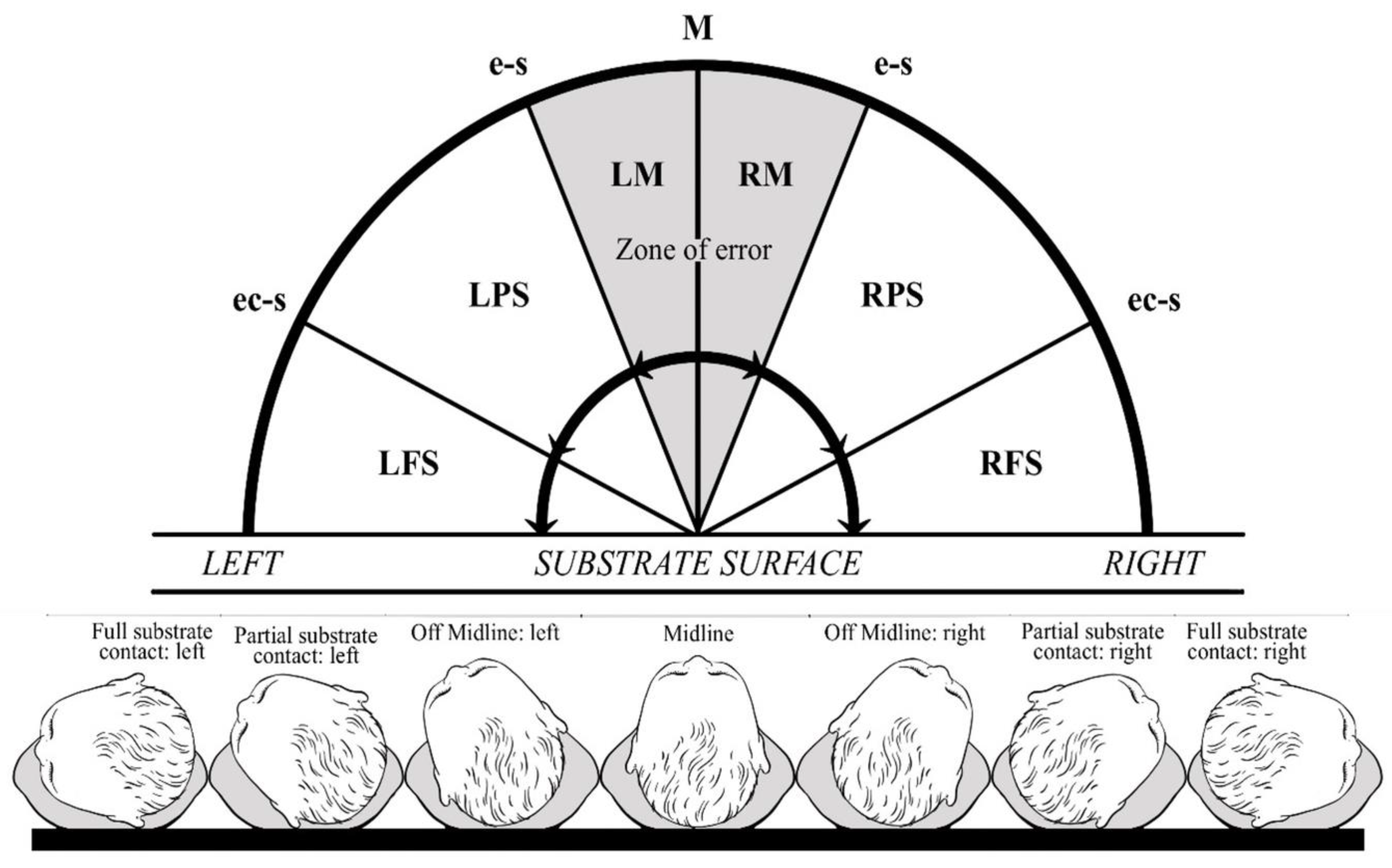
2.3. Neonatal Head Posture

The naturalistic observation of infant supine head posture was carried out in the same soundproofed examination room with no lateralised stimulation. The cot was positioned so that bilateral light sources were equidistant, thereby eliminating lateralised light stimulation. Neonates were swaddled and fed prior to the session. The time of testing was not held constant since head posture is unrelated to prandial conditions [22]. Clothing likely to restrict movement was removed. The examiner placed one hand on either side of the temporal region of the neonate’s head and gently rotated the head to a midline position. An assisted midline posture was maintained until no lateralised pressure was experienced against the examiner’s hands. The head was then released. No lateral stimulation of facial or perioral regions occurred to avoid a rooting response. Once the infant’s head was released, head posture was recorded for a five-minute period. This five-minute observation period was filmed with a GoPro Hero4 recording device (out of the line of sight of the infant) to eliminate any subjective bias in scoring.
An in-depth behavioural analysis was performed on each five-minute recording. Head posture (according to a coding scheme seen in Figure 1) together with the infant’s ongoing behavioural state [41,42] was recorded in consecutive 30 s intervals.
Postural deviations out of the midline were categorised according to the following four levels that created a seven-item coding scheme of neonatal head posture [43]:
  • Midline (M): no left/right rotation.
  • Off-midline: left (LM)/right (RM) head rotation without ipsilateral ear contact with the substrate surface.
  • Partial substrate contact: left (LPS)/right (RPS) head rotation with ipsilateral ear contact with the substrate surface.
  • Full substrate contact: left (LFS)/right (RFS) head rotation with ipsilateral ear and cheek contact with the substrate surface.
The seven items within this scheme are mutually exclusive and can be arranged in a 180° space, as shown in Figure 1. Apart from the midline posture, each item represents a segment of space. For each infant, postural amplitude is represented on an ordinal scale, representing deviation from the midline as ascending codes (e.g., RFS > RPS > RM). The coding scheme makes use of neonatal anatomical landmarks to determine the extent of postural deviation, which take individual variations in head size and shape into account.
According to the seven-item scheme, a definitively lateralised posture was considered to have occurred once there was partial ear contact with the substrate. The head typically oscillates around the midline, presumably as a result of extrapyramidal immaturity; movements between LM and RM therefore fell within a zone of error and were regarded as midline postures. Neonates who did not meet the classification criteria of partial contact with the substrate surface were removed from the analyses.
Since a distinction is made in the literature between two components of neonatal postural control, namely the kinetic assumption of head position (direction of the first head turning from the midline position) and the subsequent maintenance of a static head posture over time [44,45], both aspects were investigated in order to select the most appropriate grouping variable for subsequent arterio-postural analyses.
The direction of the initial head posture was recorded. In order to assess the maintenance of a lateralised head posture, postural amplitude and direction over the five-minute period of observation were rated at ten consecutive and equally spaced time points and used to calculate an individualised head posture score (HP score) for each neonate. The seven categories reflecting the amplitude of adopted head postures (LFS; LPS; LM; M; RM; RPS; RFS) were tallied for each neonate (Figure 1).
A laterality index for neonatal head posture preference (HP) was calculated for each assessment with the formula:
H P = ( L R ) / ( L + R )
where R equals the number of rightward postures and L the number of leftward postures. Infants with negative scores were classified as biased to the right and those with positive scores were classified as biased to the left.
Additional maintenance measures included the total time spent on the left side, total time spent on the right side, maximum postural amplitude adopted in both directions (according to the coding scheme above), and the number of midline crosses.
Since the direction of the first definitive turn (partial or full substrate contact) out of the midline best represented maintenance of the asymmetry, Ʌ = 0.146, χ2(2) = 190.785, p < 0.001, neonates were grouped as left- or right-biased according to the direction of the first turn out of the midline.

2.4. Revised Edinburgh Handedness Inventory

Parental handedness was assessed with a revised version of the Edinburgh Handedness Inventory [46,47]. Scores obtained from this inventory form a participant specific laterality quotient (LQ) range from 0 (all left) to 50 (all right) [45]. Parents with an LQ ≥ 30 were classified right-handed (91% mothers, 84% fathers); ≤20 were classified left-handed (9% mothers, 13% fathers). LQs between 21 and 29 identified those individuals who could not be classified definitively as either left- or right-handed (0% mothers, 3% fathers). This ratio is also used in the original inventory. Revisions to the inventory include the removal of the options for (1) placing a double score for extreme handedness; and (2) placing a score in both the right and the left columns for indifferent subjects [46]. These adaptations address common scoring criticisms of the traditional Edinburgh Handedness Inventory [48,49].
A hard copy of the revised form of the Edinburgh Handedness Inventory was given to each parent during the neonatal head posture assessment. This optimised the convenience of inventory completion and ensured the author remained blind to the potential handedness of the subject prior to the analysis. Each inventory consisted of ten items. Scores were recorded, an LQ calculated, and participants were grouped accordingly.

3. Statistical Analysis

Data were analysed using IBM SPSS Statistics (version 23) software. Four postural variables were subjected to a principal component analysis. These included neonatal HP score, difference in time spent between left and right sides (Time DIFF), maximum right-sided position, and maximum left-sided position. The principal axis method was used to extract the components. Only one component was extracted with an eigenvalue exceeding 1.0. An item was said to load on a given component if the component loading was 0.40 or greater for that component. Measures of the Kaiser–Meyer–Olkin Measure of Sampling Adequacy (0.799) and Bartlett’s Test of Sphericity (χ2(21) = 663.21, p < 0.001) were acceptable for this analysis.
“Postural maintenance” component scores were strongly related to the lateralisation of the first head turn, r = −0.924, p < 0.001. Group memberships according to infant “postural maintenance” were subjected to a discriminant function analysis (DFA) to assess the extent to which sustained posture could accurately classify the first head turn (i.e., to the right, midline, or left). The analysis is appropriate in the face of unequal class sizes if the smallest group exceeds the number of predictor variables [50]. All the assumptions of the analysis were upheld [50]. Accordingly, the first turn out of the midline was chosen as a grouping variable for the arterio-postural analyses.
Each haemodynamic measure of the middle cerebral artery was analysed using a mixed-design ANOVA. For each analysis, the within-subjects factor was the respective arterial parameter (of the left and right paired arteries) and the between-subjects factor was the postural dominance (left-postured; right-postured). One-tailed paired t-tests compared lateral differences in posture groups in instances of significant interactions. One-tailed independent t-tests also compared sex differences in participant demographics and haemodynamic parameters at each site of measurement. Tests of normality and homoscedasticity (namely Levene’s test of equality of variance and Box’s test of equality of covariance matrices) were run on each dataset. If the assumption of normality was not upheld, a non-parametric Wilcoxon signed-rank test was run instead.
A DFA was used to determine the predictability of lateralised neonatal posture based on laterality indices of vessel calibre and blood flow volume. A stepwise DFA determined which predictors at the two sites explained the greatest proportion of variance. Laterality indices were computed with the following equation:
L I = ( L R ) / ( L + R )
where R equals the right arterial measure and L the left arterial measure. A positive value indicated left arterial dominance, whereas a negative value indicated right arterial dominance.
The following predictor variables were used (1) MCAO diameter LI; (2) MCADT diameter LI; (3) MCAO blood flow volume LI; and (4) MCADT blood flow volume LI. All assumptions for the analysis were upheld [51].
A two-step cluster analysis was used to explore natural clusters of structural and haemodynamics arterial asymmetries in the data and their relationship with neonatal head posture. Given the assumption of variable independence for effective clustering, four arterial laterality indices were subjected to a principal component analysis. These included MCAO diameter LI, MCAO blood flow volume LI, MCADT diameter LI, and MCADT blood flow volume LI. One component was extracted. The extracted component explained 85.04% of the variance in the diametric and volumetric arterial asymmetry, thereby constituting an “arterial asymmetry” metric. The Kaiser–Meyer–Olkin Measure of Sampling Adequacy (0.662) and Bartlett’s Test of Sphericity (χ2(6) = 440.94, p < 0.001) were acceptable for this analysis.
Arterial asymmetry component scores were subjected to a two-step cluster analysis. The number of clusters formed was not specified in advance. For distance measures, the log-likelihood method with Akaike’s information criterion was used. The “silhouette measure of cohesion and separation”, which ranges from −1 to 1, was used as an estimate of the overall goodness of fit for the cluster structure: <0.25 = no substantial structure, 0.26–0.50 = weak structure that could be artificial, 0.51–0.70 = reasonable structure, and 0.71–1.0 = strong structure [52].
Cohen’s (1992) “rule of thumb” for effect size interpretations was used for between-group comparisons: d = 0.20 (small effect), d = 0.50 (medium effect), and d = 0.80 (large effect). Significance was determined with a 95% confidence level at p < 0.05.

4. Results

Observations of neonatal head posture, together with bilateral geometric and haemodynamic assessments of the middle cerebral origin and distal trunk, were recorded in 86 healthy full-term neonates. The final sample included 55 males and 31 females born via normal vaginal delivery or caesarean section (Table 1). Gestational age at birth of the sample ranged from 36 to 41 weeks, and birth weights ranged from 2200 g to 4690 g. Postnatal age at the time of scanning was 18 to 174 h (M = 49.35 h; SD = 29.43 h; Median = 41.5 h; Range = 156 h). Mean Apgar scores were 9 at one minute (IQR = 1.0; Range = 6) and 9 at five minutes (IQR = 0.0; Range = 3).
Neonatal head turning was systematically biased to the right, χ2(1) = 6.698, p = 0.010, with 64% of neonates turning to the right and 36% turning to the left. Right- and left-posture groups did not differ in neonatal birth weight, postnatal scanning age, gestation, or parental hand preference (p > 0.05).

4.1. Asymmetries in the Origin and Terminus of the Middle Cerebral Artery Trunk

The geometric and haemodynamic properties of the left and right middle cerebral arterial trunk were asymmetric and meaningfully related to the lateralised neonatal head-turning bias. In right-biased neonates, vascular asymmetry was consistent with larger left-than-right arterial diameters and higher blood flow volumes in the middle cerebral trunk origin and terminus (Table 2). Neonates with a leftward head posture were less laterally differentiated. At the vessel origin, the middle cerebral artery trunk was typically larger with higher blood flow volumes in the right hemisphere, but these differences did not reach significance at the termination of the arterial trunk (p = 0.230 and p = 0.197, respectively; Table 2).
The direction of the supine head-turning bias was differentially influenced by the middle cerebral arterial geometry and haemodynamics, in that posture interactions were found for arterial diameter and blood flow volume at both arterial sites (Table 3; Figure 2). Neonatal head orientation was not significantly associated with arterial velocity (peak systolic, end-diastolic, and mean velocities).
Bilaterally, left-postured neonates had larger overall arterial calibres (M = 2.20 mm, SE = 0.05; F(1, 84) = 5.608, p = 0.020, ηp2 = 0.063) and blood flow volumes (M = 229.67 mL/min, SE = 11.20; F(1, 84) = 11.968, p = 0.001, ηp2 = 0.125) at the trunk origin than their right-postured counterparts (M = 2.06 mm, SE = 0.04; M = 181.13 mL/min, SE = 8.42). At the trunk terminus, bilateral blood flow volumes were also greater in this group (left posture M = 185.99 mL/min, SE = 9.02; right posture M = 158.60 mL/min; SE = 6.77; F(1, 84) = 5.901, p = 0.017, ηp2 = 0.066). No lateral differences in arterial velocities (peak systolic, end-diastolic, or average blood flow velocities) were found in the trunk of either posture group (Table 2 and Table 3; Figure 2).
Resistance to blood flow caused by the microvascular bed distal to the site of measurement did not significantly interact with neonatal head-turning bias (Table 3). However, a main effect for arterial resistance was found at the most lateral aspect of the middle cerebral trunk. The resistance distal to the trunk terminus was higher in the left cerebral hemisphere across all participants, (F(1, 84) = 7.953, p = 0.006, ηp2 = 0.086). This main effect was also reflected in the pulsatility indices.
No lateral differences in arterial resistance were noted at the middle cerebral artery origin. Arterial resistance was, however, asymmetric at the distal trunk. Neonates with a leftward head-turning bias had higher resistance and pulsatility indices in the left cerebral hemisphere than the right (Table 2). No left–right differences were found in neonates with a right-sided head posture.

4.2. Predicting the First Head Turn from Arterial Characteristics

Arterial characteristics significantly predicted the direction of individual neonatal head turning. Given the multicollinearity between diametric and volumetric asymmetries of the origin and the distal trunk (r > 0.70), only one variable was retained by the discriminant function analysis. Postural lateralisation was predicted by blood flow volume asymmetries at the origin of the arterial trunk, Ʌ = 0.853, χ2(2) = 13.300, p < 0.001. Tests of equality of group means and an evaluation of the structure matrix revealed the volume flow at the origin accounted for approximately 15% of the variance in the model. The cross-validated classification showed that overall, 73.3% of cases were correctly classified, with accurate group membership predictions for 85.5% of right-biased and 51.69% of left-biased neonates (Table 4).

4.3. Natural Clusters of Arterial Asymmetry

Given the postural heterogeneity of left-biased neonates, a two-step cluster analysis was used to explore whether natural clustering was inherent in the sample according to their degree of vascular asymmetry and the direction of their respective head posture. A four-cluster model with a silhouette measure of cohesion and separation of 0.70 and size ratio of 2.75 was identified in the sample. Positive values indicated a leftward arterial dominance and negative values indicated a rightward arterial dominance.
Cluster one consisted of a small subgroup of neonates (17.4%; minority cluster) with a strong and consistent rightward arterial dominance (M = −1.55; SD = 0.38). This cluster was associated with a tendency to adopt a leftward head posture (60.0% of neonates turned to the left). Conversely, cluster three (14.0%) constituted a small subgroup of neonates with a strong and consistent leftward arterial dominance (M = 1.52; SD = 0.38), and a propensity to turn the head to the right (75.0% of these neonates turned to the right).
The remaining two clusters represented the majority of neonates (68.6%) whose arterial asymmetry component scores remained within one quartile of the sample median. The fourth and largest cluster (38.4%) constituted a subgroup of neonates with leftward arterial dominance (M = 0.51; SD = 0.25) and a strong propensity to adopt a rightward head posture (84.9% of neonates in this group turned to the right). Neonates in cluster two (30.2%; M = −0.45; SD = 0.26), however, were unlike the others. Although these neonates had a rightward arterial dominance, head turning in this group was not differentially distributed between the left (53.8%) and right (46.2%) sides.
The middle cerebral arterial geometry and haemodynamics differentially influenced cluster membership, in that significant cluster interactions were found for arterial diameter and blood flow volume in each group and at both arterial sites (p < 0.001; Figure 3). As expected, the arterio-postural relationship in right-postured neonates (clusters three and four) was homogeneous, in that both clusters were characterised by significant leftward arterial biases in calibre and blood flow volume, and a strong propensity to adopt a right-sided head posture when supine. Neonates with the highest propensity to turn the head to the left (cluster one) constituted a small subgroup of the sample and these neonates were characterised by consistent rightward arterial biases in calibre and volume flow (Figure 3; Table 4). Neonates with weaker and less consistent rightward arterial biases across the trunk were unlike the other clusters: Postural lateralisation was more heterogeneous in this group and the vascular asymmetry, although still significant, was less pronounced (Figure 3; Table 5).

5. Discussion

Systematic handedness-related anatomical asymmetries extend beyond the brain parenchyma to the vascular system in adults. We previously reported vascular correlates for hand preference and proficiency at multiple points in the left and right supra-aortic arterial tree in adults [1]. The developmental origins of this correlation are investigated in the present study. To our knowledge, this is the first investigation of the relationship between anatomical and physiological cerebro-arterial asymmetries and lateralised infant behaviour.
Our findings demonstrate that bilateral structural symmetry between the paired middle cerebral vessels of healthy neonates is the anatomical exception rather than the rule. Trunk calibre and corresponding blood flow volume is systematically biased towards the left cerebral hemisphere. As we found for carotid arterio-handedness relationships in adults [1], the structurofunctional vascular asymmetry in neonates is contralaterally related to the supine head-turning bias, which is considered a strong precursor of handedness [21,24].
The preponderance of neonates with a rightward head position was systematic in the sample and was in keeping with earlier normative studies of head-turning bias [19,20,21,22,23,24]. This study demonstrated that spontaneous sustained head posturing is coherent with the lateralisation of the first turn out of the midline, making the latter an ideal index of neonatal lateral bias for the analysis of arterio-posture relationships.
The arterio-postural relationship is contralaterally organised in neonates who turn to the right. This relationship is characterised by larger arterial diameters and higher blood flow volumes in the left hemisphere. Deviations from this typical arterio-postural association were associated with increasing lateral heterogeneity in head-turning behaviour. In left-turning newborns, the coherence of the arterio-postural relationship is dependent on the magnitude of arterial asymmetry across the midline of the brain in the opposite direction, as well as maintenance of these larger arterial diameters from the proximal to distal regions of the right trunk. Only 17.4% of the sample demonstrated the strong rightward arterial asymmetries with leftward-turning dispositions.
Right-posturing neonates demonstrate a reliable right-hand preference at 19 weeks (p < 0.001) [25], and 82% of infants with rightward-reaching preferences at 19 weeks are also right-biased at 3 years of age [20]. Conversely, infants with leftward-turning preferences are less likely to show a reliable hand preference at 19 weeks (p < 0.50). Approximately 43 to 75% of left-postured neonates show a 19-week left-hand preference [25] and only 21% of infants who are left-biased for reaching at 19 weeks remain left-biased at 3 years of age [20]. The low specification of lateralisation in left-posturing neonates is consistent with a greater heterogeneity in patterns of cerebral lateralisation and manual preferences in left-handed adults [53]. It is also in keeping with the low specification with which lateralised neonatal head postures predict handedness later in development.
The present findings suggest that the relationship between arterial asymmetry and newborn head-turning biases is graded and is not optimally accommodated within a dichotomous model. In adults, the degree of arterial dominance is associated with the degree of contralateral hand proficiency [1].
Cerebral lateralisation for language and handedness, as classically described in the mature brain, is considered a predominantly neocortical phenomenon [54]. While adult-like parenchymal asymmetries have repeatedly been observed in the neonatal cortex [11,14,55,56,57,58], lateralised head posturing has been conceptualised as a subcortically mediated behaviour [43,58,59]. It has been hypothesised that striatal asymmetry exerts postural control in newborns through extrapyramidal pathways and is progressively encephalised together with the developmental emergence and refinement of pyramidal motor function [43,53].
Evidence for a subcortical substrate largely stems from the rodent literature [60,61,62]. Human striatal asymmetries have, however, been documented in neurotransmitter [63,64], activation [65], and volumetric studies [66], and these asymmetries are also often related to adult hand preference and proficiency [63,64,65]. In human newborns, the prominent head-turning bias in the first few weeks of postnatal development is attributed to (1) anatomical and functional maturation of subcortical regions that precede those of cortical regions responsible for motoric function [43,67]; (2) immaturity of visual function and postural differentiation [43,68]; and (3) relative dominance of neural rather than environmental factors in the regulation of behaviour [20]. While the exact neural substrate of neonatal head posture asymmetries has long been debated, our findings, amongst others, lend further weight to the hypothesis that the lateralised neonatal head posture is part of the same laterally differentiated motor system as later hand preference. The propensity to adopt a lateralised head posture when supine and the direction of the head-turning asymmetry is a stable individual characteristic [20,43], is predictive of future handedness [24,25], and is uninfluenced by fluctuations in behavioural state or extraneous environmental factors [22,25].
Neonatal arterial asymmetries are meaningfully related to future manual preference and therefore to cerebral dominance for language and practic functions. From a functional point of view, the direction of this asymmetry potentially accommodates greater perfusion demands in the hemisphere dominant for language and manual dexterity [1,12,15,16,17]. Vascular asymmetries in right-posturing neonates reflect a predominantly leftward bias in the metabolic demand of the lateral surface of the frontal, parietal, and temporal lobes, as well as medial subcortical structures of the brain. The arterial asymmetry is less marked in the presence of atypical behavioural lateralisation, with a higher likelihood of a leftward postural expression in cases of extreme reversals of the arterial bias.
The present findings do not exclude the possibility of a top-down, possibly demand-driven, neurodevelopmental process, in which greater left hemispheric resource utilisation culminates in commensurate compensatory changes in vascular supply.
The continued impact of parenchymal demand in determining the form and size of the cerebrovascular system has been repeatedly demonstrated [69,70,71]. Since the earliest observation of neuroanatomical laterality has been reported at just 11 weeks post-conception [72], it remains possible that neonatal arterial asymmetries in vessels subserving lateralised functions, like those reported in this work, might result from the accumulating influence of asymmetrical top-down metabolic demands in early development, rather than the converse.
However, there is also evidence to suggest a genetic template for lateralised cortical angiogenesis. RNA sequencing analyses of human embryos and foetuses have shown that gene sets for angiogenesis are significantly asymmetric and are more strongly expressed in the cortex of the left cerebral hemisphere [73]. While blood vessels provide the developing tissue with oxygen, they can also play many roles in the development of central nervous system tissue, including guiding axon outgrowth and neuronal migration [74]. In the light of these findings, the presence of robust adult-like structural cerebrovascular asymmetries at birth might have a broader role in shaping cerebral asymmetries beyond supply.
Faint but systematic echoes of a leftward bias in angiogenic prespecification can be found in other supra-aortic extracranial vessels, but where asymmetries are meaningless for the brain. The vertebral arteries are left-dominant in approximately 54% of cases (and right-dominant in only 30%) [75]. This pattern is not related to hand preference [75] and is inconsequential for cerebral lateralisation because the left and right vertebral arteries converge to form the midline basilar artery prior to contributing to cerebral perfusion. A similar leftward bias has also been reported in the external carotid arteries (left-dominant in 58% of cases; right-dominant in 38%), with no meaningful relationship to handedness [1]. The fact that these biases exist suggest a genetic specification for the asymmetry in angiogenesis. If this is the case, existing asymmetries in arteries supplying the cerebral cortex might be enhanced by the ongoing asymmetric demand of the tissue through the top-down mechanisms described above.
The existence of a vascular correlate of the most manifest of human behavioural asymmetries has key implications for understanding the origins of human cerebral organisation from evolutionary and developmental perspectives. These findings also represent an interesting case study in the history of science, where a promising hypothesis is resoundingly rejected, only to enjoy eventual confirmation through a chronological loop spanning centuries. In spite of its early history, the hypothesis that vascular asymmetry is likely to be a vital contributor to human cerebral lateralisation is now empirically supported.
The current study was cross-sectional. A longitudinal study is the obvious next step to clarify the ontogenesis of the arterio-behavioural relationships that we have identified.

Author Contributions

A.J.v.V. contributed: conceptualisation, methodology, investigation, formal analysis, writing (original draft preparation), visualisation, project administration; M.S. (Michael Saling) contributed: conceptualisation, resources, principal supervision, writing (original draft preparation); S.R. contributed: methodology, writing (review and editing), validation, co-supervision; P.A. contributed: writing (review and editing), co-supervision; J.C. contributed: writing (review and editing), co-supervision; M.S. (Mark Solms) contributed: conceptualisation, writing (review and editing and final draft preparation). All authors have read and agreed to the published version of the manuscript.

Funding

National Health & Medical Research Council (NHMRC) Senior Research Fellowship (ID 1081288); NHMRC Career Development Fellowship 1141354; Australian Government Research Training Program (RTP) Scholarship.

Institutional Review Board Statement

The study was conducted in accordance with the Declaration of Helsinki, and approved by the Institutional Review Board of The Royal Women’s Hospital (protocol code 16/18 on 4 July 2016).

Informed Consent Statement

Informed consent was obtained from all subjects involved in the study.

Data Availability Statement

The data presented in this study are available on request from the corresponding author.

Acknowledgments

We are indebted to the Royal Women’s Hospital and Frances Perry House for providing the platform for us to conduct our study. We would also like to thank Julie Archbold for her help in calibrating the Doppler ultrasound machine.

Conflicts of Interest

The authors declare no conflict of interest.

References

  1. Jansen van Vuuren, A.; Saling, M.M.; Ameen, O.; Naidoo, N.; Solms, M. Hand preference is selectively related to common and internal carotid arterial asymmetry. Laterality Asymmetry Body Brain Cogn. 2016, 4, 377–398. [Google Scholar] [CrossRef]
  2. Broca, P. Sur le siège de la faculté du langage articulé. Bull. Société Anat. Paris 1865, 6, 377–393. [Google Scholar] [CrossRef]
  3. Dax, G. Notes sur le même sujet. Gaz. Hebd. Med. Chir. 1865, 2, 259–262. [Google Scholar]
  4. Hyrtl, J. Handbuch der Topographischen Anatomie, 4th ed.; Braumüller: Vienna, IT, USA, 1860. [Google Scholar]
  5. Magendi, F. Recherches Physiologique sur la Vie er la Mort, 4th ed.; Gabou, Bechet Jeuve: Paris, France, 1822. [Google Scholar]
  6. Ogle, W. On dextral pre-eminence. Trans. R. Med. Chir. Soc. Lond. 1871, 54, 279–301. [Google Scholar] [CrossRef] [PubMed]
  7. de Fleury, A. Du dynamisme comparé des hémisphères cérèbraux dans l’homme. Association Française Pour l’Avancement des Sciences (comptes-rendus de la première session). Congrès Bordx. 1873, 1, 834–845. [Google Scholar]
  8. Beeley, A.L. Left-handedness. Am. J. Phys. Anthropol. 1919, 2, 389–400. [Google Scholar] [CrossRef]
  9. Kellogg, G.M. The physiology of right-and left-handedness. J. Am. Med. Assoc. 1989, 30, 356–358. [Google Scholar] [CrossRef]
  10. Crichton-Browne, J. Dexterity and the bend sinister. Proc. R. Inst. Great Br. 1907, 18, 623–652. [Google Scholar]
  11. Geschwind, N.; Levitsky, W. Human brain: Left-right asymmetries in temporal speech region. Science 1968, 161, 186–187. [Google Scholar] [CrossRef] [PubMed]
  12. Gur, R.C.; Obrist, W.B.; Hungerbuhler, J.P.; Younkin, D.; Rosen, A.D.; Skolnick, B.E.; Reivich, M. Sex and head posture differences in cerebral blood flow during rest and cognitive activity. Science 1982, 217, 659–660. [Google Scholar] [CrossRef]
  13. Mazziotta, J.C.; Phelps, M.E.; Carson, R.E.; Kuhl, D.E. Tomographic mapping of human cerebral metabolism: Sensory deprivation. Ann. Neurol. 1982, 12, 435–444. [Google Scholar] [CrossRef]
  14. Wada, J.A.; Clarke, R.; Hamm, A. Cerebral hemispheric asymmetry in humans. Arch. Neurol. 1975, 32, 239–246. [Google Scholar] [CrossRef] [PubMed]
  15. Chiron, C.; Jambaque, I.; Nabbout, R.; Lounes, R.; Syrota, A.; Dulac, O. The right brain hemisphere is dominant in human infants. Brain 1997, 120, 1057–1065. [Google Scholar] [CrossRef] [PubMed]
  16. Kamath, S. Observations on the length and diameter of vessels forming the circle of Willis. J. Anat. 1981, 133 Pt 3, 419–423. [Google Scholar] [PubMed]
  17. Willis, M.W.; Ketter, T.A.; Kimbrell, T.A.; George, M.S.; Herscovitch, P.; Danielson, A.L.; Benson, B.E.; Post, R.M. Age, sex and laterality effects on cerebral glucose metabolism in healthy adults. Psychiatry Res. Neuroimaging 2002, 114, 23–37. [Google Scholar] [CrossRef] [PubMed]
  18. Krogh, A. The Anatomy and Physiology of Capillaries; Yale University Press: New Haven, CT, USA, 1929. [Google Scholar]
  19. Gesell, A.; Ames, L.B. The development of handedness. J. Genet. Psychol. 1947, 70, 155–175. [Google Scholar] [CrossRef] [PubMed]
  20. Liederman, J. Mechanisms underlying instability in the development of hand preference. In Manual Specialization and the Developing Brain; Young, G., Segalowitz, S., Corter, C., Trehub, S., Eds.; Academic Press: New York, NY, USA, 1983; pp. 71–92. [Google Scholar]
  21. Saling, M.M. Familial handedness, prenatal environmental adversity, and neonatal lateral organisation. In Manual Specialisation and the Developing Brain; Young, G., Segalowitz, S.J., Corter, C.M., Trehab, S.E., Eds.; Academic Press: New York, NY, USA, 1983; pp. 275–284. [Google Scholar]
  22. Turkewitz, G.; Creighton, S. Changes in lateral differentiation of head posture in the human neonate. Dev. Psychobiol. 1974, 8, 85–89. [Google Scholar] [CrossRef]
  23. Coryell, J.; Michel, G.F. How supine postural preference of infants can contribute towards the development of handedness. Infant Behav. Dev. 1978, 1, 245–257. [Google Scholar] [CrossRef]
  24. Michel, G.F. Right-handedness: A consequence of infant supine head-orientation preference? Science 1981, 212, 685–687. [Google Scholar] [CrossRef]
  25. Goodwin, R.; Michel, G.F. Head orientation position during birth in neonatal period, and hand preference at 19 weeks. Child Dev. 1981, 52, 819–826. [Google Scholar] [CrossRef]
  26. Oktar, S.O.; Yücel, C.; Karaosmanoglu, D.; Akkan, K.; Ozdemir, H.; Tokgoz, N.; Tali, T. Blood-flow volume quantification in internal carotid and vertebral arteries: Comparison of 3 different ultrasound techniques with phase contrast MR imaging. Am. J. Neuroradiol. 2006, 27, 363–369. [Google Scholar]
  27. Kloosterman, A.; Hierck, B.; Westerweel, J.; Poelma, C. Quantification of blood flow topology in developing vascular networks. Plos ONE 2014, 9, 1–15. [Google Scholar]
  28. Gibbons, G.H.; Dzau, V.J. The emerging concept of vascular remodelling. NEJM 1994, 330, 1431–1438. [Google Scholar] [PubMed]
  29. Lasjaunias, P.; Bernstein, A.; ter Brugge, K.G. Surgical Neuroangiography; Springer: Berlin/Heidelberg, Germany, 2001. [Google Scholar]
  30. Drayton, M.R.; Skidmore, R. Vasoactivity of the major intracranial arteries in newborn infants. Arch. Dis. Child. 1987, 62, 236–240. [Google Scholar] [CrossRef] [PubMed]
  31. van Vuuren, A.J.; Saling, M.; Rogerson, S.; Anderson, P.; Cheong, J.; Solms, M. Cerebral arterial asymmetries in the neonate: Insight into the pathogenesis of stroke. Symmetry 2022, 14, 456. [Google Scholar] [CrossRef]
  32. Hayashi, T.; Ichiyama, T.; Uchida, M.; Tashiro, N.; Tanaka, H. Evaluation by color Doppler and pulsed Doppler sonography of blood-flow velocities in intracranial-arteries during the early neonatal-period. Eur. J. Pediatr. 1992, 151, 461–465. [Google Scholar] [CrossRef] [PubMed]
  33. Kempley, S.T.; Vyas, S.; Bower, S.; Nicolaides, K.H.; Gamsu, H. Cerebral and renal artery blood flow velocity before and after birth. Early Hum. Dev. 1996, 46, 165–174. [Google Scholar] [CrossRef] [PubMed]
  34. Pourcelot, L. L’examen Doppler des Vaisseaux Périphériques; ACD Production: Paris, France, 1982. [Google Scholar]
  35. Gosling, R.C.; King, D.H. Arterial assessment by Doppler shift ultrasound. Proc. R. Soc. Med. 1988, 67, 447–449. [Google Scholar]
  36. Wu, Y.; Hsieh, W.; Hsu, C.; Chiu, N.; Chou, H.; Chen, C.; Peng, S.; Hung, H.; Chang, J.; Chen, W.J.; et al. Relationship of neonatal cerebral blood flow velocity asymmetry with early motor, cognitive and language development in term infants. Ultrasound Med. Biol. 2013, 39, 797–803. [Google Scholar] [CrossRef] [PubMed]
  37. Jahromi, A.S.; Cinà, C.S.; Liu, Y.; Clase, C.M. Sensitivity and specificity of color duplex ultrasound measurement in the estimation of internal carotid artery stenosis: A systematic review and meta-analysis. J. Vasc. Surg. 2005, 41, 962–972. [Google Scholar] [CrossRef]
  38. Kamouchi, M.; Kishikawa, K.; Okada, Y.; Inoue, T.; Ibayashi, S.; Iida, M. Reappraisal of flow velocity ratio in common carotid artery to predict hemodynamic change in carotid stenosis. Am. J. Neuroradiol. 2005, 26, 957–962. [Google Scholar]
  39. Hansen, N.B.; Stonestreet, B.S.; Rosenkrantz, T.S.; Oh, W. Validity of Doppler measurements of anterior cerebral artery blood flow velocity: Correlation with brain blood flow in piglets. Pediatrics 1983, 72, 526–531. [Google Scholar] [CrossRef]
  40. Julkunen, M.; Uotila, J.; Eriksson, K.; Janas, M.; Luukkaala, T.; Tammela, O. Obstetric parameters and Doppler findings in cerebral circulation as predictors of 1 year neurodevelopmental outcome in asphyxiated infants. J. Perinatol. 2012, 32, 631–638. [Google Scholar] [CrossRef]
  41. Prechtl, H.F.R. The behavioural states of the newborn infant (a review). Brain Res. 1974, 76, 185–212. [Google Scholar] [CrossRef]
  42. Wolf, P.H. The Causes, Controls, and Organization of Behaviour in the Neonate. Psychol. Issues 1966, 5, 1–105. [Google Scholar]
  43. Saling, M.M. Determinants of Lateral Organisation in Neonates. Unpublished Doctoral Dissertation, University of Witwatersrand, Johannesburg, South Africa, 1982. [Google Scholar]
  44. Turkewitz, G. The development of lateral differences in the human infant. In Lateralization in the Nervous System; Harnad, S.R., Doty, R.W., Goldstein, L., Jaynes, J., Krauthamer, G., Eds.; Academic Press: San Diego, CA, USA, 1977; pp. 251–259. [Google Scholar]
  45. Turkewitz, G.; Birch, H.G. Neurobehavioral organisation of the human newborn. In Exceptional Infant: Studies in Abnormalities; Hellmuth, I., Ed.; Bruner/Mazel: New York, NY, USA, 1971; pp. 24–40. [Google Scholar]
  46. Oldfield, R.C. The assessment and analysis of handedness: The Edinburgh inventory. Neuropsychologia 1971, 9, 97–113. [Google Scholar] [CrossRef]
  47. White, K.; Ashton, R. Handedness assessment inventory. Neuropsychologia 1976, 14, 261–264. [Google Scholar] [CrossRef] [PubMed]
  48. Edlin, J.M.; Leppanen, M.L.; Fain, R.J.; Hackländer, R.P.; Hanaver-Torrez, S.D.; Lyle, K.B. On the use (and misuse?) if the Edinburgh Handedness Inventory. Brain Cogn. 2015, 94, 44–51. [Google Scholar] [CrossRef] [PubMed]
  49. Williams, S.M. Handedness inventories: Edinburgh versus Annett. Neuropsychology 1991, 5, 43–48. [Google Scholar] [CrossRef]
  50. Huberty, C.J. Discriminant Analysis. Rev. Educ. Res. 1975, 45, 543–598. [Google Scholar] [CrossRef]
  51. Klecka, W.R. Discriminant Analysis: Quantitative Applications in the Social Sciences Series; Sage Publications: Thousand Oaks, CA, USA, 1980. [Google Scholar]
  52. Kaufman, L.; Rousseeuw, P.J. Finding Groups in Data: An Introduction to Cluster Analysis; John Wiley & Sons, Inc.: Hoboken, NJ, USA, 1990. [Google Scholar]
  53. Hervé, P.Y.; Zago, L.; Petit, L.; Mazoyer, B.; Tzourio-Mazoyer, N. Revisiting human hemispheric specialization with neuroimaging. Trends Cogn. Sci. 2013, 17, 69–80. [Google Scholar] [CrossRef] [PubMed]
  54. Galaburda, A.M.; Le May, M.; Kemper, T.L.; Geschwind, N. Right-left asymmetries in the brain. Science 1978, 199, 852–856. [Google Scholar] [CrossRef] [PubMed]
  55. Sowell, E.R.; Thompson, P.M.; Rex, D.; Kornsand, D.; Tessner, K.D.; Jernigan, T.L.; Toga, A.W. Mapping sulcal pattern asymmetry and local cortical surface gray matter distribution in vivo: Maturation in perisylvian cortices. Cereb. Cortex 2002, 12, 17–26. [Google Scholar] [CrossRef] [PubMed]
  56. Chi, J.G.; Dooling, E.C.; Gilles, F.H. Left-right asymmetries of the temporal speech areas of the human fetus. Arch. Neurol. 1977, 34, 346–348. [Google Scholar] [CrossRef]
  57. Dehaene-Lambertz, G.; Hertz-Pannier, L.; Dubois, J. Nature and nurture in language acquisition: Anatomical and functional brain-imaging studies in infants. Trends Neurosci. 2006, 29, 367–373. [Google Scholar] [CrossRef] [PubMed]
  58. Kinsbourne, M. A model for the ontogeny of cerebral organisation in non-right-handers. In Neuropsychology of Left-Handedness; Herron, J., Ed.; Raven Press: New York, NY, USA, 1980. [Google Scholar]
  59. Peters, M. Differentiation and lateral specialisation in motor development. In Manual Specialisation and the Developing Brain; Young, G., Segalowitz, S.J., Corter, C.M., Trehab, S.E., Eds.; Academic Press: New York, NY, USA, 1983; pp. 141–159. [Google Scholar]
  60. Glick, S.D.; Jerussi, T.P.; Zimmerberg, B. Behavioural and neuropharmacological correlates of nigrostriatal asymmetry in rats. In Lateralisation in the Nervous System; Harnad, S., Doty, R.W., Goldstein, L., Jaynes, J., Krauthamer, G., Eds.; Academic Press: New York, NY, USA, 1977. [Google Scholar]
  61. Andrade, C.; Alwarshetty, M.; Sudha, S.; Chandra, J.S. Effect of innate direction bias on T-maze learning in rats: Implications for research. J. Neurosci. Methods 2001, 110, 31–35. [Google Scholar] [CrossRef] [PubMed]
  62. Rodriguez, M.; Martin, L.; Santana, C. Ontogenic development of brain asymmetry in dopaminergic neurons. Brain Res. Bull. 1994, 33, 163. [Google Scholar] [CrossRef] [PubMed]
  63. de la Fuente-Fernández, R.; Kishore, A.; Calne, D.B.; Ruth, T.J.; Stoessl, A.J. Nigrostriatal dopamine system and motor lateralization. Behav. Brain Res. 2000, 112, 63–68. [Google Scholar] [CrossRef]
  64. Tomer, R.; Slagter, H.A.; Christian, B.T.; Fox, A.S.; King, C.R.; Murali, D.; Davidson, R.J. Dopamine asymmetries predict orienting bias in healthy individuals. Cereb. Cortex 2013, 23, 2899–2904. [Google Scholar] [CrossRef]
  65. Scholza, V.H.; Flaherty, A.W.; Kraft, E.; Keltner, J.R.; Kwong, K.K.; Chen, Y.I.; Rosen, B.R.; Jenkins, B.G. Laterality, somatotopy and reproducibility of the basal ganglia and motor cortex during motor tasks. Brain Res. 2000, 879, 204–215. [Google Scholar]
  66. Kang, X.; Herron, T.J.; Ettlinger, M.; Woods, D.L. Hemispheric asymmetries in cortical and subcortical anatomy. Laterality Asymmetries Body Brain Cogn. 2015, 20, 658–684. [Google Scholar] [CrossRef] [PubMed]
  67. Teicher, M.H.; Andersen, S.L.; Hostetter, J.C., Jr. Evidence for dopamine receptor pruning between adolescence and adulthood in striatum but not nucleus accumbens. Brain Res. Dev. 1995, 89, 167–172. [Google Scholar] [CrossRef] [PubMed]
  68. Hopkins, B.; Lems, Y.L.; van Wulfften Palthe, T.; Hoeksma, J.; Kardaun, O.; Butterworth, G. Development of head position preference during early infancy: A longitudinal study in the daily life situation. Dev. Psychobiol. 1990, 23, 39–53. [Google Scholar] [CrossRef] [PubMed]
  69. Pries, A.R.; Reglin, B.; Secomb, T.W. Modelling of angioadaptation: Insights for vascular development. Int. J. Dev. Biol. 2011, 55, 399–405. [Google Scholar] [CrossRef]
  70. van Overbeeke, J.J.; Hillen, B.; Tulleken, C.A.F. A comparative study of the circle of Willis in fetal and adult life: The configuration of the posterior communicating artery. J. Anat. 1991, 178, 45–54. [Google Scholar]
  71. Rossitti, S.; Lofgren, J. Vascular dimensions of cerebral arteries follow the principle of minimum work. Stroke 1993, 24, 371–377. [Google Scholar] [CrossRef]
  72. Kasprian, G.; Langs, G.; Brugger, P.C.; Bittner, M.; Weber, M.; Arantes, M.; Prayer, D. The Prenatal origin of hemispheric asymmetry: An in utero neuroimaging study. Cereb. Cortex 2011, 21, 1076–1083. [Google Scholar] [CrossRef]
  73. De Kovel, C.G.F.; Lisgo, S.N.; Fisher, S.E.; Francks, C. Subtle left-right asymmetry of gene expression profiles in embryonic and foetal human brains. Sci. Rep. 2018, 8, 12606. [Google Scholar] [CrossRef]
  74. Eichmann, A.; Thomas, J.L. Molecular parallels between neural and vascular development. Cold Spring Harb. Perspect. Med. 2013, 3, a006551. [Google Scholar] [CrossRef]
  75. Cagnie, B.; Petrovic, M.; Voet, D.; Barbaix, E.; Cambie, D. Vertebral artery dominance and hand preference: Is there a correlation? Man. Ther. 2006, 11, 153–156. [Google Scholar] [CrossRef]
Figure 1. The seven-item scheme for coding of neonatal head postures arranged within a 180° space as determined by a multidimensional analysis [43]. Corresponding illustrations of postural amplitudes are also presented. M = midline; RM = off-midline right; LM = off-midline left; RPS = right partial substrate contact; LPS = left partial substrate contact; RFS = right full substrate contact; LFS = left full substrate contact; e-s = ear substrate contact; ec-s = ear and cheek substrate contact.
Symmetry 16 00161 g001
Figure 2. Interaction effects of middle cerebral artery origin (top) and trunk terminus (bottom). Diameter (A), VMEAN (B), blood flow volume (C) at initial head turn. Error bars show the 95% confidence interval. LMCA = left middle cerebral artery; RMCA = right middle cerebral artery; VMEAN = mean velocity; index; Q = blood flow volume.
Symmetry 16 00161 g002
Figure 3. Interaction effects of bilateral middle cerebral artery origin and distal trunk diameter (A), Q (B), and cluster membership. Majority clusters are represented by a solid line and minority clusters by a stippled line. Error bars show the 95% confidence interval. LMCA = left middle cerebral artery; RMCA = right middle cerebral artery; Q = blood flow volume.
Symmetry 16 00161 g003
Table 1. Neonatal and maternal characteristics as a function of head posture asymmetry. Gender distributions are expressed as percentages. All other characteristics are means with standard deviations.
Right PostureLeft PostureTotal
N a553186
Neonatal Gender (%)
        Male54.564.558.1
        Female45.535.541.9
Neonatal Characteristics (M, SD)
Gestational age at birth (weeks)
38.9 (1.4)39.1 (1.6)39.0 (1.4)
Birth weight (grams)3402.4 (552.3)3458.9 (553.0)3422.7 (550.09)
Age at scan (hours)48.1 (28.5)51.6 (31.4)49.35 (29.4)
AS1min b9 (1.0)9 (1.0)9 (1.0)
AS5min b9 (0.0)9 (0.0)9 (0.0)
Heart rate (beats/minute)113.1 (12.1)114.4 (13.6)113.6 (12.6)
Maternal Characteristics (M, SD)
Maternal age
35.1 (4.2)34.3 (5.0)34.8 (4.5)
Maternal LQ46.38 (8.45)44.52 (12.39)45.71 (10.02)
Paternal LQ45.82 (8.64)40.42 (14.01)43.87 (11.11)
Notes. Apart from the N and Sex data, the figures in this table are mean values, with standard deviations given in brackets. AS1min = Apgar score at 1 min; AS5min = Apgar score at 5 min; LQ = laterality quotient from Edinburgh Handedness Inventory. a Neonatal posture groups are determined by the first turn out of the midline according to the criteria of the seven-item coding scheme [43]. b Median and interquartile ranges reported.
Table 2. Comparisons of geometric and haemodynamic parameters between left and right middle cerebral arteries according to neonatal head posture.
Left HemisphereRight Hemisphere
ArteryPostureParameterMSDMSDt/Zdfpd/r
MCAORightDiameter (mm)2.150.351.980.32 3.100 54 0.001 * 0.411
PSV (cm/s)53.4610.8952.4012.06 0.851 54 0.199 0.116
EDV (cm/s) 18.53 5.09 18.44 5.62 0.156 54 0.438 0.021
VMEAN (cm/s) 30.17 6.56 29.76 7.32 0.546 54 0.294 0.074
RI0.650.070.650.07 0.627 54 0.266 0.000
PI 0.98 0.14 0.97 0.15 0.612 54 0.272 0.095
Q (mL/min)198.3672.48163.9161.47 2.866 54 0.003 * 0.388
LeftDiameter (mm)2.080.342.320.44 −2.163 30 0.019 * −0.394
PSV (cm/s)57.3815.0558.4014.06 −0.530 30 0.300 −0.096
EDV (cm/s)19.597.1119.716.71 −0.147 30 0.442 −0.026
VMEAN (cm/s) 32.19 9.27 32.61 8.82 −0.387 30 0.351 −0.070
RI 0.66 0.07 0.66 0.06 −0.500 30 0.310 0.000
PI0.990.151.000.14 −0.439 30 0.332 −0.076
Q (mL/min) a200.8989.10258.45132.55 −2.332 30 0.020 * −0.296
MCADTRightDiameter (mm) 2.04 0.341.890.33 3.070 54 0.002 * 0.432
PSV (cm/s) 52.38 11.3551.2513.09 0.948 54 0.174 0.129
EDV (cm/s) 17.65 5.14 17.63 5.64 0.045 54 0.482 0.005
VMEAN (cm/s) 29.23 6.76 28.83 7.69 0.562 54 0.288 0.078
RI 0.66 0.060.660.07 1.556 54 0.063 0.000
PI 1.00 0.14 0.98 0.15 1.520 54 0.067 0.238
Q (mL/min) 172.69 60.00144.5150.19 3.080 54 0.002 * 0.418
LeftDiameter (mm) 2.00 0.332.070.32 −0.864 30 0.197 −0.154
PSV (cm/s) 55.69 15.2455.7713.60 −0.059 30 0.476 −0.011
EDV (cm/s) 17.80 6.6519.0113.60 −1.398 30 0.086 −0.163
VMEAN (cm/s) 30.69 9.42 31.26 8.35 −0.572 30 0.286 −0.104
RI 0.68 0.060.660.07 1.998 30 0.027 * 0.333
PI 1.04 0.15 0.99 0.16 1.922 30 0.032 * 0.345
Q (mL/min) a 179.27 79.27192.7177.23 −1.333 30 0.183 −0.167
Note. * p < 0.05; MCAO = middle cerebral artery origin; MCADT = middle cerebral artery distal trunk; PSV = peak systolic velocity; cm/s = centimetres/second; EDV = end-diastolic velocity; VMEAN = mean velocity; RI = resistance index; PI = pulsatility index; Q = blood flow volume; mL/min = millimetres/minute. a A Wilcoxon signed-rank test was run on these variables.
Table 3. Relations between head posture and haemodynamic parameters of the middle cerebral artery.
Middle Cerebral OriginMiddle Cerebral Distal Trunk
SourcedfFpηp2dfFpηp2
DiameterDiameter10.3170.5750.00410.6720.4150.008
Posture15.6080.020 *0.06311.4620.2300.017
Diameter * Posture113.707<0.001 *0.14015.9890.016 *0.067
PSVPSV10.0000.9850.00010.3060.5810.004
Posture13.5760.0620.04111.9870.1620.023
PSV * Posture10.8940.3470.01110.4090.5240.005
EDVEDV10.0010.9740.00011.6320.2050.019
Posture10.8820.3500.01010.3920.5330.005
EDV * Posture10.0460.8310.00111.7580.1890.020
VMEANVMEAN10.0000.9950.00010.0230.8800.000
Posture12.2590.1370.02611.3690.2450.016
VMEAN * Posture10.4130.5220.00510.6510.4220.008
RIRI10.0090.9230.00017.9530.006 *0.086
Posture10.6400.4260.00810.6840.4110.008
RI * Posture10.6350.4280.00711.8900.1730.022
PIPI10.0050.9430.00017.4450.008 *0.081
Posture10.6380.4270.00810.7330.3940.009
PI * Posture10.5430.4630.00611.7630.1880.021
Q Q 10.7440.3910.00910.6590.4190.008
Posture111.9680.001 *0.12515.9010.017 *0.066
Q * Posture111.8000.001 *0.12315.2580.024 *0.059
Note. * p < 0.05; PSV = peak systolic velocity; EDV = end-diastolic velocity; VMEAN = mean velocity; RI = resistance index; PI = pulsatility index; Q = blood flow volume.
Table 4. Classification table for arterial asymmetry and initial head posture.
Initial Head TurnPredicted Group MembershipTotal
RightLeft
Original aCountRight47855
Left151631
%Right85.514.5100
Left48.451.6100
Cross-validated b,cCountRight47855
Left151631
%Right85.514.5100
Left48.451.6100
Notes. a A total of 73.3% of original grouped cases correctly classified. b Cross-validation is performed only for those cases in the analysis. In cross-validation, each case is classified by the functions derived from all cases other than that case. c A total of 73.3% of cross-validated grouped cases correctly classified.
Table 5. Comparisons of diameter and blood flow volume parameters between left and right middle cerebral arteries according to the four clusters.
Left HemisphereRight Hemisphere
ArteryClusterPostural TendencyParameterMSDMSDt/Zdfpd/r
MCAO1LeftDiameter (mm)1.890.292.590.50 −6.115 14 0.001 * −1.710
Q (mL/min) a151.9353.04317.83156.083.409140.001 * −0.622
2HeterogeneousDiameter (mm) a1.990.212.170.303.094250.002 * −0.429
Q (mL/min)170.7643.57219.6474.20 −5.575 25 0.001 * −1.449
3RightDiameter (mm)2.700.181.940.21 12.882 11 0.001 * 3.733
Q (mL/min)326.5669.28148.5842.50 10.371 11 0.001 * 3.257
4RightDiameter (mm)2.130.271.870.21 6.647 32 0.001 * 1.214
Q (mL/min)196.9664.46144.4242.63 6.238 32 0.001 * 1.192
MCADT1LeftDiameter (mm) 1.80 0.292.340.38 −12.477 14 0.001 * −3.799
Q (mL/min) 138.80 57.40240.0678.02 −9.739 14 0.001 * −2.889
2HeterogeneousDiameter (mm) 1.93 0.201.980.26 −1.893 25 0.035 * −0.380
Q (mL/min) 157.32 46.00174.8653.84 −2.962 25 0.003 * -0.599
3RightDiameter (mm)2.540.261.900.22 8.358 11 0.001 * 2.461
Q (mL/min) 274.85 70.51126.0936.04 9.096 11 0.001 * 3.130
4RightDiameter (mm) 2.02 0.271.790.25 7.312 32 0.001 * 3.137
Q (mL/min) 169.23 50.47129.1436.32 6.806 32 0.001 * 1.286
Notes. * p < 0.05; MCAO = middle cerebral artery origin; MCADT = middle cerebral artery distal trunk; Q = blood flow volume; mm = millimetres; mL/min = millilitres/minute. a A Wilcoxon signed-rank test was run on these variables.
Disclaimer/Publisher’s Note: The statements, opinions and data contained in all publications are solely those of the individual author(s) and contributor(s) and not of MDPI and/or the editor(s). MDPI and/or the editor(s) disclaim responsibility for any injury to people or property resulting from any ideas, methods, instructions or products referred to in the content.

