Article

Vertebral Slip Morphology in Dysplastic Spondylolisthesis as a Criterion for the Choice of the L5/S1 Support (ALIF, PLIF, Fibular Graft) in Surgical Treatment

1
Department of Orthopaedic Surgery, University Hospital Brno, 625-00 Brno, Czech Republic
2
Department of Orthopaedic Surgery, Faculty of Medicine, Masaryk University, 625-00 Brno, Czech Republic
3
Institute of Solid Mechanics, Mechatronics and Biomechanics, University of Technology, 612-00 Brno, Czech Republic
*
Author to whom correspondence should be addressed.
Symmetry 2022, 14(7), 1466; https://doi.org/10.3390/sym14071466
Submission received: 5 June 2022 / Revised: 28 June 2022 / Accepted: 7 July 2022 / Published: 18 July 2022
(This article belongs to the Special Issue Symmetry/Asymmetry in Musculoskeletal Science)

Abstract

:
Dysplastic spondylolisthesis is a severe pathological condition, based on dysplastic changes in the lumbosacral part of the spine, that causes the asymmetry of the lumbosacral junction. The appropriate therapeutic algorithm remains controversial. As the gold standard, the surgical reposition of the slipped vertebra and 360° fusion of the affected spinal segment is preferred. Thirty-two patients were operated on between the years 2005 and 2018. Different techniques of 360° fusion, based on the severity of the displacement of the affected vertebral segment, were used. Herein, the advantages and disadvantages of different techniques of interbody fusion are discussed. The slippage and retention after reduction in the vertebrae are evaluated prior to the operation, postoperatively, one year after the surgery, and during follow-up, which was 7 years on average (minimum 2 years for a follow-up). Complications associated with the surgery are evaluated, in addition to the operation time, blood loss, spinopelvic parameters, and patient satisfaction with the surgery. All surgical techniques improved the slippage compared to preoperative conditions. The retention of the reposition was not changed significantly in postoperative controls. The incidence of neurological complications reached 12.5%. Surgical treatment is the only treatment option that successfully addressed the pathological principle of dysplastic spondylolisthesis. All of the surgical methods used led to restoring the symmetry of the lumbar spine, and to the improvement in both radiological parameters and the alleviation of subjective difficulties. The aim of this article is to summarize surgical methods in patients having dysplastic spondylolisthesis with a slip of more than 25%, who were operated on, and to determine the optimal treatment algorithm according to the severity of the slip.

1. Introduction

Spondylolisthesis is defined as a pathological condition of the spine in which the ventral to the ventrocaudal displacement of the cranial vertebra relative to the more caudal vertebra occurs. A specific type of spondylolistheses is dysplastic spondylolisthesis, which is based on dysplastic changes most commonly in the lumbosacral junction (further referred to as LS). According to the degree of dysplastic changes and the extent of the slip, dysplastic spondylolisthesis is further divided into high dysplastic and low dysplastic types. As a result of the pathological shift, anatomical changes lead to the asymmetry of the lumbosacral junction and decompensation of the sagittal profile of the entire spine, which affects the posture when standing and walking [1].
Several classification systems have been developed to divide spondylolisthesis into subtypes. In 1974, a classification was created in Montreal by the team of Wiltse, Newman, and MacNab, which divided spondylolisthesis into six types according to the etiology of the disability, namely dysplastic, isthmic, degenerative, traumatic, pathological, and iatrogenic spondylolisthesis [2]. Another classification was developed by Marchetti and Bartolozzi in 1997 [3]. This classification includes two basic types: developmental and acquired spondylolisthesis. Acquired spondylolisthesis is divided into traumatic, pathologic, degenerative, and postsurgical. Developmental spondylolisthesis is further divided into the subgroups of low dysplastic and high dysplastic types.
The division into low dysplastic and high dysplastic spondylolisthesis is an important prognostic factor and indicator of subsequent treatment. In low dysplastic spondylolisthesis (further referred to as LDDS), the slip is up to 50% and the parallel position of the endplates of the vertebral body and the sacrum is maintained. There is no significant kyphotisation of the LS junction. The upper edge of the sacrum is usually without a defect in the front of the vertebral body. The risk of deformity progression is low. Due to the low risk of progression, patients may be treated conservatively. In the case of asymptomatic patients, clinical and radiological conditions are checked at regular intervals and surgical treatment is indicated by worsening subjective difficulties and by the increasing extent of the slip as seen on X-ray scans.
The high dysplastic type (further referred to as HDDS), on the contrary, is characterized by a slip of more than 50% and should be always indicated for surgical treatment. The L5 vertebral body assumes a trapezoidal shape in the sagittal plane with a concave lower endplate. The S1 upper endplate resembles the shape of a dome and the front part of the S1 body is defective, thus contributing to the progression of the slip (Figure 1). The risk of slip progression in HDDS is high. There is a gradual forward and downward movement of the L5 body, leading to lumbar hyperlordosis, segmental kyphotisation of the LS junction, pelvic retroversion, and ventral movement of the femoral heads. These changes lead to alterations in spinopelvic balance and normal standing and walking [
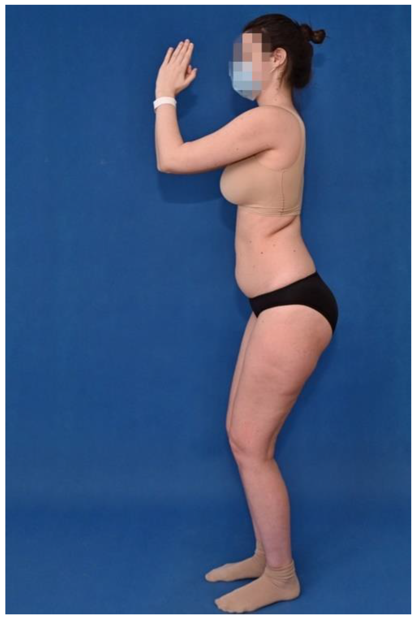
4]. Vertical position of the sacrum with a ventral displacement of the femoral heads and the hamstring contracture leads to semiflexion of the hip and knee joints, leading to a change in the sagittal posture profile, the so-called “monkey’s gait” [5]. The change in posture is documented in Figure 2. The incidence of highly dysplastic spondylolisthesis is 11.3% in adults diagnosed with spondylolisthesis [6]. Subjective difficulties most often include lumbar spine pain with possible neurological complications due to nerve root irritation.
The measurement of the extent of the slip is evaluated on X-ray scans using the Meyerding scale [7]. Meyerding differentiated the sacral endplate into quarters and categorized spondylolisthesis into grades I–IV according to the direction of the slip in a certain quarter. Later, two more grades were added: grade 0, characterized by a normal state without displacement; and grade V, corresponding to spondyloptosis. The percentage expression of the extent of the slip is evaluated by measuring according to Taillard [8].
Despite significant progress in the diagnosis and development of indication patterns, the question of the appropriate therapeutic algorithm remains controversial. With the development of surgical methods, conservative therapy is declining in favor of surgical therapy. The aim of the surgery should be to prevent the progression of the slip and to correct the asymmetry of lumbosacral parameters, leading to an improvement in the overall sagittal balance of the spine, and the consequent improved standing and walking posture. The clinical effect should be to reduce pain in the LS junction. The cosmetic effect is also significant.
There is significant variability in individual surgical procedures, from fixation in situ, with no reduction in the slipped vertebra, to extensive surgeries with an effort to completely reduce the slipped vertebra; these procedures use different surgical approaches and include more vertebral segments in stabilization [9].
The aim of this paper is to describe the individual surgical methods used for patients having dysplastic spondylolisthesis with a shift of more than 25%, who were operated on at our institution, and to determine the ideal surgical therapeutic algorithm according to the extent of the vertebral slip.

2. Materials and Methods

2.1. Patient Selection

In the period 2005–2018, a total of 32 patients (22 women) were operated on for dysplastic spondylolisthesis with a slip over 25%. The average age of the patients at the time of surgery was 18.5 years (range 10–38 years).
Complete radiological data were obtained from 26 patients. Pre- and postoperative X-ray scans of the LS spine in the posterior-anterior and lateral projections were performed in all patients. All patients underwent standing-bending X-rays in hyperextension and hyperflexion to assess the instability of the affected segment. Another routine examination performed in all patients undergoing surgical treatment of the LS spine is a long-standing X-ray of the entire spine in sagittal projection including femoral heads. This examination is necessary to assess the sagittal balance of the spine, both preoperatively and in the postoperative controls. Furthermore, an MRI of the affected section of the spine is performed to assess dysplastic changes in the LS junction. MRI is also recommended for the identification of nerve structures and to access the pathology of the vertebral body and intervertebral disc [10]. The extent of the slip and displacement of the L5 vertebra compared to the S1 vertebra is expressed as a percentage according to Taillard and according to the Meyerding classification. Twelve patients were classified as Meyerding grade III and 6 patients as grade IV. We also included 8 patients with dysplastic changes in the L5/S1 segment, Meyerding grade II.

2.2. Surgical Technique

In our study, two basic groups were defined, based on the surgical approach used in the treatment of dysplastic spondylolistheses. The first of these was patients operated on using only the posterior approach; this group was further divided into two subgroups according to the type of fusion of the L5/S1 interbody space. The first subgroup consisted of patients who underwent treatment of the L5/S1 segment with the posterior lumbar interbody fusion technique (further referred to as PLIF); in the second subgroup of patients, a dowel fibular strut graft was used for L5/S1 fusion. The second basic group comprised patients operated on using the combination of anterior and posterior approaches, where the L5/S1 fusion was treated with anterior lumbar interbody fusion (further referred to as ALIF).

2.2.1. Posterior Approach Only

The procedure using the posterior approach only is performed in the prone position of the patient. Posterior elements of the spine are exposed through the posterior midline approach. Then, transpedicular screws are inserted into the L5/S1 vertebrae.
In the PLIF technique, the next step is to perform a laminectomy, visualizing the dura mater. By retracting the dural sac, the intervertebral space is reached. After resection of the posterior longitudinal ligament, the mass of the intervertebral disc is removed. Next, the dome of the upper endplate of the S1 vertebra is resected. L5 vertebral reduction is achieved using reduction instruments under direct visual control of the L5 nerve roots. Interbody fusion is most often performed with bone allografts or using an interbody fusion cage. Stabilization is completed by bone decortication and using grafts to perform the posterolateral fusion.
An alternative to this procedure is, as mentioned above, the use of a bone auto- or allograft from the fibular shaft [11]. In this technique, the posterior approach is extended distally to the S1/S2 segment; after that, stabilization and reduction are performed in the L5/S1 level without opening the spinal canal in this segment. The next step is the S2 laminectomy and the interbody fusion using a fibular autograft or an allograft. The fibular graft, measuring approximately 5 cm, is then introduced through a drilled “tunnel” through the S2 and S1 vertebrae into the L5 vertebral body (Figure 3). The preoperative, intraoperative, and final postoperative X-rays are shown in Figure 4.

2.2.2. Combination of Posterior and Anterior Approaches

The other option used in the surgical treatment of dysplastic spondylolisthesis was a combination of posterior and anterior approaches utilizing the ALIF technique [12]. Using the posterior approach, the slipped vertebra is reduced and stabilized without opening the spinal canal. The position of the patient is then changed from pronation to supination. The interbody space is treated from the front with the ALIF technique. The incision is made transversely above the symphysis. The fascia is intersected longitudinally, and the retroperitoneal space is reached along the posterior surface of the straight abdominal muscle. It is possible to access this area from the right and from the left. The peritoneum is retracted medially, exposing the LS junction. While protecting the vascular bundle, the L5/S1 disc and the dome-shaped prominence of the S1 vertebra are resected. Either an auto- or allograft is used to replace the disc. The advantage of this surgical technique lies in the absence of opening the spinal canal and manipulation of the nerve structures. This method has been shown to provide excellent results in the long term, with 83% union rates at the 10-year follow-up [13]. The disadvantage is the need for another surgical approach and the possibility of injury to the vascular bundle. Another disadvantage, especially in more severe dysplasia, is the anatomical situation and poor view of the area being operated on, which greatly complicates the treatment of the L5/S1 interbody space.
In our set of patients, 25 patients were operated on only using the posterior approach. In 9 of these, the L5/S1 interbody fusion was treated with the PLIF technique; the fibular dowel graft was used in 16 cases.
The combination of posterior and anterior approaches employing the ALIF technique was used in 7 cases. Postoperative treatment in all patients consisted of fixation with a lumbar belt for 3 months.

2.3. Evaluation

The preoperative extent of the slip and the extent of the reduction immediately after the surgery were evaluated. The retention of the reduction was evaluated using X-rays one year after the surgery and during long-term follow-up, which was an average of 7 years after the surgery (range 2–15 years postoperatively). On X-rays, we focused on measuring sagittal balance parameters such as Sacral Slope (SS), Pelvic Incidence (PI), Pelvic Tilt (PT), Lumbar Lordosis (LL), Slip Angle (SA), and Incidence L5 (IL5). The evaluation included a review of complications associated with the surgery, the length of the surgery itself, and the extent of blood loss. Complications were divided into early and late. Early complications were defined until 3 months after surgery, and late complications were those occurring after 3 months postoperatively. Subjective perception and clinical evaluation of surgical therapy were obtained using the Oswestry Disability Index (ODI), and information on the overall benefit of treatment and surgery used the Overall Treatment Evaluation (OTE) questionnaire.

3. Results

3.1. Slip Reduction and Retention

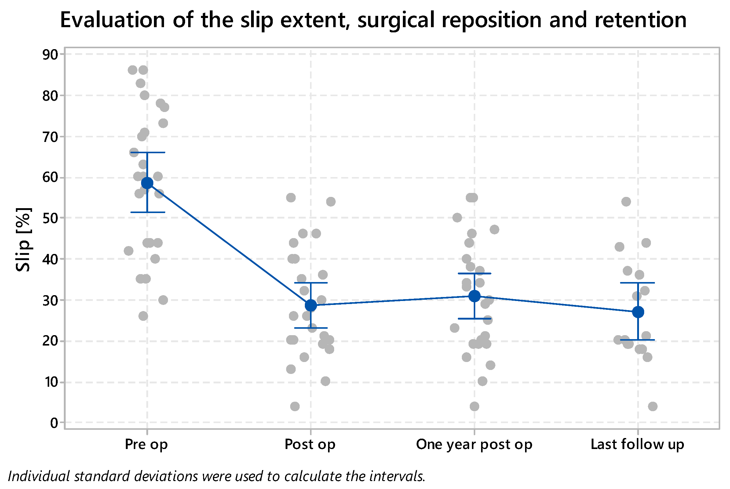
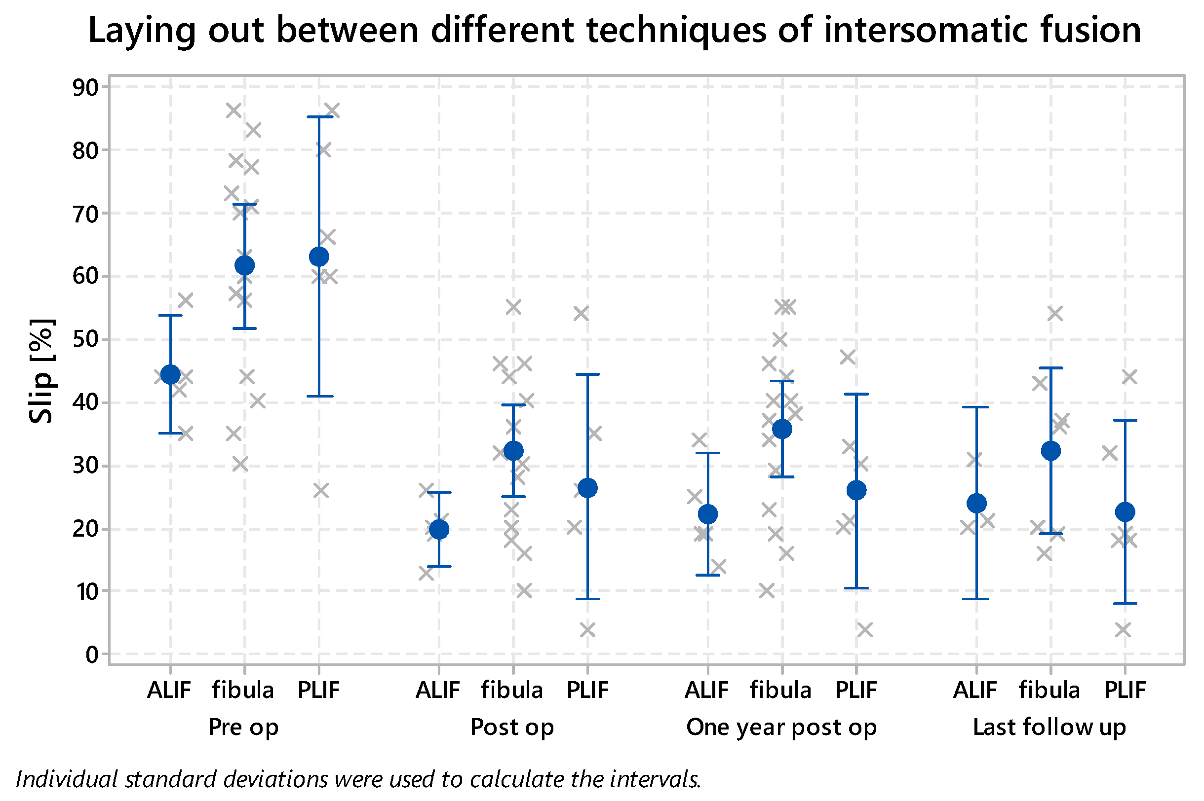
The average preoperative extent of the slip was 60% (27–86%). Postoperatively, the extent of the slip was reduced to 28% (4–55%). The reduction, therefore, reduced the size of the slip by more than 50% (32% statistically significant slip decrease, p = 0.000). Overall, the extent of the slip was 32% one year after the surgery and 28% during the long-term observation. The given results are summarized in Figure 5.
With the PLIF technique, the slip was reduced and decreased from 67% (54–86%) to 26% postoperatively (4–54%) (31% statistically significant slip decrease, p = 0.000). When using fibula, there was a decrease from 62% (27–86%) to 32% (10–55%) (30% statistically significant slip decrease, p = 0.000), and when combining the approaches and using ALIF interbody fusion, the slip decreased from 44% (35–56%) to 20% (13–26%) (24% statistically significant slip decrease, p = 0.001).
Slip retention was evaluated one year after surgery and, on average, 7 years after surgery. The distribution among the individual methods of interbody fusion, i.e., the PLIF technique, the use of a fibular graft, and the ALIF technique is shown in Figure 6. The only increase in the slip extent—from 32% to 35%—was observed with the use of fibula, between the postoperative period and one year after surgery. However, during the long-term follow-up, the extent of the slip again corresponded to the postoperative value of the slip, i.e., 32%. After repeated evaluation of the X-ray images, we concluded that this was most likely caused by an incorrect measurement of the extent of the slip, given by the rotation of X-ray scans.
All performed surgical methods led to statistically significant improvement in radiological parameters. In repeated postoperative examinations, there were no recurrences of slips, due to the creation of a firm fusion in the affected segment. For large slips, of over 50%, we achieved a better reduction in the slip with the PLIF technique compared to the use of the fibular graft.

3.2. Surgical Time and Complications

The time of the surgery was measured from the skin incision to the end of the surgical procedure. The average surgery time corresponded to 220 min. When employing the PLIF technique, the surgery lasted an average of 227 min; when using the fibula allograft, this was 204 min. With the ALIF technique, i.e., the combination of anterior and posterior approaches, the surgery took 250 min. The combination of approaches, the need to turn the patient from the prone to the supine position, and the need to re-drape the operating field causes a time delay, which is reflected in the total surgery time.
The extent of blood loss corresponded to an average of 740 mL in all techniques. When employing the dorsal approach only and the PLIF technique, this was 871 mL; when using a fibular graft, it was 709 mL. When combining the posterior and the anterior approaches, the extent of blood loss was 658 mL, i.e., less than in the posterior approach requiring the opening of the spinal canal.
Early complications, occurring up to 3 months after the surgery, were present in 5 patients (5/32; 16%). Of these, neurological complications occurred in 4 patients (4/32; 12.5%). In 3 patients, surgical revision was necessary with the need to reposition screws because of their malposition; subsequent clinical improvement was reported. One patient had transitory buttock hypesthesia with spontaneous regression over time, without the need for surgical revision. One patient required revision of the surgical wound because of secondary healing in the early postoperative period.
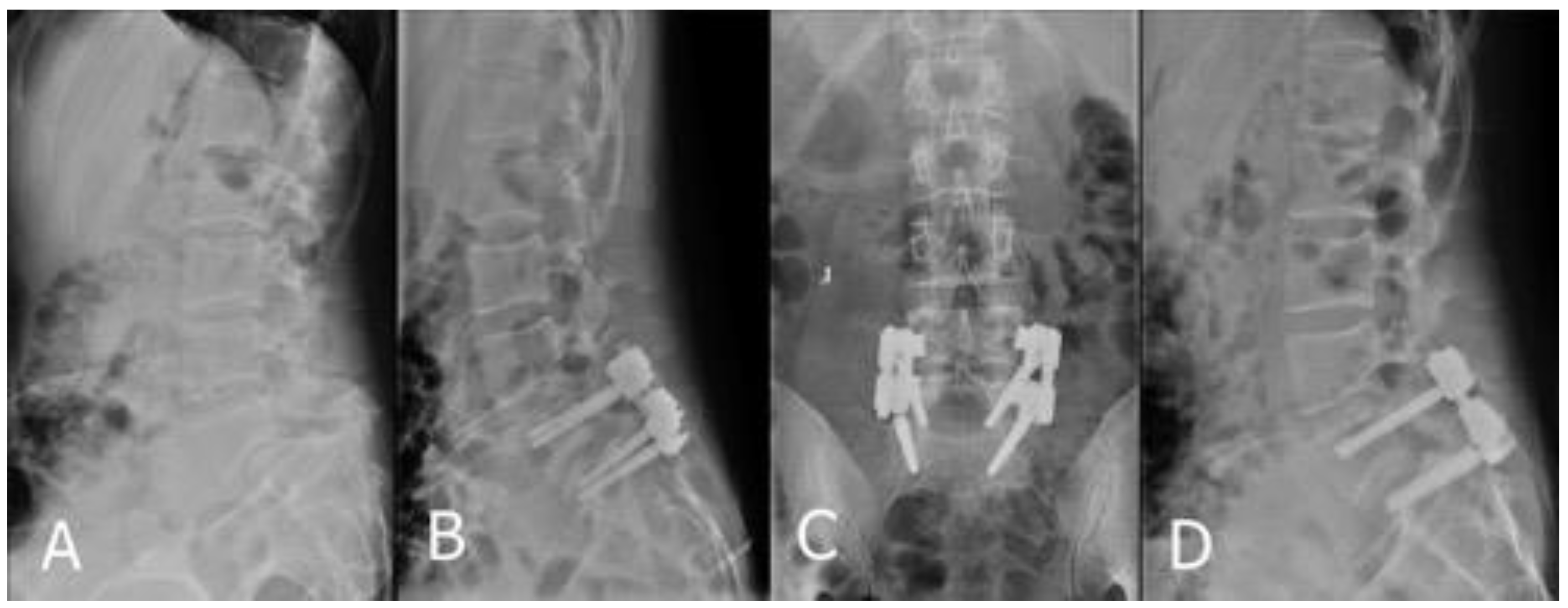
We noted that two patients experienced late complications. In the first patient, the screw in the S1 vertebra broke (Figure 7). Due to negative symptoms and the unchanged position of the instrumentation, surgical revision was not indicated, and the patient has been and still is treated conservatively.
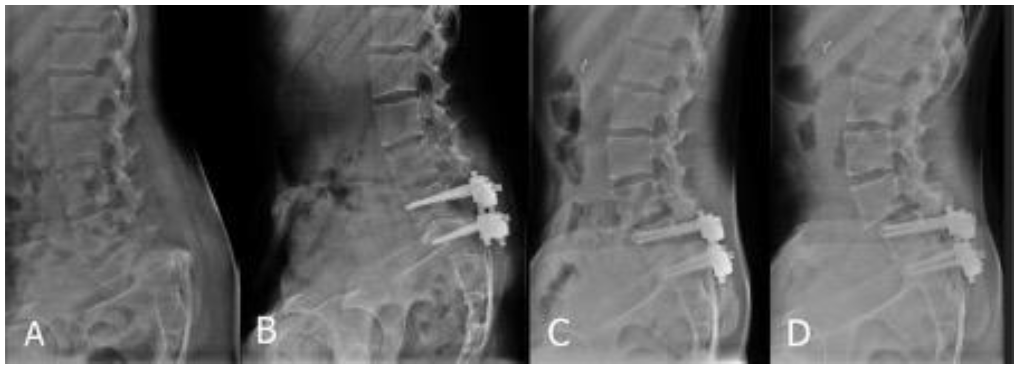
In the other patient, we observed a ventral displacement of the L4 vertebra over the L5/S1 fusion (Figure 8). The patient was operated on using the PLIF technique. Thus, this was most likely an iatrogenic involvement and overload of the segment above the fusion after the release of the dorsal structures of the L5/S1 segment. At the current time, 6 years after surgery, the patient’s problems are only mild and intermittent. If difficulties progress, we will have to indicate a surgical revision with an extension of the instrumentation to L4 and a 360° fusion of the L4/L5 segment.

3.3. Sagittal Balance

Another part of the evaluation was the radiological measurement of sagittal balance in operated patients. The Slip Angle (SA) was 16° on average (min 0°–max 53°) before surgery; it was reduced to 9° on average (min 0°–max 28°) postoperatively. The Pelvic Incidence (PI) was 70° on average (min 55°–max 84°) preoperatively and 70° on average (min 46°–max 84°) postoperatively. The Pelvic Tilt (PT) was 31° on average (min 5°–max 54°) preoperatively and 24° on average (min 5°–max 48°) postoperatively. The Sacral Slope (SS) was 39° on average (min 20°–max 67°) preoperatively and 43° on average (min 22°–max 75°) postoperatively. The Incidence of L5 (IL5) was 55° on average (min 20°–max 111°) preoperatively and 43° on average (min 21°–max 74°) postoperatively. The lumbar lordosis angle (Lumbar Lordosis, LL) corresponded to a preoperative magnitude of 56° on average (min 20°–max 82°) and postoperatively to 51° on average (min 24°–max 82°). Results of the evaluation of spinopelvic parameters are summarized in Table 1.

3.4. Satisfaction Questionnaires

Patient-reported outcomes after surgical therapy were evaluated using the Oswestry Disability Index (ODI) during a long-term follow-up [14]. ODI evaluates lower-back pain according to different physiological situations, such as resting pain, personal care, lifting weights, walking, sitting, standing, sex life, social life, and traveling. The score of the pain is 0–5 in all situations, where 0 corresponds to minimal pain and 5 to maximal pain. The final score is expressed as a percentage. The interpretation of the score corresponds to 0–20%: minimal disability; 21–40%: moderate disability; 41–60%: severe disability; 61–80%: crippled; 81–100%: bed-bound or exaggerating symptoms. Patients who had posterior approach surgery using the PLIF technique rated the outcome on average at 18%, patients with a posterior approach surgery using the fibula at 8%, and patients on the combined approach at 10%. As mentioned above, the range of 0–20% corresponds to minimal disability. Patients can cope with most living activities. None of the surgical methods resulted in a restriction of normal daily activities due to postoperative LS spine pain.
The overall satisfaction was evaluated by using the Overall Treatment Evaluation (OTE) questionnaire [15]. In this questionnaire, the patient is asked about the change in the quality of life following the surgical treatment, and the importance of this change to the patient’s normal daily activities. All patients (100%) reported a change for the better compared to the preoperative condition, and 64% of the respondents reported a much better condition in their evaluation of the treatment. The perception of this change was assessed by 64% of patients as being important, and by 36% of patients as being extremely/very important. As a conclusion, we can state that all patients reported an improvement in their quality of life following the surgical therapy.

4. Discussion

Dysplastic spondylolisthesis is a specific type of involvement of the spinal segment, which affects the L5/S1 segment and occurs twice as often in women [16]. Clinical symptoms and signs often appear in childhood and adolescence during the growth spurt [17]. Our group of patients was also dominated by women, who made up almost 70% of the total number of patients. The age distribution also corresponds to the above-mentioned statement, with the majority of patients being mostly adolescents and young adults.
In order to correctly determine the therapeutic algorithm of this pathology, a thorough diagnosis and the correct classification into classification groups are necessary. The long-format X-ray of the spine is necessary to assess the spinopelvic balance, which is pathologically altered in patients with dysplastic spondylolisthesis [18]. There is an increase in lumbar lordosis. The lumbosacral profile and ventral displacement of the femoral heads also lead to a significant increase in PI and PT [19]. The values of the given spinopelvic parameters were also significantly increased in our group. The SS value does not have to be significantly changed; it depends on the position of the sacrum. In the case of large slips, where the sacrum is verticalized, i.e., it takes the form of pelvic retroversion, the SS values are lower than those in the physiological state [20]. The position and angulation of the lumbosacral junction can be measured in various ways. One of these is to form an angle between the L5 upper vertebral endplate and the S1 upper vertebral endplate. This angle corresponds to the lumbosacral angle (LSA). Another possibility is to measure the Slip Angle (SA), as described by Boxall [21]. There is also variability in the formation of a given angle. This variability is caused by the pathological situation of the lumbosacral junction, the dome shape of the upper endplate of the S1 vertebra, which makes it difficult to form straight lines. In our group, we created SA as the angle between the perpendicular to the posterior edge of the S1 vertebra and the lower endplate of the L5 vertebral body.
Dysplastic changes in vertebral bodies act as a predisposing factor for the progression of vertebral slip. The method of measuring and determining the progress of the slip involves calculating the Severity Index (SI) and defining the unstable zone (UZ). To calculate the SI, it is necessary to make a horizontal line pointing from the center of the S2 vertebra. Next, two vertical lines are created. The first starts from the center of the lower endplate of the L5 vertebra, and the second passes through the center of the femoral heads. In a physiological state, these verticals are identical, as the vertical from the L5 body should pass through the center of the femoral heads. The distance from the center of S2 to the vertical line drawn from the center of the femoral heads is D2. The distance of the vertical line from the L5 body to the vertical line from the center of the femoral heads is D1. The equation for the calculation is SI = D1 × 100/D2. With advanced slip progression and gradual verticalization of the sacrum and ventral displacement of the femoral heads, D1 and, therefore, SI, increases. An SI above 20% is determined as a significant factor corresponding to highly dysplastic spondylolisthesis with a high risk of slip and clinical condition progression. The creation of a square, the side of which is given by the size of the distance D1, corresponds to an unstable zone. If the L4 vertebra extends into this square, it should be part of instrumented stabilization [22].
Opinions on the treatment of dysplastic spondylolisthesis remain inconsistent and controversial. It is necessary to distinguish between low and high dysplastic spondylolisthesis. HDDS is, in contrast to LDDS, dangerous because of the risk of slip progression, which may possibly progress as far as spondyloptosis [23]. Prevention of progression is one of the important aspects of treatment; therefore, HDDS should, in our view, always be indicated for surgical treatment.
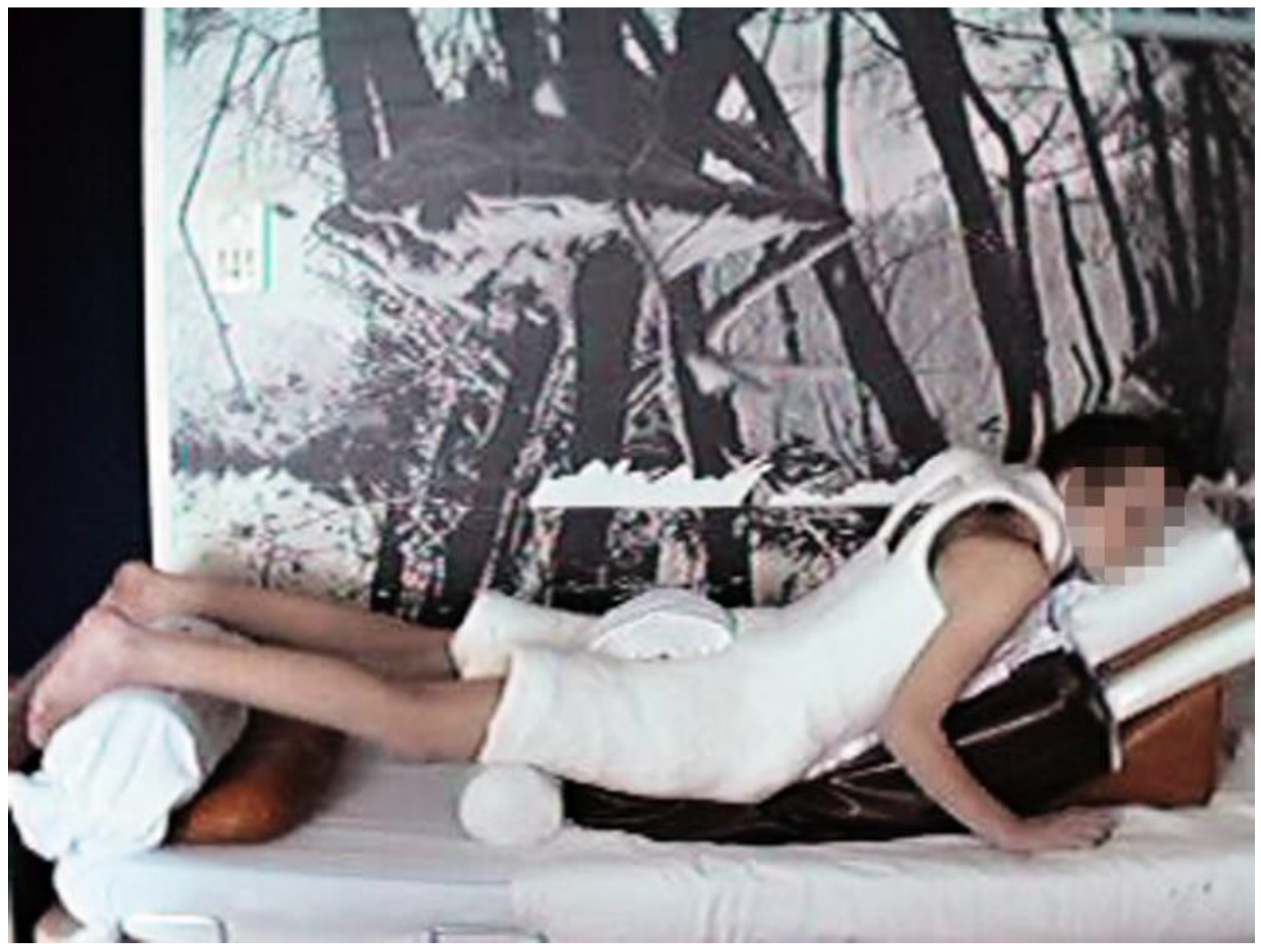
According to Scaglietti, conservative therapy consists of long-term immobilization in a plaster corset in hyperextension with pressure on the sacrum [24]. The plaster corset results in significant discomfort for the patient and its reduction ability is only minimal (Figure 9).
Rehabilitation and physiotherapy act as supportive therapies that strengthen the deep stabilization system of the spine but do not address the pathological merits of the problem. A comparison of surgical and conservative therapies in pediatric patients with highly dysplastic spondylolisthesis was performed by Bourassa-Moreau based on a follow-up of 28 patients, 23 of whom were treated surgically and 5 conservatively. Improved quality of life and a better score in the SRS-22 questionnaire were achieved in the group treated surgically [25].
However, opinions on the surgery to be performed also differ in the literature, from mere in situ fixation to the effort to fully reduce the affected segment. Proponents of mere in situ fixation argue that neurological complications are minor compared to those resulting from the reduction and overstretching of nerve structures. In addition, more frequent postoperative complications and a higher frequency of reoperations when attempting reduction are reported [26]. However, results of the in situ fusion are unclear, with a high risk of nonunion development and increased probability of slip deterioration [27]. On the contrary, the advantage of reduction over in situ fusion is the correction of lumbosacral kyphosis, thereby improving the overall sagittal profile and the symmetry of the lumbosacral junction, leading to correction of gait and standing, and, furthermore, the possibility of direct decompression of nerve structures [28]. A comparison of both methods, i.e., reduction and in situ fixation, was performed by Longo [29]. In his study, it was found that there is an improvement in biomechanical conditions and local kyphosis in patients with a reduction in the slipped vertebra. The reduction did not increase the risk of neurological complications associated with in situ fixation.
There is a consensus that, in the case of reduction, it is necessary to perform an interbody fusion of the anterior column; otherwise, there is a risk of failure of the reduction and instrumentation with the formation of a nonunion [30]. An interbody fusion can be performed by the PLIF technique. Lamartina [1] presents satisfactory short-term clinical outcomes with the reduction in the deformity using the PLIF technique with cages for interbody fusion. In the case of smaller slips, good results were achieved by a combination of posterior reduction and anterior interbody fusion using the ALIF technique [31]. A new technique presented in the literature describes a combination of approaches with posterior mono-segmental stabilization and anterior direct reduction in the slip using the anterior cantilever procedure [32]. The authors achieved a high fusion rate and partial reduction in the slip, and improved the anatomy of the LS junction. Good clinical and radiological results have also been obtained using a trans-sacral strut graft to perform interbody fusion [9,33]. According to the authors, there was no significant difference in the use of an allograft or autograft, and remodeling and firm fusion occurred in all patients. An integral part of surgical therapy is the correction of segmental kyphosis and restoration of the symmetry of the lumbosacral junction [34].
The range of instrumentation and reduction extent have also been discussed. Some authors recommend extending the instrumentation to the L4 vertebra in the case of large slips [35]. Conversely, prolongation of instrumentation distally using iliac screws into iliac bone reduces the risk of instrumentation failure [36]. Lombardi [37] recommends mono-segmental stabilization with the reposition of the slipped vertebra in all cases of dysplastic spondylolisthesis with a slip over 25%. Authors achieved a high rate of spinal fusion and restored proper lumbosacral anatomy while improving the whole sagittal balance. According to the extent of reduction, most authors recommend partial reduction in the slip instead of total reduction, because of the high risk of nerve root irritation [38]. At our institution, we prefer reduction and mono-segmental stabilization. We agree with Stulik et al. [39], who, in their group of patients with highly dysplastic spondylolisthesis, achieved very good results in the effort to complete reduction and mono-segmental stabilization of the affected segment.
The frequency of neurological complications resulting from the possible overstretching of nerve roots is one of the arguments of opponents of reduction. Intraoperative neuromonitoring is recommended to reduce the incidence of iatrogenic L5 radiculopathy [40]. Lamartina [1] reviewed publications on the treatment of highly dysplastic spondylolisthesis, involving a total of 224 patients. Twenty-eight patients, i.e., 12.5%, suffered from neurological postoperative symptomatology. Of these, 20 patients had solely transient disabilities and 8 patients (3.6%) suffered from verified radiculopathy. In our group, the frequency of neurological complications was 12.5%, as diagnosed in 4 patients of a total of 32.

5. Conclusions

In this single institutional retrospective study, we showed that surgical treatment of dysplastic spondylolisthesis leads to a significant improvement in both radiological and clinical parameters. All performed surgical methods showed a statistically significant decrease in the slip and firm fusion that remained unchanged in postoperative follow-up. We achieved a better reduction in the slip using the PLIF technique compared to the use of the fibular graft. For patients with slips over 50%, we prefer surgical treatment using the posterior approach and utilizing the PLIF technique, in which an improvement toward the correct anatomical position can be achieved under direct control with the decompression of nerve roots. For patients with slips below 50% and in a good anatomical situation, we use a combination of posterior and anterior surgical approaches and the ALIF interbody fusion technique. The number of neurological complications did not exceed the average complication rate reported by other authors. The goal for the future is to continue to achieve consistent diagnostics, improve surgical techniques, and reduce neurological complications.

Author Contributions

R.V.: Writing—original draft. Preparation, creation, and/or presentation of the published work, specifically writing the initial draft (including substantive translation). M.P.: Writing—original draft. Preparation, creation, and/or presentation of the published work, specifically writing the initial draft (including substantive translation). L.R.: Validation. Verification, whether as a part of the activity or separate, of the overall replication/reproducibility of results/experiments and other research outputs. J.S.: Investigation. Conducting a research and investigation process, specifically performing the experiments, or data/evidence collection. J.K.: Investigation Conducting a research and investigation process, specifically performing the experiments, or data/evidence collection. M.L.: Resources Provision of study materials, reagents, materials, patients, laboratory samples, animals, instrumentation, computing resources, or other analysis tools. P.V.: Formal analysis; application of statistical, mathematical, computational, or other formal techniques to analyze or synthesize study data. M.R.: Supervision. Oversight and leadership responsibility for the research activity planning and execution, including mentorship external to the core team. All authors have read and agreed to the published version of the manuscript.

Funding

This work was supported by the Ministry of Health, Czech Republic—Conceptual Development of Research Organization (FNBr, 65269705). The funder had no role in the design of the study; in the collection, analyses, or interpretation of data; in the writing of the manuscript, or in the decision to publish the results.

Institutional Review Board Statement

All procedures performed in studies involving human participants were in accordance with the ethical standards of the institutional and/or national research committee and with the 1964 Helsinki declaration and its later amendments or comparable ethical standards. Ethical committee name: The Ethics Committee for Multicentre Clinical Trials of the University Hospital Brno, Jihlavská 20, 62500 Brno, Czech Republic; Evidence number of ethical approval: 01-300622/EK. Date of approval: 30 June 2022.

Informed Consent Statement

Informed consent was obtained from all individual participants included in the study.

Data Availability Statement

The data presented in this study are available on request from the corresponding author.

Conflicts of Interest

All authors certify that they have no affiliations with or involvement in any organization or entity with any financial interest or non-financial interest in the subject matter or materials discussed in this manuscript.

References

  1. Lamartina, C.; Zavatsky, J.M.; Petruzzi, M.; Specchia, N. Novel concepts in the evaluation and treatment of high-dysplastic spondylolisthesis. Eur. Spine J. 2009, 18, 133–142. [Google Scholar] [CrossRef] [PubMed] [Green Version]
  2. Wiltse, L.L.; Newman, P.H.; Macnab, I. Classification of spondylolysis and spondylolisthesis. Clin. Orthop. 1976, 117, 23–29. [Google Scholar]
  3. Marchetti, P.C.; Bartolozzi, P. Classification of spondylolisthesis as a guideline for treatment. In The Textbook of Spinal Surgery, 2nd ed.; Bridwell, K.H., DeWald, R.L., Hammerberg, K.W., Eds.; Lippincott-Raven: Philadelphia, PA, USA, 1997; pp. 1211–1254. [Google Scholar]
  4. Vidal, J.; Marnay, T. Morphology and anteroposterior body equilibrium in spondylolisthesis L5-S1. Rev. Chir. Orthop. Reparatrice Appar. Mot. 1983, 69, 17–28. [Google Scholar] [PubMed]
  5. Osterman, K.; Schlenzka, D.; Poussa, M.; Seitsalo, S.; Virta, L. Isthmic spondylolisthesis in symptomatic and asymptomatic subjects, epidemiology, and natural history with special reference to disk abnormality and mode of treatment. Clin. Orthop. 1993, 297, 65–70. [Google Scholar] [CrossRef]
  6. Kunze, K.N.; Lilly, D.T.; Khan, J.M.; Louie, P.K.; Ferguson, J.; Basques, B.A.; Nolte, M.T.; Dewald, C.J. High-Grade Spondylolisthesis in Adults: Current Concepts in Evaluation and Management. Int. J. Spine Surg. 2020, 14, 327–340. [Google Scholar] [CrossRef]
  7. Wright, I.P. Who Was Meyerding? Spine 2003, 28, 733–735. [Google Scholar] [CrossRef]
  8. Taillard, W.F. Etiology of spondylolisthesis. Clin. Orthop. 1976, 117, 30–39. [Google Scholar] [CrossRef]
  9. Rivollier, M.; Marlier, B.; Kleiber, J.-C.; Eap, C.; Litre, C.-F. Surgical treatment of high-grade spondylolisthesis: Technique and results. J. Orthop. 2020, 22, 383–389. [Google Scholar] [CrossRef]
  10. Mac-Thiong, J.-M.; Labelle, H. A proposal for a surgical classification of pediatric lumbosacral spondylolisthesis based on current literature. Eur. Spine J. 2006, 15, 1425–1435. [Google Scholar] [CrossRef]
  11. Sasso, R.C.; Shively, K.D.; Reilly, T.M. Transvertebral Transsacral Strut Grafting for High-grade Isthmic Spondylolisthesis L5-S1 With Fibular Allograft. J. Spinal Disord. Technol. 2008, 21, 328–333. [Google Scholar] [CrossRef] [Green Version]
  12. Suk, K.S.; Jeon, C.H.; Park, M.S.; Moon, S.H.; Kim, N.H.; Lee, H.M. Comparison between posterolateral fusion with pedicle screw fixation and anterior interbody fusion with pedicle screw fixation in adult spondylolytic spondylolisthesis. Yonsei Med. J. 2001, 42, 316–323. [Google Scholar] [CrossRef] [PubMed]
  13. Ishihara, H.; Osada, R.; Kanamori, M.; Kawaguchi, Y.; Ohmori, K.; Kimura, T.; Matsui, H.; Tsuji, H. Minimum 10-year follow-up study of anterior lumbar interbody fusion for isthmic spondylolisthesis. J. Spinal Disord. 2001, 14, 91–99. [Google Scholar] [CrossRef] [PubMed]
  14. Fairbank, J.C.T.; Pynsent, P.B. The Oswestry Disability Index. Spine 2000, 25, 2940–2953. [Google Scholar] [CrossRef] [PubMed]
  15. Jaeschke, R.; Singer, J.; Guyatt, G.H. Measurement of health status: Ascertaining the minimal clinically important difference. Control. Clin. Trials 1989, 10, 407–415. [Google Scholar] [CrossRef]
  16. Suchomel, P.; Krbec, M. Spondylolisthesis—Diagnostics and Therapy; Galen: Prague, Czech Republic, 2007; pp. 109–111. [Google Scholar]
  17. Dewald, C.J.; Vartabedian, J.E.; Rodts, M.F.; Hammerberg, K.W. Evaluation and Management of High-Grade Spondylolisthesis in Adults. Spine 2005, 30, S49–S59. [Google Scholar] [CrossRef] [Green Version]
  18. Labelle, H.; Roussouly, P.; Berthonnaud, E.; Transfeldt, E.; O’Brien, M.; Chopin, D.; Hresko, T.; Dimnet, J. Spondylolisthesis, Pelvic Incidence, and Spinopelvic Balance. Spine 2004, 29, 2049–2054. [Google Scholar] [CrossRef]
  19. Hanson, D.S.; Bridwell, K.H.; Rhee, J.M.; Lenke, L.G. Correlation of Pelvic Incidence with Low- and High-Grade Isthmic Spondylolisthesis. Spine 2002, 27, 2026–2029. [Google Scholar] [CrossRef]
  20. Hresko, M.T.; Labelle, H.; Roussouly, P.; Berthonnaud, E. Classification of High-Grade Spondylolistheses Based on Pelvic Version and Spine Balance. Spine 2007, 32, 2208–2213. [Google Scholar] [CrossRef]
  21. Boxall, D.; Bradford, D.S.; Winter, R.B.; Moe, J.H. Management of severe spondylolisthesis in children and adolescents. J. Bone Jt. Surg. Am. 1979, 61, 479–495. [Google Scholar] [CrossRef]
  22. Lamartina, C. A square to indicate the unstable zone in severe spondylolisthesis. Eur. Spine J. 2001, 10, 444–448. [Google Scholar] [CrossRef] [Green Version]
  23. Newman, P.H. Spondylolisthesis, its cause and effect. Ann. R. Coll. Surg. Engl. 1955, 16, 305–323. [Google Scholar] [PubMed]
  24. Scaglietti, O.; Frontino, G. Technique of anatomical reduction of lumbar spondylolisthesis and its surgical stabilization. Clin. Orthop. 1976, 117, 165–175. [Google Scholar] [CrossRef]
  25. Bourassa-Moreau, É.; Mac-Thiong, J.-M.; Joncas, J.; Parent, S.; Labelle, H. Quality of life of patients with high-grade spondylolisthesis: Minimum 2-year follow-up after surgical and nonsurgical treatments. Spine J. 2013, 13, 770–774. [Google Scholar] [CrossRef] [PubMed]
  26. Poussa, M.; Schlenzka, D.; Seitsalo, S.; Ylikoski, M.; Hurri, H.; Osterman, K. Surgical Treatment of Severe Isthmic Spondylolisthesis in Adolescents: Reduction or Fusion: In Situ. Spine 1993, 18, 894–901. [Google Scholar] [CrossRef]
  27. Agabegi, S.S.; Fischgrund, J.S. Contemporary management of isthmic spondylolisthesis: Pediatric and adult. Spine J. 2010, 10, 530–543. [Google Scholar] [CrossRef]
  28. Bradford, D.S.; Boachie-Adjei, O. Treatment of severe spondylolisthesis by anterior and posterior reduction and stabilization. A long-term follow-up study. J. Bone Jt. Surg. Am. 1990, 72, 1060–1066. [Google Scholar] [CrossRef]
  29. Longo, U.G.; Loppini, M.; Romeo, G.; Maffulli, N.; Denaro, V. Evidence-Based Surgical Management of Spondylolisthesis: Reduction or Arthrodesis in Situ. J. Bone Jt. Surg. 2014, 96, 53–58. [Google Scholar] [CrossRef] [PubMed] [Green Version]
  30. Boos, N.; Marchesi, D.; Zuber, K.; Aebi, M. Treatment of Severe Spondylolisthesis by Reduction and Pedicular Fixation: A 4–6-Year Follow-Up Study. Spine 1993, 18, 1655–1661. [Google Scholar] [CrossRef]
  31. Spruit, M.; Pavlov, P.; Leitao, J.; De Kleuver, M.; Anderson, P.; Den Boer, F. Posterior reduction and anterior lumbar interbody fusion in symptomatic low-grade adult isthmic spondylolisthesis: Short-term radiological and functional outcome. Eur. Spine J. 2002, 11, 428–433. [Google Scholar] [CrossRef] [Green Version]
  32. Tu, K.-C.; Shih, C.-M.; Chen, K.-H.; Pan, C.-C.; Jiang, F.-C.; Hsu, C.-E.; Wang, Y.-M.; Lee, C.-H. Direct reduction of high-grade lumbosacral spondylolisthesis with anterior cantilever technique—Surgical technique note and preliminary results. BMC Musculoskelet. Disord. 2021, 22, 1–10. [Google Scholar] [CrossRef]
  33. Hanson, D.S.; Bridwell, K.H.; Rhee, J.M.; Lenke, L.G. Dowel Fibular Strut Grafts for High-Grade Dysplastic Isthmic Spondylolisthesis. Spine 2002, 27, 1982–1988. [Google Scholar] [CrossRef] [PubMed]
  34. Shufflebarger, H.L.; Geck, M.J. High-Grade Isthmic Dysplastic Spondylolisthesis. Spine 2005, 30, S42–S48. [Google Scholar] [CrossRef] [PubMed]
  35. Min, K.; Liebscher, T.; Rothenfluh, D. Sacral dome resection and single-stage posterior reduction in the treatment of high-grade high dysplastic spondylolisthesis in adolescents and young adults. Eur. Spine J. 2011, 21, 785–791. [Google Scholar] [CrossRef] [PubMed] [Green Version]
  36. Molinari, M.R.W.; Bridwell, K.H.; Lenke, L.G.; Ungacta, F.F.; Riew, K.D. Complications in the Surgical Treatment of Pediatric High-Grade, Isthmic Dysplastic Spondylolisthesis: A Comparison of Three Surgical Approaches. Spine 1999, 24, 1701. [Google Scholar] [CrossRef]
  37. Lombardi, F.; Custodi, V.M.; Pugliese, R.; Risso, A.; Gaetani, P.; Butti, G. Treatment of high-grade spondylolisthesis with Schanz recoil screws: Our experience. Eur. Spine J. 2013, 22, 914–918. [Google Scholar] [CrossRef] [Green Version]
  38. Martiniani, M.; Lamartina, C.; Specchia, N. “In situ” fusion or reduction in high-grade high dysplastic developmental spondylolisthesis (HDSS). Eur. Spine J. 2012, 21, 134–140. [Google Scholar] [CrossRef] [Green Version]
  39. Štulík, J.; Nesnidal, P.; Kozelnicka, K.; Barna, M. Surgical Treatment of High-Grade High-Dysplastic Spondylolistheses in Young Patients—Prospective Monocentric Study of 29 Patients. Acta Chir. Orthop. Traumatol. Cech 2018, 85, 305–318. [Google Scholar]
  40. Schär, R.T.; Sutter, M.; Mannion, A.F.; Eggspühler, A.; Jeszenszky, D.; Fekete, T.F.; Kleinstück, F.; Haschtmann, D. Outcome of L5 radiculopathy after reduction and instrumented transforaminal lumbar interbody fusion of high-grade L5–S1 isthmic spondylolisthesis and the role of intraoperative neurophysiological monitoring. Eur. Spine J. 2017, 26, 679–690. [Google Scholar] [CrossRef]
Figure 1. Dysplastic changes in the L5/S1 vertebral segment. The red line represents the trapezoidal shape of the L5 vertebral body, and the blue line represents the dome shape of the S1 superior endplate.
Symmetry 14 01466 g001
Figure 2. Change in the sagittal balance due to HDDS. Hyperlordosis of the lumbar spine and semiflexion of hips and knees.
Symmetry 14 01466 g002
Figure 3. S2 vertebra laminectomy and insertion of the fibular graft through the S1 to L5 vertebral body.
Symmetry 14 01466 g003
Figure 4. High dysplastic spondylolisthesis. Man, 22 years old at the time of surgery: (A) pre-op X-ray, slip 66%; (B) per-op X-ray, preparation of the space for the graft; (C) post-op X-ray, reduction in the slip to 37%.
Symmetry 14 01466 g004
Figure 5. Illustration of the slip extent prior to the surgery, immediately after surgery, one year after surgery, and in the last follow-up (7 years, on average).
Symmetry 14 01466 g005
Figure 6. Laying out between different techniques of interbody fusion: immediately after surgery, one year after surgery, and in the last follow-up.
Symmetry 14 01466 g006
Figure 7. Breaking of screws in S1 vertebra after 360° ALIF technique fusion: (A) pre-op X-ray; (B) post-op X-ray; (C,D) breaking of screws in S1 vertebra, 4 years after surgery.
Symmetry 14 01466 g007
Figure 8. Ventrolisthesis of L4 vertebra after L5/S1 PLIF technique: (A) pre-op X-ray; (B) post-op X-ray; (C) one year after surgery; (D) deterioration in the ventrolisthesis of L4 in the last follow-up.
Symmetry 14 01466 g008
Figure 9. Plaster cast used in the past for HDDS treatment.
Symmetry 14 01466 g009
Table 1. Summary of spinopelvic parameters.
Spinopelvic ParametersPreoperativePostoperative
MeanMin–MaxMeanMin–Max
Slip angle (SA)16°0–53°0–28°
Pelvic Incidence, PI70°55–84°70°46–84°
Pelvic Tilt, PT31°5–54°24°5–48°
Sacral slope, SS39°20–67°43°22–75°
Incidence L5, IL555°20–111°43°21–74°
Lumbar Lordosis, LL56°20–82°51°24–82°
Publisher’s Note: MDPI stays neutral with regard to jurisdictional claims in published maps and institutional affiliations.

