Article

The Practitioner’s Eye: The Ricketts Technique Elements in Non-Extraction Treatment Camouflaging Skeletal Class III with Bite Asymmetry—A Case Series Presentation

1
Private practice, ul. Sowia 17/5, 53-329 Wroclaw, Poland
2
Department of Dentofacial Orthopedics and Orthodontics, Wroclaw Medical University, Krakowska 26, 50-425 Wroclaw, Poland
*
Author to whom correspondence should be addressed.
Symmetry 2022, 14(2), 316; https://doi.org/10.3390/sym14020316
Submission received: 6 January 2022 / Revised: 20 January 2022 / Accepted: 1 February 2022 / Published: 4 February 2022
(This article belongs to the Special Issue Dentofacial Asymmetry — Challenges and Perspectives)

Abstract

:
The study presents four cases of camouflage treatment of skeletal Class III with occlusal asymmetry in adult patients. Cephalometric analysis was performed using two different reference lines, S-N and FH. The treatment was carried out without the use of additional fixed appliances, no extraoral elastics for maxillary protection, and no extraction of teeth in the mandible. In addition to the characteristic elements and archwires taken from the Ricketts technique, NiTi, TMA, Wilcock archwires, Class III asymmetric intraoral elastic and criss-cross as well as individualized biomechanical systems were used. It has been proven that mild and moderate skeletal Class III with occlusal asymmetry can be treated with orthodontic camouflage, without additional fixed appliances for expansion or protraction of the maxilla. Moreover, it has been shown that it is possible to effectively treat this defect without extracting the teeth in the mand.

1. Introduction

Skeletal Class III is a disorder that occurs relatively often among the population of the Far East, where its incidence reaches even 19% [1], while in the Caucasian race it ranges between 1.5 and 5% [2]. Despite the lack of detailed population data on the frequency of asymmetry in this malocclusion, according to Yuan et al. [3], it is 31% in patients with mandibular prognathism. The asymmetric form of Class III may be confined to the dental-alveolar part but may also coexist with the asymmetric structure of the skull base.
Patients with asymmetric skeletal Class III require careful and unconventional orthodontic treatment. The increased expectations of these patients are confronted with actual therapeutic possibilities, concerning both changes in occlusal conditions and facial features. Due to the phenotypic diversity of skeletal Class III [4], in adult patients with such a malocclusion, it is possible to abandon the routine treatment scheme, which often requires tooth extraction in the mandible [5], and scope it out through the prism of (a) thorough diagnosis of the main malocclusion, (b) the cause, degree, and scope of the asymmetry, and (c) the type of frequently coexisting with malpositioned teeth.

1.1. Aim of the Study

The study aimed to present four examples of an effective camouflage treatment of the skeletal Class III with occlusal asymmetry, without performing extractions in the mandible and without the use of additional fixed appliances, thus relatively simple mechanically and easily agreed to by the patients. In all the cases presented in the article, after visualizing the differences between the results of orthodontic and interdisciplinary (orthodontic and surgical) treatments, patients have chosen the no-surgery one. The treatments were carried out in 2010–2012.

1.2. Ethical Committee

This paper required no ethical committee approval because the study was not conducted on human beings but their radiograms. The treatment process is presented not in the form of the original studies, but a description of cases treated with an ethically accepted orthodontic method, the use of which requires no extra consent of the ethical committee. The presented cases were treated applying practical and widely accepted biomechanics, with clinical tips aimed at solving the problem of asymmetry.

2. A Case Series Presentation

To present the data, selected results of the cephalometric analysis of the initial cephalometric X-rays of all patients presented in the article were summarized in Table 1.
Key: F (°)–Facial angle, the angle between NPg line and the Frankfurt Plane (FH); XY (°)–Facial Axis angle, the angle between the Ricketts facial axis and the NBa line; NSBa (°)–skull base angle, the angle between SN and SBa lines; A: N-Pg (mm)–maxillary convexity, distance from the Downs point A and the NPg line; SNA (°)–Se-N-A angle; SNB (°)–Se-N-B angle; ANB (mm)–difference between SNA and SNB angles; Wits (mm)–the distance between the projections of points A and B Downs on the occlusal plane; Mx / MP (°)–base angle; FMA (°)–angle between the FH line and the base of the mandible (MP); SN / MP (°)–angle between the SN line and the base of the mandible (MP); SN / FH (°)–angle between the line SN and FH; IMPA (°)–angle between the axis of the lower incisor and MP; UI / XY (°)–angle between the axis of the upper incisor and the XY axis; OJ (mm)–overjet; Sn (mm)–the distance between the sub-nasal region (Sn) and the Ricketts aesthetic line (E); UL (mm)–the distance between the most protruding point of the upper lip and the Ricketts aesthetic line (E); LL (mm)–the distance between the most protruding point of the lower lip and the Ricketts aesthetic line (E)
The measurements were performed using the Ricketts method and some measurements were taken from the Steiner and Tweed analysis (Figure 1.)
Key: SN–Sella-Nasion line; FH–Frankfurt horizontal plane; XY line–facial axis; UI and LI lines–long axes of maxillary and mandibular incisors, respectively; Mx, Oc, and Mp–maxillary, occlusal and mandibular lines, respectively; E–Rickett’s aesthetic line.

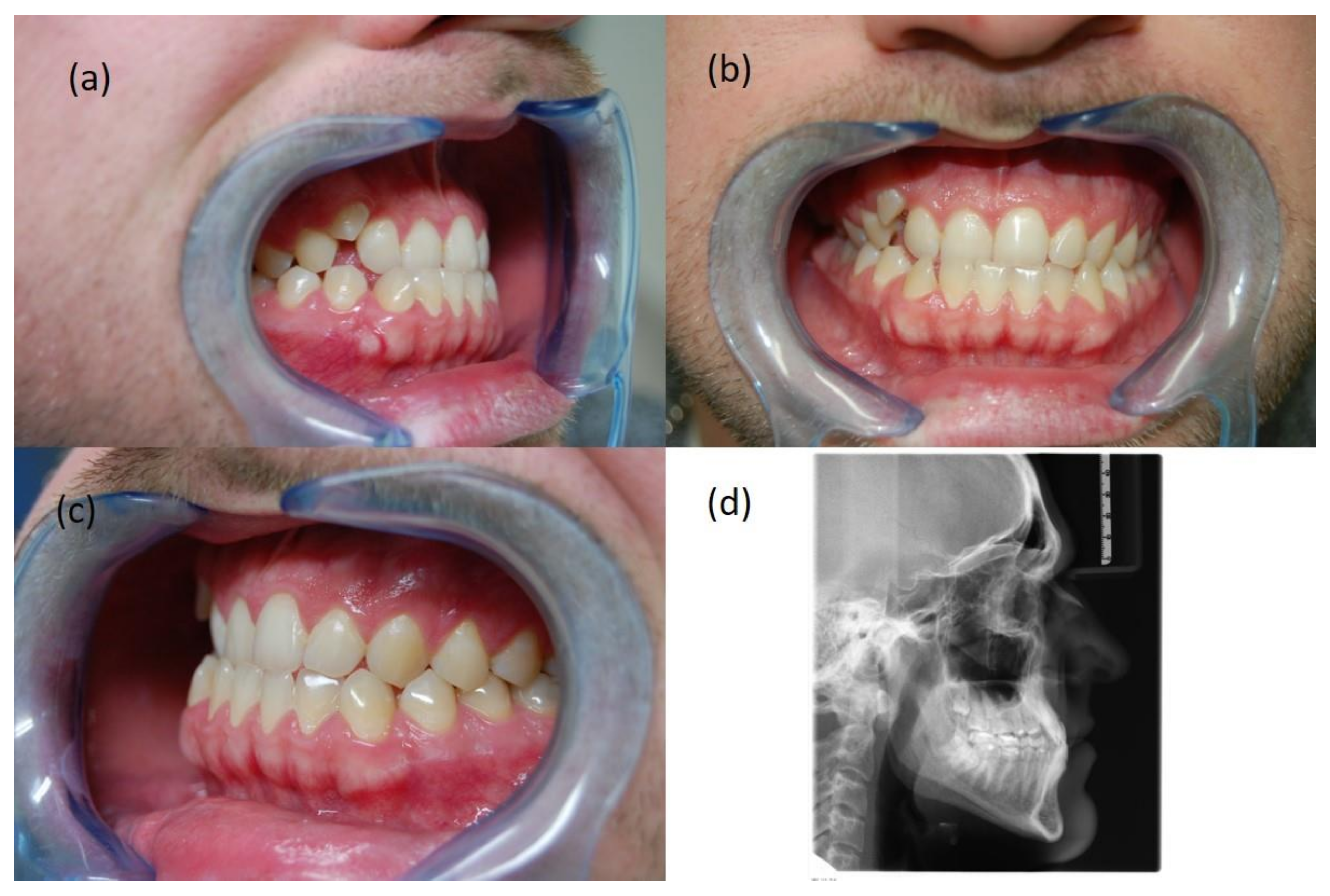
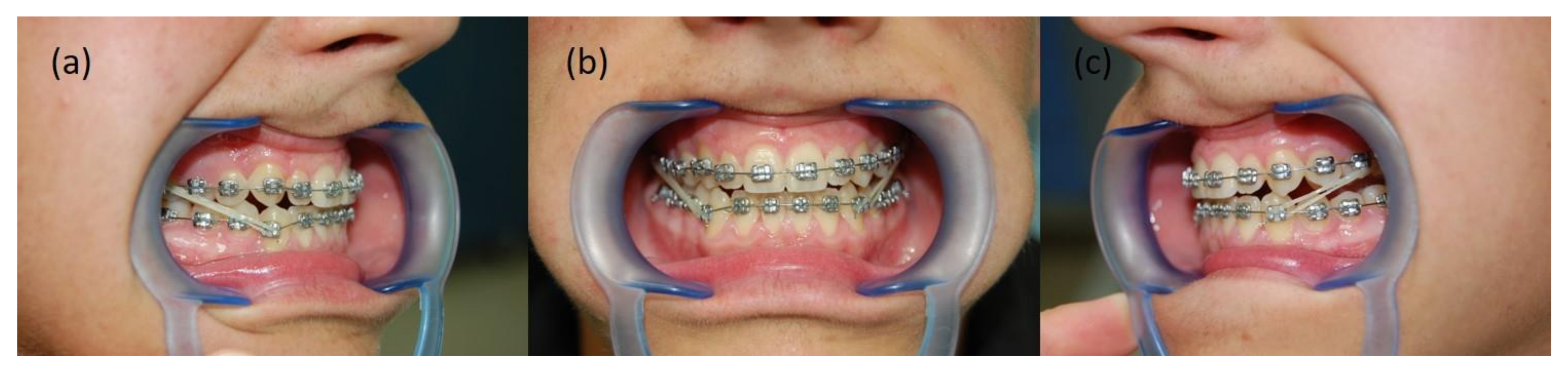
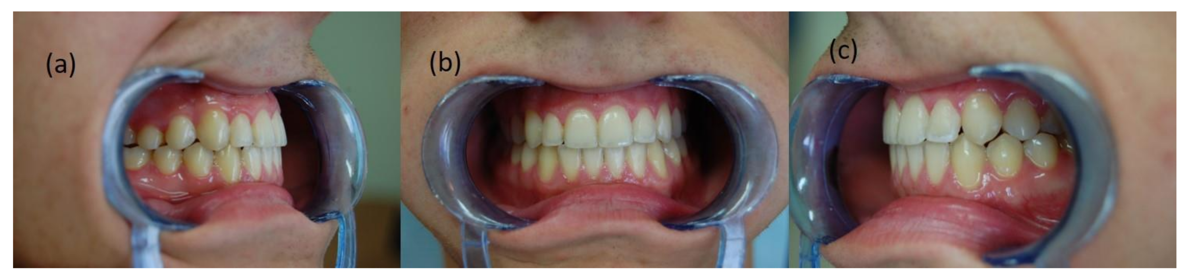
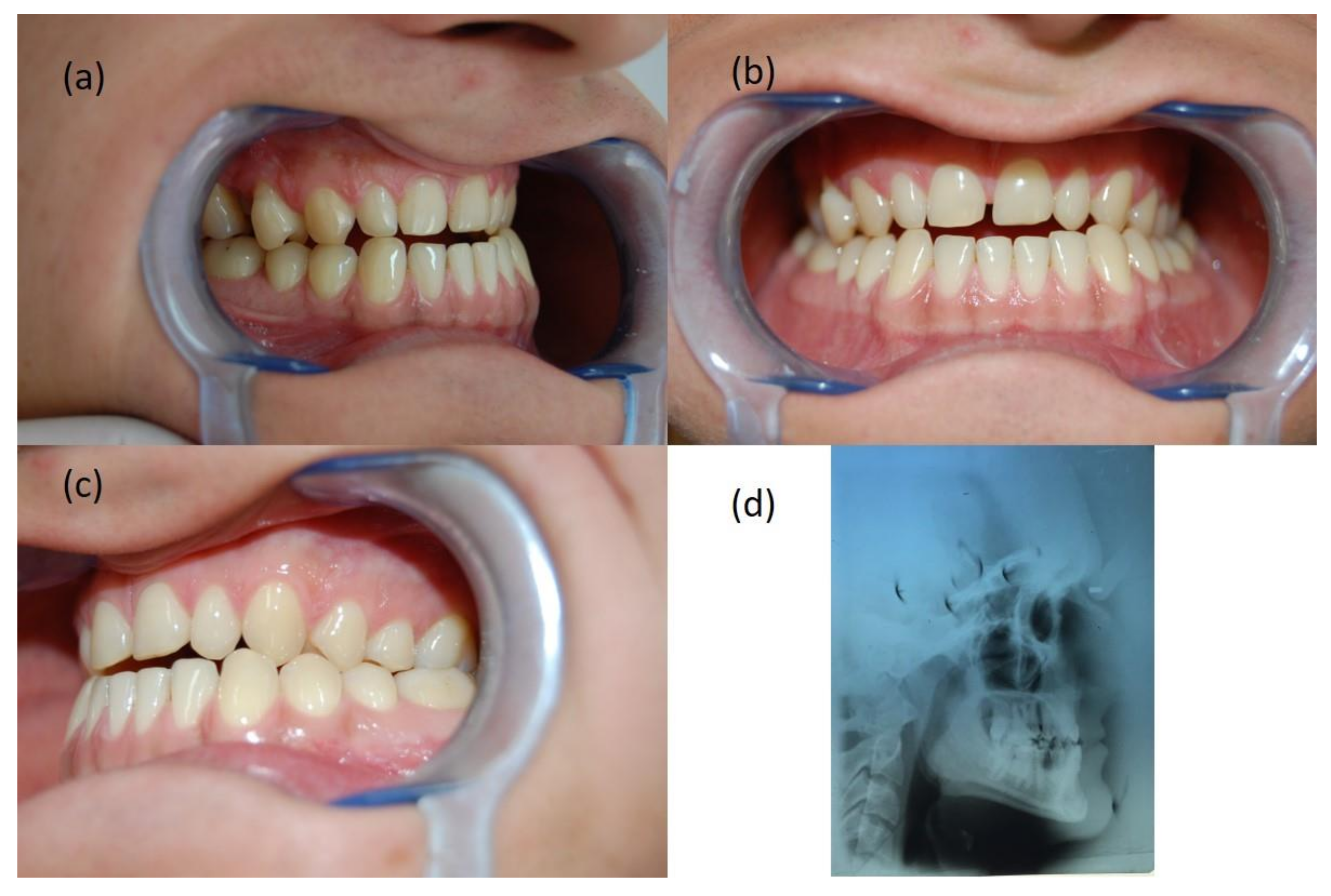
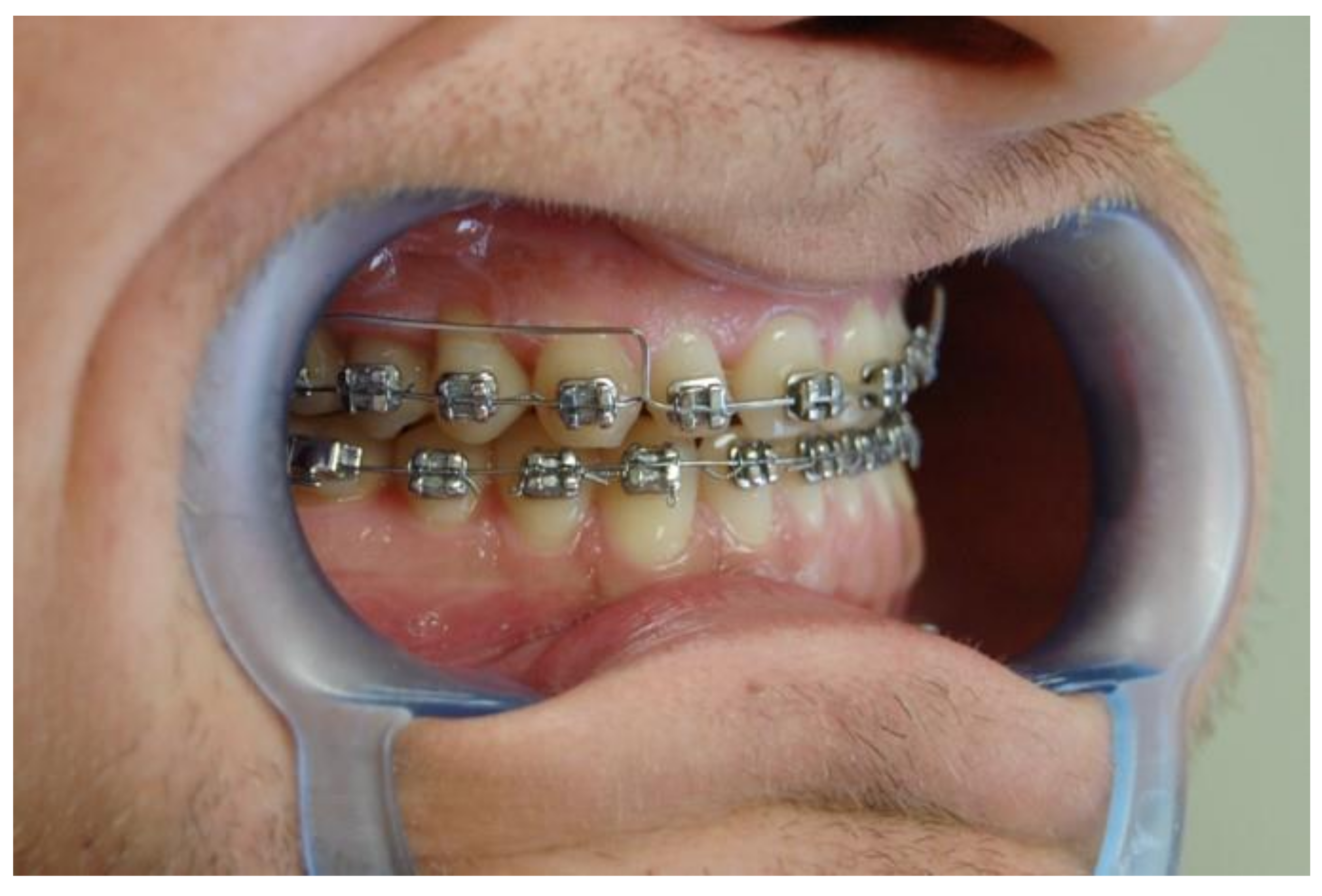
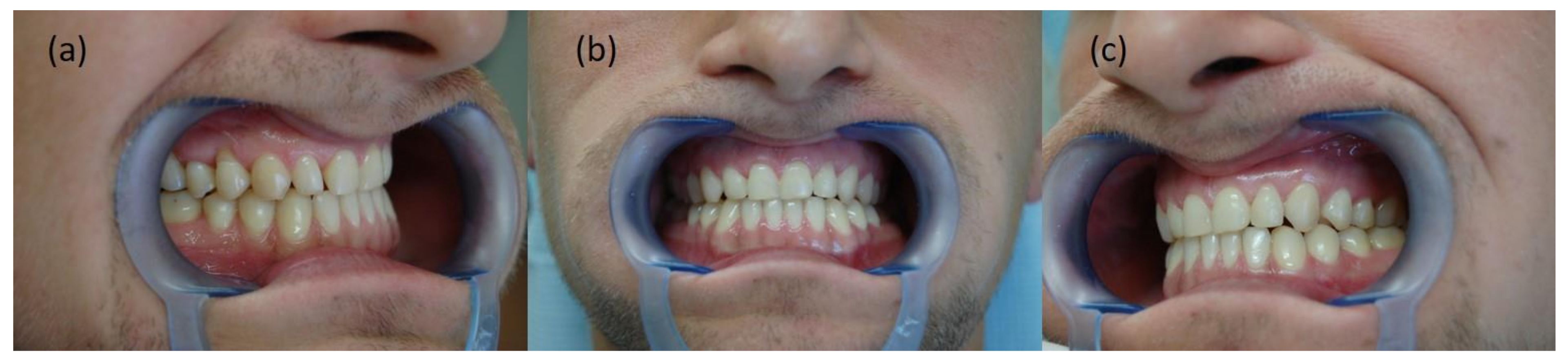
2.1. Patient KK, 26 Years Old

Diagnosis

The extraoral examination made it possible to recognize the retraction of the subnasal area, without the features typical of severe mandibular prognathism, without smoothing the mento-labial fold. Based on the intraoral examination, a narrowing of the upper dental arch, right-sided Angle Class III, partial lack of space for tooth 13, and a reversed overjet was found (Figure 2a–c). The analysis of the cephalometric X–ray allowed the diagnosis of maxillary retrognathism and skeletal Class III (Figure 2d).

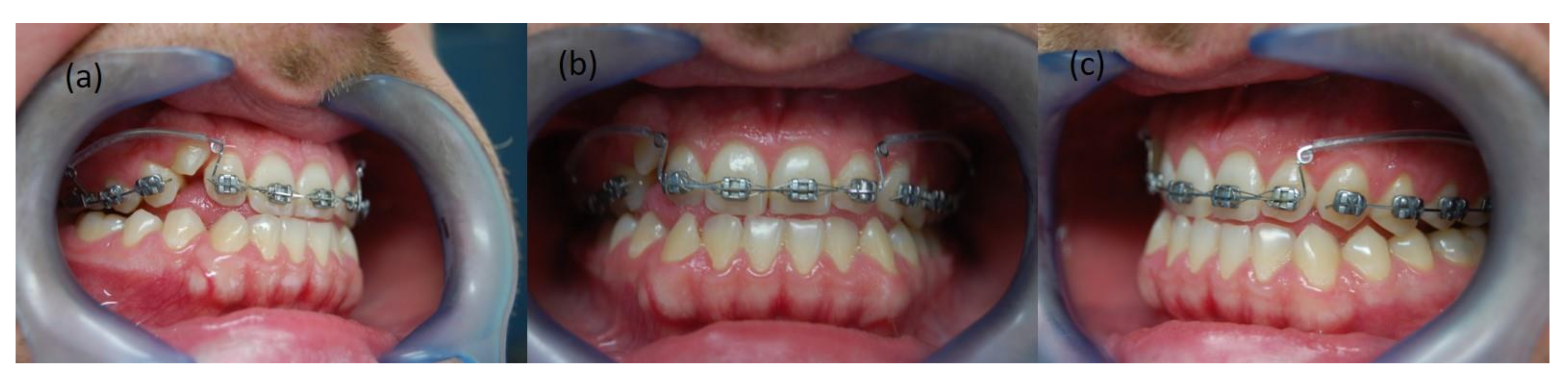
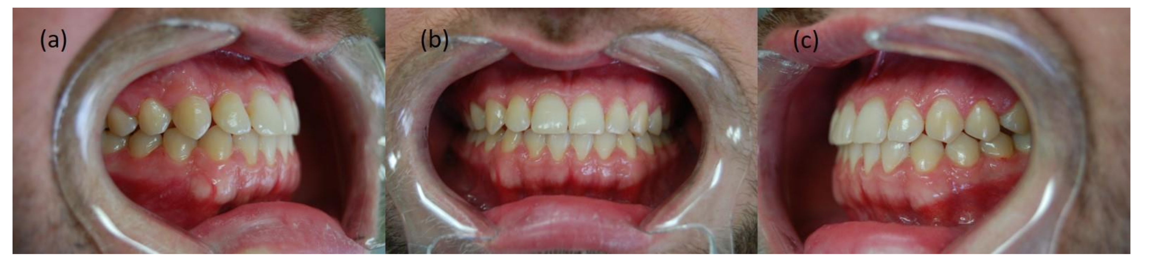
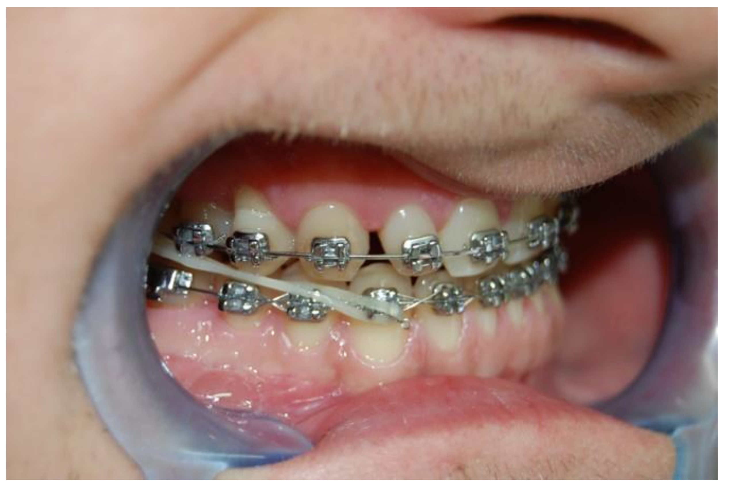
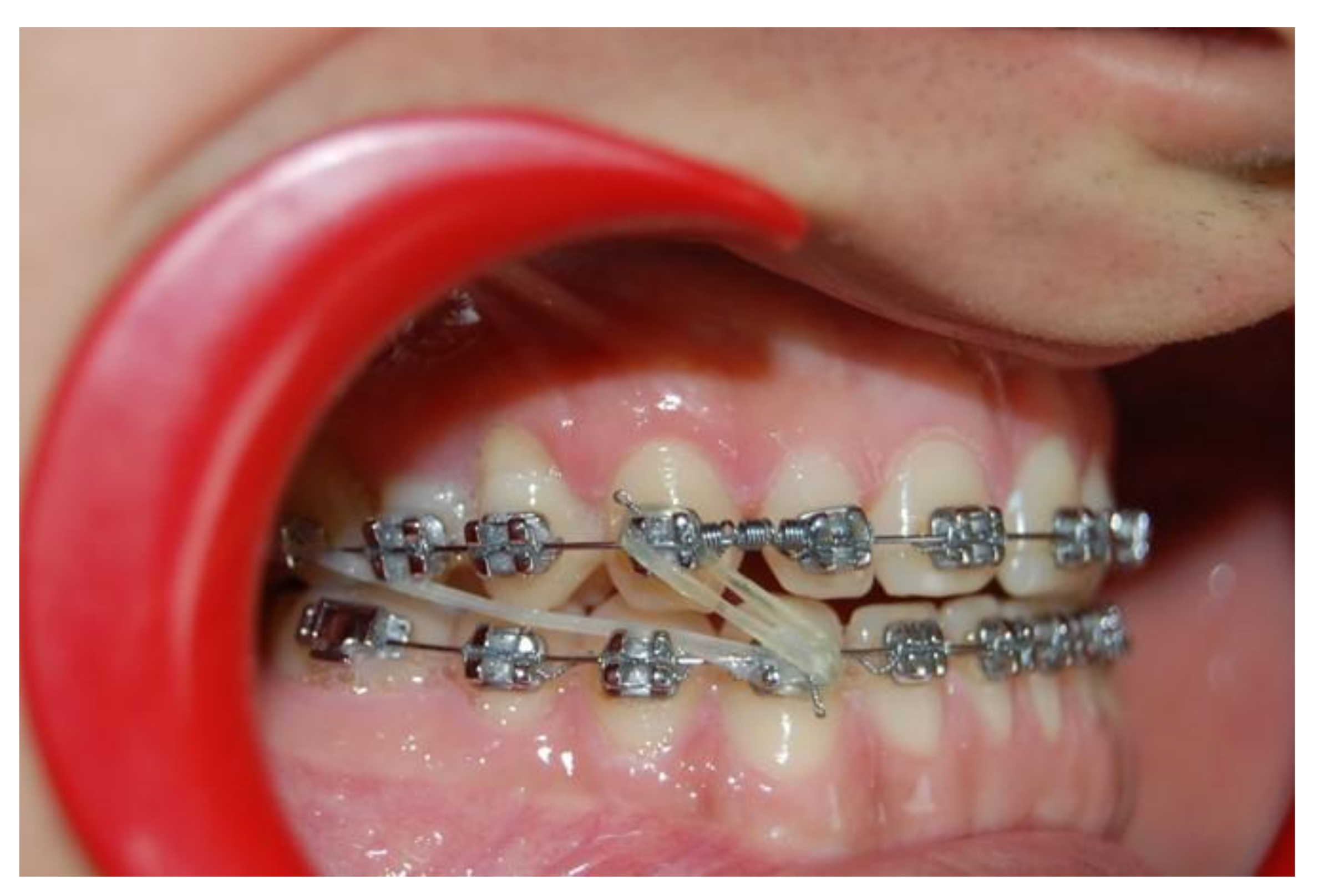
2.2. Treatment Plan and Process

The treatment aimed to create a place for tooth 13 and bring it to the dental arch, as well as Class III camouflage in the form of a traumatic bite elimination, and to obtain at least a partial occlusal standard. Therapy was commenced with lengthening the upper dental arch with an expansive stainless steel 0.016 × 0.016″ utility (Elgiloy blue, Rocky Mountain Orthodontics, Denver, CO, USA). It was used for the first seven months of treatment in combination with segmented archwires without bracing on the lower teeth (Figure 3a–c). In the eighth month of treatment, the basic archwire was replaced with continuous NiTi arches with diameters 0.012“ and 0.014″, respectively, with which the lateral sections of the upper dental arch were widened, and tooth 13 was brought to the place created as a result of forwarding movement of the upper incisors. At the same time, an appliance for the lower teeth was placed and a triangular intraoral elastic on the right side was inserted immediately, covering the following teeth: 44, 13, and 43 (diameter 1/8″, heavy). After 8 weeks, it was replaced with a single-sided, long, Class III elastics (1/4″ diameter, heavy). After lining the lower teeth with NiTi archwires with diameters of 0.012″ and 0.014″, respectively, a steel archwire 0.016 × 0.016″ (Elgiloy blue, Rocky Mountain Orthodontics, Denver, CO, USA) was used with a lingual torque root of the incisors. Active treatment was completed with the ideal archwires (Elgiloy yellow, Rocky Mountain Orthodontics, Denver, CO, USA). The final results of the therapy, which lasted 30 months, are presented in Figure 4a–c. Comparing the initial and final photos, it can be concluded that not only the impacted tooth 13 was inserted into the dental arch, but the occlusal norm was achieved by restoring the correct overjet and Angle Class I on the right side, while keeping Angle Class I on the left side.

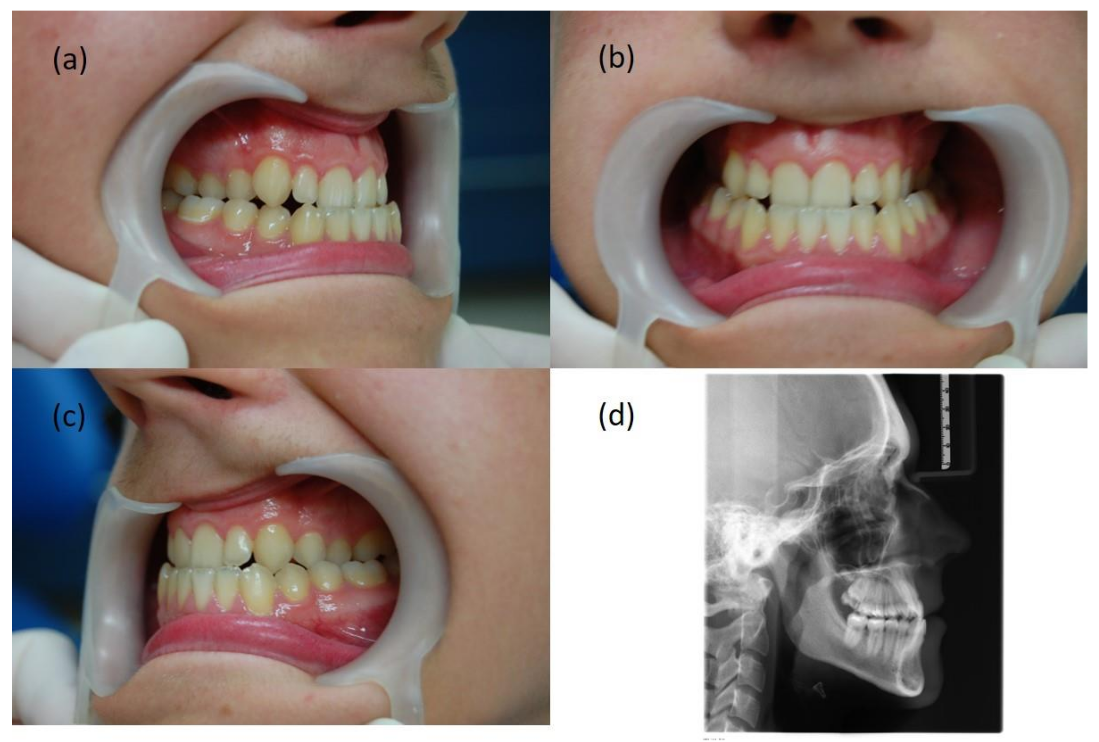
3. Patient IG, 18 Years Old

3.1. Diagnosis

The extraoral examination revealed a straight profile with a slightly recessed subnasal area. Based on intraoral examination (Figure 5a–c), a narrowed upper dental arch, a total crossbite with an inverse overjet, Angle Class III and canine Class III on the right side, and a 4 mm shift of the centerline of the upper dental arch to the right were found. The analysis of the cephalometric X-ray revealed the coexisting retrusion of the upper incisors (Figure 5d).

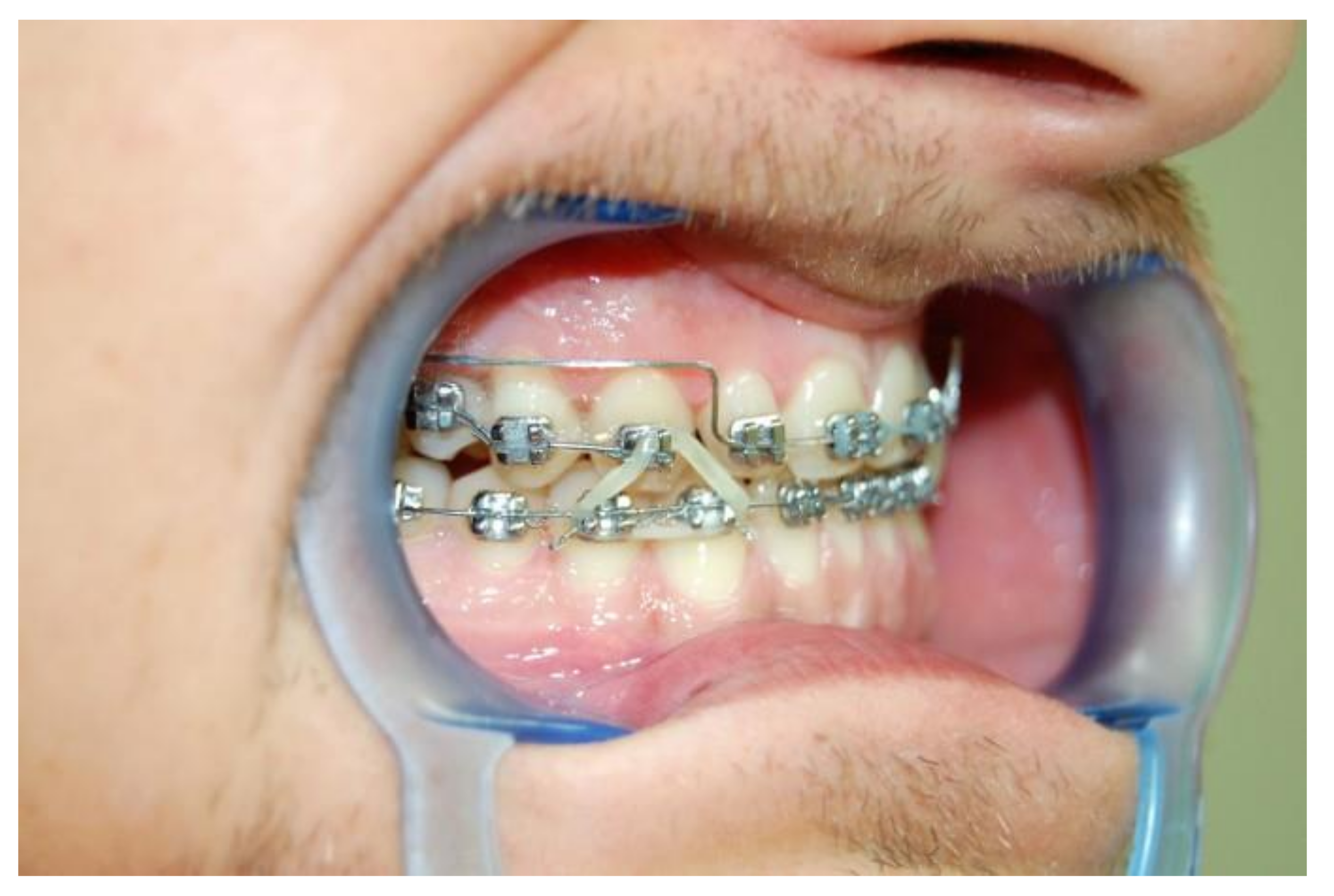
3.2. Treatment Plan and Process

The treatment aimed to widen and extend the upper dental arch, achieve its symmetry and restore Class I (Angle and canine) on the right side with the use of elastics. Ricketts technique was used in the treatment. After 4 months of widening the upper dental arch with the Utility archwire, it was replaced with a wide continuous NiTi archwire 0.017 × 0.025″ and then a TMA 0.018 × 0.022″. At the same time, the braces were fitted to the lower teeth, initially only in sections from teeth 46 to 43, and long right-sided Class III intraoral elastics (1/4″ diameter, heavy) were introduced immediately. In the fifth month of treatment, the remaining part of the appliance in the lower dental arch was set up and the patient was advised to wear elastics on both sides. His discipline in this matter and, at the same time, non-compliance with the planned dates of follow-up visits led to visible hypercorrection (Figure 6a–c). After the use of elastics was discontinued, Angle Class I and a positive overjet were obtained. Unfortunately, at the same time, there was a narrowing of the upper dental arch in the lateral sections, therefore, in the 18th month of treatment, criss-cross elastics were used from the palatal surface of teeth 16 and 26 to the buccal surfaces of the teeth—46 and 36, respectively. The treatment was completed after 30 months, and the correct results were obtained—occlusion and proper width of the upper dental arch (Figure 7a–c).

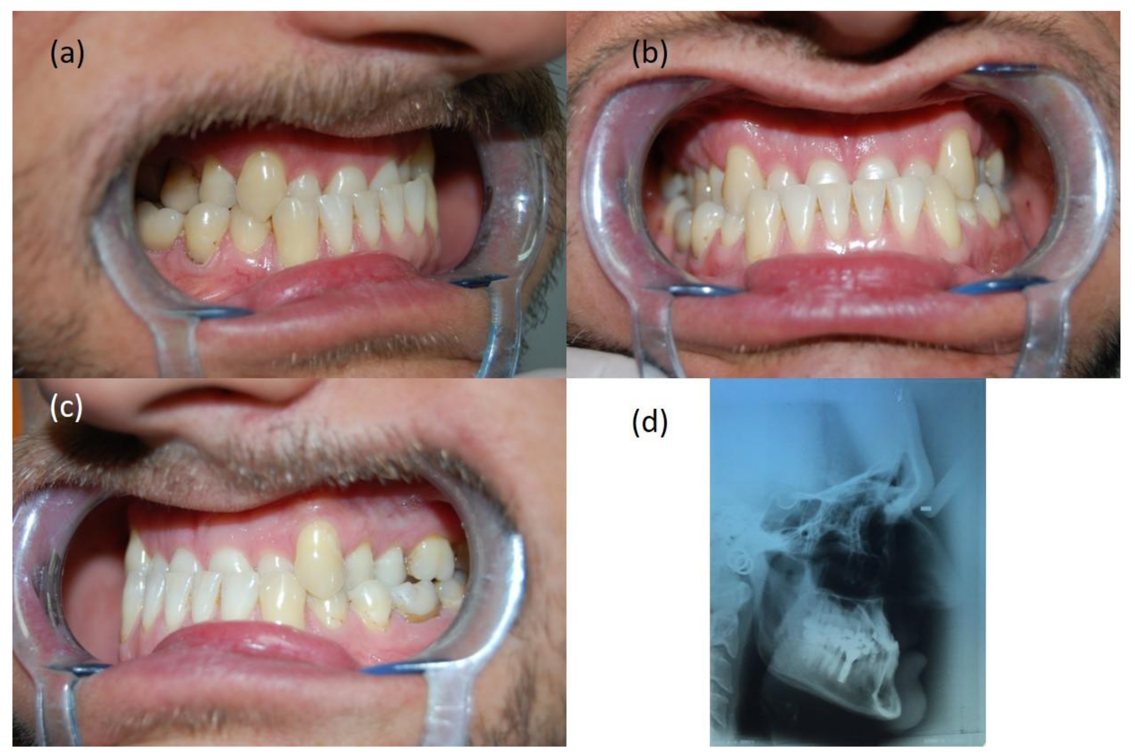
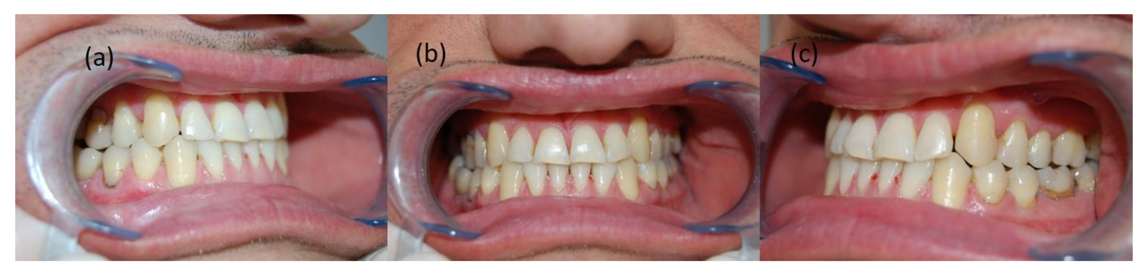
4. Patient PJ, 46 Years Old

4.1. Diagnosis

During the extraoral examination, a concave maxillary profile with a retraced sub nasal area and a shortening of the maxillary section was found. The functional test was negative. Intraoral examination revealed narrowing of the upper dental arch, severe negative overjet, deepened Spee curve (extrusion of lower incisors, i.e., deepened overbite), and bilateral Angle Class III (Figure 8a–c). After the analysis of the cephalometric X-ray, skeletal Class III was found, resulting from the underdevelopment of the maxilla, along with mandibular prognathism and upper and lower incisors retrusion (Figure 8d).

4.2. Treatment Plan and Process

The treatment aimed to improve the occlusal conditions by obtaining the correct overjet and overbite, as well as correct occlusion of the teeth in the lateral sections. It was planned to widen the upper dental arch, intrude the lower incisors and eliminate dental malposition. Treatment began with the simultaneous setting of an upper expansive 0.016 × 0.016″ utility archwire (Elgiloy yellow, Rocky Mountain Orthodontics, Denver, CO, USA) and a modified Schwarz removable appliance on the lower dental arch. The modification consisted of a slight enlargement of the acrylic behind the lower incisors, creating a small flat surface and removal of the labial bow. The patient’s strong motivation and understanding of the potential dangers of not wearing braces allowed the use of it as a removable appliance. After the first three months of treatment, the remaining brackets in the upper dental arch were fitted, a basic expansive archwire was several times activated (also thermally), replaced with a continuous NiTi archwire, first with a diameter of 0.016″, then with 0.016 × 0.016″. After seven months of treatment, four brackets were fitted on the lower incisors and a segmented archwire was placed, fixed in the slots with a figure-eight ligature to block them. A month later, the remaining brackets of the lower appliance were fixed. After 12 months of treatment and the use of intraoral criss-cross elastics, an Australian archwire (Wilcock) with a diameter of 0.018″ was placed in the upper dental arch, which was to stabilize the obtained width of the dental arch. From that moment on, there began the correction of the position of the upper canines with the use of medium chains elastics and the mesialization of the upper premolars and molars. After 18 months of treatment, at the request of the patient, satisfied with the result, it was decided to discontinue the active phase (Figure 9a–c).

5. Patient BB, 30 Years Old

5.1. Diagnosis

During the external and intraoral physical examination, a straight jaw profile and hypotension of the upper lip were found, as well as partial anterior open bite, underjet, narrowing of the upper dental arch, asymmetrical positioning of the teeth in the upper dental arch causing the centerline to shift by 3 mm to the right, palatopositioned tooth 15 with a complete lack of space for this tooth, along with persistent infantile swallowing (Figure 10a–c). After the analysis of the cephalometric X-ray, the patient was diagnosed with skeletal Class III, mainly due to maxillary retrognathism (Figure 10d).

5.2. Treatment Plan and Process

The treatment aimed to obtain the correct bite by expanding the upper dental arch, obtaining the correct overjet and overbite, leveling the asymmetry, and simultaneously introducing tooth 15 into the dental arch. Next, it was planned to obtain a canine Class I relationship, close the diastema and eliminate the open bite, together with learning and consolidation of the correct respiratory and muscle habits.
The treatment started with ligating the upper and lower 0.014″ NiTi arches wires. After two months, NiTi archwires 0.016 × 0.016″ were introduced, and after another three ones a basic utility archwire 0.016 × 0.016″ (Elgiloy blue, Rocky Mountain Orthodontics, Denver, CO, USA) with lateral segmented archwires was used. Before that, however, as a result of widening the upper dental arch with continuous NITI archwires, enough space was recovered for tooth 15. An additional 0.012″ NiTi archwire, tied piggy-back began to introduce this tooth into the occlusion. Originally, the bracket on UR5 was glued right next to its chewing surface. Six months into the treatment, Wilcock’s archwire 0.016″ was introduced together with a system of elements that interacted with each other—a Class III elastics and a passive spring. This mechanism aided mesialization of tooth 13 when the elastic was worn and moved it towards the mesial part of tooth 12, and aided the recovery and then the maintenance of Angle Class I on the right side (Figure 11). The next sequence of photos shows the final process of introducing tooth 15 into the dental arch and closing the gap next to 13, created thanks to utility archwire and after diastema closure (Figure 12, Figure 13 and Figure 14). The treatment was completed after 36 months, and the following photos show its final result (Figure 15a–c). On the left side (Figure 15c), there is a noticeable tendency for bite opening as a result of persistent infantile swallowing. The extended duration of treatment with fixed braces did not bring the expected improvement of this habit, so it was decided to use a Schwarz removable retainer with a tongue crib.

6. Discussion

Virtually every detailed diagnosis of skeletal malocclusion, including Class III, is based on the analysis of the cephalometric X-ray, despite the lack of clear scientific evidence for the usefulness of this diagnostic technique [6]. The strategy of using various planes and reference lines adopted in the current study was aimed at reducing the risk of making a mistake when marking measurement points on the cephalometric X-rays, which in turn was to reduce the risk of taking wrong therapeutic decisions.
According to the results of the Ricketts analysis, all patients in our studies were characterized by skeletal Class III. The skeletal measurements indicated a different degree of mandibular prognathism, because in each case the Downs facial angle was greater than 90°, regardless of the position of the upper jaw [4]. In all our patients, point A was also behind the Nasion–Pogonion (N-Pg) line, so the linear parameter describing the relation of this point to the N-Pg line was always negative. Nevertheless, analysis of the cephalometric X-rays using the Steiner approach did not give such unambiguous results. Only in patients KK and PJ did the SNB angle indicate a protruded position of the mandible, whilst the SNA angle in those patients was within the generally accepted norm [1]. The presented difference in the diagnosis of a skeletal malocclusion may be related to the greater differentiation of the inclination of this line, raised in orthodontic literature, for example, individual variability of Nasion point position, which reduces the reliability of the S-N line—compared to the FH line—in determining the antero-posterior position of the maxilla and mandible and the associated malocclusion [7]. This is confirmed by the research of Ricketts et al. [8] and Ricketts and Langlade [9], emphasizing the low stability of the S-N line compared to the FH references line. Schulhof [10] had a similar opinion, stressing that this lability is particularly evident in skeletal Class III cases when determining the antero-posterior position of the mandible and maxilla. Bourriau et al. [11], claimed that the determination of the points that make up the Frankfurt FH plane is burdened with a greater number of errors than the determination of the S-N line, which is a significant reason to use the S-N line for the analysis of the cephalometric X-ray instead of the FH reference line. Indeed, the S-N reference line is much more commonly used than the FH reference line to establish the relationship between the maxilla and the mandible. Proffit [5], like Bourriau et al. [11], explains the difficulties in locating the Porion point and recommends the use of the S-N line with an appropriate correction. Currently, however, this view seems a bit anachronistic due to the development of modern imaging techniques, which provide much more precise possibilities of locating structures and measurement points on cephalometric X-rays.
The diagnostic significance of the S-N line in the case of suspicion of Class III is also emphasized by Beltrao [12], according to which the value of the NSBa angle (Nasion-Sella-Basion) is closer to 120° indicates skeletal Class III. This is also confirmed by the meta-analysis of the NSBa cranial base angle value and its correlation with the anterior–posterior malocclusion, conducted by Aixiu Gong et al. [13]. It proved—based on analysis of approximately 3000 cases—that in almost 25% (n = 730) of patients with Class III there is a negative and statistically significant correlation between the NSBa angle and the dominance of indices characterizing negative overjet. The strength of this correlation is confirmed by the fact that in one of our patients KK—despite the lack of obvious Class III teeth and extraoral features—the lowest value of the NSBa angle was found.
Regarding the Class III asymmetric therapy, in 2010, Janson et al. [14] described a relatively simple and minimally invasive method, without extraction and the use of additional intra or extraoral appliances. It was inspiring for us, because it stood in clear opposition to the prevailing “arms race” in the search for more and more sophisticated and complicated systems aimed at improving the effectiveness of treatment of skeletal Class III, resulting in a large variety of techniques and methods used in the treatment of the discussed malocclusion.
Already at the turn of the 1960s and 1970s, Dr. Rolf Frankel [15] developed a system of innovative, removable appliances called functional regulators, of which type III was intended for the treatment of a reversed overjet. The wide side shields and the vestibular pads stimulated the formation of bone tissue as a result of pulling the oral vestibule mucosa at the border of its movable and immovable parts, which resulted in a favorable remodeling of the jaws. In addition, the active effect of the wire elements of the appliance on the teeth supported the entire treating process of jaws’ underdevelopment [16]. Unfortunately, this therapeutic success was and is strictly dependent on the cooperation of most adolescent patients, as is the case with any removable appliance. Simultaneously, with the evolution of functional regulators, scientific research was carried out confirming the effectiveness of the use of extraoral appliances for the treatment of Class III. The chin cup, which was first described in 1803 [17], returned to the world of modern scientific studies at the beginning of the 1970s [18]. Then, thanks to the studies by Graber [19] and Wendell et al. [20], along with the individual Delaire face mask often used together with intraoral devices for rapid expansion of the upper jaw (RME), it became a permanent fixture in the canon of instruments used in the treatment of Class III in adolescent and adult patients [21,22,23,24]. Baccetti et al. [25], by comparing the effectiveness of RME associated with a face mask (FM) to a reverse headgear (MCH), proved that the first system is more appropriate, in the case of maxillary retrognathia, and the second—in the case of mandibular prognathism. An interesting way to use RME was proposed by Liou EJ and Tsai WC. They proved that alternating—in appropriate time intervals—expanding and constricting the two-hinge appliances intended for RME together with the use of a face mask results in a more effective protraction of the upper jaw than one direction expanding of the screw [26]. The effectiveness of this protocol is confirmed by the results of the studies by Franchi et al. [27] and Sycinska-Dziarnowska et al. [28]. On the other hand, the fact that in the treatment of skeletal Class III it is not always necessary to use the protraction appliance and the jaw widening at the same time was proved by Vaughn et al. [29], who, when examining 45 adolescent patients, found no significant differences between the results of treatment of patients with RME/FM and FM alone.
Currently, many researchers describe the use of micro-implants—Temporary Intraoral Skeletal Anchorage Devices (TISAD) in the treatment of Class III, including its severe, and therefore asymmetric, forms. TISADs are used as fasteners for (a) Class I elastics activating the individual mechanism of lower molar retraction [30], (b) Class III elastics [31], and (c) individual extraoral devices for maxillary protraction [32]. An interesting treatment of an asymmetry case with the use of TISAD and without extraction, but with an inter-proximal reduction (IPR) of the enamel was described by Tseng et al. [33] in 2016. Using self-ligating brackets and micro-implants fitted unilaterally under the zygomatic arch for side segment retraction, they obtained a change in the ANB angle by 5° (from −3° to 2°) and symmetrically positioned equal dental arches. Regardless of the undisputed benefits of using absolute anchorage in the treatment of skeletal malocclusions [34], supporters of skeletal Class III treatment without TISAD seem to agree that the Carriere’s CM3 (Carriere Motion Class III) appliance [35], as well as the multiloop edgewise arch wire (MEAW) technique, are among the least burdensome for the patient, and at the same time effective treatment methods. The aforementioned CM3 appliances are activated by long, Class III elastics, stretched between CM3 and the upper molars. They do not require upper fixed braces and are replaced by the 1 mm Essix splint. Such a hybrid allows for very good results, also in patients with permanent dentition [36]. As for MEAW, already in 1994, Sato [37] presented four cases of the effective use of this technique in the treatment of skeletal Class III treatment with enlarged base angle, without the use of additional fixed appliances, but with molar extractions. At the same time, he criticized the use of Class III long elastics, demonstrating greater effectiveness of short elastics. Twenty years later, He et al. [38], who also used the MEAW technique for Class III treatment, showed a beneficial effect of long intermaxillary elastics, but in patients with deepened vertical bite and reduced base angle. Researchers divided 44 patients into two almost equal groups, where in the first group they used intraoral elastics attached to TISAD placed between teeth 16 and 17 and 26 and 27, and in the second—on teeth 17 and 27. They obtained a very good treatment result in all patients. The only differences were an average of 4.5 months longer treatment duration in the first group and the loss and need for re-implantation of TISAD in the second group.
The necessity of extracting molars to reconstruct the occlusal plane and reposition the posterior part of the mandible during MEAW treatment was questioned by Rubin [39]. On the other hand, however, he complemented the very good results of Class III treatment with this technique and suggested that they could also be obtained using, for example, a continuous TMA archwire, due to its greater resilience. The fact that the occlusion plane can be reconstructed with continuous NiTi archwires and without extraction was proved in 1999 by Kucukkeles et al. [40]. Gurgel et al. in their work described the effective widening of the jaws with the use of an additional TMA archwire [41]. In the current study, similar mechanisms in the presented cases were used, where instead of numerous loops, continuous archwires with high resilience were used, in addition to Utility archwires with segmented archwires of reduced stiffness, characteristic for the Ricketts technique. The Ricketts utility archwire is—due to its structure and chemical composition of the alloy from which it is made—intended for tooth movements in the anterior sector of the dental arches [42]. This set (utility and two segmented lateral wires) is very useful where dental malpositions in the lateral sections coexist with an underjet, and additionally when the incisors are in retrusion. The use of the Australian archwire in some of our patients, which is characterized by high stiffness and hardness due to the relatively high proportion of carbon atoms in its composition and which provides little resistance during the sliding movement of the brackets [43], made it possible to use chain elastics without fear of uncontrolled narrowing of the dental arches or the undesirable extrusion of the upper molars when long Class III intraoral elastics were used.
The treatment plan in each of our cases assumed no extraction in the lower dental arch and provided for the extension of the upper dental arch with the use of highly flexible TMA and NiTi archwires. The justification of waiving extractions in lower dental arches is the fact that—apart from the restrictive use of the MEAW protocol and Proffit’s guidelines [5]—there are virtually no other reports explicitly obliging extraction in the inoperable treatment of skeletal Class III. On the contrary, over the last 20 years, when analyzing selected publications in which the course of treatment of a group of several dozen patients was analyzed, there is a noticeable trend that reduces the need for extraction of lower premolars in this type of therapy. When Lin and Gu [44] presented their 2003 publication in which camouflage treatment was implemented in a group of 18 patients with skeletal Class III, 66.6% of them required extraction. Then, 12 years later, in 2015, indications for extraction still dominated, because they were performed in 58% of cases [45]. In 2018, in a group of 36 patients, thus twice as large as in the work of Lin and Gu [42], the percentage treated with the camouflage method with premolars extractions decreased significantly to 16.6% [46].
Our recommended use of Class III long extracts, of various strength and trajectory, or the implementation of non-standard, individual biomechanical systems, simple in their design, but effective and useful—especially in the treatment of the observed asymmetry in the positioning of the teeth—was aimed at simplifying the treatment process thanks to reducing the need for a face mask or other additional fixed appliances/expanders.
The examples of various procedures in the treatment of skeletal Class III with occlusal asymmetry presented in the discussion confirm that the therapeutic procedure in the cases of camouflage presented in this article not only complies with the “primum non nocere” principle but also meets modern therapeutic standards. The positive result of the treatment of the presented cases shows that there are many different methods of dealing with this complicated and difficult to treat a malocclusion. The current study has proved that by using a diverse range of techniques and orthodontic archwires, as well as by using individual biomechanical solutions, very good therapeutic results can be obtained. As was mentioned in the introduction, the phenotypic form of asymmetric skeletal Class III is so diverse that there exists no universal method of its treatment. Therefore, creating a priori obligatory schemes, e.g., removing teeth in the lower dental arch or using additional devices supporting treatment, in some situations may be an incorrect or at least unnecessary assumption.

7. Conclusions

The presented and successfully treated Class III cases with occlusal asymmetry prove that neither tooth extractions nor the use of additional intra- and extraoral elements is the condition sine qua non for obtaining a very good occlusion. Moreover, they should not be used without a detailed analysis of the skeletal parameters, in addition to different, and not just one, reference lines. The key to success in the treatment of this malocclusion, additionally complicated by asymmetry, is the use of individual therapeutic solutions at the material, biomechanical and conceptual levels. Skillful use of the utility archwire for the Ricketts technique allows for anterior–posterior extension of the anterior segment of the maxilla without deflecting the incisors. Moreover, the use of different types of archwires, low friction technique, long and short unsymmetrical elastics, as well as individual biomechanical systems increase the chances of therapeutic success of this complicated malocclusion.
Finally, it should be underlined that the choice of the treatment method should only be made after the differences between skeletal Class III treatment have been carefully explained to the patient, either employing camouflage or using interdisciplinary surgical and orthodontic procedures. Only then can you get an understanding of the compromise, fully accept the proposed treatment plan, and together enjoy success.

8. Limitations

The lack of all the photos documenting the treatment process may be considered as a minor drawback of this paper. However, the aim of this work was not to present stage-by-stage treatment leading to normal occlusion. Instead, it was focused on key phases serving as good clinical tips for orthodontic practitioners. Furthermore, “overloading” the paper with pictures bringing nothing new or spectacular from the clinical point of view was not the aim of the case series demonstrated in the current study.

Author Contributions

Conceptualization, J.I.; Methodology, J.I.; Formal analysis, B.K. and J.L; Validation, J.I., B.K., J.L.; Writing–original draft preparation, J.I.; Writing–review and editing, B.K. and J.L.; Supervision, B.K. and J.L.; All authors have read and agreed to the published version of the manuscript.

Funding

This research received no external funding.

Institutional Review Board Statement

Not applicable.

Informed Consent Statement

Informed consent was obtained from all subjects involved in the study.

Data Availability Statement

Patients’ medical documentation.

Conflicts of Interest

The authors declare no conflict of interest.

References

  1. Chan, G.K. Class III malocclusion in Chinese (Cantonese): Etiology and treatment. Am. J. Orthod. 1974, 65, 152–156. [Google Scholar] [CrossRef]
  2. Baccetti, T.; Reyes, B.C.; McNamara, J.A., Jr. Gender differences in Class III malocclusion. Angle Orthod. 2005, 75, 510–520. [Google Scholar] [CrossRef]
  3. Yuan, J.T.; Teng, E.; Heller, J.B.; Kawamoto, H.K.; Bradley, J. Asymmetric Class III Malocclusion Association With Cranial Base Deformation and Occult Torticollis. J. Craniofacial Surg. 2012, 23, 1421–1424. [Google Scholar] [CrossRef]
  4. Langlade, M. Diagnostique Orthodontique; Maloine: Paris, France, 1981; p. 708. [Google Scholar]
  5. Proffit, W.R.; Fields, H.W.; Sarver, D.M. Ortodoncja Współczesna Tom 1, 2nd ed.; Komorowska, A., Ed.; Elsevier: Wroclaw, Poland, 2009; p. 297. [Google Scholar]
  6. Durão, A.R.; Pittayapat, P.; Rockenbach, M.I.B.; Olszewski, R.; Ng, S.; Ferreira, A.P.; Jacobs, R. Validity of 2D lateral cephalometry in orthodontics: A systematic review. Prog. Orthod. 2013, 14, 31. [Google Scholar] [CrossRef] [Green Version]
  7. Pancherz, H.; Sack, B. Kritische Analyse der Winkel SNA, SNB und ANB bei der Auswertung von kieferorthopädischen Behandlungen. Fortschritte der Kieferorthopädie 1990, 51, 309–317. [Google Scholar] [CrossRef]
  8. Ricketts, R.M.; Schulhof, R.J.; Bagha, L. Orientation Sella-Nasion or Frankfort horizontal. Am. J. Ortho. 1976, 69, 648. [Google Scholar] [CrossRef]
  9. Ricketts, R.M.; Langlade, M. Plaidoyer pour une orientation cephalometrique. Rev. D’orthopédie Dento-Faciale 1977, 2, 161–172. [Google Scholar] [CrossRef]
  10. Schulhof, R.J. When S-N is Abnormal. J. Clin. Orthod. 1977, 11, 343. [Google Scholar]
  11. Bourriau, J.; Bidange, G.; Foucart, J.-M. Measurement errors in 2D cephalometrics. L’Orthodontie Fr. 2012, 83, 23–36. [Google Scholar] [CrossRef]
  12. Beltrão, P. Non-Surgical Treatment of Class III with Multiloop Edgewise Arch-Wire (MEAW) Therapy. In Emerging Trends in Oral Health Sciences and Dentistry [Internet]; Virdi, M.S., Ed.; IntechOpen: London, UK, 2015; Available online: https://www.intechopen.com/chapters/47810 (accessed on 11 March 2021). [CrossRef]
  13. Gong, G.; Li, J.; Wang, Z.; Li, Y.; Hu, F.; Li, Q.; Miao, D.; Wang, L. Cranial base characteristics in anteroposterior malocclusions: A meta-analysis. Angle Orthod. 2016, 86, 668–680. [Google Scholar] [CrossRef] [Green Version]
  14. Janson, G.; De Freitas, M.R.; Araki, J.; Franko, E.J.; Barros, S.E.C. Class III subdivision malocclusion corrected with asymmetric intermaxillary elastics. Am. J. Orthod. Dentofac. Orthop. 2010, 138, 221–230. [Google Scholar] [CrossRef]
  15. Frankel, R. Maxillary retrusion on class III and treatment with the function corrector III. Trans. Eur. Orthod. Soc. 1970, 46, 249–259. [Google Scholar]
  16. McNamara, J.A., Jr. The functional regulator (FR-3) of Frankel. Am. J. Orthod. Dentofac. Orthop. 1985, 88, 409–424. [Google Scholar] [CrossRef]
  17. Barrington, E.; Haswell, G.D. The Natural History and Diseases of the Human Teeth, 2nd ed.; Fox, J., Ed.; The Natural History and Diseases of the Human Teeth: London, UK, 1803; p. 331. [Google Scholar]
  18. Suzuki, N. A cephalometric observation on the effect of the chin cap. J. Jpn. Orthod. Soc. 1972, 31, 64–74. [Google Scholar]
  19. Graber, L.W. Chin cup therapy for mandibular prognathism. Am. J. Orthod. 1977, 72, 23–41. [Google Scholar] [CrossRef] [Green Version]
  20. Wendell, P.D.; Nanda, R.; Sakamoto, T.; Nakamura, S. The effects of chin therapy on the mandibule: A longitudinal study. Am. J. Orthod. 1985, 87, 265–274. [Google Scholar] [CrossRef]
  21. Delaire, J. Confection du masque orthopedique. Rev. Stomat. 1971, 72, 579–584. [Google Scholar]
  22. Haas, A.J. The treatment of Maxillary Deficiency by Opening the Midpalatal Suture. Angle Orthod. 1965, 35, 200. [Google Scholar]
  23. Westwood, P.V.; McNamara, J.A., Jr.; Baccetti, T.; Franch, L.; Sarver, D.M. Long-term effects of Class III treatment with rapid maxillary expansion and facemask therapy followed by fixed appliances. Am. J. Orthod. Dentofac. Orthop. 2003, 123, 306–320. [Google Scholar] [CrossRef] [Green Version]
  24. Baccetti, T.; McGill, J.S.; Franchi, L.; McNamara, J.A., Jr.; Tollaro, I. Skeletal effects of early treatment of Class III malocclusion with maxillary expansion and face mask therapy. Am. J. Orthod. Dentofac. Orthop. 1998, 113, 333–343. [Google Scholar] [CrossRef]
  25. Baccetti, T.; Rey, D.; Angel, D.; Oberti, G.; McNamara, A.J., Jr. Mandibular cervical headgear vs rapid maxillary expander and facemask for orthopedic treatment of Class III malocclusion. Angle Orthod. 2007, 77, 619–624. [Google Scholar] [CrossRef]
  26. Liou, E.J.; Tsai, W.C. A new protocol for maxillary protraction in cleft patients: Repetitive weekly protocol of alternate rapid maxillary expansions and constrictions. Cleft Palate Craniofac. J. 2005, 42, 21–27. [Google Scholar] [CrossRef]
  27. Franchi, L.; Baccetti, T.; Masucci, C.; Defraia, E. Early Alt-RAMEC and facial mask protocol in class III malocclusion. J. Clin. Orthod. 2011, 45, 601–609. [Google Scholar]
  28. Sycinska-Dziarnowska, M.; Janiszewska-Olszowska, J.; Grocholewicz, K. Leczenie przodozgryzu rzekomego z wykorzystaniem protokołu Alt-Ramec. Clin. Orthod. 2018, 1, 41–53. [Google Scholar]
  29. Vaughn, G.A.; Mason, B.; Moon, H.B.; Turley, P.K. The effects of maxillary protraction therapy with or without rapid palatal expansion: A prospective, randomized clinical trial. Am. J. Orthod. Dentofac. Orthop. 2005, 128, 299–309. [Google Scholar] [CrossRef]
  30. Chung, K.R.; Kim, S.H.; Choo, H.; Kook, Y.A.; Cope, J.B. Distalization of the mandibular dentition with mini-implants to correct a Class III malocclusion with a midline deviation. Am. J. Orthod. Dentofac. Orthop. 2010, 137, 135–146. [Google Scholar] [CrossRef]
  31. De Clerck, H.J.; Cornelis, M.A.; Cevidanes, L.H.; Heymann, G.C.; Tulloch, C.J. Orthopedic traction of the maxilla with miniplates: A new perspective for treatment of midface deficiency. J. Oral. Maxillofac. Surg. 2009, 67, 2123–2129. [Google Scholar] [CrossRef] [Green Version]
  32. Elnagar, M.H.; Elshourbagy, E.; Ghobashy, S.; Khedr, M.; Evans, C.A. Dentoalveolar and arch dimension changes in patients treated with miniplate-anchored maxillary protraction. Am. J. Orthod. Dentofac. Orthop. 2017, 151, 1092–1106. [Google Scholar] [CrossRef]
  33. Tseng, L.; Chang, C.H.; Roberts, W.E. Diagnosis and conservative treatment of skeletal Class III malocclusion with anterior crossbite and asymmetric maxillary crowding. Am. J. Orthod. Dentofac. Orthop. 2016, 149, 555–566. [Google Scholar] [CrossRef] [Green Version]
  34. Kucukkeles, N.; Yilmaz, H.N. Antoszewska, S., Ed.; Class III malocclusion. In Skeletal Anchorage in Orthodontic Treatment. Class II, III & Transverse malocclusion; Elamed: Katowice, Poland, 2014; pp. 35–52. ISBN 978-83-61190-53-0. [Google Scholar]
  35. Carriere, L. Nonsurgical correction of severe skeletal class III malocclusion. J. Clin. Orthod. 2016, 50, 216–230. [Google Scholar]
  36. McNamara, J.A., Jr.; Franchi, L.; McNamara-McClatchey, L.; Kowalski, S.E.; Cheesemane, C.C. Evaluation of adolescent and adult patients treated with the Carriere Motion Class III appliance followed by fixed appliances. Angle Orthod. 2021, 91, 149–156. [Google Scholar] [CrossRef]
  37. Sato, S. Case Report: Developmental characterization of skeletal Class III malocclusion. Angle Orthod. 1994, 64, 105–111. [Google Scholar] [CrossRef]
  38. He, S.; Gao, J.; Wamalwa, P.; Wang, Y.; Zou, S.; Chen, S. Camouflage treatment of skeletal Class III malocclusion with multiloop edgewise archwire and modified Class III elastics by maxillary mini-implant Anchorage. Angle Orthod. 2013, 83, 630–640. [Google Scholar] [CrossRef] [Green Version]
  39. Rubin, R.M. Commentary: Skeletal class III maloccusion. Angle Orthod. 1994, 64, 111. [Google Scholar] [CrossRef]
  40. Kucukkles, N.; Acar, A.; Demirkaya, A.A.; Evrenol, B.; Enacar, A. Cephalometric evaluation of open bite treatment with NiTi archwires and anterior elastics. Am. J. Dentofac. Orthop. 1999, 116, 555–562. [Google Scholar] [CrossRef]
  41. Gurgel, J.A.; Pinzan-Vercelino, C.R.M.; Leon-Salazar, V. Maxillary and mandibular dentoalveolar expansion with an auxiliary beta-titanium arch. Am. J. Orthod. Dentofac. Orthop. 2017, 152, 543–552. [Google Scholar] [CrossRef] [PubMed]
  42. Langlade, M. Therapeutique Orthodontique; Maloine: Paris, France, 1986; pp. 135–158. [Google Scholar]
  43. Pelsue, B.M.; Zinelis, S.; Bradley, T.G.; Berzins, D.W.; Eliades, T.; Eliades, G. Structure, Composition and Mechanical Properties of Australian Orthodontic Wire. Angle Orthod. 2009, 79, 97–101. [Google Scholar] [CrossRef]
  44. Lin, J.; Gu, Y. Preliminary investigation of nonsurgical treatment of severe skeletal class III malocclusion in the permanent dentition. Angle Orthod. 2003, 73, 401–410. [Google Scholar] [CrossRef]
  45. Georgalis, K.; Woods, M.G. A study of Class III treatment: Orthodontic camouflage vs. orthognathic surgery. Aust. Orthod. J. 2015, 31, 138–148. [Google Scholar] [CrossRef]
  46. Eslami, S.; Faber, J.; Fateh, A.; Sheikholaemmeh, F.; Grassia, V.; Jamilian, A. Treatment decision in adult patients with class III malocclusion: Surgery versus orthodontics. Prog. Orthod. 2018, 19, 28. [Google Scholar] [CrossRef] [PubMed] [Green Version]
Figure 1. The reference points and lines applied in the cephalometric analyses.
Symmetry 14 00316 g001
Figure 2. Patient KK’s initial records. Intraoral pictures: (a) right side, (b) en face, (c) left side, and (d) lateral cephalogram.
Symmetry 14 00316 g002
Figure 3. Patient KK’s Utility archwire stage. Intraoral pictures: (a) right side, (b) en face, (c) left side.
Symmetry 14 00316 g003
Figure 4. Patient KK’s final stage. Intraoral pictures: (a) right side, (b) en face, (c) left side.
Symmetry 14 00316 g004
Figure 5. Patient IG’s initial records. Intraoral pictures: (a) right side, (b) en face, (c) left side, and (d) lateral cephalogram.
Symmetry 14 00316 g005
Figure 6. Patient IG’s records. Just before when intraoral elastics were getting off the teeth. Intraoral pictures: (a) right side, (b) en face, (c) left side.
Symmetry 14 00316 g006
Figure 7. Patient IG’s final stage. Intraoral pictures: (a) right side, (b) en face, (c) left side.
Symmetry 14 00316 g007
Figure 8. Patient PJ’s initial records. Intraoral pictures: (a) right side, (b) left side, (c) left side, and (d) lateral cephalogram.
Symmetry 14 00316 g008
Figure 9. Patient PJ’s final stage. Intraoral pictures: (a) right side, (b) en face, (c) left side.
Symmetry 14 00316 g009
Figure 10. Patient BB’s initial records. Intraoral pictures: (a) right side, (b) left side, (c) left side, and (d) lateral cephalogram.
Symmetry 14 00316 g010
Figure 11. Patient BB’s intermediate stage. Launching of long class III elastic. Intraoral picture: side right.
Symmetry 14 00316 g011
Figure 12. Patient BB’s intermediate stage. Triangle class III elastic and a passive spring were added. Intraoral picture: side right.
Symmetry 14 00316 g012
Figure 13. Patient BB’s intermediate stage. Retention of canine I Class and beginning of introducing 15. Intraoral picture: side right.
Symmetry 14 00316 g013
Figure 14. Patient BB’s intermediate stage. Retention of both canine I Class and 15 in right place. Intraoral picture: side right.
Symmetry 14 00316 g014
Figure 15. Patient BB’s final stage. Intraoral pictures: (a) right side, (b) en face, (c) left side.
Symmetry 14 00316 g015
Table 1. Results of the initial ceph-analyses Cephalometric measurements.
Patient KKPatient IGPatient PJPatient BB
F (°)94.494.19591
XY (°)90.085.89591
NSBa (°)114128125130
A:NPg (mm)−5.28−0.5−11−8
SNA (°)81.879.48073
SNB (°)85.880.28779
ANB (°)−4−0.8−7−6
Wits (mm)−7.31−3.33−13−5
Mx/MP (°)23.831.61118
FMA (°)23.525.31420
SN/MP (°)28.839.62130
SN/FH (°)6.715510
IMPA (°)85.589.87286
UI/XY (°)0−8.4−13−9
OJ (mm)−1,5−2−7−3
Sn (mm)−15−12−32−21
UL (mm)−11−9−28−17
LL (mm)−6−4−9−12
Publisher’s Note: MDPI stays neutral with regard to jurisdictional claims in published maps and institutional affiliations.

