Article

Exploring Parametric and Mechanistic Differences between Expi293FTM and ExpiCHO-STM Cells for Transient Antibody Production Optimization

BioMedicine Design, Medicinal Sciences, Pfizer Worldwide R&D, 610 Main Street, Cambridge, MA 02139, USA
*
Author to whom correspondence should be addressed.
These authors contributed equally to this work.
Antibodies 2023, 12(3), 53; https://doi.org/10.3390/antib12030053
Submission received: 10 July 2023 / Revised: 26 July 2023 / Accepted: 8 August 2023 / Published: 10 August 2023
(This article belongs to the Section Antibody Discovery and Engineering)

Abstract

:
Rapidly producing drug-like antibody therapeutics for lead molecule discovery and candidate optimization is typically accomplished by large-scale transient gene expression technologies (TGE) with cultivated mammalian cells. The TGE methodologies have been extensively developed over the past three decades, yet produce significantly lower yields than the stable cell line approach, facing the technical challenge of achieving universal high expression titers for a broad range of antibodies and therapeutics modalities. In this study, we explored various parameters for antibody production in the TGE cell host Expi293FTM and ExpiCHO-STM with the transfection reagents ExpiFectamineTM and polyethylenimine. We discovered that there are significant differences between Expi293FTM and ExpiCHO-STM cells with regards to DNA complex formation time and ratio, complex formation buffers, DNA complex uptake trafficking routes, responses to dimethyl sulfoxide and cell cycle inhibitors, as well as light-chain isotype expression preferences. This investigation mechanistically dissected the TGE processes and provided a new direction for future transient antibody production optimization.

1. Introduction

Therapeutic antibodies are the largest class of new drugs occupying half of the global top 10 therapeutics products sold in 2022 [1]. These therapeutic interventions have achieved significant efficacies with reduced side effects in a number of disease areas especially for cancer, autoimmune, and inflammatory disorders. There is a huge and growing demand for recombinant antibodies in biopharmaceutical research and development pipelines. Expediting antibody discovery processes and shortening the development cycle are critical in effectively bringing valuable antibody drug products to the needed patients in clinics. Since antibodies are therapeutic protein drugs that are produced by cultivated mammalian cells, the antibody protein production process is an essential step for biotherapeutics research and development, e.g., lead candidate identification and optimization, in vitro and in vivo efficacy studies, pharmacokinetics, and toxicology studies, as well as clinical applications. Tremendous research efforts have been put into method development for robust transgene production over the decades.
While the chromosomal-integrated stable cell line approach is routinely utilized for generating clinical-grade recombinant antibodies, transient gene expression technologies (TGE) with episomal plasmid transfection in mammalian cells have been widely used for the rapid production of research-grade antibodies in milligrams and grams quantities [2,3,4,5,6]. Without the need for gene recombination into the host genomes which usually takes weeks to be stabilized, transient transfection of target DNAs complexed with cationic lipids, cationic polymers, or calcium phosphate, enables a fast and efficient protein scale-up production for a large number of antibody candidates in days. Mammalian cell hosts employed by the TGE technologies ensure not only functional protein folding and assembly but also proper post-translational modifications. The predominant host cell line for TGE is human embryonic kidney (HEK)-293 cells, which are known to be easy to use, highly transfectable to plasmid DNAs, and adaptable to robust high cell density growth in serum-free suspension. Another major TGE host of choice is Chinese hamster ovary (CHO) cells, the most common stable cell hosts for commercial and clinical protein production, which offers a potential advantage of end-to-end product alignment.
The TGE technologies have made a tremendous stride in improving expression titers and protein yields over the past thirty years. Through cell line engineering, vector engineering, and process engineering, antibody titers greater than 1 g/L produced transiently by HEK293 cells on a large scale have been reported [7,8]. Similarly, despite the initial modest expression yields [9,10], the transient CHO systems also reach the expression titer range of 1–3 g/L for a number of antibodies [11,12,13,14,15,16,17]. However, TGE technologies still face significant challenges. Transient expression levels of many recombinant antibody constructs remain 1–2 orders of magnitude lower than those from the corresponding stable cell lines. For some low-expression antibodies, reasonable expression titers still cannot be obtained. To meet the demands for rapid hit screenings and for increasing molecular complexities of multispecific modalities [18,19,20], the TGE workflow and protocols need constant optimization. In addition, while the above-mentioned proprietary HEK293 and CHO cell lines have significant advantages for protein production, only commercial cell lines Expi293FTM and ExpiCHO-STM [8,13,21,22], are readily available to the scientific communities. Research efforts on investigating and further optimizing these highly productive cell hosts [23] are imperatively needed, especially in the areas like transfection parameters and mechanisms as well as new strategies for production enhancements. The outcomes from such studies with impacts on the production of various biotherapeutics hits should help improve the overall TGE technologies in varied systems.
In this study, we set out to fine-tune several key aspects of the transfection processes by the reductionist “one factor at a time” approach [7,9,10,11,24,25,26,27,28] in both Expi293FTM and ExpiCHO-STM cells. Transfection reagents of ExpiFectamineTM and polyethyleneimine (PEI) were both employed for plasmid DNA transfection. The TGE protocols with optimized parameters that can significantly boost antibody production were developed. We have uncovered that the Expi293FTM and ExpiCHO-STM systems are different from each other in multiple aspects, e.g., complex formation time and volume, DNA-to-PEI ratio, responses to dimethyl sulfoxide (DMSO) and cell cycle inhibitors, as well as antibody light-chain isotype preferences. These observations should provide new insight into the mechanistic differences between the TGE in Expi293FTM and ExpiCHO-STM.

2. Materials and Methods

2.1. Chemicals and Reagents

DMSO (CAT#02650), sodium butyrate (CAT#156547), valproic acid sodium salt (VPA, CAT#P4543), carbenicillin disodium salt (CAT#C1389), Genestein (CAT# G6649), chlorpromazine (CAT# C8138), Filipin (CAT# F9765), and methyl-β-cyclodextrin (CAT# C4555) were purchased from Sigma-Aldrich (Burlington, MA, USA). Sodium butyrate (CAT#M19-137) was from EMD Millipore (Billerica, MA, USA). Polyethyleneimine Max powder (CAT#24765-1, Polysciences Inc., Warrington, PA, USA) was dissolved in cell culture grade sterile water, adjusted to pH 7.0, sterile-filtered to the final stock solution of 1.5 mg/mL, aliquoted and stored at −20 °C. Tryptone N1 powder (CAT#19553, Organotechnie, La Courneuve, France) was dissolved into Expi293 expression medium to a final concentration of 20% (w/v) and sterile filtered. ExpiFectamine™ 293 Transfection Kit (CAT#A14524) and ExpiFectamineTM CHO Transfection Kit (CAT#A29131) were purchased from Thermo Fisher Scientific (Waltham, MA, USA).

2.2. Cell Culture

Expi293F™ cells (Cat#A14527) and ExpiCHO-S™ cells (CAT#A29127) were purchased from Thermo Fisher Scientific. Expi293FTM expression medium (Thermo Fisher, CAT#A1435101) and ExpiCHO-STM expression media (Thermo Fisher, Cat#A2910001) were used to culture the corresponding cells in Multitron incubator shakers (INFORS HT, Bottmingen, Switzerland) or Kuhner incubator shakers (Kuhner, Birsfelden, Switzerland) with 8% CO2 at 37 °C. The Expi293F™ cells were routinely maintained in Thomson Optimum Growth® shake flasks (Thomson, Oceanside, CA, USA) at 120 rpm and ExpiCHO-S™ Cells were cultured at 68 rpm in Erlenmeyer flasks (Corning Inc., Corning, NY, USA). To monitor cell density and viability, a Vi-CELLTM cell viability analyzer (XR Model, Beckman Coulter Life Sciences, Indianapolis, IN, USA) was used routinely and all cells were maintained over 99% viability during culturing.

2.3. Plasmids and Plasmid Preparation

Plasmid DNAs encoding antibody heavy chain (HC) or light chains (LC) were in a cytomegalovirus (CMV)-promoter-based DNA expression vector pTT5. cDNA clones encoding human p21 (pCMV3-p21, Ref Seq number NM_000389.3, CAT#HG11108-UT) and p27 (pCMV3-p27, Ref Seq number NM_004064.3, CAT #HG11109-UT) were purchased from SinoBiological (Wayne, PA, USA). All plasmids carry the ampicillin-resistant gene. To prepare plasmid DNA for cell transfection, plasmid DNAs were transformed into OneShotTM Top10 chemically competent E.coli (Thermo Fisher, CAT#C404003), then expanded into 1 L terrific broth medium with 50 µg/mL carbenicillin from single colonies at 37 °C with 210 rpm in New Brunswick Innova TM44 incubator shaker (Eppendorf AG, Hamburg, Germany) for overnight culturing. The plasmid DNAs were purified with GenEluteTM HP Select Plasmid Gigaprep Kit (Sigma-Aldrich, CAT#NA0800-1KT). DNA concentrations were determined by UV-spectrophotometer Nanodrop OneTM (Thermo Fisher) and DNA sequences were confirmed with Sanger DNA sequencing.

2.4. Transient Transfection Procedures for Expi293FTM and ExpiCHO-STM

The ExpiFectamine™ 293 Transfection Kit and ExpiFectamineTM CHO Transfection Kit were utilized according to the manufacturer’s instructions with modifications. The basic transfection procedures with ExpiFectamineTM were described previously [13]. Essentially, the total DNA concentration used for transfection was 1.0 µg/mL of cell culture. ExpiFectamine™ 293 was used as 2.7 µL/mL of cell culture and ExpiFectamineTM CHO was as 3.2 µL/mL. Both the DNAs and the transfection reagents were first diluted in Opti-MEMTM medium (Thermo Fisher, CAT#31985070) prior to the mixing incubation at room temperature (15 min for ExpiFectamine™ 293 and 2.5 min for ExpiFectamine™ CHO). The DNA/ExpiFectamineTM complexes were added to either Expi293FTM at 3.0 × 10⁶/mL of viable cell density (VCD) or ExpiCHO-STM at 6.0 × 10⁶/mL VCD with >99% viability. 16 h post-transfection, 5 mL/L of enhancer-1 and 50 mL/L of enhancer-2 were added to the Expi293FTM culture, whereas 6 mL/L of ExpiFectamineTM CHO enhancer and 200 mL/L of ExpiCHO Feed were added to the ExpiCHO-STM culture. For ExpiCHO-STM, the cell culture was shifted to 32 °C and 80 rpm on the same day, with an additional 120 mL/L ExpiCHO Feed at 96 h post-transfection. The conditioned medium (CM) for Expi293FTM was harvested at 120 h post-transfection and the CM for ExpiCHO-STM was harvested at 168 h post-transfection.
For PEI-mediated Expi293FTM transfection, 8 mg/L of PEI prediluted into the Opti-MEMTM medium was mixed with 1 mg/L DNAs prediluted into the Opti-MEMTM medium. The DNA-PEI complexes formed after a 15-min-incubation at room temperature were then added to the Expi293FTM culture at 3.0 × 10⁶/mL VCD. 16 h post-transfection, 25 mL/L of Tryptone N1 (20%) and 8 mL/L of VPA (500 mM) were added to the culture. The CM was harvested at 120 h post-transfection. For the PEI-based ExpiCHO-STM transfection, the procedures were previously described [29]. In essence, 7 mg/L of PEI was mixed with 2 mg/L DNAs for a 2.5-min-incubation at room temperature, prior to the mixture inoculation to the ExpiCHO-STM culture; 16 h post-transfection, 20 mL/L of sodium butyrate (4 mM), 10 mL/L of DMSO, and 100 mL/L of CHO CD EfficientFeedTM-B medium (Thermo Fisher, CAT#A10240-01) were added. Then, the cell culture was shifted to 32 °C and 80 rpm on the same day, with an additional 100 mL/L CHO CD EfficientFeedTM-B at 96 h post-transfection. The CM was harvested at 168 h post-transfection.

2.5. Expression Titer Determination and Quality Control for Expressed Antibodies

Titers for expressed antibodies were determined by ÄKTA chromatography system (Cytiva, Marlborough, MA, USA) or Agilent HPLC-1260 (Agilent Technologies, Santa Clara, CA) with HiTrapTM MabSelectSuReTM 1 mL column (Cytiva, CAT#11003493). The wash buffer was phosphate-buffered saline (PBS, 137 mM NaCl, 2.7 mM KCl, 8.1 mM Na2HPO4, 2.7 mM KH2PO4, pH 7.2) and elution buffer was 150 mM Glycine, 40 mM NaCl, pH 3.5. The eluted antibody solution was neutralized with 10% of 2M HEPES, pH 8.0, and subjected to a size exclusion chromatography analysis with Superdex200 10/300 GL (Cytiva, CAT# GE28-9909-44) with 40 min at 0.75 mL/min flow rate in Agilent HPLC-1200. The antibody eluants were also analyzed by SDS-PAGE and visualized by coomassie blue staining.

2.6. Liquid Chromatography (LC)–Mass Spectrometry (MS) Protein Sample Preparation

Protein samples were initially diluted to 1.0 mg/mL in PBS (calcium/magnesium-free, pH 7.2) and deglycosylated using recombinant PNGaseF (New England BioLabs, Ipswich, MA, USA). Dithiothreitol (Thermo Fisher Scientific) was added to the reduced samples at a final concentration of 50 mM. Intact and reduced samples were incubated for 2 h at 37 °C and then diluted to 0.25 mg/mL using 0.1% formic acid in water (Thermo Fisher Scientific).

2.7. LC-MS Instrument Methods

Prepared protein samples were processed using an ACQUITY UPLC I-Class PLUS System (Waters) and Waters UNIFI software (version 1.9.4.053). Each sample was injected at 250 ng and separated over a BioResolve mAb Polyphenyl reverse-phase column (450 Å, 2.7 µm) held at 65 °C. Mobile phases A and B were LC-MS Grade 0.1% formic acid in Water and LC-MS Grade 0.1% formic acid in acetonitrile (Thermo Fisher Scientific), respectively. Each run was performed at 0.400 mL/min with the following gradient % B settings: 0 min 5% B, 3 min 5% B, 6 min 85% B, 7 min 85% B, 7.10 min 95% B, 9 min 95% B, 9.10 min 5% B, 10 min 5% B. The ACQUITY RDa Detector for mass spectrometry was set to full scan and positive ion mode with high mass range scanning (400–4000 m/z). The cone voltage for intact analysis was 70 V and 45 V for reduced analysis. The data were processed using Maximum Entropy settings.

2.8. Dynamic Light Scattering

Dynamic Light Scattering (DLS) studies were performed using a Malvern Zetasizer UltraRed DLS instrument (Malvern Panalytical Ltd., Malvern, UK) and ZS XPLORER (v 3.1.0.64) software. Transfection reagents and transfection reagent-DNA complexes were mixed at room temperature and transferred (2 mL) to disposable cuvettes immediately prior to DLS measurement. Complex sizing was monitored at 25 °C for 30–40 min, plotting Z-average diameter (nm) versus time (min).

3. Results

3.1. The Volume of Opti-MEMTM Dilution Buffer for DNAs and Transfection Reagents Was Critical for the TGE Protein Productivity in Expi293FTM and ExpiCHO-STM Cells

It is reasonable to hypothesize that each experimental step in the TGE processes can have an influence on the final protein productivity. The DNA/transfection reagent complex formation step is the beginning part of the transfection process, and we first sought to optimize this reaction step. Prior to the mixing and incubation, both plasmid DNAs and transfection reagents are typically diluted into Opti-MEMTM medium in a separate tube. This manipulation was originally used for enabling easy mixing, as the volumes of DNAs and transfection agents used are normally too small to be mixed thoroughly. In Figure 1A, Opti-MEMTM volumes from 10 mL/L transfection scale to 100 mL/L were used each for the dilution processes of plasmid DNAs encoding antibody-A with either ExpiFectamine™ 293 or PEI for the transfection in Expi293FTM cells. In contrast to the common assumption that the higher the concentrations of the reagents, the better transfection efficiency, the expression titers at 10 mL/L Opti-MEMTM were the lowest for both ExpiFectamine™ 293 and PEI. The expression titers in Expi293FTM cells continued to increase drastically up to Opti-MEMTM dilution at 70 mL/L and then leveled off at 100 mL/L. As shown in Figure 1B, a similar volume effect was observed for both ExpiFectamine™CHO and PEI-mediated transfection in ExpiCHO-STM cells. The titers continued to increase up to the Opti-MEMTM dilution at 80 mL/L and then decreased at 160 mL/L. These data together indicate that the proper dilution of plasmid DNAs and the transfection reagents prior to the complexation can significantly increase the titers of transient expression in both Expi293FTM and ExpiCHO-STM cells.
To investigate the pH effect on the DNA/transfection reagents formation, 50 mM Citrate, pH 3.0, 200 mM Histidine pH 5.8, PBS pH 7.2, Tris Buffered Saline (TBS, pH 8.0), and TBS pH 9.0 were used for buffer dilution, along with Opti-MEMTM as the control. As shown in Figure 1C, PBS pH 7.2 was interchangeable with Opti-MEMTM for ExpiFectamine™ 293-mediated transfection in Expi293FTM cells, whereas the transfection was drastically decreased in TBS pH 8.0 and there was nearly no expression in 50 mM Citrate, pH 3.0, 200 mM Histidine pH 5.8, and TBS pH 9.0. The PEI-mediated transfection in Expi293FTM cells retained substantial expression at PBS pH 7.2 and TBS pH 8.0, with no expression in other pHs. As shown in Figure 1D, reasonable expression was detected in ExpiCHO-STM with PBS pH 7.2 in the PEI-mediated transfection, similar to that in Expi293FTM cells. However, for the ExpiFectamine™CHO-mediated transfection, no buffer other than Opti-MEMTM, not even PBS pH 7.2, achieved any reasonable expression in ExpiCHO-STM cells. These data together reveal that the complexation processes for DNAs/transfection reagents in both Expi293FTM and ExpiCHO-STM cells are sensitive to the changes in pH and that Opti-MEMTM remains the only dilution buffer for ExpiCHO-STM whereas PBS can be used for the ExpiFectamine™ 293-mediated transfection in Expi293FTM cells.

3.2. Expi293FTM and ExpiCHO-STM Cells Responded Differently to PEI/DNA Ratios

PEI is a cost-effective cationic polymer transfection reagent alternative to the commercial cationic lipid transfection reagent such as ExpiFectamine™ for transient expression, even though its transfection efficiency is usually lower than that of cationic lipids [30]. In order to further optimize the PEI-mediated expression protocols under the new Opti-MEMTM dilution condition, different ratios of PEI to plasmid DNAs were titrated for the transfection in Expi293FTM and ExpiCHO-STM cells with antibody-A. As shown in Figure 2, the best PEI/DNA ratio for Expi293FTM was 8. Under both this ratio condition and the ratio condition of 10, the PEI-mediated expression was as good as the ExpiFectamine™293-mediated expression. When the PEI/DNA ratio increased over 10, the expression titer decreased gradually. For ExpiCHO-STM, the best PEI/DNA ratio was 3.5. Under this condition, the titer for the PEI-mediated expression was about 80% of the ExpiFectamine™CHO-mediated expression. In contrast to those in Expi293FTM, the expression titer in ExpiCHO-STM decreased sharply as the PEI/DNA ratio increased over 3.5. These findings indicate that PEI can substitute ExpiFectamine™ better for Expi293FTM than for ExpiCHO-STM cells and that these two cell types responded differently to the PEI/DNA ratios.

3.3. ExpiCHO-STM Cells Preferred a Shorter Complex Formation Time Than That of Expi293FTM Cells

Complex formation time for DNA and transfection reagents is another important parameter for the transient transfection process. In order to ascertain the optimal DNA complex formation time for either ExpiFectamine™ or PEI, a time course study in Expi293FTM and ExpiCHO-STM cells was conducted for the effect on expression titers. As shown in Figure 3A, there was little expression in Expi293FTM or ExpiCHO-STM when ExpiFectamine™293 or ExpiFectamine™CHO was added directly to cell culture followed by immediate addition of the plasmid DNA encoding antibody-A (−0.5 min). However, significant expression was detected for both cell hosts when the complexes were added to the culture immediately after the mixing without incubation (0 min). For Expi293FTM cells, the expression titer continued to increase and reached the highest peak at 5 min, then gradually leveled off up to 30 min. In contrast, ExpiCHO-STM’s titer raised sharply and reached the highest peak at 2.5 min. After that, the titer decreased sharply up to 15 min and then leveled off to 30 min.
As shown in Figure 3B, the time course study for PEI in Expi293FTM and ExpiCHO-STM cells revealed a pattern similar to those of ExpiFectamine™. There was little expression in Expi293FTM or ExpiCHO-STM when PEI was added directly to cell culture followed immediately with the addition of plasmid DNAs (−0.5 min). Different from that of ExpiFectamine™ at 0 min (Figure 3A), the expression levels in both cell lines were still low when the complexes were added to the culture immediately after the mixing without incubation (0 min) (Figure 3B). Then for Expi293FTM, the titer continued to increase as the incubation time increased. It reached its highest peak at 15 min and leveled off up to 30 min. In contrast, ExpiCHO-STM cells’ titer rose sharply, reached the highest peak at 2.5 min, and then decreased significantly afterward up to 30 min. These observations on PEI mimicked the patterns of ExpiFectamine™ in Expi293FTM and ExpiCHO-STM cells, respectively. These data together indicate that for both ExpiFectamine™ and PEI, ExpiCHO-STM cells expressed better with a shorter DNA/transfection reagent complex formation time whereas Expi293FTM cells preferred a longer one.

3.4. The Size of the DNA Complexes with Either ExpiFectamine™CHO or PEI Increased as a Function of the Incubation Time

Various complex formation times for the DNA/transfection reagent might have an impact on the size of the complexes, which might be attributed to the transfection efficiency differences observed between Expi293FTM and ExpiCHO-STM cells. To determine whether or not the size of the DNA/transfection agent complexes changes during the incubation time, we set out to perform the DLS analysis. As shown in Figure 4, the DNA complex with ExpiFectamine™CHO increased with a Z-average diameter ranging from 600 nM to 1200 nM, whereas the diameter for the ExpiFectamine™CHO alone remained constant. Similarly but to a lesser extent, the DNA complex with PEI also continued to increase over time with a Z-average diameter ranging from 400 nM to 800 nM, while the diameter for the PEI alone scattered dynamically. Yet the DNA condensation by ExpiFectamine™293 did not result in dominant homogeneous complexes such as those of ExpiFectamine™CHO and PEI. These data together indicate that the sizes for the DNA complexes with ExpiFectamine™CHO and PEI increased over time, whereas those for the ExpiFectamine™293 DNA complexes scattered dynamically.

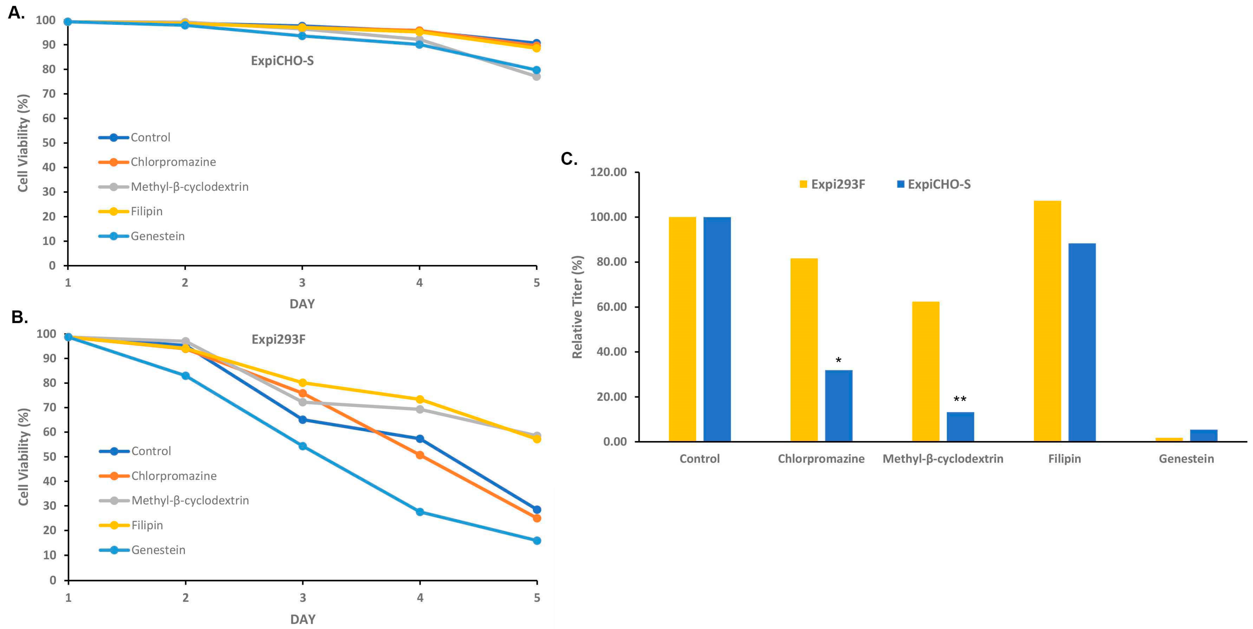
3.5. Expi293FTM and ExpiCHO-STM Cells Responded Differently to the Endocytosis Blockers

Different complexation time preferences by Expi293FTM and ExpiCHO-STM, as well as the different size changes in the complexes with ExpiFectamine™CHO and ExpiFectamine™293, imply that Expi293FTM and ExpiCHO-STM cells might utilize different DNA uptake pathways. To investigate how the endocytosis blockers affect the TGE protein productivity in these two cell lines, a pharmacological experiment was performed. The Expi293FTM and ExpiCHO-STM cells were pretreated with endocytic pathway inhibitors and then transfected with plasmid DNAs encoding antibody-A three hours later. Four types of endocytosis inhibitors were tested: 100 µM chlorpromazine which is known to disrupt clathrin assembly and block the clathrin-dependent endocytosis [31], 1 mM methyl-β-cyclodextrin and 5 µg/mL Filipin that are inhibitors of caveolae formation through cholesterol binding and depletion [32,33], and 200 µM Genestein, a tyrosine kinase inhibitor that can also inhibit caveolar uptake [34,35]. As shown in Figure 5A, ExpiCHO-STM cells maintained high viability after the treatments of these inhibitors, whereas the cell viability of Expi293FTM decreased over time in all conditions (Figure 5B). Interestingly, protein expression in ExpiCHO-STM was drastically inhibited by the treatments of methyl-β-cyclodextrin and Genestein, substantially by that of chlorpromazine (Figure 5C). This suggests that both the caveolae and clathrin-dependent pathways might be involved in the DNA complex uptake in ExpiCHO-STM cells. In Expi293FTM cells, antibody expression was completely eliminated by the treatment of Genestein, but not affected by other treatments, suggesting that the caveolae pathway is the major route in Expi293FTM cells. These data together indicate that Expi293FTM and ExpiCHO-STM cells respond differently to the endocytosis blockers for the TGE process.

3.6. Expi293FTM and ExpiCHO-STM Cells Responded Differently to the Cotransfection of Cell Cycle Inhibitor p21 and p27

Inducing controlled proliferation is a strategy for improving protein yield, and co-transfection with cell cycle inhibitors p21/p27 has been previously reported to enhance TGE in HEK293 and CHO-K1 cells [27,36,37]. We sought to determine whether or not this cotransfection strategy can improve TGE efficiency in Expi293FTM and ExpiCHO-STM cells. As shown in Figure 6, antibodies-A, B, and C along with three different ratios of p21:p27 (5:1, 1:1, 1:5) have been tested. At a 1:1 ratio, p21:p27 enhanced the expression of antibody-A in Expi293FTM by 50% and in ExpiCHO-STM by 2.5 folds. It also enhanced antibody-C expression in Expi293FTM cells by 40%, and in ExpiCHO-STM antibody-B by 2 folds and antibody-C by 1.5 fold. At a 5:1 ratio, antibody-C expression was enhanced in Expi293FTM by 20%, and antibody-A expression was enhanced in ExpiCHO-STM by 2 folds. At 1:5 ratio, expression of antibody-A and antibody-B were enhanced in ExpiCHO-STM by 40% and 60%, respectively, while no enhancement effect was detected in Expi293FTM cells. These results together suggest that ExpiCHO-STM seemed to respond better than Expi293FTM to the cotransfection of p21/p27 when transiently producing various antibodies.

3.7. DMSO Could Enhance More Transient Expression in ExpiCHO-STM Cells Than in Expi293FTM Cells as the Concentration Increased

Adding expression enhancers during cell culture is an important step for the TGE processes. We tested several reported chemicals in the Expi293FTM and ExpiCHO-STM systems. The addition of lithium acetate (LiOAc) was reported to enhance transient expression in transient CHO cells [10]. Adding potent CDK4/6 inhibitors such as Palbociclib [38] to the cell culture did not result in a meaningful expression titer enhancement at the concentrations of 1–40 µM . However, when the DMSO effect [10,39] was explored in the Expi293FTM and ExpiCHO-STM cells, significant beneficial results were detected in ExpiCHO-STM cells but not in Expi293FTM cells. As shown in Figure 7, 0.2–2% of DMSO, added to the cell culture three hours before the transfection, significantly increased expression in ExpiCHO-STM cells, with the highest effect seen in the concentration of 2%. The enhancement effect was detected in both ExpiFectamine™CHO- and PEI-mediated transfections, with a bigger improvement with ExpiFectamine™CHO. For Expi293FTM cells, 0.75% DMSO enhanced the PEI-mediated expression of antibody-A, with less effect at higher concentrations. For the ExpiFectamine™293-mediated transfection, no enhancement was detected. These data indicate that ExpiCHO-STM cells responded better to the expression-enhancing DMSO activity than Expi293FTM cells.

3.8. Expression Preferences for Kappa and Lambda Light Chain Isotype Were Detected in Expi293FTM and ExpiCHO-STM Cells

Antibodies contain two types of light chains, kappa and lambda, while their ratios in animals vary. Therapeutic antibody hits could come from different animal sources. For humans, the ratio of kappa and lambda is between 1.7 and 2 with more lambda during early infancy and old age [40]. Murine has a ratio of about 17 [41], whereas the ratios in animals like dogs, horses, and cattle are around 0.1 [42]. There is no report about whether or not Expi293FTM and ExpiCHO-STM cells express differently the antibodies with different light chain isotypes. In this study, when a set of human antibodies sharing a common light chain (CLC antibodies Kappa A-C and CLC antibodies Lambda D-F) were produced in Expi293FTM and ExpiCHO-STM cells, we found ExpiCHO-STM cells produced reasonably good expression titers to the antibodies with a common kappa light chain (CLC antibodies Kappa A–C) but very poorly to the antibodies with a common lambda light chain (CLC antibodies Lambda D–F, Figure 8A). In contrast, Expi293FTM cells produced reasonably well the same set of antibodies with either lambda or kappa light chains (Figure 8A).
To further confirm that there is indeed a kappa light chain expression bias for ExpiCHO-STM cells, CLC antibody-E with one heavy chain, one kappa light chain, and one lambda light chain were co-transfected into either ExpiCHO-STM or Expi293FTM, respectively. Consistently, the titer in Expi293FTM was significantly higher than that in ExpiCHO-STM (Figure 8B). The purified antibody protein was then analyzed by mass spectrometry. As shown in Figure 8B–D, Expi293FTM-derived CLC antibody-E contained a similar amount of kappa and lambda light chains, whereas ExpiCHO-STM-derived CLC antibody-E only contained kappa light chain with almost no detectable lambda light chain. These results indicate that ExpiCHO-STM cells can preferably express certain antibodies with kappa light chains over those with lambda light chain, while Expi293FTM cells seems producing similarly well the same set of antibodies with either light chain isotypes.

4. Discussion

The TGE technologies in mammalian cells are a powerful tool for the rapid generation of many high-quality therapeutic hits supplied from the ever-evolving antibody discovery platforms, such as display technologies, hybridoma, and single-cell technologies, as well as computational and rational engineering. Yet, the TGE technologies still face major challenges like low production yield and batch-to-batch variation. To improve the TGE production yield and robustness, this study has examined the TGE production processes by investigating the parametric and mechanistic differences between the commercial cell host Expi293FTM and ExpiCHO-STM. While both systems could be optimized by increasing the volume for the complex dilution medium, ExpiCHO-STM appeared to be more sensitive to the subtle changes in the DNA complex formation process and preferred a much shorter complexation time than Expi293FTM. Also, ExpiCHO-STM was found to be more responsive to the changes in buffers and DNA-to-PEI ratios as well as the treatments of DMSO and cell cycle inhibitors. In addition, this study has revealed that for certain antibodies with common light chains, ExpiCHO-STM exhibited an expression preference towards those with kappa light-chain isotype over the lambda isotype during the TGE production, whereas Expi293FTM did not seem to have such a preference with the same set of antibodies tested. These interesting observations might reflect the mechanistic differences between the TGE processes in Expi293FTM and ExpiCHO-STM, providing a future direction for optimization.
Several interesting findings from the optimization efforts of this study have been unexpectedly centering on the DNA complex formation step. Firstly, the dilution effects observed on both cationic lipid ExpiFectamineTM and cationic polymer PEI appeared to be counterintuitive. The common sense is that the higher concentration of the plasmid DNAs and the transfection reagents should result from a more efficient transfection and therefore produce a higher production yield. Nonetheless, this study revealed that a further dilution actually gave much-improved expression titers. More dilutions on DNA and transfection reagents imply that smaller sizes of the DNA complexes are formed and preferred by the TGE processes. Secondly, Expi293FTM and ExpiCHO-STM were found to differ significantly in the DNA complex formation time. ExpiCHO-STM preferred a much shorter incubation time than Expi293FTM, indicating that ExpiCHO-STM uptakes more efficiently a smaller size of the DNA complexes than Expi293FTM. This observation is consistent with the finding that ExpiCHO-STM and Expi293FTM responded differently to the treatments of endocytosis inhibitors. It has been reported that the particle sizes for the caveolae pathways and the clathrin-dependent pathways are different [34,43].
This study confirms the beneficial effects of DMSO, whereas published potential enhancer LiOAc showed no expression improvement, likely due to the different host systems utilized by the study [25]. DMSO is a small molecule with polar, aprotic, and amphiphilic properties, which has a diverse impact on cells. Low-dose DMSO (0.1–1.5%) treatment can alter membrane lipids and increase membrane permeability [44,45]. Its impacts on DNA conformation, nucleic acid content, protein beta-sheet structure, and cell cycle progression have been reported [45,46]. DMSO can also serve as a chemical chaperon [25,47], and increase cellular mRNA level [39]. The enhancement effects of low-dose DMSO on the transient production in Expi293FTM and ExpiCHO-STM could be attributed to these above-mentioned mechanisms. Interestingly, this study further uncovered that the ExpiCHO-STM cells were more responsive to the DMSO treatment than the Expi293FTM cells, suggesting that this effect could be cell-type specific.
This study also validated the beneficial effects of co-transfecting cell cycle inhibitors on the transient production of certain antibodies. P21/p27 are cyclin-dependent kinase inhibitors that regulate cell cycle progression at the G1/S phase [48], and their expression booster effects in FreeStyleTM HEK293F systems were reported previously [27,37]. Our studies showed that co-transfecting p21/p27 at the ratio of 1:1 was significantly better than the published optimized ratio of 1:5 in the Expi293FTM while both ratios worked well in the ExpiCHO-STM system. Inducing cell growth arrest is a common strategy for improving recombinant protein production, which also includes mild hypothermia by temperature shift [49,50], the addition of histone deacetylase inhibitor (HDACi) [51], and cell cycle kinase chemical inhibitors [52,53]. Since HDACi valproic acid is routinely employed by the Expi293FTM workflow and the 32 °C temperature shift is in the ExpiCHO-STM workflow, the further enhancement by co-transfecting p21/p27 indicates a synergistic and compatibility effect among these strategies.
It is known that increasing PEI concentration to a peak can improve protein expression but decline afterward. In this study, while the best PEI/DNA ratio was 8 for Expi293FTM and 3.5 for ExpiCHO-STM, the final PEI concentrations for both cell lines were actually similar (8 mg/L and 7 mg/L) due to the twice amounts of DNAs used for ExpiCHO-STM. This PEI concentration range might represent the most tolerated PEI concentration for both cell lines. Increasing PEI concentration can cause cellular toxicities which include endosome membrane rupture and mitochondria dysfunction [54]. This study further revealed that ExpiCHO-STM seemed to be more sensitive to free PEI than Expi293FTM, as its expression titer dropped more sharply when the PEI concentration increased. A larger size of the PEI/DNA complex could also be formed when the PEI/DNA ratio increases. This is consistent with the hypothesis that ExpiCHO-STM cells prefer a smaller complex size as the expression titer decreased drastically when the complex formation time was over 2.5 min. Expi293FTM might prefer a larger complex size as its expression peaked in 15 min and remained constant for up to 30 min.
Antibody production is known to be affected by factors such as HC:LC ratios [55], variable regions [56], and constant regions [57] of HC and LC. One surprising finding from this study is that ExpiCHO-STM seems to express much better the Kappa light chain than the lambda isotype for a few antibodies, whereas Expi293FTM has a similar expression preference towards both isotypes. Interestingly, this observation correlates well with the fact that CHO is rodent derived which has much more kappa isotype (95% vs. 5%) than human (60% vs. 40%). Even though CHO does express well many lambda-containing antibodies and variable regions of the light chains also play a role in its expression, there might be species-specific factors that can facilitate the expression of the light-chain isotype. Recently, it was reported that the lambda isotype, but not the kappa light chain, can drastically stimulate the expression of IgMs and IgA2 in Expi293FTM cells [58]. This enhancement does not affect the expression of IgG1 and IgA1. Our data reveal for the first time that CHO cells have an expression preference towards the kappa isotype.
The TGE process in mammalian cells is more complex than expected, as a recent system biology study on transcriptomic analysis of CHO and HEK293 during transient production has revealed their profound differences in the secretary pathway utilization [59]. The findings from this study further support the notion that the TGE processes in CHO and HEK293 might employ additional mechanisms which may include the uptake pathways for the DNA complexes and the expression preference for antibody light-chain isotype. Process improvements through understanding and exploiting these differences need to be evaluated and implemented. With improved titers in the TGE processes, more antibody hits can be generated with a lower culture volume. This should enable a faster, more effective, and economical way to triage new therapeutics designs churning out from machine learning and artificial intelligence, along with the conventional antibody discovery platforms.

5. Conclusions

This study discovered the parametric and mechanistic differences in Expi293FTM and ExpiCHO-STM cells with regard to the TGE processes. The resulting findings generated a further-optimized workflow and parameters (Table 1) for rapid generating antibody candidates, as well as a new direction for improvement.

Author Contributions

Conceptualization, J.Z., A.M.D. and X.Z.; methodology, J.Z., G.G.Y., D.C., C.M., M.R., R.S., A.S.T., S.L., C.P., L.L., A.M.D. and X.Z.; formal analysis, J.Z., G.G.Y., D.C., C.M., M.R., R.S., A.S.T., S.L., C.P., L.L., A.M.D. and X.Z.; investigation, J.Z., G.G.Y., D.C., C.M., M.R., R.S., A.S.T., S.L., C.P., L.L., A.M.D. and X.Z.; resources, L.L., A.M.D. and X.Z.; writing—original draft preparation, J.Z., G.G.Y., M.R., R.S., A.M.D. and X.Z.; writing—review and editing, J.Z., G.G.Y., M.R., R.S., A.M.D. and X.Z.; supervision, A.M.D. and X.Z.; project administration, A.M.D. and X.Z.; funding acquisition, L.L. All authors have read and agreed to the published version of the manuscript.

Funding

This research received no external funding.

Institutional Review Board Statement

Not applicable.

Informed Consent Statement

Not applicable.

Data Availability Statement

Data is contained within the article.

Acknowledgments

The authors would like to thank Molica Abel, Eric Sousa, and Will Somers for the discussion.

Conflicts of Interest

All authors are employed by Pfizer research. All authors declare that the research was conducted in the absence of any commercial or financial relationships that could be construed as a potential conflict of interest.

References

  1. Urquhart, L. Top product forecasts for 2022. Nat. Rev. Drug Discov. 2022, 21, 11. [Google Scholar] [CrossRef]
  2. Pham, P.L.; Kamen, A.; Durocher, Y. Large-scale transfection of mammalian cells for the fast production of recombinant protein. Mol. Biotechnol. 2006, 34, 225–237. [Google Scholar] [CrossRef]
  3. Baldi, L.; Hacker, D.L.; Adam, M.; Wurm, F.M. Recombinant protein production by large-scale transient gene expression in mammalian cells: State of the art and future perspectives. Biotechnol. Lett. 2007, 29, 677–684. [Google Scholar] [PubMed] [Green Version]
  4. Geisse, S.; Voedisch, B. Transient expression technologies: Past, present, and future. Methods Mol. Biol. 2012, 899, 203–219. [Google Scholar]
  5. Gutiérrez-Granados, S.; Cervera, L.; Kamen, A.A.; Gòdia, F. Advancements in mammalian cell transient gene expression (TGE) technology for accelerated production of biologics. Crit. Rev. Biotechnol. 2018, 38, 918–940. [Google Scholar] [CrossRef]
  6. Jager, V.; Bussow, K.; Schirrmann, T. Transient Recombinant Protein Expression in Mammalian Cells. In Animal Cell Culture; Cell Engineering; Al-Rubeai, M., Ed.; Springer Interantional Publishing: Cham, Switzerland, 2015; Volume 9, pp. 27–64. [Google Scholar]
  7. Backliwal, G.; Hildinger, M.; Chenuet, S.; Wulhfard, S.; De Jesus, M.; Wurm, F.M. Rational vector design and multi-pathway modulation of HEK 293E cells yield recombinant antibody titers exceeding 1 g/L by transient transfection under serum-free conditions. Nucleic Acids Res. 2008, 36, e96. [Google Scholar] [CrossRef] [Green Version]
  8. Jain, N.K.; Barkowski-Clark, S.; Altman, R.; Johnson, K.; Sun, F.; Zmuda, J.; Liu, C.Y.; Kita, A.; Schulz, R.; Neill, A.; et al. A high density CHO-S transient transfection system: Comparison of ExpiCHO and Expi293. Protein Expr. Purif. 2017, 134, 38–46. [Google Scholar]
  9. Liu, C.; Dalby, B.; Chen, W.; Kilzer, J.M.; Chiou, H.C. Transient transfection factors for high-level recombinant protein production in suspension cultured mammalian cells. Mol. Biotechnol. 2008, 39, 141–153. [Google Scholar] [CrossRef]
  10. Ye, J.; Kober, V.; Tellers, M.; Naji, Z.; Salmon, P.; Markusen, J.F. High-level protein expression in scalable CHO transient transfection. Biotechnol. Bioeng. 2009, 103, 542–551. [Google Scholar] [CrossRef] [PubMed]
  11. Daramola, O.; Stevenson, J.; Dean, G.; Hatton, D.; Pettman, G.; Holmes, W.; Field, R. A high-yielding CHO transient system: Coexpression of genes encoding EBNA-1 and GS enhances transient protein expression. Biotechnol. Prog. 2014, 30, 132–141. [Google Scholar] [PubMed]
  12. Rajendra, Y.; Hougland, M.D.; Alam, R.; Morehead, T.A.; Barnard, G.C. A high cell density transient transfection system for therapeutic protein expression based on a CHO GS-knockout cell line: Process development and product quality assessment. Biotechnol. Bioeng. 2015, 112, 977–986. [Google Scholar] [PubMed]
  13. Zhong, X.; Ma, W.; Meade, C.L.; Tam, A.S.; Llewellyn, E.; Cornell, R.; Cote, K.; Scarcelli, J.J.; Marshall, J.K.; Tzvetkova, B.; et al. Transient CHO expression platform for robust antibody production and its enhanced N-glycan sialylation on therapeutic glycoproteins. Biotechnol. Prog. 2019, 35, e2724. [Google Scholar] [CrossRef] [PubMed] [Green Version]
  14. Stuible, M.; Burlacu, A.; Perret, S.; Brochu, D.; Paul-Roc, B.; Baardsnes, J.; Loignon, M.; Grazzini, E.; Durocher, Y. Optimization of a high-cell-density polyethylenimine transfection method for rapid protein production in CHO-EBNA1 cells. J. Biotechnol. 2018, 281, 39–47. [Google Scholar] [CrossRef] [PubMed]
  15. Schmitt, M.G.; White, R.N.; Barnard, G.C. Development of a high cell density transient CHO platform yielding mAb titers greater than 2 g/L in only 7 days. Biotechnol. Prog. 2020, 36, e3047. [Google Scholar]
  16. Rodriguez-Conde, S.; Inman, S.; Lindo, V.; Amery, L.; Tang, A.; Okorji-Obike, U.; Du, W.; Bosch, B.-J.; Schreur, P.J.W.; Kortekaas, J.; et al. Suitability of transiently expressed antibodies for clinical studies: Product quality consistency at different production scales. mAbs 2022, 14. [Google Scholar]
  17. Steger, K.; Brady, J.; Wang, W.; Duskin, M.; Donato, K.; Peshwa, M. CHO-S antibody titers >1 gram/liter using flow electroporation-mediated transient gene expression followed by rapid migration to high-yield stable cell lines. J. Biomol. Screen. 2015, 20, 545–551. [Google Scholar]
  18. Zhong, X.; D’Antona, A.M. Recent Advances in the Molecular Design and Applications of Multispecific Biotherapeutics. Antibodies 2021, 10, 13. [Google Scholar] [CrossRef]
  19. Brinkmann, U.; Kontermann, R.E. The making of bispecific antibodies. mAbs 2017, 9, 182–212. [Google Scholar] [CrossRef] [Green Version]
  20. Labrijn, A.F.; Janmaat, M.L.; Reichert, J.M.; Parren, P.W.H.I. Bispecific antibodies: A mechanistic review of the pipeline. Nat. Rev. Drug Discov. 2019, 18, 585–608. [Google Scholar]
  21. Chiou, H.C.; Vasu, S.; Liu, C.Y.; Cisneros, I.; Jones, M.B.; Zmuda, J.F. Scalable transient protein expression. Methods Mol. Biol. 2014, 1104, 35–55. [Google Scholar]
  22. Liu, C.Y.; Spencer, V.; Kumar, S.; Liu, J.; Chiou, H.; Zmuda, J.F. Attaining high transient titers in CHO cells: Case study involving the use of the ExpiCHOTM mammalian transient expression system. Genet. Eng. Biotechnol. News 2015, 35, 34–35. [Google Scholar] [CrossRef]
  23. Zhong, X.; Schenk, J.; Sakorafas, P.; Chamberland, J.; Tam, A.; Thomas, L.M.; Yan, G.; D’Antona, A.M.; Lin, L.; Nocula-Lugowska, M.; et al. Impacts of fast production of afucosylated antibodies and Fc mutants in ExpiCHO-S for enhancing FcgammaRIIIa binding and NK cell activation. J. Biotechnol. 2022, 360, 79–91. [Google Scholar] [CrossRef]
  24. Heng, Z.S.-L.; Yeo, J.Y.; Koh, D.W.-S.; Gan, S.K.-E.; Ling, W.-L. Augmenting recombinant antibody production in HEK293E cells: Optimizing transfection and culture parameters. Antib. Ther. 2022, 5, 30–41. [Google Scholar]
  25. Johari, Y.B.; Estes, S.D.; Alves, C.S.; Sinacore, M.S.; James, D.C. Integrated cell and process engineering for improved transient production of a “difficult-to-express” fusion protein by CHO cells. Biotechnol. Bioeng. 2015, 112, 2527–2542. [Google Scholar] [CrossRef] [PubMed]
  26. Rajendra, Y.; Hougland, M.D.; Schmitt, M.G.; Barnard, G.C. Transcriptional and post-transcriptional targeting for enhanced transient gene expression in CHO cells. Biotechnol. Lett. 2015, 37, 2379–2386. [Google Scholar] [CrossRef] [PubMed]
  27. Kiszel, P.; Fiesel, S.; Voit, S.; Waechtler, B.; Meier, T.; Oelschlaegel, T.; Schraeml, M.; Engel, A.M. Transient gene expression using valproic acid in combination with co-transfection of SV40 large T antigen and human p21(CIP)/p27(KIP). Biotechnol. Prog. 2019, 35, e2786. [Google Scholar] [CrossRef] [PubMed]
  28. Pham, P.L.; Perret, S.; Cass, B.; Carpentier, E.; St-Laurent, G.; Bisson, L.; Kamen, A.; Durocher, Y. Transient gene expression in HEK293 cells: Peptone addition posttransfection improves recombinant protein synthesis. Biotechnol. Bioeng. 2005, 90, 332–344. [Google Scholar]
  29. Zhong, X.; Schwab, A.; Ma, W.; Meade, C.L.; Zhou, J.; D’antona, A.M.; Somers, W.; Lin, L. Large-Scale Transient Production in ExpiCHO-S with Enhanced N-Galactosylation-Sialylation and PEI-Based Transfection. Methods Mol. Biol. 2022, 2313, 143–150. [Google Scholar]
  30. Nomani, A.; Hyvönen, Z.; Pulkkinen, E.; Hiekkala, M.; Ruponen, M. Intracellular gene delivery is dependent on the type of non-viral carrier and defined by the cell surface glycosaminoglycans. J. Control Release 2014, 187, 59–65. [Google Scholar] [CrossRef]
  31. Wang, L.H.; Rothberg, K.G.; Anderson, R.G. Mis-assembly of clathrin lattices on endosomes reveals a regulatory switch for coated pit formation. J. Cell Biol. 1993, 123, 1107–1117. [Google Scholar] [CrossRef]
  32. Lamaze, C.; Schmid, S.L. The emergence of clathrin-independent pinocytic pathways. Curr. Opin. Cell Biol. 1995, 7, 573–580. [Google Scholar] [CrossRef] [PubMed]
  33. Parton, R.G.; Richards, A.A. Lipid rafts and caveolae as portals for endocytosis: New insights and common mechanisms. Traffic 2003, 4, 724–738. [Google Scholar] [PubMed] [Green Version]
  34. Khalil, I.A.; Kogure, K.; Akita, H.; Harashima, H. Uptake pathways and subsequent intracellular trafficking in nonviral gene delivery. Pharmacol. Rev. 2006, 58, 32–45. [Google Scholar] [CrossRef] [Green Version]
  35. Tiruppathi, C.; Song, W.; Bergenfeldt, M.; Sass, P.; Malik, A.B. Gp60 activation mediates albumin transcytosis in endothelial cells by tyrosine kinase-dependent pathway. J. Biol. Chem. 1997, 272, 25968–25975. [Google Scholar] [CrossRef] [PubMed] [Green Version]
  36. Coqueret, O. New roles for p21 and p27 cell-cycle inhibitors: A function for each cell compartment? Trends Cell Biol. 2003, 13, 65–70. [Google Scholar] [PubMed]
  37. Vink, T.; Oudshoorn-Dickmann, M.; Roza, M.; Reitsma, J.J.; de Jong, R.N. A simple, robust and highly efficient transient expression system for producing antibodies. Methods 2014, 65, 5–10. [Google Scholar] [CrossRef]
  38. Toogood, P.L.; Harvey, P.J.; Repine, J.T.; Sheehan, D.J.; VanderWel, S.N.; Zhou, H.; Keller, P.R.; McNamara, D.J.; Sherry, D.; Zhu, T.; et al. Discovery of a potent and selective inhibitor of cyclin-dependent kinase 4/6. J. Med. Chem. 2005, 48, 2388–2406. [Google Scholar] [CrossRef]
  39. Rajendra, Y.; Balasubramanian, S.; Kiseljak, D.; Baldi, L.; Wurm, F.M.; Hacker, D.L. Enhanced plasmid DNA utilization in transiently transfected CHO-DG44 cells in the presence of polar solvents. Biotechnol. Prog. 2015, 31, 1571–1578. [Google Scholar] [CrossRef]
  40. Molé, C.M.; Béné, M.C.; Montagne, P.M.; Seilles, E.; Faure, G.C. Light chains of immunoglobulins in human secretions. Clin. Chim. Acta 1994, 224, 191–197. [Google Scholar] [CrossRef]
  41. Popov, A.V.; Zou, X.; Xian, J.; Nicholson, I.C.; Brüggemann, M. A human immunoglobulin lambda locus is similarly well expressed in mice and humans. J. Exp. Med. 1999, 189, 1611–1620. [Google Scholar] [CrossRef]
  42. Arun, S.S.; Breuer, W.; Hermanns, W. Immunohistochemical examination of light-chain expression (lambda/kappa ratio) in canine, feline, equine, bovine and porcine plasma cells. Zentralbl Vet. A 1996, 43, 573–576. [Google Scholar]
  43. Conner, S.D.; Schmid, S.L. Regulated portals of entry into the cell. Nature 2003, 422, 37–44. [Google Scholar] [CrossRef]
  44. de Ménorval, M.A.; Mir, L.M.; Fernández, M.L.; Reigada, R. Effects of dimethyl sulfoxide in cholesterol-containing lipid membranes: A comparative study of experiments in silico and with cells. PLoS ONE 2012, 7, e41733. [Google Scholar]
  45. Tunçer, S.; Gurbanov, R.; Sheraj, I.; Solel, E.; Esenturk, O.; Banerjee, S. Low dose dimethyl sulfoxide driven gross molecular changes have the potential to interfere with various cellular processes. Sci. Rep. 2018, 8, 14828. [Google Scholar]
  46. Liu, C.H.; Chen, L.H. Promotion of recombinant macrophage colony stimulating factor production by dimethyl sulfoxide addition in Chinese hamster ovary cells. J. Biosci. Bioeng. 2007, 103, 45–49. [Google Scholar] [CrossRef] [PubMed]
  47. Hwang, S.J.; Jeon, C.J.; Cho, S.M.; Lee, G.M.; Yoon, S.K. Effect of chemical chaperone addition on production and aggregation of recombinant flag-tagged COMP-angiopoietin 1 in Chinese hamster ovary cells. Biotechnol. Prog. 2011, 27, 587–591. [Google Scholar] [CrossRef]
  48. Lim, S.; Kaldis, P. Cdks, cyclins and CKIs: Roles beyond cell cycle regulation. Development 2013, 140, 3079–3093. [Google Scholar] [PubMed] [Green Version]
  49. Fox, S.R.; Patel, U.A.; Yap, M.G.; Wang, D.I. Maximizing interferon-gamma production by Chinese hamster ovary cells through temperature shift optimization: Experimental and modeling. Biotechnol. Bioeng. 2004, 85, 177–184. [Google Scholar] [PubMed]
  50. Kaufmann, H.; Mazur, X.; Fussenegger, M.; Bailey, J.E. Influence of low temperature on productivity, proteome and protein phosphorylation of CHO cells. Biotechnol. Bioeng. 1999, 63, 573–582. [Google Scholar] [CrossRef]
  51. Hao, C.; Liu, P.; Zhao, L.; Zhu, C.; Tian, X.; Wang, L.; Zhang, Z. Inhibition of leukemic cells by valproic acid, an HDAC inhibitor, in xenograft tumors. Onco Targets Ther. 2013, 6, 733–740. [Google Scholar] [CrossRef] [Green Version]
  52. Meyer, H.J.; Turincio, R.; Ng, S.; Li, J.; Wilson, B.; Chan, P.; Zak, M.; Reilly, D.; Beresini, M.H.; Wong, A.W. High throughput screening identifies novel, cell cycle-arresting small molecule enhancers of transient protein expression. Biotechnol. Prog. 2017, 33, 1579–1588. [Google Scholar] [CrossRef]
  53. Du, Z.; Treiber, D.; McCarter, J.D.; Fomina-Yadlin, D.; Saleem, R.A.; McCoy, R.E.; Zhang, Y.; Tharmalingam, T.; Leith, M.; Follstad, B.D.; et al. Use of a small molecule cell cycle inhibitor to control cell growth and improve specific productivity and product quality of recombinant proteins in CHO cell cultures. Biotechnol. Bioeng. 2015, 112, 141–155. [Google Scholar] [CrossRef]
  54. Moghimi, S.M.; Symonds, P.; Murray, J.C.; Hunter, A.; Debska, G.; Szewczyk, A.C. A two-stage poly(ethylenimine)-mediated cytotoxicity: Implications for gene transfer/therapy. Mol. Ther. 2005, 11, 990–995. [Google Scholar]
  55. Schlatter, S.; Stansfield, S.H.; Dinnis, D.M.; Racher, A.J.; Birch, J.R.; James, D.C. On the optimal ratio of heavy to light chain genes for efficient recombinant antibody production by CHO cells. Biotechnol. Prog. 2005, 21, 122–133. [Google Scholar]
  56. Ling, W.L.; Lua, W.H.; Poh, J.J.; Yeo, J.Y.; Lane, D.P.; Gan, S.K.E. Effect of VH-VL Families in Pertuzumab and Trastuzumab Recombinant Production, Her2 and FcgammaIIA Binding. Front. Immunol. 2018, 9, 469. [Google Scholar] [CrossRef] [PubMed] [Green Version]
  57. Lua, W.H.; Ling, W.L.; Yeo, J.Y.; Poh, J.J.; Lane, D.P.; Gan, S.K.E. The effects of Antibody Engineering CH and CL in Trastuzumab and Pertuzumab recombinant models: Impact on antibody production and antigen-binding. Sci. Rep. 2018, 8, 718. [Google Scholar] [PubMed] [Green Version]
  58. Gong, S.; Gautam, S.; Coneglio, J.D.; Scinto, H.B.; Ruprecht, R.M. Antibody Light Chains: Key to Increased Monoclonal Antibody Yields in Expi293 Cells? Antibodies 2022, 11, 37. [Google Scholar]
  59. Malm, M.; Kuo, C.-C.; Barzadd, M.M.; Mebrahtu, A.; Wistbacka, N.; Razavi, R.; Volk, A.-L.; Lundqvist, M.; Kotol, D.; Tegel, H.; et al. Harnessing secretory pathway differences between HEK293 and CHO to rescue production of difficult to express proteins. Metab. Eng. 2022, 72, 171–187. [Google Scholar] [CrossRef] [PubMed]
Figure 1. Optimizing the dilution buffer processes for the DNA complexation with ExpiFectamine™ and PEI for Expi293FTM and ExpiCHO-STM. As described in Section 2.4, the target DNAs and the transfection reagents were diluted separately in different volumes of Opti-MEMTM (mL/L cell culture volume per tube); prior to the mixing of the two components for the complexation formation. For Expi293FTM, the expression titers were determined five days post-transfection. For ExpiCHO-STM, the expression titers were measured seven days post-transfection. (A) The Opti-MEMTM medium dilution conditions for the DNA complexation with ExpiFectamine™ 293 and PEI (*, p < 0.05) in Expi293FTM for antibody-A. The titers obtained under the conditions of 10 mL/L per tube were set as 100%. (B) The Opti-MEMTM medium dilution conditions for the DNA complexation with ExpiFectamine™ CHO (**, p < 0.05) and PEI in ExpiCHO-STM for antibody-A. The titers obtained under the conditions of 10 mL/L per tube were set as 100%. (C) The pH effects of the dilution buffers for the DNA complexation with ExpiFectamine™ 293 (#, p < 0.05) and PEI (#, p < 0.05) in Expi293FTM for antibody-A. The Opti-MEMTM medium and other indicated dilution buffers in different pHs were used for diluting the DNAs and the transfection agents in 100 mL/L per tube. The titers obtained under the conditions of Opti-MEMTM medium were set as 100%. (D) The pH effects of the dilution buffers for the DNA complexation with ExpiFectamine™-CHO (&, p < 0.05) and PEI (&, p < 0.05) in ExpiCHO-STM for antibody-A. The titers obtained under the conditions of Opti-MEMTM medium were set as 100%.
Antibodies 12 00053 g001
Figure 2. Optimizing the ratios between PEI and DNA in Expi293FTM and ExpiCHO-STM. As described in Materials & Methods, the target DNAs encoding antibody-A were incubated with PEI in various ratios and transfected into Expi293FTM and ExpiCHO-STM cells. For Expi293FTM, the expression titers were determined five days post-transfection. For ExpiCHO-STM, the expression titers were measured seven days post-transfection. The titers obtained for those with ExpiFectamine™ were set as 100% (n = 4 ± S.D., ##, p < 0.05)).
Antibodies 12 00053 g002
Figure 3. Optimizing the DNA complex formation time in Expi293FTM and ExpiCHO-STM with ExpiFectamine™ and PEI. As described in Section 2.4, the target DNAs encoding antibody-A were incubated with either ExpiFectamine™ (Panel A) or PEI (Panel B) for various time points (−0.5 min: the transfection reagents and the DNAs were directly added into the cell culture without mixing; 0 min: the transfection reagents and the DNAs were mixed, but without incubation prior to the addition to cell culture; incubation time after mixing: 0.5 min, 1 min, 2.5 min, 5 min, 10 min, 15 min, and 30 min), and transfected into Expi293FTM and ExpiCHO-STM cells. The highest titers with each transfection reagent in each cell host were set as 100% (n = 3 ± S.D., ***, p < 0.05).
Antibodies 12 00053 g003
Figure 4. Size measurement of the DNA: ExpiFectamine™CHO or PEI complex with DLS. DLS measurements for size determination were described in Section 2.8. ExpiFectamine™CHO alone, or PEI alone, or in complex with the target DNAs encoding antibody-A (DNA:PEI = 1:3.5; DNA: ExpiFectamine™CHO = 1 µg:3.2 µL) performed in Opti-MEMTM medium (#, p < 0.05, ##, p < 0.05).
Antibodies 12 00053 g004
Figure 5. Expi293FTM and ExpiCHO-STM cells responded differently to the endocytosis blockers. As described in Section 2.4, various endocytosis blockers were added to the cell culture of Expi293FTM and ExpiCHO-STM prior to the transfection of the target DNAs encoding antibody-A complexed with ExpiFectamine™. Cell viability in ExpiCHO-STM (pane A), and Expi293FTM (panel B) as well as expression titers (panel C) were determined. The titers for the mock-treated control in each cell host were set as 100% (*, p < 0.05, **, p < 0.05).
Antibodies 12 00053 g005
Figure 6. Co-transfecting the genes encoding cell cycle inhibitor p21 and p27 enhanced transient expression in Expi293FTM and ExpiCHO-STM cells, with a bigger effect in ExpiCHO-STM. As described in Section 2.4, the target DNAs encoding antibody-A, antibody-B, and antibody C complexed with ExpiFectamine™ along with different amounts of plasmid DNAs encoding p21/p27 were transfected into Expi293FTM (Panel A) and ExpiCHO-STM (Panel B) cells. The control titers without p21/p27 DNAs were set as 100% (n = 3 ± S.D, #, p < 0.05).
Antibodies 12 00053 g006
Figure 7. DMSO with increased concentrations could enhance more transient expression in ExpiCHO-STM cells than in Expi293FTM cells. As described in Section 2.4, Expi293FTM (Panel A) and ExpiCHO-STM (Panel B) cells were pretreated with different concentrations of DMSO prior to the DNA transfection for antibody-A. The control titers without DMSO treatment were set as 100% (n = 3 ± S.D., *, p < 0.05, #, p < 0.05).
Antibodies 12 00053 g007
Figure 8. Expression preferences for kappa and lambda light chain isotypes were detected in Expi293FTM and ExpiCHO-STM. As described in Section 2.4, the target DNAs encoding common light chain (CLC) antibodies A, B, and C with a kappa light chain or CLC antibody-D, E, and F with a lambda light chain were transfected into either Expi293FTM or ExpiCHO-STM cells. (A) Expi293FTM produced high titers for CLC antibodies A-C with kappa light chain and for CLC antibodies D-F with a lambda light chain, yet ExpiCHO-STM only expressed well for CLC antibodies A-C with kappa light chain (#, p < 0.05). (B) Co-transfecting heavy chain (HC) of CLC antibody E with both kappa (LCK) and lambda (LCL) light chain into either Expi293FTM or ExpiCHO-STM cells resulted in 1:1 kappa/lambda expression ratio in Expi293FTM cells but 99:1 expression ratio in ExpiCHO-STM cells. (C) Mass spectrometry analysis of the kappa and lambda co-transfection in Expi293FTM cells. (D) Mass spectrometry analysis of the kappa and lambda co-transfection in ExpiCHO-STM cells.
Antibodies 12 00053 g008
Table 1. Optimized parameters for the TGE processes in Expi293FTM and ExpiCHO-STM cells.
ParametersExpi293FTMExpiCHO-STM
Viable Cell Density3.0 × 106/mL6.0 × 106/mL
Complexation Time
(ExpiFectamineTM)
5 min2.5 min
Complexation Time
(PEI)
15 min2.5 min
Opti-MEMTM volume
(ExpiFectamineTM/PEI)
100 mL/L100 mL/L
1% DMDO
(ExpiFectamineTM/PEI)
-+
P27/p21 1:1-+
Preferred Light ChainLambda/KappaKappa
PBS as dilution buffer +-
DNA:PEI ratio1:81:3.5
Disclaimer/Publisher’s Note: The statements, opinions and data contained in all publications are solely those of the individual author(s) and contributor(s) and not of MDPI and/or the editor(s). MDPI and/or the editor(s) disclaim responsibility for any injury to people or property resulting from any ideas, methods, instructions or products referred to in the content.

