Article

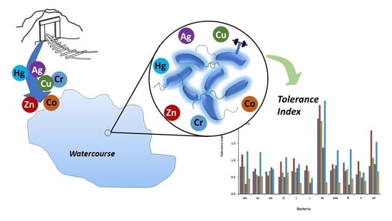
Evidence of Resistance of Heavy Metals from Bacteria Isolated from Natural Waters of a Mining Area in Mexico

by
Alondra Escamilla-Rodríguez
,
Salvador Carlos-Hernández
and
Lourdes Díaz-Jiménez
*
Waste Revaluation Laboratory, Natural Resources Sustainability and Energy, Cinvestav Saltillo, Ramos Arizpe 25900, Mexico
*
Author to whom correspondence should be addressed.
Water 2021, 13(19), 2766; https://doi.org/10.3390/w13192766
Submission received: 10 September 2021 / Revised: 1 October 2021 / Accepted: 3 October 2021 / Published: 6 October 2021
(This article belongs to the Special Issue Assessment of Water Quality)

Abstract

:
This study focuses on identifying relationships between the content of heavy metals in water and the resistance patterns of different bacteria. Samples from watercourses in one of the most important mining areas in Mexico were collected. Seventy-one bacteria were isolated, and their resistance to Cr, Zn, Cu, Ag, Hg, and Co was studied. The Minimum Inhibitory Concentration range was determined, and a Multiple Metal Resistant index was calculated. After that, 11 isolated bacteria were chosen to estimate kinetic parameters. The obtained results show differences in the behavior of the studied bacteria concerning the presence of heavy metals in the media: (1) without effect, (2) inhibited growth; and (3) considerable inhibited growth. Finally, a Performance Index was proposed to select adequate bacteria for heavy metals removal; five bacteria were selected. Among them, Pseudomonas koreensis was identified as a good candidate for a future biosorption system since these bacteria can stimulate growth in the presence of all the metals tested.

Graphical Abstract

1. Introduction

The presence of heavy metals in drinking water causes health damage because of their toxicity and non-biodegradability [1,2]. These elements are prone to bioaccumulation in tissues. Besides, they are persistent in the food chain, which induces long-term problems such as anemia, brain damage, hypertension, epigastric pain, vomiting, diarrhea, and lung tumors, among other diseases [3]. Unfortunately, the symptoms are expressed when the heavy metal concentrations reach high levels in the organism.
Several anthropogenic activities produce large amounts of waste with heavy metals. Frequently, those wastes are released into the environment without adequate treatment, thereby increasing water pollution and health risks [4]. Furthermore, since the population grows continuously, the water demand also increases, reducing water availability. In some regions of the world, the aquifers are in overexploitation conditions; this situation requires extracting deeper water, which could increase the concentration of salts and heavy metals.
In Mexico, heavy metals have been identified in underground and surface water bodies, as well as in animal fauna for human consumption [5,6,7]. As in other regions, mining is one of the main activities delivering residual heavy metals to water bodies in different country zones [7,8,9,10]. An example is found in Mazapil, Zacatecas, Mex., where the largest gold mine in the Americas is located. In this mine, the exploitation process is done through open-pit mining; 50,000 tons of rocks are processed daily, with high concentrations of gold, silver, zinc, and copper, among others [11]. Consequently, all nearby lakes and streams began to show symptoms of metal contamination.
In this context, it is necessary to design efficient systems to remove metals from drinking water. Different methods have been developed for this issue, such as chemical precipitation, adsorption, ion exchange, membrane filtration, flocculation, and electro-dialysis [12,13,14,15,16]. Processes for heavy metal removal are often neither effective nor economical, especially for water with heavy metal content below 100 mg L−1 [17,18,19]. In addition, the disposal of toxic sub-products has serious repercussions.
Bioremediation has been identified as a promising alternative for the treatment of large water bodies with low metal concentrations. This technique attempts to eliminate, reduce, isolate, or stabilize a contaminant or a group of pollutants by using a biological medium such as bacteria, fungi, or algae [20,21]. Among the advantages of bioremediation that stand out are the ease of implementation for metal recovery from elution and the operation in a wide range of scales. Also, it is a low-cost method [21,22], and it can be used as an alternative for the recovery of strategic metals and other valuable resources from wastes [13,23,24,25]. On the other hand, bioremediation methods can be affected by the properties of the water to be treated, such as pH, ionic strength, coexisting ions, and suspended solids. Moreover, the presence of several heavy metals can inhibit the biological activity of bacteria degrading other metal species of interest. Then, bioremediation requires more research in order to overcome these issues.
Heavy metals generally cause an inhibitory effect on microorganisms by blocking essential functional groups, displacing essential metal ions, or modifying active conformations of biological molecules. However, some metals are essential (e.g., Co, Cu, Zn, Ni) since they provide vital cofactors for metalloproteins and enzymes [26]. Nevertheless, heavy metals found in their environment can affect microorganisms (growth, morphology, and biochemical activities), resulting in decreased biomass and diversity [27]. Besides, it is known that microorganisms living in polluted areas by effluents from mining could develop tolerance to heavy metals. Therefore, they are candidates to be used in water purification systems.
One way to design efficient processes for the decontamination of water by biological methods is by isolating and identifying microorganisms present in contaminated water or wastewater [28]. Thus, numerous research has been carried out on the isolation of microorganisms tolerant to heavy metals from wastewater. For example, 38 microorganisms were isolated from different sources of wastewater in Egypt, of which 14 showed tolerances to Hg, Cu, Co, Ni, and Zn. The bacterium Stenotrophomonas maltophilia was identified as multi-metal resistant, and this can be useful in bioremediation treatments [29]. In another study, 15 bacteria were isolated from wastewater from an electroplating wastewater treatment plant. These bacteria proved to be tolerant to the presence of Cu2+, Ni2+, Mn2+, Co2+, and Cr2O72− [30]. Bacteria tolerant to Cr, Ag, and Hg have also been isolated from wastewater from a university hospital and a chemical technical school [31]. On the other hand, two bacteria resistant to chromium and lead could be isolated from wastewater from a tannery [32].
This paper focuses on the study of water samples from the watercourses around Mazapil, Zacatecas. The specific purposes are to isolate bacteria from the natural microbiome, determine the metal resistance or tolerance of isolated bacteria, select the most dominant group, and investigate in vitro its resistance to chromium, copper, zinc, mercury, silver, and cobalt. The final objective of this research is to complement the obtained results presented in this paper to implement new purifying systems for drinking water; the dominant group will be considered candidate microorganisms for a biofilter able to remove heavy metals from water.

2. Materials and Methods

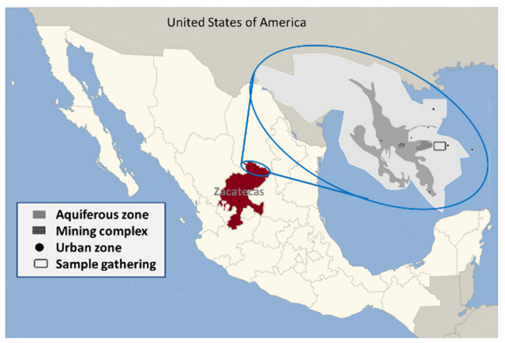
2.1. Samples and Gathering

The sampling area is in Mazapil, Zacatecas, Mexico (23°41′–25°04′ N, 101°11′–102°41′ W) in both the watercourse “Arroyo Grande” (24°43′001″ N, 101°52′01.2″ W) and the spring “Las Goteras” (Figure 1). This zone is a micro-watershed receiver of superficial and subterranean water. The town is near deposits of zinc, gold, silver, copper, mercury, phosphorite, limestone, lulite, calcite, onyx, and marble. The sampling points were chosen according to the anthropogenic activities and eventual sources of contamination; these areas include springs, dams, intakes, streams, and overflows.
A total of 15 samples of water were taken using glass bottles according to the local regulation NMX-AA-003-1980 [33]. The samples were kept at 4 °C in the dark, and they were analyzed within 24 h, according to the Standard Methods for Examination of Water and Wastewater [34]. Nitrate, nitrite content, total hardness, total alkalinity, and pH were determined in situ using Pond Test Strips. The identification of metal ions was performed using a Thermo Model iCE 3000 atomic absorption spectrometer equipped with single element hollow cathode lamps and an air-acetylene-nitrous oxide burner. Water samples were filtered, digested with HNO3, and analyzed directly to determine metals concentration, according to the standard NMX-AA-051-SCFI-2001 [35].

2.2. Identification of Bacteria Tolerance to Heavy Metals

A serial dilution from a saline solution of 0.85% (10−1 to 10−4) was conducted on the collected samples; they were then cultured by duplicate in nutrient agar, EMB, MacConkey, and Mannitol salt agar. MSA is mainly a selective culture medium useful in the staphylococci isolation, while EMB and MacConkey’s agar are differential media. MSA contains mannitol and a high salt concentration (NaCl 7.5%) that inhibits most bacteria. EMB contains lactose and saccharose as carbon sources and dyes as indicators that differentiate Enterobateriaceae and coliform microorganisms. MacConkey’s agar is used to differentiate between Gram negative bacteria by the capacity to ferment lactose. For the assays, plates were incubated at 30–35 °C for 24 h, and then an agar plate count was performed. Colonies were examined visually and chosen based on their dominance in each culture medium; the best ones were selected to be replanted in a nutrient agar supplemented with heavy metals solutions. Two concentrations were employed: minimum and maximum. The corresponding values were selected according to a Mexican normative [36], the former considers the minimum concentration detected by other methods already researched, and the last one corresponds to the maximum permissible concentration in this normative [36].
The heavy metals solutions were prepared in plastic bottles washed with HNO3 65%, and employed the following salts: K2Cr2O7 (0.05, 100 mg L−1), ZnSO4 (5, 100 mg L−1), CuSO4 (2, 100 mg L−1), AgNO3 (0.05, 100 mg L−1), Hg2SO4 (0.001, 100 mg L−1) and CoCl2 (0.02, 100 mg L−1). Plates were incubated at 30–35 °C until they showed bacterial growth. Microscopic and macroscopic morphology were evaluated on each colony and transferred to nutrient agar to cultivate pure colonies. Gram-stained samples were found confirming purity. Specie identification of isolate was determined according to Bergey’s Manual of Determinative Bacteriology. For the molecular identification of bacteria, five isolates were chosen among those with the highest metal tolerance. The molecular identification was carried out by 16S rDNA sequencing.

2.3. Assessment of Metal Toxicity

The heavy metals tested were taken from the following precursors: K2Cr2O7 at 10–1250 mg L−1, ZnSO4 at 10–2500 mg L−1, CuSO4 at 5–1250 mg L−1, Hg2SO4 at 5–100 mg L−1, CoCl2 at 5–1250 mg L−1 and AgNO3 at 5–2500 mg L−1. Stock solutions were prepared in distilled water slightly acidified (2 to 4 drops of HNO3) and were sterilized at 110 °C for 15 min. These solutions were kept at 4 °C for no longer than one month. The glassware used was leached in 65% HNO3 and rinsed several times with distilled water avoiding metal contamination. In order to quantitatively assess the effect of heavy metals, plate diffusion and susceptibility were tested, and a multiple metal resistance index (MMR) was determined.
Plate diffusion method. 0.25 mL of the appropriate metal salt solution was added to each plate of nutrient agar in a central well of 1 cm in diameter and 4 mm in depth. Plates were then incubated at 35 °C for 24 h in order to allow diffusion of the metal into the agar. At this time, it was expected that a concentration gradient of the metal had been formed. On each plate, eight strains were inoculated into radial streaks. Plates were then incubated at 35 °C for five days. After incubation, the diameter of colony growth (in mm) was measured, and its percentage of growth was calculated [37].
Susceptibility test. A modified Kirby-Bauer method was used for the susceptibility of the selected isolates to chromium, zinc, copper, silver, mercury, and cobalt [38]. The bacteria were suspended in 5 mL of sterile nutrient broth and incubated for 18–24 h, 32 °C at 100 rpm, until the suspension was adjusted to 106 CFU mL−1 (No. 5 in the McFarland standard). A sterile swab was dipped into the inoculums tube, inoculating the dried surface of a nutrient agar plate by streaking the swab three times over the entire agar surface and allowing the plate to be at room temperature for 3–5 min to dry. The appropriately impregnated disks with metal solutions were placed on the surface of the agar afterward. Once all disks were in place, the plates were inverted and placed in a 35 °C incubator for 16–18 h. Following incubation, the inhibitory zone sizes were measured to the nearest millimeter using a Vernier. Escherichia coli K12 SMG123 was used as a control strain verifying the metal susceptibility. The results of the diffusion susceptibility test were reported as susceptible (S), intermediate (I), or resistant (R) [39].
Multiple Metal Resistance (MMR) evaluation. The used method is based on that proposed by Krumperman [40] for multiple antibiotic resistance. The main equation is adapted to multiple metal resistance determination as follows Equation (1):
M M R = a / b
where a stands for the number of metals the isolate was resistant to, and b represents the total number of metals the isolate was tested against. A similar equation was applied by Matyar [41] and Kimiran-Erdem [42].

2.4. Determination of Kinetic Parameters and Tolerance Index

One loop full of microbial growth obtained from nutrient agar plates after 48 h incubation of selected species was inoculated in Nutrient Broth. Subsequently, 1 mL of the culture was transferred to Nutrient Broth contaminated with 50, 100, and 200 mg L−1 of the metals studied (one by one); then, it was incubated seven days at 32 °C and inspected each day through UV-visible spectrometry at 600 nm verifying microbial growth. The tolerance index is defined as the ratio of the CFU mL−1 in the contaminated medium and the control medium [43]. Once bacteria reached the steady-state, it was possible to determine the kinetic parameters: growth rate (µ) and doubling time (td) according to the next Equations (2) and (3):
μ = l n N f l n N 0 t f t 0
t d = 0.693 μ
where N0 and Nf are the total CFU mL−1 at the beginning and the end of the exponential phase, respectively, and tf and t0 is the final and initial time, respectively.
Finally, the Tolerance index (TI) represents the relative growth rate of the bacteria, and it is computed as follows Equation (4):
T I = μ m s μ c
where µms is the growth rate in metal-containing solution and µc is the growth rate in a control solution at 72 h. TI is commonly used to quantify metal tolerance in the organisms [43]: the higher the TI value, the greater the tolerance.
Selection of candidate bacteria for future water purification systems. After the previous analysis, a Performance Index (PI) is proposed to select the bacteria that could remove heavy metals from water. The PI is obtained by combining the MMR, the TI, and the tdm as expressed in the Equation (5):
P I = M M R × 1 t d m × i T I i
where i stands for Cr, Zn, Cu, Ag, Hg, and Co, which are the tested heavy metals; dtm is the mean double time of each bacterium, the inverse of this term is included in the performance index since it concerns the bacteria growth rate, a short double-time implies a fast adaptation to the environment and then a better behavior.
The criteria used to select the bacteria correspond to the value of PI: the larger the PI, the more adequate the bacteria are for adsorption systems.

3. Results and Discussion

3.1. Water Characteristics

The samples from all the water sources were clear and odorless; some presented minimal soil content. Some vegetation was detected at each site, usually along the periphery. The pH measure showed values ranging from 6.9 to 8.1, and the temperature was between 9.5 and 13 °C (Table 1). According to local regulations, all these data are within the range of values expected in a natural water source.
Regarding microbiological parameters, low counts of total coliform bacteria were found in water samples collected from the spring, the near intake, the stream, the first tank, and the overflow. Once the data was verified with local regulations, the results showed that these sources of water should not be used for human consumption. In the rest of the samples, total coliform bacteria were not found. The presence of coliforms and Enterobacteriaceae species has shown that water sources are polluted with intestinal microbiota found in humans and other animal species, indicating its continuous traffic in these places. The rest of the bacteria are ubiquitous aerobic, Gram-negative, and positive bacteria common in aquatic environments, soils, vegetation, and even some animals.
All samples were classified as hard and very hard water from the other physicochemical parameters, indicating the pass of water through deposits such as limestone, increasing the presence of Ca2+ and Mg2+ ions. Also, high levels of total alkalinity have been detected, indicating the presence of compounds such as bicarbonates, carbonates, and hydroxides influenced by the existence of rocks, soils, salts, certain plant activities, and certain industrial wastewater discharges. Both results suggest a rich salt content in the water. The values obtained for nitrite and nitrate follow the values established by local norms NOM-127-SSA1-1994 and NOM-001-ECOL-1996 [36,44]. Assessing the content of heavy metals in the water samples showed traces of Mn, Cr, and Co (below 0.01 mg L−1). These results are acceptable for human consumption of water, according to NMX-AA-051-2001 [35].

3.2. Isolated Bacteria

Once the physicochemical analyses of water samples were concluded, the research was focused on the isolation of metal tolerant and resistant bacteria. To increase the metal toxicity, a total of 71 dominant bacteria were isolated and tested in nutrient agar supplemented with Cu, Co, Cr, Zn, Hg, and Ag (one by one). Out of all the isolates, 47 grew in all the supplemented agars; this implies the metals encourage the growth of these bacteria and avoid the toxic effects of certain forms of metals in a specific dose. The ability of the bacteria to overcome the metal toxicity is an inherent characteristic probably due to the near contact between the soil, sediment, and water-rich in salt and metal content [45,46,47].
After that, the bacteria isolated were classified based on morphological, cultured and biochemical characteristics, compared with the standard description in the Bergey’s Manual of Determinative Bacteriology for a primary identification. The isolates were identified as follows: 10 belong to the Pseudomonadaceae family, 10 fit the Enterobacteriaceae family, and three are members of the Staphylococcus family; the rest of the bacteria remain unidentified (Table 2). The growth percentage corresponds to the increase of bacteria compared to the initial population; bacteria with high tolerance to heavy metals presented large percentage growth. Some microorganisms reached the population of the generation time or doubling time (100% in comparison with the initial population) in these experiments, and some of them even grew more than 100%. The generation time depends on several factors, including the type of microorganism; it is an important parameter to analyze the bacteria behavior further [48]. In this study, the bacteria with high growth percentages are designated as Tolerant due to their ability to cope with metal toxicity through the intrinsic properties of microorganisms [49].

3.3. Heavy Metal Resistance Evaluation

From the Plate diffusion analysis, it was found that 17 bacteria grew in the presence of all the tested metals, more than in the control agar without metals supplementation; meanwhile, the rest of the bacteria grew at least in three of the substrates supplemented with the tested metals (Table 3).
It was also noticeable that silver, copper, mercury, cobalt, and chromium modified the macroscopic morphology on some bacterial strains. Similar results have been reported with copper and mercury, which caused an increase in the total CFU [50] and with sub-lethal concentrations of several metals, which indicated structural abnormalities and changes in the cellular distribution of bacteria [51]. The change in the morphology was probably due to a period of adaptation where cells synthesized some enzymes essential for the uptake of metals [52]. Also, it is important to remark that the metal biosorption by living cells is performed in two steps: passive and active. The passive step concerns the metal ion adsorption on the cell surface, which could explain the morphology changes. The adsorption mechanism is based on the interaction between the metal ion and the functional groups (carboxyl, hydroxyl, amino, phosphate, and sulfide) which are included in the polysaccharides, lipids, and proteins composing the cell wall. The processes occurring in this step (electrostatic attraction, ion exchange, complexation, and precipitation) are independent of the cellular metabolism [53]. On the other side, the active step involves the ion metal intracellular uptake, following similar mechanisms for uptake nutrients such as Na2+, K+, and Ca2+ [54]. In the case of the bacteria isolated in this work, further studies are required to elucidate the specific mechanisms of heavy metal biosorption. Moreover, it was detected that several bacterial strains have mercury tolerance, mercury being the most toxic metal tested. This behavior increases the possibility of using these strains to detoxify the polluted environment by anthropogenic activities.
On the other side, the susceptibility test was used to classify the bacteria as:
(A)
Susceptible, if the bacteria growth was inhibited by the tested concentration, which is characterized by an inhibitory zone higher than 18 mm,
(B)
Resistant, if the bacteria growth persisted in the presence of heavy metal ions; that is: if they showed an inhibitory zone lower than 13 mm,
(C)
Intermediate, if the bacteria showed an inhibitory zone between 13–18 mm, which indicates the bacteria metal tolerance [39],
(D)
Susceptible Dose-Dependent (SDD), this term is related to those bacteria without an inhibitory zone; that means a higher concentration of a heavy metal solution is necessary to determine the Minimum Inhibitory Concentration (MIC), as shown in Table 3. The MIC is defined as the lowest metal concentration, which completely averts bacterial growth (the presence of an inhibitory zone).
Most of the bacteria were resistant to zinc and mercury, presented an intermediate behavior against the rest of the metals, and only a few strains were sensitive to chromium. It is important to notice that all the bacteria have an SDD behavior for silver and copper. Considering that silver and copper are the main products of exploitation of the mining area [11], it is possible that some traces of this metal migrate to the natural waters and become a part of the normal water microbiome, resulting in the SDD behavior.
The main sources of resistant bacteria were the dams (41.37%), followed by the springs (24.13%) and the intake (13.79%). This situation suggests the need for close contact with the soil in order to generate a resistance mechanism in the bacteria [45,46,47]. The frequency of mercury resistant bacteria at concentrations below 100 mg L−1 was higher than those of the other metals tested (93.10%); 14 bacteria showed a multi-resistance pattern with an MMR index higher than 0.2; this implies bacteria were exposed to a rich metal environment [41]. The most predominant multi-resistant bacterium is the Pseudomonas AH found in the last dam; it presents a multi-resistance pattern for Zn, Ag, Hg, and Co with MIC of 2500, 2500, 85, 105 ppm, respectively. Also, this isolate exhibited intermediate and susceptible responses to the other two metals. Similarly, 10 bacteria exhibited a multi-resistant pattern to Zn, Hg, and Co, and two bacteria for Zn, Hg and Cr. Besides, six bacteria showed a bi-resistant pattern to Zn and Hg, one to Hg and Co, and one to Hg and Cr.
On the other side, the MIC was generally centered at 650–1250 mg L−1 for Cr, 650–2500 mg L−1 for Zn, 65–85 mg L−1 for Hg, and 650–1250 mg L−1 for Co, covering a wide range of concentrations and consequently offering a potential advantage over other biological systems for metal removal previously studied. These results showed the most of the isolates that were considered resistant to Co because the MIC values exceeded that of the E. coli K-12 (400 mg L−1). Similar behavior was found for Cr and Hg, which the MIC values for E. coli were 600 and 800 mg L−1, respectively. Since it was not possible to determine the MIC for silver and copper, these metals were considered less toxic to all bacteria than the other metals (Table 3); this could indicate a relationship in the resistance process for both components in the isolated bacteria or an unspecific mechanism. Other investigations have shown that bivalent metals facilitate their bioavailability in combination with chlorine by promoting metal-protein interactions and the transport across the membrane [55]; that was the case for the Ag and Cu tested in this work.
The toxic effects and the doses tested in these assays allow for the consideration of a specific pattern or toxicity order for the selected bacteria: Hg > Co > Cr > Zn > Ag = Cu, which are in agreement with the plate diffusion method and does not differ from that reported in other studies [37,56]. It was found that the multi-resistance occurs only to toxic compounds that have similar mechanisms underlying their toxicity, attributed to a variety of detoxifying processes [57]. Mercury is considered highly toxic due to lipid solubility, which could affect cell membrane permeability, causing bacterial cell death [58]. Cr, Co, and Zn are less toxic; a small concentration of these metals can be used by normal bacterial metabolism to produce cofactors, enzymes, or vitamins useful for bacterial growth. However, the substance becomes toxic when the metabolism is overcome [56,59]. Several authors have suggested that heavy metal rich environments could improve adaptation mechanisms in bacteria, as in this case. And although mercury, silver, zinc, and copper were not analyzed in the water samples, a significant number of the bacteria were found resistant to high concentrations of the salts tested, suggesting that this mechanism dealing with metal toxicity may be linked to the tolerance to other metallic ions because metal toxicity does not only occur in the environment [26]. Then, from the previous analysis, eleven isolates were selected for the next step in this work.

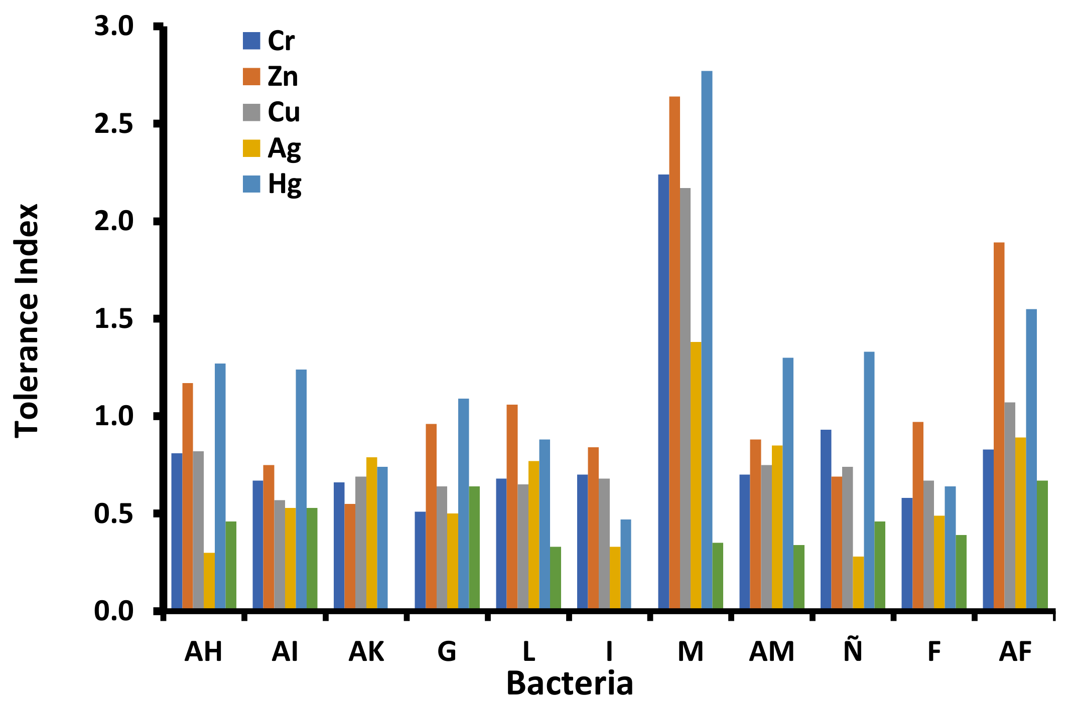
3.4. Metal Tolerance Index

The metal tolerance index (TI) is a metric used to identify metal tolerant organisms [43]. TI has been determined for the eleven selected bacteria that show multi-resistance patterns to the metals tested (Figure 2). The bacterium M shows the highest TI, followed by the bacterium AF. Cobalt was the most toxic metal, and mercury was the most tolerated by the analyzed bacteria (Co > Ag, Cr = Cu > Zn, Hg). The Pseudomonas M was the most tolerant of all tested metals, promoting its growth compared with the liquid culture without any heavy metals. However, with cobalt, it exhibited less growth. The rest of the bacteria showed similar behavior, high tolerance to Hg and Zn without affecting its growth, and inhibition by Ag and Co, especially at concentrations higher than 50 mg L−1. It is also possible to note that low concentrations of any metal can stimulate bacterial growth. This was probably due to synergic effects in the enzymatic catalysis; as the concentration increases the metal toxicity, the stimulating effect decreases, and the system is eventually affected severely or inhibited [60]. The resistance of these bacteria to metals is probably related to incorporating those elements into the cytoplasm, once they bind to specific metallothionein [61,62]. Moreover, Gram-negative bacteria have a higher resistance to the toxic effects of heavy metals due to their cellular wall, which hinders their incorporation into inner cells [62]. These significant differences in the resistance to heavy metals could be related to variances in the isolation site and adaptation mechanisms developed by bacteria. Other studies reveal a relationship in tolerance to Zn, Cr, and Cu because they are used as micronutrients for different metabolic pathways and tend to bioaccumulate when they exceed certain levels close to MIC [43,58]; this could explain the increasing growth for all bacteria in the presence of these metals. In the case of Ag, the tendency to bioaccumulate into the inner cell has been reported previously, which causes several toxic effects [43]. In consequence, the response of the bacteria to the presence of metals in their environment may depend on metal concentration, bacteria, isolation site, and the nature of the metal (micronutrient or toxic).

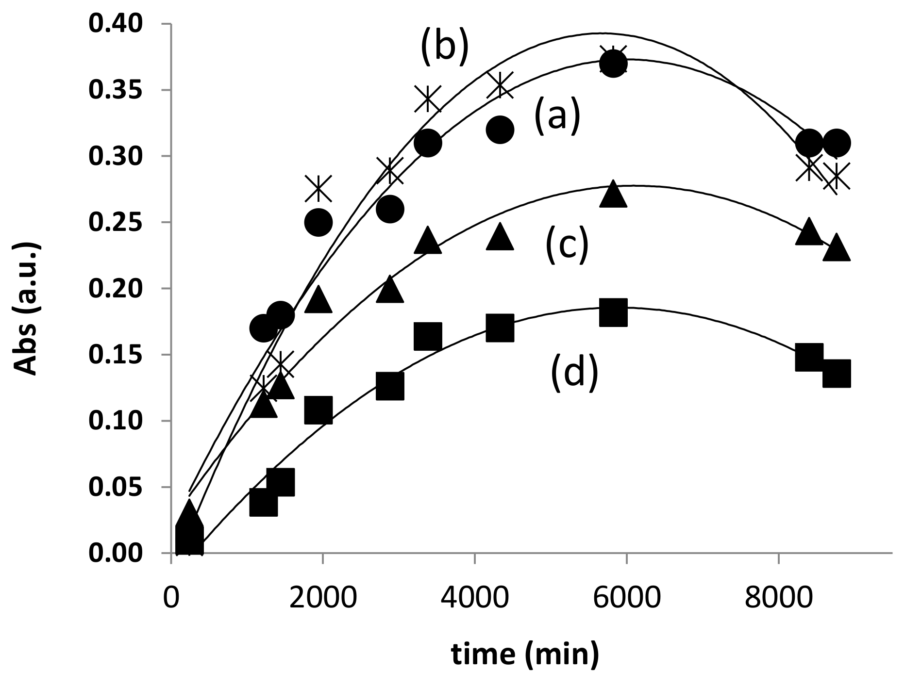
3.5. Kinetic Parameters

At this point, a growth model describing the behavior of the bacteria in the presence of the heavy metals tested is obtained. Single growth curves for each bacterium versus each tested metal have been performed. The general behavior of bacteria is presented in Figure 3. Two different conditions were considered (control substrate without metals, bacterial growth in the presence of metals).
Curve (b) corresponds to the bacterial growth without effect related to Zn and Hg; as can be seen, there exists a slight increase (5–10%) in the total amount of bacteria. In (c), inhibited growth for Cr and Cu is observed; the total amount of bacteria decreases between 10 and 25%. Meanwhile, (d) shows a considerable inhibited growth for Ag and Co, a decrease of 56% on the bacterial growth is identified; it implies toxic effects of these metals to the bacteria.
The qualitative behavior of all bacteria are similar. It is characterized by a prolonged lag phase, indicating an adaptation process by the bacteria induced by the metals; after that, a fast exponential phase followed by a stationary phase are observed; finally, a drastic dead phase is reached. Specifically, bacteria which reacted to cobalt and silver are characterized by a very long adaptation phase, followed quickly by an exponential and dead phase. From the specific growth curves for each bacterium and metal tested, it was possible to determine the growth rate (µ) and the doubling time (td) (Table 4). Bacteria exhibited the same values for µ and td for the inhibited growth behavior (c); a higher µ and lower td in the case of the behavior without effect (b), characterized by a contracted cell duplication over time and an increase in the growth rate; and a low µ and higher td for the considerable inhibited growth (d), this implies an increase in the doubling time and a decrease in the growth rate due to toxic effects of metals. This behavior is summarized in Table 5. Only two bacteria (Pseudomonas Ñ and Staphylococcus F) have without effect growth (b) for Co, Ag, and Hg, respectively, and an inhibited growth (c) for the rest of the metals. Otherwise, Enterobacteria I only shows a considerable inhibited growth (d) for all metals. The rest of the bacteria presented a mixture of the three behaviors described previously. These results have also evidenced a co-resistance behavior for the metals tested and the analyzed bacteria. However, further studies are required in order to analyze the biosorption potential of these microorganisms to remove heavy metals at low concentrations from contaminated waters and to implement an immobilization system for domestic water treatment. And finally, economic feasibility studies about market factors should be done for a successful application for large-scale ecological restoration.
Finally, summarizing the previous analysis, the performance index of the eleven bacteria is presented in Table 6. It is considered from these data that five bacteria (PI ≥ 0.1) show adequate characteristics for the assimilation of heavy metals. They can then can be studied further to design biosorption systems for the purification of water. Three bacteria correspond to the gene Pseudomonas, and two are identified as Enterobacter. This agrees with the findings of other authors [18,63], indicating that these species are well situated for the removal of heavy metals.

4. Conclusions

The present study investigates the tolerance behavior of 71 bacteria isolated from natural waters. Thirty-one bacteria exhibited a tolerance mechanism to Cr, Cu, Ag, Hg, Zn, Co at low concentrations; 28 bacteria were classified as tolerant, intermediate, and resistant to each metal tested. Moreover, it was possible to determine an overall toxicity pattern: Hg > Co > Cr > Zn > Ag = Cu for all bacteria tested. Eleven bacteria were classified as metal resistant, and on the basis of its behavior, a growth curve model was proposed; this model represents curves without effect, an inhibited and considerable inhibited growth for the metals tested in this work. The Pseudomonas M showed the best response for all the metals tested without presenting significant signs of toxicity. Five bacteria are selected as candidates for future biosorption systems (Pseudomonas M, Enterobacteriaceae AH, Enterobacteriaceae AF, Pseudomonas Ñ, and Pseudomonas AM) since they showed a large performance index regarding their resistance to heavy metals.

Author Contributions

Conceptualization, L.D.-J.; Formal analysis, A.E.-R.; Funding acquisition, L.D.-J. and S.C.-H.; Investigation, A.E.-R.; Supervision, S.C.-H. and L.D.-J.; Writing—original draft, A.E.-R.; Writing—review & editing, S.C.-H. and L.D.-J. All authors have read and agreed to the published version of the manuscript.

Funding

This research was funded by Consejo Nacional de Ciencia y Tecnología (CONACYT) through the Projects number 196005 and 149646.

Institutional Review Board Statement

Not applicable.

Informed Consent Statement

Not applicable.

Data Availability Statement

The data presented in this study are available on request from the corresponding author.

Acknowledgments

A.E.R. thanks the support received by Consejo Nacional de Ciencia y Tecnología (CONACYT) under Grant number 173971.

Conflicts of Interest

The authors declare no conflict of interest.

References

  1. Ahluwalia, S.S.; Goyal, D. Microbial and plant derived biomass for removal of heavy metals from wastewater. Bioresour. Technol. 2007, 98, 2243–2257. [Google Scholar] [CrossRef]
  2. Chowdhury, S.; Mazumder, M.A.J.; Al-Attas, O.; Husain, T. Heavy metals in drinking water: Occurrences, implications, and future needs in developing countries. Sci. Total Environ. 2016, 569–570, 476–488. [Google Scholar] [CrossRef] [PubMed]
  3. Farooq, U.; Kozinski, J.A.; Khan, M.A.; Athar, M. Biosorption of heavy metal ions using wheat based biosorbents-A review of the recent literature. Bioresour. Technol. 2010, 101, 5043–5053. [Google Scholar] [CrossRef] [PubMed]
  4. Pérez Castresana, G.; Castañeda Roldán, E.; García Suastegui, W.A.; Morán Perales, J.L.; Cruz Montalvo, A.; Handal Silva, A. Evaluation of health risks due to heavy metals in a rural population exposed to Atoyac River pollution in Puebla, Mexico. Water 2019, 11, 277. [Google Scholar] [CrossRef] [Green Version]
  5. Razo, I.; Carrizales, L.; Castro, J.; Díaz-Barriga, F.; Monroy, M. Arsecin and heavy metal pollution of soil, water and sediments in a semi-arid climate mining area in Mexico. Water. Air. Soil Pollut. 2004, 152, 129–152. [Google Scholar] [CrossRef]
  6. García-Hernández, J.; Cadena-Cárdenas, L.; Betancourt-Lozano, M.; García-De-La-Parra, L.M.; García-Rico, L.; Márquez-Farías, F. Total mercury content found in edible tissues of top predator fish from the Gulf of California, Mexico. Toxicol. Environ. Chem. 2007, 89, 507–522. [Google Scholar] [CrossRef]
  7. Covarrubias, S.A.; Peña Cabriales, J.J. Contaminación ambiental por metales pesados en México: Problemática y estrategias de fitorremediación. Rev. Int. Contam. Ambient. 2017, 33, 7–21. [Google Scholar] [CrossRef]
  8. Gómez-Alvarez, A.; Meza-Figueroa, D.; Villalba-Atondo, A.I.; Valenzuela-García, J.L.; Ramírez-Hernández, J.; Almendariz-Tapia, J. Estimation of potential pollution from mine tailings in the San Pedro River (1993–2005), Mexico-US border. Environ. Geol. 2009, 57, 1469–1479. [Google Scholar] [CrossRef]
  9. Levresse, G.; Lopez, G.; Tritlla, J.; López, E.C.; Chavez, A.C.; Salvador, E.M.; Soler, A.; Corbella, M.; Sandoval, L.G.H.; Corona-Esquivel, R. Phytoavailability of antimony and heavy metals in arid regions: The case of the Wadley Sb district (San Luis, Potosí, Mexico). Sci. Total Environ. 2012, 427–428, 115–125. [Google Scholar] [CrossRef]
  10. Lizárraga-Mendiola, L.; González-Sandoval, M.R.; Durán-Domínguez, M.C.; Márquez-Herrera, C. Geochemical behavior of heavy metals in a Zn-Pb-Cu mining area in the State of Mexico (central Mexico). Environ. Monit. Assess. 2009, 155, 355–372. [Google Scholar] [CrossRef]
  11. Vdovin, V.; Pareja, G.; Lind, P. Peñasquito Polymetallic Operation; Zacatecas State Mexico; NI 43-101 Technical Report; Goldcorp Inc.: Vancouver, BC, Canada, 2010. [Google Scholar]
  12. Joseph, L.; Jun, B.M.; Flora, J.R.V.; Park, C.M.; Yoon, Y. Removal of heavy metals from water sources in the developing world using low-cost materials: A review. Chemosphere 2019, 229, 142–159. [Google Scholar] [CrossRef]
  13. Nancharaiah, Y.V.; Venkata Mohan, S.; Lens, P.N.L. Metals removal and recovery in bioelectrochemical systems: A review. Bioresour. Technol. 2015, 195, 102–114. [Google Scholar] [CrossRef]
  14. Abdullah, N.; Yusof, N.; Lau, W.J.; Jaafar, J.; Ismail, A.F. Recent trends of heavy metal removal from water/wastewater by membrane technologies. J. Ind. Eng. Chem. 2019, 76, 17–38. [Google Scholar] [CrossRef]
  15. Sharma, S.K. Heavy Metals in Water; Royal Society of Chemistry: Cambridge, UK, 2014; ISBN 9781782620174. [Google Scholar]
  16. Wadhawan, S.; Jain, A.; Nayyar, J.; Mehta, S.K. Role of nanomaterials as adsorbents in heavy metal ion removal from waste water: A review. J. Water Process Eng. 2020, 33, 101038. [Google Scholar] [CrossRef]
  17. Banerjee, G.; Pandey, S.; Ray, A.K.; Kumar, R. Bioremediation of heavy metals by a novel bacterial strain Enterobacter cloacae and its antioxidant enzyme activity, flocculant production, and protein expression in presence of lead, cadmium, and nickel. Water. Air. Soil Pollut. 2015, 226, 1–9. [Google Scholar] [CrossRef]
  18. De Niederhäusern, S.; Bondi, M.; Anacarso, I.; Iseppi, R.; Sabia, C.; Bitonte, F.; Messi, P. Antibiotics and heavy metals resistance and other biological characters in enterococci isolated from surface water of Monte Cotugno Lake (Italy). J. Environ. Sci. Health—Part A Toxic/Hazardous Subst. Environ. Eng. 2013, 48, 939–946. [Google Scholar] [CrossRef] [PubMed]
  19. Dey, S.; Paul, A.K. Hexavalent chromium reduction by aerobic heterotrophic bacteria indigenous to chromite mine overburden. Braz. J. Microbiol. 2013, 44, 307–315. [Google Scholar] [CrossRef] [Green Version]
  20. Oyetibo, G.O.; Miyauchi, K.; Huang, Y.; Chien, M.F.; Ilori, M.O.; Amund, O.O.; Endo, G. Biotechnological remedies for the estuarine environment polluted with heavy metals and persistent organic pollutants. Int. Biodeterior. Biodegrad. 2017, 119, 614–625. [Google Scholar] [CrossRef]
  21. Qin, H.; Hu, T.; Zhai, Y.; Lu, N.; Aliyeva, J. The improved methods of heavy metals removal by biosorbents: A review. Environ. Pollut. 2020, 258, 113777. [Google Scholar] [CrossRef]
  22. Vijayaraghavan, K.; Balasubramanian, R. Is biosorption suitable for decontamination of metal-bearing wastewaters? A critical review on the state-of-the-art of biosorption processes and future directions. J. Environ. Manag. 2015, 160, 283–296. [Google Scholar] [CrossRef]
  23. Won, S.W.; Kotte, P.; Wei, W.; Lim, A.; Yun, Y.S. Biosorbents for recovery of precious metals. Bioresour. Technol. 2014, 160, 203–212. [Google Scholar] [CrossRef] [PubMed]
  24. Rathna, R.; Nakkeeran, E. Biological Treatment for the Recovery of Minerals from Low-Grade Ores; Elsevier B.V.: Amsterdam, The Netherlands, 2020; ISBN 9780444643216. [Google Scholar]
  25. Işıldar, A.; van Hullebusch, E.D.; Lenz, M.; Du Laing, G.; Marra, A.; Cesaro, A.; Panda, S.; Akcil, A.; Kucuker, M.A.; Kuchta, K. Biotechnological strategies for the recovery of valuable and critical raw materials from waste electrical and electronic equipment (WEEE)—A review. J. Hazard. Mater. 2019, 362, 467–481. [Google Scholar] [CrossRef] [PubMed]
  26. Christopher, M.; Paul, O.; Hamadi, B. Association of metal tolerance with multidrug resistance among environmental bacteria from wetlands of Lake Victoria basin. Agric. Biol. J. N. Am. 2014, 5, 24–32. [Google Scholar] [CrossRef]
  27. Barkay, T. Adaptation of Aquatic Microbial Communities to Hg 2+ Stress. Appl. Environ. Microbiol. 1987, 53, 2725–2732. [Google Scholar] [CrossRef] [Green Version]
  28. Sharma, P.; Tripathi, S.; Chandra, R. Metagenomic analysis for profiling of microbial communities and tolerance in metal-polluted pulp and paper industry wastewater. Bioresour. Technol. 2021, 324, 124681. [Google Scholar] [CrossRef]
  29. Naguib, M.M.; Khairalla, A.S.; El-Gendy, A.O.; Elkhatib, W.F. Isolation and characterization of mercury-resistant bacteria from wastewater sources in egypt. Can. J. Microbiol. 2019, 65, 308–321. [Google Scholar] [CrossRef]
  30. Cai, X.; Zheng, X.; Zhang, D.; Iqbal, W.; Liu, C.; Yang, B.; Zhao, X.; Lu, X.; Mao, Y. Microbial characterization of heavy metal resistant bacterial strains isolated from an electroplating wastewater treatment plant. Ecotoxicol. Environ. Saf. 2019, 181, 472–480. [Google Scholar] [CrossRef]
  31. Lima e Silva, A.A.; Carvalho, M.A.; de Souza, S.A.; Dias, P.M.; Silva Filho, R.G.; Saramago, C.S.; Bento, C.A.; Hofer, E. Heavy metal tolerance (Cr, Ag and Hg) in bacteria isolated from sewage. Braz. J. Microbiol. 2012, 43, 1620–1631. [Google Scholar] [CrossRef] [Green Version]
  32. Nokman, W.; Benluvankar, V.; Maria Packiam, S.; Vincent, S. Screening and molecular identification of heavy metal resistant Pseudomonas putida S4 in tannery effluent wastewater. Biocatal. Agric. Biotechnol. 2019, 18, 101052. [Google Scholar] [CrossRef]
  33. Secretaría de Comercio y Fomento Industrial. Residual Waters—Sampling; NMX-AA-003-1980; Diario Oficial de la Federación: Mexico City, Mexico, 1980; p. 9.
  34. Clesceri, L.S.; Greenberg, A.E.; Eaton, A.D. Anionic Surfactants as MBAS. In Standard Methods for the Examination of Water and Wastewater; APHA: Washington, DC, USA, 1999; pp. 1027–1033. [Google Scholar]
  35. Secretaría de Economía. Water Analisis—Determination of Metals by Atomic Absorption in Natural, Drinking, Wastewaters and Wastewaters Treated—Test Method; NMX-AA-051-SCFI-2001; Diario Oficial de la Federación: Mexico City, Mexico, 2001.
  36. Secretaría de Salud. Salud Ambiental, Agua para Uso y Consumo Humano-Limites Permisibles de Calidad y Tratamientos a que Debe Someterse el Agua para su Potabilización; NOM-127-SSA1-1994; Diario Oficial de la Federación: Mexico City, Mexico, 1994.
  37. Hassen, A.; Saidi, N.; Cherif, M.; Boudabous, A. Effects of heavy metals on Pseudomonas aeruginosa and Bacillus thuringiensis. Bioresour. Technol. 1998, 65, 73–82. [Google Scholar] [CrossRef]
  38. Bauer, A.W.; Kirby, W.M.M.; Sherris, J.C.; Turck, M. Antibiotic susceptibility testing by a standardized single dosk method. Am. J. Clin. Pathol. 1966, 36, 493–496. [Google Scholar] [CrossRef]
  39. Cockerill, F.R.; Patel, J.B.; Alder, J.; Brandford, P.B.; Dudley, M.N.; Hardy, D.J.; Hecht, D.W.; Hindler, J.A.; Powell, M.; Swenson, J.M.; et al. Performance Standards for Antimicrobial Susceptibility Testing; Twenty-Third Informational Supplement; Clinical and Laboratory Standards Institute: Wayne, NJ, USA, 2013; ISBN 1562388975. [Google Scholar]
  40. Krumperman, P.H. Multiple antibiotic resistance indexing of Escherichia coli to identify high-risk sources of faecal contamination of foods. Appl. Environ. Microbiol. 1983, 46, 165–170. [Google Scholar] [CrossRef] [Green Version]
  41. Matyar, F.; Kaya, A.; Dinçer, S. Antibacterial agents and heavy metal resistance in Gram-negative bacteria isolated from seawater, shrimp and sediment in Iskenderun Bay, Turkey. Sci. Total Environ. 2008, 407, 279–285. [Google Scholar] [CrossRef]
  42. Kimiran-Erdem, A.; Arslan-Aydoğdu, E.Ö.; Gürün, S.; Altun, Ö. Determination of multiple antibiotic and heavy metal resistance of the bacteria isolated from the Küçükçekmece Lagoon, Turkey. Polish J. Environ. Stud. 2015, 24, 1077–1084. [Google Scholar] [CrossRef]
  43. Muñoz, A.J.; Ruiz, E.; Abriouel, H.; Gálvez, A.; Ezzouhri, L.; Lairini, K.; Espínola, F. Heavy metal tolerance of microorganisms isolated from wastewaters: Identification and evaluation of its potential for biosorption. Chem. Eng. J. 2012, 210, 325–332. [Google Scholar] [CrossRef]
  44. Secretaría de Ecología. Que Establece los Límites Máximos Permisibles de Contaminantes en las Desacrgas de Aguas Residuales en Aguas y Bienes Nacionales; NOM-001-ECOL-1996; Diario Oficial de la Federación: Mexico City, Mexico, 1996; p. 13.
  45. Roane, T.M.; Kellogg, S.T. Characterization of bacterial communities in heavy metal contaminated soils. Can. J. Microbiol. 1996, 42, 593–603. [Google Scholar] [CrossRef]
  46. Nithya, C.; Gnanalakshmi, B.; Pandian, S.K. Assessment and characterization of heavy metal resistance in Palk Bay sediment bacteria. Mar. Environ. Res. 2011, 71, 283–294. [Google Scholar] [CrossRef]
  47. Karadede, H.; Ünlü, E. Concentrations of some heavy metals in water, sediment and fish species from the Ataturk Dam Lake (Euphrates), Turkey. Chemosphere 2000, 41, 1371–1376. [Google Scholar] [CrossRef]
  48. Gibson, B.; Wilson, D.J.; Feil, E.; Eyre-Walker, A. The distribution of bacterial doubling times in the wild. Proc. R. Soc. B Biol. Sci. 2018, 285. [Google Scholar] [CrossRef]
  49. Zouboulis, A.I.; Loukidou, M.X.; Matis, K.A. Biosorption of toxic metals from aqueous solutions by bacteria strains isolated from metal-polluted soils. Process Biochem. 2004, 39, 909–916. [Google Scholar] [CrossRef]
  50. Singleton, F.L.; Guthrie, R.K. Aquatic bacterial populations and heavy metals-I. Composition of aquatic bacteria in the presence of copper and mercury salts. Water Res. 1977, 11, 639–642. [Google Scholar] [CrossRef]
  51. Sterritt, R.M.; Lester, J.N. Interactions of heavy metals with minerals. Sci. Total Environ. 1980, 14, 5–17. [Google Scholar] [CrossRef]
  52. Ezzouhri, L.; Castro, E.; Moya, M.; Espinola, F.; Lairini, K. Heavy metal tolerance of filamentous fungi isolated from polluted sites in Tangier, Morocco. Afr. J. Microbiol. Res. 2009, 3, 35–48. [Google Scholar]
  53. Das, N. Recovery of precious metals through biosorption—A review. Hydrometallurgy 2010, 103, 180–189. [Google Scholar] [CrossRef]
  54. Priyadarshanee, M.; Das, S. Biosorption and removal of toxic heavy metals by metal tolerating bacteria for bioremediation of metal contamination: A comprehensive review. J. Environ. Chem. Eng. 2021, 9, 104686. [Google Scholar] [CrossRef]
  55. Florence, T.M. The Speciation of trace eElements in waters. Talanta 1982, 29, 345–364. [Google Scholar] [CrossRef]
  56. Duxbury, T.; Bicknell, B. Metal-tolerant bacterial populations from natural and metal-polluted soils. Soil Biol. Biochem. 1983, 15, 243–250. [Google Scholar] [CrossRef]
  57. Rajbanshi, A. Study on Heavy Metal Resistant Bacteria in Guheswori Sewage Treatment Plant. Our Nat. 2008, 6, 52–57. [Google Scholar] [CrossRef]
  58. Malik, A. Metal bioremediation through growing cells. Environ. Int. 2004, 30, 261–278. [Google Scholar] [CrossRef] [PubMed]
  59. Ansari, M.I.; Malik, A. Biosorption of nickel and cadmium by metal resistant bacterial isolates from agricultural soil irrigated with industrial wastewater. Bioresour. Technol. 2007, 98, 3149–3153. [Google Scholar] [CrossRef] [PubMed]
  60. Beyenal, N.Y.; Özbelge, T.A.; Özbelge, H.Ö. Combined effects of Cu2+ and Zn2+ on activated sludge process. Water Res. 1997, 31, 699–704. [Google Scholar] [CrossRef]
  61. Chojnacka, K. Biosorption and bioaccumulation-the prospects for practical applications. Environ. Int. 2010, 36, 299–307. [Google Scholar] [CrossRef] [PubMed]
  62. Gadd, G.M. Biosorption: Critical review of scientific rationale, environmental importance and significance for pollution treatment. J. Chem. Technol. Biotechnol. 2009, 84, 13–28. [Google Scholar] [CrossRef]
  63. Mclean, J.; Beveridge, T.J. Chromate Reduction by a Pseudomonad Isolated from a Site Contaminated with Chromated Copper Arsenate Chromate Reduction by a Pseudomonad Isolated from a Site Contaminated with Chromated Copper Arsenate. Appl. Environ. Microbiol. 2001, 67, 1076–1084. [Google Scholar] [CrossRef] [PubMed] [Green Version]
Figure 1. Localization of natural sources of water.
Water 13 02766 g001
Figure 2. Tolerance index of selected bacteria to heavy metal tested.
Water 13 02766 g002
Figure 3. Growth curve proposed for the bacterial behaviour to heavy metals tested. (a) Metal-free, (b) Stimulating growth, (c) Un-inhibited growth and (d) Inhibited growth. (symbol = experimental data, line = fitting data).
Water 13 02766 g003
Table 1. Physico-chemical and microbiological characteristics of the water samples.
Sample OriginpHTemp. (°C)Total CFU/mLColiforms CFU/mLHardness (ppm CaCO3)Alkalinity (ppm)
Spring7.410.01.89 × 1042.00 × 102150300
Intake7.79.53.00 × 1032.80 × 103150300
Stream8.110.0C2.00 × 103150180
Tank7.610.51.04 × 1051.00 × 102150300
Overflow8.110.5C1.05 × 1041000300
Overflow7.210.6C0150300
Dam7.311.31.42 × 1050300300
Dam7.911.6C0150300
Dam6.913.0C0150300
Dam711.37.20 × 1030150300
Dam712.31.23 × 1040150720
Dam87.62.60 × 1030300300
Dam813.31.65 × 1040150180
Dam712.3C01000300
C = countless.
Table 2. Bacterial strains isolated and percentage of growth in metals tested.
Bacteria CodeGrowth %Primary ID
CrZnCuAgHgCo
AE10012010014012020Staphylococcus spp
AI2001501251751250Enterobacteriaceae
AK1751251751751500Pseudomonas spp
AN15025017522510050No identified
AP14314314314310014.3Enterobacteriaceae
AQ10016014020010020Gram (-) bacterium
G16711710013366.766.7Staphylococcus spp
L731001361361090Pseudomonas spp
AG200167672331330No identified
AH100601401201600Enterobacteriaceae
AR8610010014310071.4Enterobacteriaceae
J10085.714311442.90Enterobacteriaceae
Q7112911412971.40Enterobacteriaceae
Z5612210011188.977.8Enterobacteriaceae
I501257510037.525Enterobacteriaceae
K100601406000Enterobacteriaceae
M11783.316783500Pseudomonas spp
N10071.48610014.342.9Pseudomonas spp
Y5655.64411110055.6Enterobacteriaceae
AM8671.97112911428.6Pseudomonas spp
01201004000No identified
A5677.86711155.655.6Pseudomonas spp
Ñ12985.7868628.60Pseudomonas spp
V7171.4100577.140No identified
X7866.76712255.655.6Pseudomonas spp
AC85115928553.80No identified
AD83117838383.30No identified
AO042.91004314.371.4No identified
F7185.7437157.157.1Staphylococcus spp
H6733.36710055.655.6Pseudomonas spp
AF8366.7838383.350Enterobacteriaceae
Table 3. Minimum inhibitory concentration and sensibility of the bacterial strains.
BacteriaMIC (mg L−1)MMR
CrCuZnAgHgCo
AESDD, ISDD, I260, RSDD, I65, R650, I0.33
AI650, ISDD, I2500, RSDD, I65, R65, R0.50
AKSDD, ISDD, I2500, RSDD, I65, R650, R0.50
ANSDD, ISDD, ISDD, ISDD, I65, R1250, I0.16
APSDD, ISDD, ISDD, ISDD, I65, RSDD, I0.16
G650, SSDD, I2500, RSDD, I65, R650, R0.50
LSDD, ISDD, I2500, RSDD, I65, R105, R0.50
AG650, SSDD, I650, ISDD, I65, R650, I0.16
AHSDD, ISDD, I2500, R2500, R85, R105, R0.66
AR260, SSDD, I2500, RSDD, I65, RSDD, I0.33
J650, RSDD, ISDD, ISDD, I85, RSDD, I0.33
KSDD, ISDD, ISDD, ISDD, I85, RSDD, I0.16
Z650, SSDD, ISDD, ISDD, I85, RSDD, I0.16
ISDD, ISDD, I2500, RSDD, I85, R1250, R0.50
M650, ISDD, I2500, RSDD, I65, R65, R0.50
NSDD, ISDD, I2500, RSDD, I85, R1250, R0.50
Y650, ISDD, I2500, RSDD, I65, RSDD, I0.33
AM650, ISDD, I2500, RSDD, I85, R650, R0.50
SDD, ISDD, I2500, RSDD, I65, R105, I0.33
ASDD, ISDD, I650, RSDD, I85, R650, I0.33
Ñ650, ISDD, I2500, RSDD, I65, R650, R0.50
X1250, RSDD, I2500, RSDD, I85, RSDD, I0.50
ACSDD, ISDD, I2500, RSDD, I65, R650, I0.33
AD1250, RSDD, I2500, RSDD, I65, R650, I0.50
AO650, ISDD, I650, SSDD, I85, I650, SNA
FSDD, ISDD, I2500, RSDD, I85, R650, R0.50
HSDD, ISDD, ISDD, ISDD, I85, R105, I0.16
AFSDD, ISDD, I2500, RSDD, I85, R650, R0.50
SDD = Susceptible-dose dependent, R = Resistant, I = Intermediate, S = Sensitive, NA = undetermined, MMR = multiple metal resistance index.
Table 4. Kinetic parameters determined for the bacteria and heavy metals tested.
BacteriaMetal-FreeCrZnCuAgHgCo
µtdµtdµtdµtdµtdµtdµtd
AH0.0323.50.0323.30.0323.30.02923.70.0323.40.03420.60.02923.9
AI0.027260.03122.50.01740.80.03122.30.03519.80.02924.20.02824.3
AK0.03221.50.03320.90.02824.40.02428.40.03321.10.02924.20.02428.3
G0.0322.90.02824.40.028250.02825.20.02626.50.01643.30.0818.5
L0.02924.10.02824.70.02725.30.02626.30.03321.10.02725.40.02725.6
I0.032220.02725.30.02923.60.02923.90.028250.02725.300
M0.02725.40.02725.70.02924.20.02725.80.028250.02824.50.0323.4
AM0.02824.60.02824.70.02824.90.02824.90.02626.20.0323.30.02725.3
Ñ0.02626.90.03420.50.03420.50.03122.30.02824.60.03321.20.02626.3
F0.02626.60.02725.40.028250.0323.20.02626.40.02626.20.02725.3
AF0.02725.50.02725.50.02825.10.02725.30.03122.10.02725.40.02627.0
µ = growth rate (h−1), td = doubling time (h).
Table 5. Type of behavior for each bacterium tested in presence of heavy metals.
BacteriaInhibited Growth Curve (c)Without Effect Growth Curve (b)Considerable Inhibited Growth (d)Identification
AI--Cr, Cu, Ag, Hg, CoZnPseudomonas koreensis
L--AgCr, Zn, Cu, Hg, CoPseudomonas azotoformans
AMCr, Zn, CuHgAg, CoPseudomonas fluorences
ÑCoCr, Zn, Cu, Ag, Hg--Pseudomonas koreensis
AHZn, Cr, AgHgCu, CoNot determined
AK--Cr, AgZn, Cu, Hg, CoPseudomonas azotoformans
G--CoCr, Zn, Cu, Ag, HgNot determined
MZn, Ag, CoCr, Cu HgNot determined
FAg, HgCr, Zn, Cu, Co--Not determined
I----Cr, Zn, Cu, Ag, Hg, CoNot determined
AFCr, Cu, HgZn, AgCoNot determined
Table 6. Candidate bacteria for future adsorption systems for heavy metals removal.
BacteriaGenusMMR1/tdmTolerance IndexPI
CrZnCuAgHgCo
MPseudomonas spp0.50.0402.242.642.171.382.770.350.23
AHEnterobacteriaceae0.660.0430.811.170.820.31.270.460.14
AFEnterobacteriaceae0.50.0400.831.891.070.891.550.670.14
ÑPseudomonas spp0.50.0440.930.690.740.281.330.460.10
AMPseudomonas spp0.50.0400.70.880.750.851.30.340.10
LPseudomona spp0.50.0400.681.060.650.770.880.330.09
GStaphylococcus spp0.50.0390.510.960.640.51.090.640.09
AIEnterobacteriaceae0.50.0390.670.750.570.531.240.530.08
FStaphylococcus spp0.50.0400.580.970.670.490.640.390.07
IEnterobacteriaceae0.50.0490.70.840.680.330.4700.07
KEnterobacteriaceae0.50.0410.660.550.690.790.740.010.07
Publisher’s Note: MDPI stays neutral with regard to jurisdictional claims in published maps and institutional affiliations.

