
Review of Microplastic Distribution, Toxicity, Analysis Methods, and Removal Technologies

Abstract
:1. Introduction
2. Microplastic Distribution
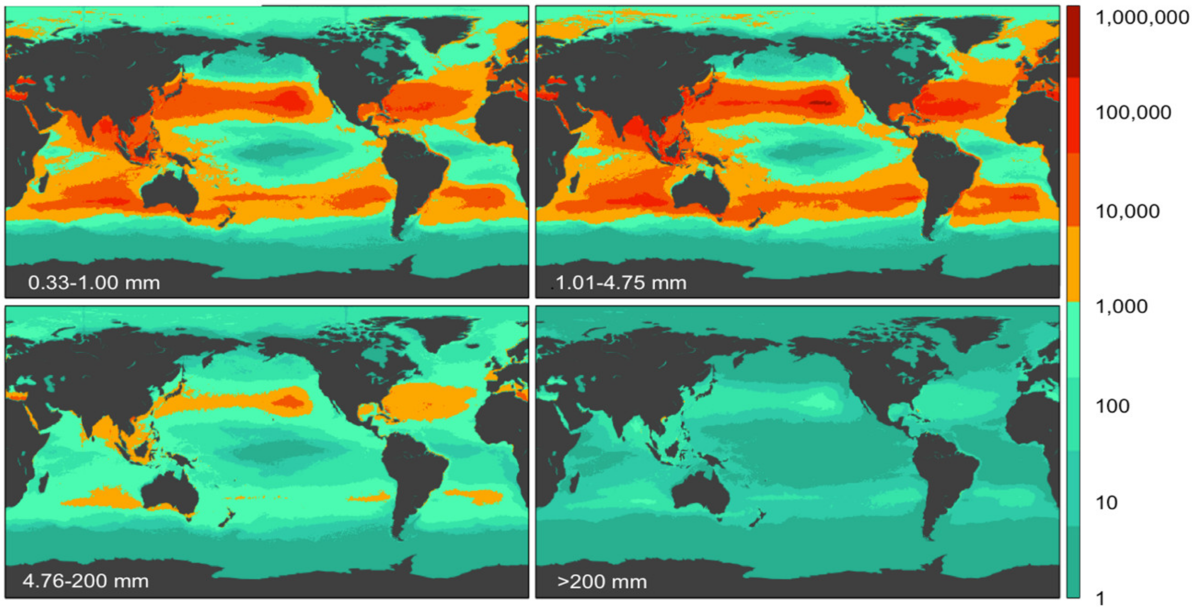
2.1. Microplastic Distribution in Marine
2.2. Microplastic Distribution in Land
2.3. Microplastic Distribution in Air
3. Microplastic Toxicity
4. Microplastic Analysis
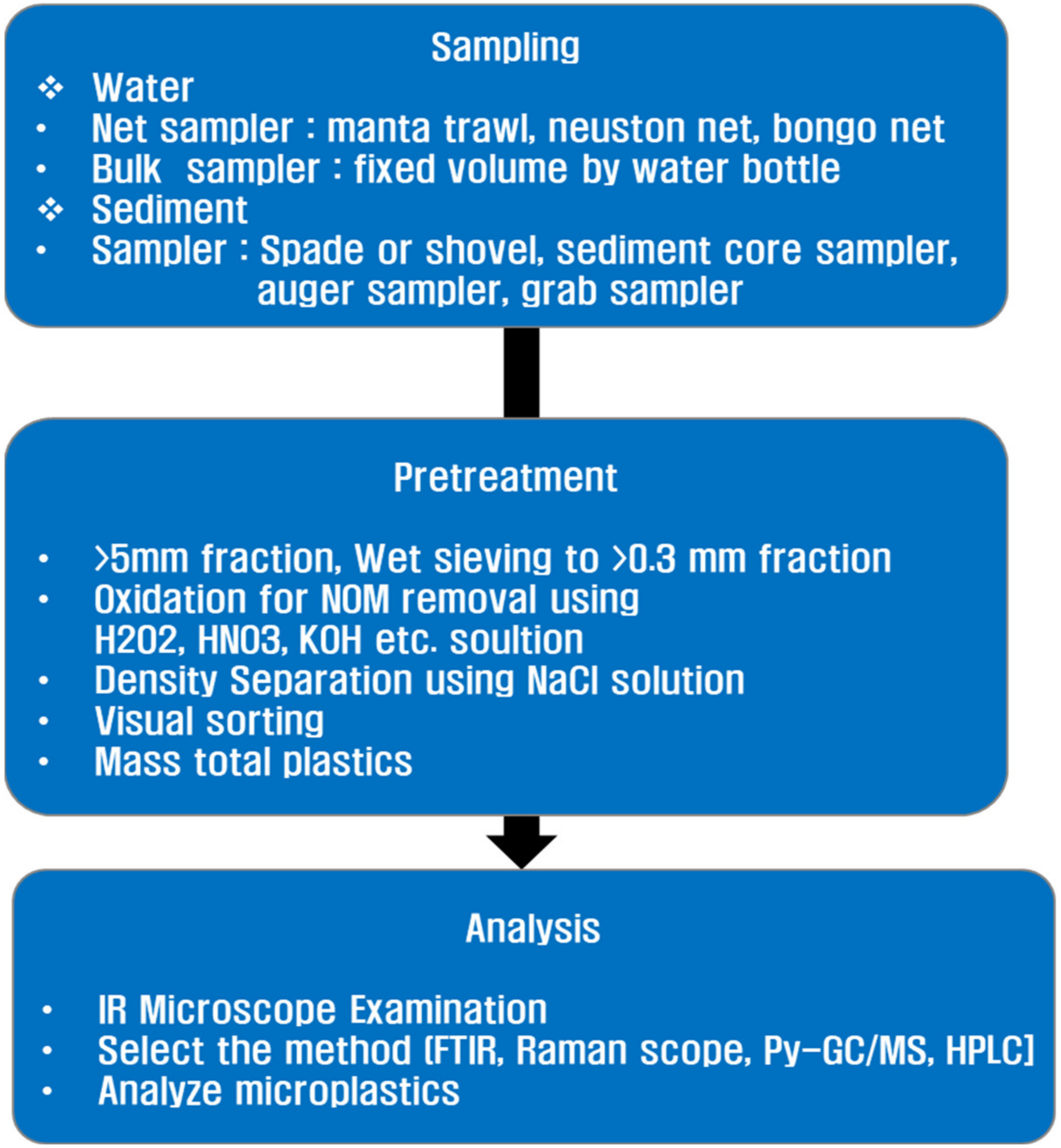
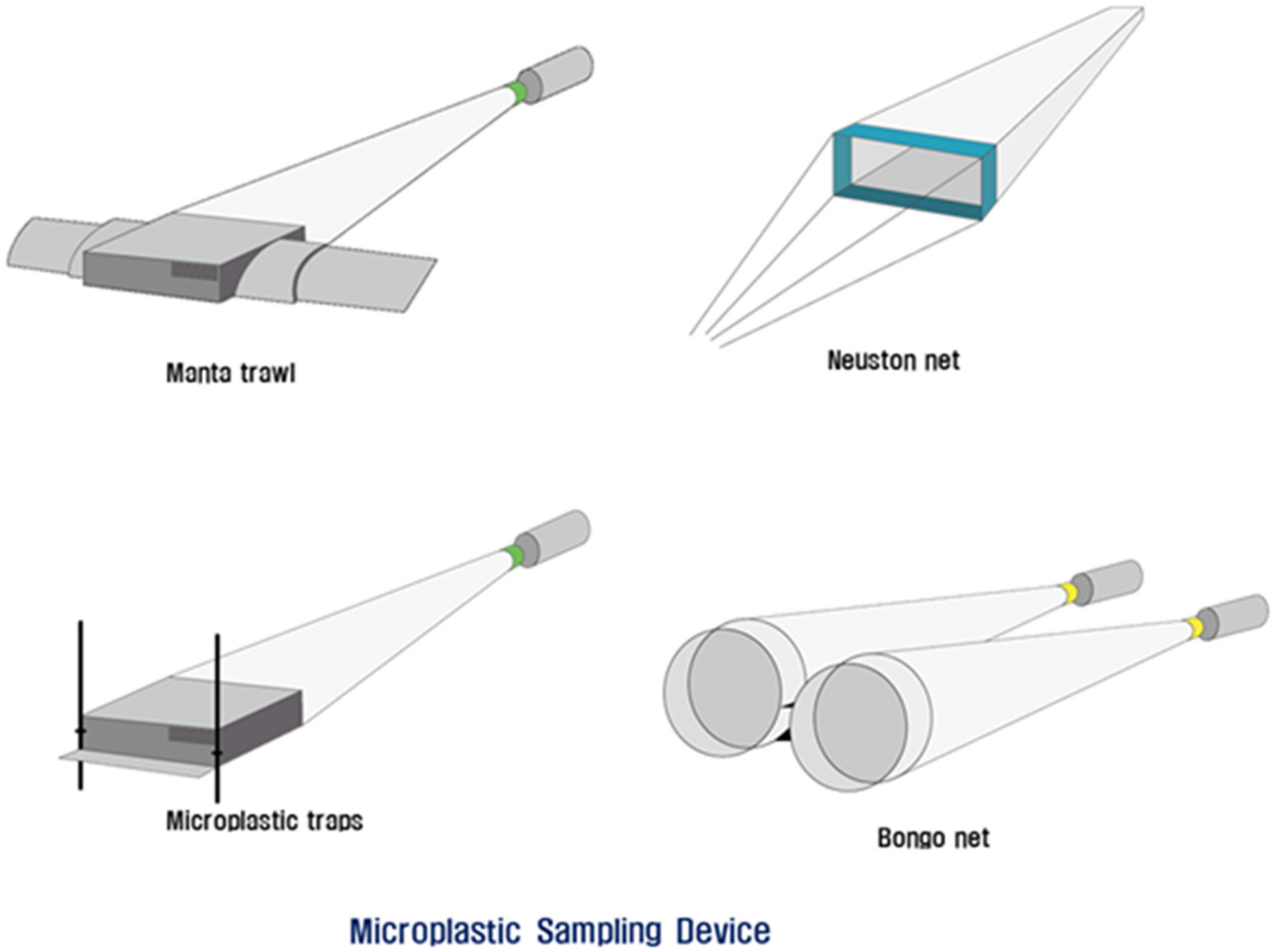
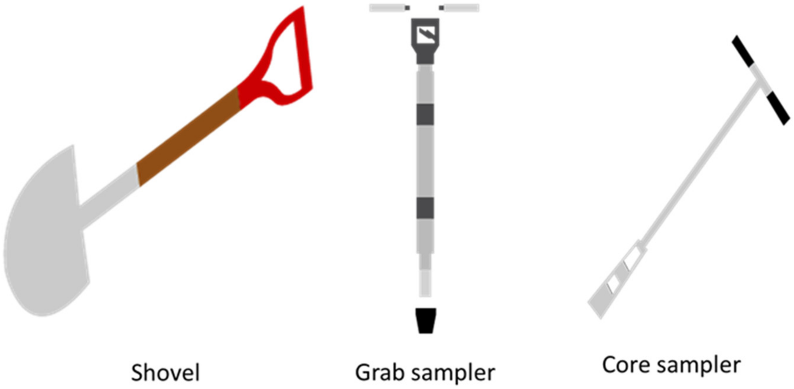
4.1. Sampling
4.1.1. Water Sampling
4.1.2. Sediment Sampling
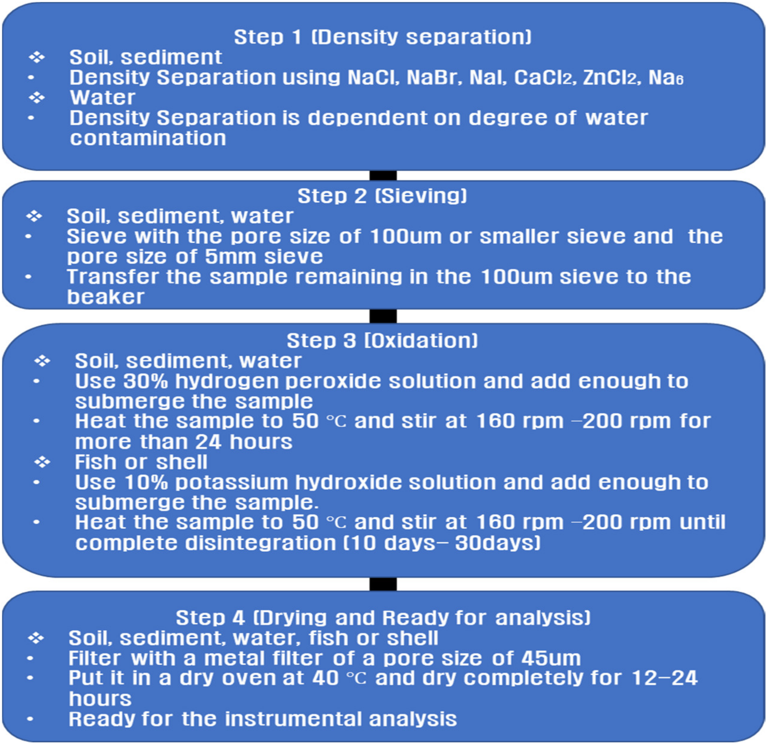
4.2. Pretreatment
4.3. Analysis Method
4.3.1. FTIR Spectroscopy
4.3.2. Raman Spectroscopy Method
4.3.3. GC/MS Method
5. Microplastic Removal Technology
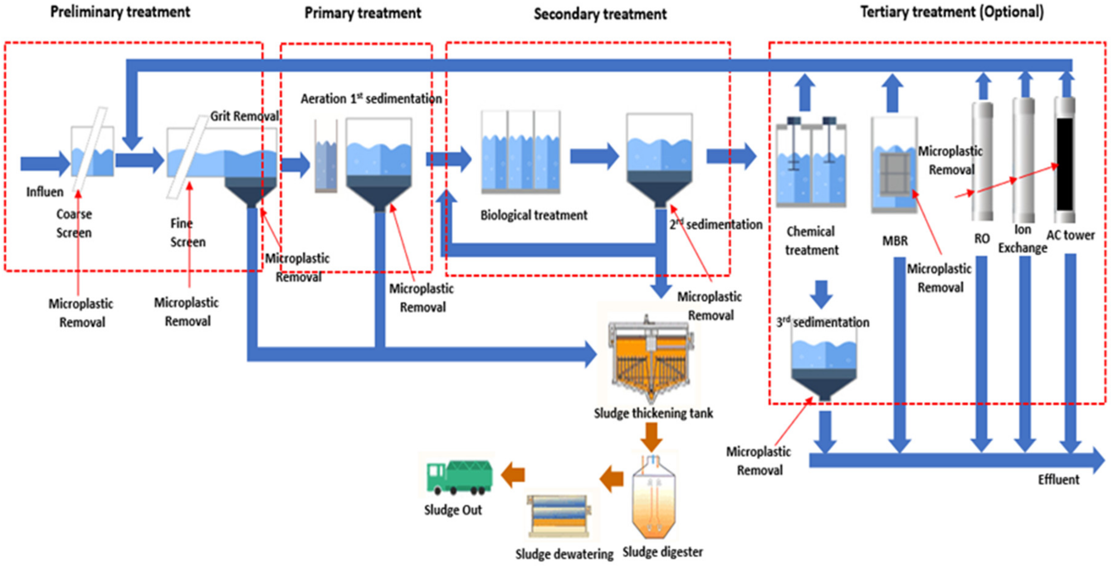
5.1. Wastewater Treatment Plants (WWTP)
5.2. Physical Removal Technology
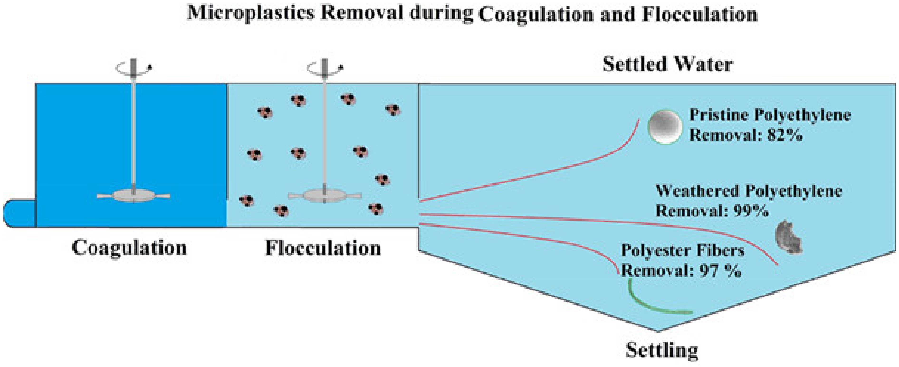
5.3. Chemical Removal Technology
5.4. Biological Removal Technology
6. Conclusions
- -
- Microplastics with the high contamination were reported as PET, PU, PS, PVC, PP, PE, and PA.
- -
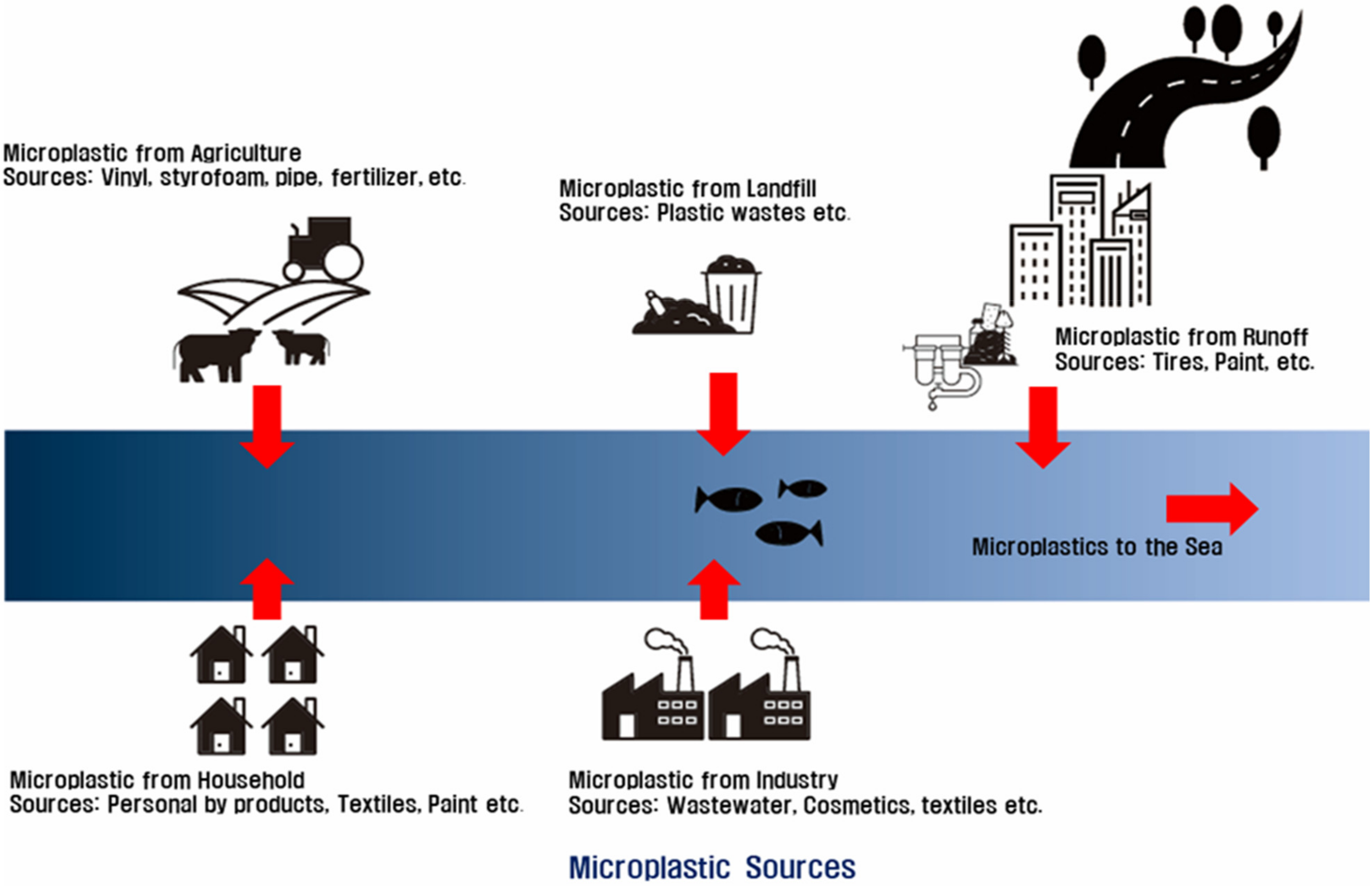
- Contamination paths of microplastics include agricultural wastewater, industrial wastewater, litter, sewage treatment plant, household personal products, road runoff, fishing waste, and atmosphere decomposition, which finally flow into the sea to pollute sea creatures and are absorbed by humans.
- -
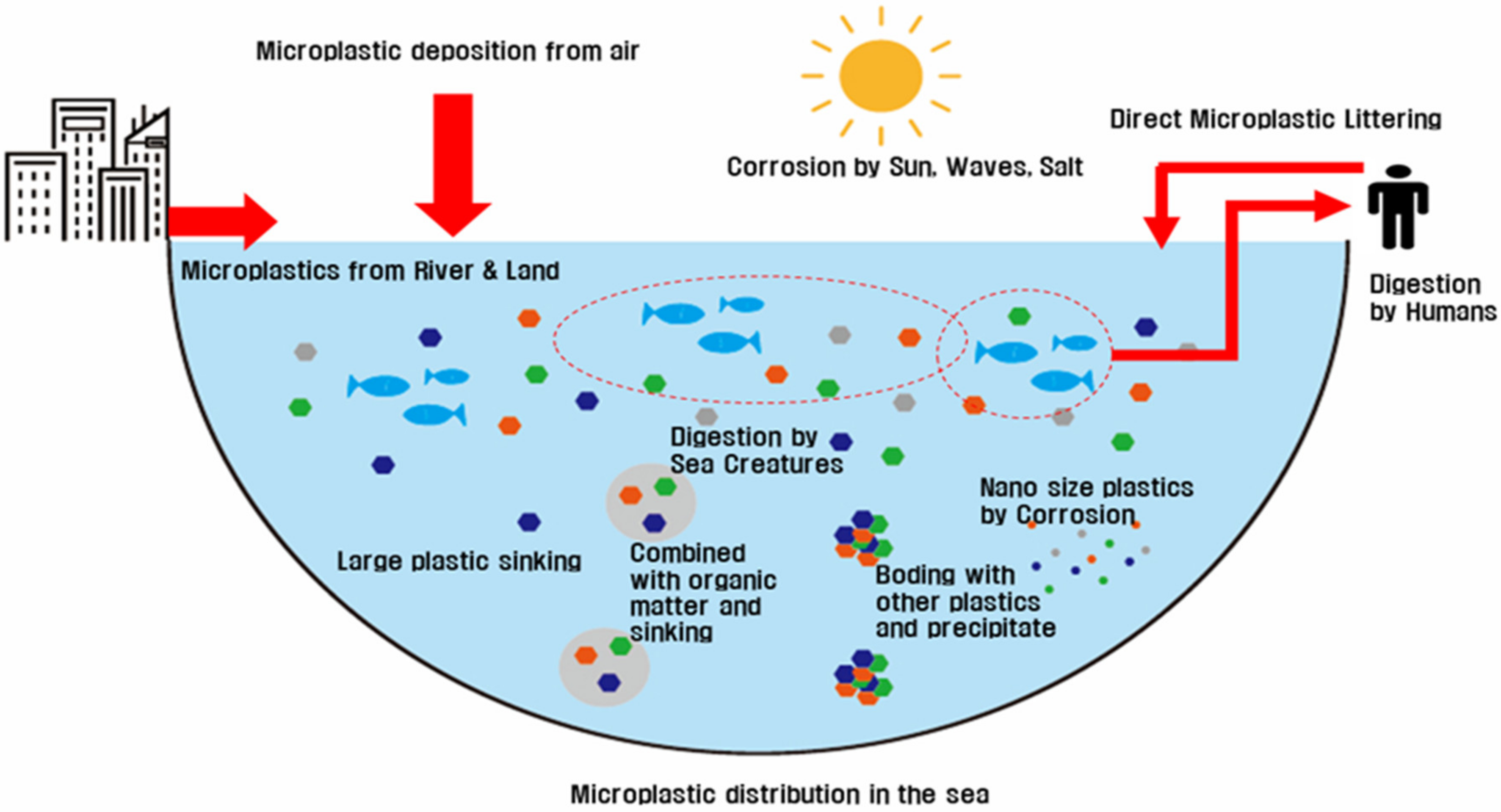
- The pollution of microplastics around the world is sharply increasing, and it is appearing in drinking water, sewage water, rivers, seas, soil, and everywhere. Finally, microplastics will cause a huge problem in the near future.
- -
- Although the toxicity of microplastics has not been studied much, plastics, such as PS, PVC, PP, etc., could still cause problems in human health, and several researchers are conducting research on the risk of microplastics.
- -
- Analysis of microplastics were divided into sampling, pretreatment, and analysis parts. Water sampling and sediment sampling were discussed in the sampling part. In the pretreatment part, how to deal with the density difference separation and how to remove other contaminants rather than microplastics were discussed in detail. In the analysis method part, most of used microplastic analytical methods and methods for a possible application for a microplastic analysis were summarized in detail.
- -
- Non-destructive analytical methods of FTIR and Raman methods were summarized by instrument settings and analytical results from other researchers. Destructive analytical methods of Pyr/GC/MS and LC/MS are summarized by instrument settings and analytical results from other researchers.
- -
- The various techniques for the removal method of microplastics were summarized in WWTP, physical, chemical, and biological technologies. Each technique for microplastic removal rate was summarized in several tables. In particular, the microplastic removal rate in WWTP was found to be more than 70% after secondary treatment, although there was a difference depending on the research papers.
Author Contributions
Funding
Institutional Review Board Statement
Informed Consent Statement
Data Availability Statement
Conflicts of Interest
References
- Plastics Europe. Plastics the Facts 2019: An Analysis of European Plastics Production, Demand and Waste Data. PlasticsEurope, Brussels. 2019. Available online: https://www.plasticseurope.org/application/files/9715/7129/9584/FINAL_web_version_Plastics_the_facts2019_14102019.pdf (accessed on 1 May 2021).
- Jambeck, J.R.; Geyer, R.; Wilcox, C.; Siegler, T.R.; Perryman, M.; Andrady, A.; Narayan, R.; Law, K.L. Plastic waste inputs from land into the ocean. Science 2015, 347, 768–771. [Google Scholar] [CrossRef]
- Barnes, D.K.A.; Galgani, F.; Thompson, R.C.; Barlaz, M. Accumulation and fragmentation of plastic debris in global environments. Philos. Trans. R. Soc. B Biol. Sci. 2009, 364, 1985–1998. [Google Scholar] [CrossRef] [Green Version]
- Rocha-Santos, T.; Duarte, A.C. A critical overview of the analytical approaches to the occurrence, the fate and the behavior of microplastics in the environment. Trends Anal. Chem. 2015, 65, 47–53. [Google Scholar] [CrossRef]
- Ng, E.L.; Huerta-Lwanga, E.; Eldridge, S.M.; Johnston, P.; Hu, H.-W.; Geissen, V.; Chen, D. An overview of microplastic and nanoplastic pollution in agroecosystems. Sci. Total Environ. 2018, 627, 1377–1388. [Google Scholar] [CrossRef] [PubMed]
- Cole, M.; Lindeque, P.; Halsband, C.; Galloway, T.S. Microplastics as contaminants in the marine environment: A review. Mar. Pollut. Bull. 2011, 62, 2588–2597. [Google Scholar] [CrossRef] [PubMed]
- Da Costa, J.P.; Santos, P.S.M.; Duarte, A.C.; Rocha-Santos, T. (Nano)plastics in the environment-Sources, fates and effects. Sci. Total Environ. 2016, 566, 15–26. [Google Scholar] [CrossRef]
- Andrady, A.L. Microplastics in the marine environment. Mar. Pollut. Bull. 2011, 62, 1596–1605. [Google Scholar] [CrossRef] [PubMed]
- Stephens, B.; Azimi, P.; El Orch, Z.; Ramos, T. Ultrafine particle emissions from desktop 3D printers. Atmos. Environ. 2013, 79, 334–339. [Google Scholar] [CrossRef]
- Ajith, N.; Arumugam, S.; Parthasarathy, S.; Manupoori, S.; Janakiraman, S. Global distribution of microplastics and its impact on marine environment-a review. Environ. Sci. Pollut. Res. Int. 2020, 27, 25970–25986. [Google Scholar] [CrossRef]
- Jones, J.I.; Vdovchenko, A.; Cooling, D.; Murphy, J.F.; Arnold, A.; Pretty, J.L.; Spencer, K.L.; Markus, A.A.; Vethaak, A.D.; Resmini, M. Systematic Analysis of the Relative Abundance of Polymers Occurring as Microplastics in Freshwaters and Estuaries. Int. J. Environ. Res. Public Health 2020, 17, 9304. [Google Scholar] [CrossRef]
- Efimova, I.; Bagaeva, M.; Bagaev, A.; Kileso, A.; Chubarenko, I.P. Secondary Microplastics Generation in the Sea Swash Zone With Coarse Bottom Sediments: Laboratory Experiments. Front. Mar. Sci. 2018, 5, 313. [Google Scholar] [CrossRef] [Green Version]
- Zhang, K.; Hamidian, A.H.; Tubić, A.; Zhang, Y.; Fang, J.K.H.; Wu, C.; Lam, P.K.S. Understanding plastic degradation and microplastic formation in the environment: A review. Environ. Pollut. 2021, 274, 116554. [Google Scholar] [CrossRef]
- Pop, C.-E.; Draga, S.; Măciucă, R.; Niță, R.; Crăciun, N.; Wolff, R. Bisphenol A Effects in Aqueous Environment on Lemna minor. Processes 2021, 9, 1512. [Google Scholar] [CrossRef]
- Bhatnagar, A.; Anastopoulos, I. Adsorptive removal of bisphenol A (BPA) from aqueous solution: A review. Chemosphere 2017, 168, 855–902. [Google Scholar] [CrossRef]
- Boyle, K.; Örmeci, B. Microplastics and Nanoplastics in the Freshwater and Terrestrial Environment: A Review. Water 2020, 12, 2633. [Google Scholar] [CrossRef]
- Sun, Q.; Ren, S.Y.; Ni, H.G. Incidence of microplastics in personal care products: An appreciable part of plastic pollution. Sci. Total Environ. 2020, 742, 140218. [Google Scholar] [CrossRef]
- Carr, S.A.; Liu, J.; Tesoro, A.G. Transport and fate of microplastic particles in wastewater treatment plants. Water Res. 2016, 91, 174–182. [Google Scholar] [CrossRef]
- Talvitie, J.; Mikola, A.; Setälä, O.; Heinonen, M.; Koistinen, A. How well is microlitter purified from wastewater?-A detailed study on the stepwise removal of microlitter in a tertiary level wastewater treatment plant. Water Res. 2017, 109, 164–172. [Google Scholar] [CrossRef] [PubMed] [Green Version]
- Simon, M.; Van Alst, N.; Vollertsen, J. Quantification of microplastic mass and removal rates at wastewater treatment plants applying Focal Plane Array (FPA)-based Fourier Transform Infrared (FT-IR) imaging. Water Res. 2018, 142, 1–9. [Google Scholar] [CrossRef]
- Sun, J.; Dai, X.; Wang, Q.; van Loosdrecht, M.C.M.; Ni, B.J. Microplastics in wastewater treatment plants: Detection, occurrence and removal. Water Res. 2019, 152, 21–37. [Google Scholar] [CrossRef]
- Mason, S.A.; Garneau, D.; Sutto, R.; Chu, Y.; Ehmann, K.; Barnes, J.; Fink, P.; Papazissimos, D.; Rogers, D.L. Microplastic pollution is widely detected in US municipal wastewater treatment plant effluent. Environ. Pollut. 2016, 218, 1045–1054. [Google Scholar] [CrossRef]
- Wang, W.; Ndungu, A.W.; Li, Z.; Wang, J. Microplastics pollution in inland freshwaters of China: A case study in urban surface waters of Wuhan, China. Sci. Total Environ. 2017, 575, 1369–1374. [Google Scholar] [CrossRef] [PubMed]
- Wang, T.; Wang, L.; Chen, Q.; Kalogerakis, N.; Ji, R.; Ma, Y. Interactions between microplastics and organic pollutants: Effects on toxicity, bioaccumulation, degradation, and transport. Sci. Total Environ. 2020, 748, 142427. [Google Scholar] [CrossRef] [PubMed]
- Piarulli, S.; Vanhove, B.; Comandini, P.; Scapinello, S.; Moens, T.; Vrielinck, H.; Sciutto, G.; Prati, S.; Mazzeo, R.; Booth, A.M.; et al. Do different habits affect microplastics contents in organisms? A trait-based analysis on salt marsh species. Mar. Pollut. Bull. 2020, 153, 110983. [Google Scholar] [CrossRef]
- Avio, C.G.; Gorbi, S.; Regoli, F. Plastics and microplastics in the oceans: From emerging pollutants to emerged threat. Mar. Environ. Res. 2017, 128, 2–11. [Google Scholar] [CrossRef] [PubMed]
- Isobe, A.; Iwasaki, S.; Uchida, K.; Tokai, T. Abundance of non-conservative microplastics in the upper ocean from 1957 to 2066. Nat. Commun. 2019, 10, 417. [Google Scholar] [CrossRef] [Green Version]
- Kim, H.J.; Lee, J.Y. Emerging Concerns about Microplastic Pollution on Groundwater in South Korea. Sustainability 2020, 12, 5275. [Google Scholar] [CrossRef]
- Lebreton, L.C.M.; van der Zwet, J.; Damsteeg, J.-W.; Slat, B.; Andrady, A.; Reisser, J. River plastic emissions to the world’s oceans. Nat. Commun. 2017, 8, 15611. [Google Scholar] [CrossRef]
- Eriksen, M.; Lebreton, L.C.M.; Carson, H.S.; Thiel, M.; Moore, C.J.; Borerro, J.C.; Galgani, F.; Ryan, P.G.; Reisser, J. Plastic Pollution in the World’s Oceans: More than 5 Trillion Plastic Pieces Weighing over 250,000 Tons Afloat at Sea. PLoS ONE 2014, 9, e111913. [Google Scholar] [CrossRef] [Green Version]
- Bakir, A.; Rowland, S.; Thompson, R. Enhanced desorption of persistent organic pollutants from microplastics under simulated physiological conditions. Environ. Pollut. 2014, 185, 16–23. [Google Scholar] [CrossRef]
- Isobe, A.; Uchida, K.; Tokai, T.; Iwasaki, S. East Asian seas: A hot spot of pelagic microplastics. Mar. Pollut. Bull. 2015, 101, 618–623. [Google Scholar] [CrossRef]
- Russell, M.; Webster, L. Microplastics in sea surface waters around Scotland. Mar. Pollut. Bull. 2021, 116, 112210. [Google Scholar] [CrossRef]
- Pan, Z.; Guo, H.; Chen, H.; Wang, S.; Sun, X.; Zou, Q.; Zhang, Y.; Lin, H.; Cai, S.; Huang, J. Microplastics in Northwestern Pacific: Abundance, distribution, and characteristics. Sci. Total Environ. 2019, 650, 1913–1922. [Google Scholar] [CrossRef] [PubMed]
- Lindeque, P.K.; Cole, M.; Coppock, R.L.; Lewis, C.N.; Miller, R.Z.; Watts, A.J.R.; Wilson-McNeal, A.; Wright, S.L.; Galloway, T.S. Are we underestimating microplastic abundance in the marine environment? A comparison of microplastic capture with nets of different mesh-size. Environ. Pollut. 2020, 265, 114721. [Google Scholar] [CrossRef] [PubMed]
- Jones, K.L.; Hartl, M.G.J.; Bell, M.C.; Capper, A. Microplastic accumulation in a Zostera marina L. bed at Deerness Sound, Orkney, Scotland. Mar. Pollut. Bull. 2020, 152, 110883. [Google Scholar] [CrossRef] [PubMed]
- Seng, N.; Lai, S.; Fong, J.; Saleh, M.F.; Cheng, C.; Cheok, Z.Y.; Todd, P.A. Early evidence of microplastics on seagrass and macroalgae. Mar. Freshw. Res. 2020, 71, 922–928. [Google Scholar] [CrossRef]
- Goss, H.; Jaskiel, J.; Rotjan, R. Thalassia testudinum as a potential vector for incorporating microplastics into benthic marine food webs. Mar. Pollut. Bull. 2018, 135, 1085–1089. [Google Scholar] [CrossRef]
- Cozzolino, L.; Nicastro, K.R.; Zardi, G.I.; de Los Santos, C. B Species-specific plastic accumulation in the sediment and canopy of coastal vegetated habitats. Sci. Total Environ. 2020, 723, 138018. [Google Scholar] [CrossRef]
- Tibbetts, J.; Krause, S.; Lynch, I.; Smith, G.H.S. Abundance Distribution and Divers of Microplastic Contamination in Urban River Environments. Water 2018, 10, 1597. [Google Scholar] [CrossRef] [Green Version]
- Park, T.-J.; Lee, S.-H.; Lee, M.-S.; Lee, J.-K.; Park, J.-H.; Zoh, K.-D. Distributions of Microplastics in Surface Water, Fish, and Sediment in the Vicinity of a Sewage Treatment Plant. Water 2020, 12, 3333. [Google Scholar] [CrossRef]
- Kosuth, M.; Mason, S.A.; Wattenberg, E.V. Anthropogenic contamination of tap water, beer, and sea salt. PLoS ONE 2018, 13, e0194970. [Google Scholar] [CrossRef]
- Mason, S.A.; Welch, V.G.; Neratko, J. Synthetic Polymer Contamination in Bottled Water. Front. Chem. 2018, 6, 407. [Google Scholar] [CrossRef] [PubMed] [Green Version]
- Ockelford, A.; Cundy, A.; Ebdon, J.E. Storm Response of Fluvial Sedimentary Microplastics. Sci. Rep. 2020, 10, 1865. [Google Scholar] [CrossRef]
- Galloway, T.; Cole, M.; Lewis, C. Interactions of microplastic debris throughout the marine ecosystem. Nat. Ecol. Evol. 2017, 1, 0116. [Google Scholar] [CrossRef] [PubMed]
- Vermeiren, P.; Muñoz, C.C.; Ikejima, K. Sources and sinks of plastic debris in estuaries: A conceptual model integrating biological, physical and chemical distribution mechanisms. Mar. Pollut. Bull. 2016, 113, 7–16. [Google Scholar] [CrossRef] [PubMed]
- Duis, K.; Coors, A. Microplastics in the aquatic and terrestrial environment: Sources (with a specific focus on personal care products), fate and effects. Environ. Sci. Eur. 2016, 28, 2. [Google Scholar] [CrossRef] [Green Version]
- Hitchcock, J.N. Storm events as key moments of microplastic contamination in aquatic ecosystems. Sci. Total Environ. 2020, 734, 139436. [Google Scholar] [CrossRef]
- Van den Berg, P.; Huerta-Lwanga, E.; Corradini, F.; Geissen, V. Sewage sludge application as a vehicle for microplastics in eastern Spanish agricultural soils. Environ. Poll. 2020, 261, 114198. [Google Scholar] [CrossRef]
- Huang, Y.; Liu, Q.; Jia, W.; Yan, C.; Wang, J. Agricultural plastic mulching as a source of microplastics in the terrestrial environment. Environ. Pollut. 2020, 260, 114096. [Google Scholar] [CrossRef]
- Weithmann, N.; Möller, J.N.; Löder, M.G.J.; Piehl, S.; Laforsch, C.; Freitag, R. Organic fertilizer as a vehicle for the entry of microplastic into the environment. Sci. Adv. 2018, 4, eaap8060. [Google Scholar] [CrossRef] [Green Version]
- Dioses-Salinas, D.C.; Pizarro-Ortega, C.I.; De-la-Torre, G.E. A methodological approach of the current literature on microplastic contamination in terrestrial environments: Current knowledge and baseline considerations. Sci. Total Environ. 2020, 730, 139164. [Google Scholar] [CrossRef]
- Wong, G.; Löwemark, L.; Kunz, A. Microplastic pollution of the Tamsui River and its tributaries in northern Taiwan: Spatial heterogeneity and correlation with precipitation. Environ. Pollut. 2020, 260, 113935. [Google Scholar] [CrossRef]
- Fuller, S.; Gautam, A. A Procedure for Measuring Microplastics using Pressurized Fluid Extraction. Environ. Sci. Technol. 2016, 50, 5774–5780. [Google Scholar] [CrossRef] [Green Version]
- Scheurer, M.; Bigalke, M. Microplastics in Swiss Floodplain Soils. Environ. Sci. Technol. 2018, 52, 3591–3598. [Google Scholar] [CrossRef]
- Corradini, F.; Meza, P.; Equiluz, R.; Casado, F.; Huerta-Lwanga, E.; Geissen, V. Evidence of microplastic accumulation in agricultural soils from sewage sludge disposal. Sci. Total Environ. 2019, 671, 411–420. [Google Scholar] [CrossRef] [PubMed]
- Harris, P.T. Review: The fate of microplastic in marine sedimentary environments: A review and synthesis. Mar. Poll. Bull. 2020, 158, 111398. [Google Scholar] [CrossRef]
- Yukioka, S.; Tanaka, S.; Nabetani, Y.; Suzuki, Y.; Ushijima, T.; Fujii, S.; Takada, H.; Van Tran, Q.; Singh, S. Occurrence and characteristics of microplastics in surface road dust in Kusatsu (Japan), Da Nang (Vietnam), and Kathmandu (Nepal). Environ. Pollut. 2020, 256, 113447. [Google Scholar] [CrossRef] [PubMed]
- Rhodes, C.J. Plastic pollution and potential solutions. Sci. Prog. 2018, 101, 207–260. [Google Scholar] [CrossRef] [PubMed]
- Vogelsang, C.; Lusher, A.; Sundvor, I.; Umar, M. Microplastics in Road Dust-Characteristics, Pathways and Measures; Technical Report, M-959; Norwegian Institute for Water Research: Oslo, Norway, 2018; Available online: https://www.miljodirektoratet.no/globalassets/publikasjoner/M959/M959.pdf (accessed on 10 July 2021).
- Liu, C.; Li, J.; Zhang, Y.; Wang, L.; Deng, J.; Gao, Y.; Yu, L.; Zhang, J.; Sun, H. Widespread distribution of PET and PC microplastics in dust in urban China and their estimated human exposure. Environ. Int. 2019, 128, 116–124. [Google Scholar] [CrossRef]
- Eriksen, M.; Thiel, M.; Prindiville, M.; Kiessling, T. Microplastic: What Are the Solution. Freshw. Microplast. 2018, 58, 273–298. [Google Scholar]
- Zhou, R.; Lu, G.; Yan, Z.; Jiang, R.; Bao, X.; Lu, P. A review of the influences of microplastics on toxicity and transgenerational effects of pharmaceutical and personal care products in aquatic environment. Sci. Total Environ. 2020, 732, 139222. [Google Scholar] [CrossRef] [PubMed]
- Webber, A.; van Randow, M.; Voigt, A.-L.; van der Au, M.; Fischer, E.; Meermann, B.; Wagner, M. Ingestion and toxicity of microplastics in the freshwater gastropod Lymnaea stagnalis: No microplastic-induced effects alone or in combination with copper. Chemosphere 2021, 263, 128040. [Google Scholar] [CrossRef] [PubMed]
- Gao, F.; Li, J.; Sun, C.; Zhang, L.; Jiang, F.; Cao, W.; Zheng, L. Study on the capability and characteristics of heavy metals enriched on microplastics in marine environment. Mar. Poll. Bull. 2019, 144, 61–67. [Google Scholar] [CrossRef] [PubMed]
- Xu, K.; Zhang, Y.; Huang, Y.; Wang, J. Toxicological effects of microplastics and phenanthrene to zebrafish (Danio rerio). Sci. Total Environ. 2021, 757, 143730. [Google Scholar] [CrossRef] [PubMed]
- Lu, K.; Qiao, R.; An, H.; Zhang, Y. Influence of microplastics on the accumulation and chronic toxic effects of cadmium in zebrafish (Danio rerio). Chemosphere 2018, 202, 504–520. [Google Scholar] [CrossRef] [PubMed]
- Yang, W.; Gao, X.; Wu, Y.; Wan, L.; Tan, L.; Yuan, S.; Ding, H.; Zhang, W. The combined toxicity influence of microplastics and nonylphenol on microalgae Chlorella pyrenoidosa. Ecotoxicol. Environ. Saf. 2020, 195, 110484. [Google Scholar] [CrossRef]
- Stock, V.; Böhmert, L.; Lisicki, E.; Block, R.; Cara-Carmona, J.; Pack, L.K.; Selb, R.; Lichtenstein, D.; Voss, L.; Henderson, C.J.; et al. Uptake and effects of orally ingested polystyrene microplastic particles in vitro and in vivo. Arch. Toxicol. 2019, 93, 1817–1833. [Google Scholar] [CrossRef]
- Wang, Q.; Bai, J.; Ning, B.; Fan, L.; Sun, T.; Fang, Y.; Wu, J.; Li, S.; Duan, C.; Zhang, Y.; et al. Effects of bisphenol A and nanoscale and microscale polystyrene plastic exposure on particle uptake and toxicity in human Caco-2 cells. Chemosphere 2020, 254, 126788. [Google Scholar] [CrossRef]
- Wang, Y.; Qian, H. Phthalates and Their Impacts on Human Health. Healthcare 2021, 9, 603. [Google Scholar] [CrossRef]
- Adam, V.; Yang, T.; Nowack, B. Toward an ecotoxicological risk assessment of microplastics: Comparison of available hazard and exposure data in freshwaters. Environ. Toxicol. Chem. 2018, 38, 436–447. [Google Scholar] [CrossRef] [Green Version]
- Besseling, E.; Redondo-Hasselerharm, P.; Foekema, E.M.; Koelmans, A.A. Quantifying ecological risks of aquatic micro-and nanoplastic. Crit. Rev. Environ. Sci. Technol. 2019, 49, 32–80. [Google Scholar] [CrossRef] [Green Version]
- Xu, P.; Peng, G.; Su, L.; Gao, Y.; Li, D. Microplastic risk assessment in surface waters: A case study in the Changjiang Estuary, China. Mar. Poll. Bull. 2018, 113, 647–654. [Google Scholar] [CrossRef] [PubMed]
- Zhou, Y.; Wang, J.; Zou, M.; Jia, Z.; Zhou, S.; Li, Y. Microplastics in soils: A review of methods, occurrence, fate, transport, ecological and environmental risks. Sci. Total Environ. 2020, 748, 141368. [Google Scholar] [CrossRef] [PubMed]
- Dey, T.K.; Uddin, M.E.; Jamal, M. Detection and removal of microplastics in wastewater: Evolution and impact. Environ. Sci. Pollut. Res. 2021, 28, 16925–16947. [Google Scholar] [CrossRef] [PubMed]
- Campanale, C.; Savino, I.; Pojar, I.; Massarelli, C.; Uricchio, V.F. A Practical Overview of Methodologies for Sampling and Analysis of Microplastics in Riverine Environments. Sustainability 2020, 12, 6755. [Google Scholar] [CrossRef]
- Barrows, A.P.W.; Neumann, C.A.; Berger, M.L.; Shaw, S.D. Grab vs. neuston tow net: A microplastic sampling performance comparison and possible advances in the field. Anal. Methods 2017, 9, 1446–1453. [Google Scholar] [CrossRef]
- Cutroneo, L.; Reboa, A.; Besio, G.; Borgogno, F.; Canesi, L.; Canuto, S.; Dara, M.; Enrile, F.; Forioso, I.; Greco, G.; et al. Microplastics in seawater: Sampling strategies, laboratory methodologies, and identification techniques applied to port environment. Environ. Sci. Pollut. Res. 2020, 27, 8938–8952. [Google Scholar] [CrossRef]
- Abeynayaka, A.; Kojima, F.; Miwa, Y.; Ito, N.; Nihei, Y.; Fukunaga, Y.; Yashima, Y.; Itsubo, N. Rapid Sampling of Suspended and Floating Microplastics in Challenging Riverine and Coastal Water Environments in Japan. Water 2020, 12, 1903. [Google Scholar] [CrossRef]
- Von Ammon, U.; Jeffs, A.; Zaiko, A.; van der Resi, A.; Goodwin, D.; Beckley, L.E.; Malpot, E.; Pochon, X. A Portable Cruising Speed Net: Expanding Global Collection of Sea Surface Plankton Data. Front. Mar. Sci. 2020, 7, 1109. [Google Scholar] [CrossRef]
- Vermaire, J.C.; Pomeroy, C.; Herczegh, S.M.; Haggart, O.; Murphy, M. Microplastic Abundance and Distribution in the Open Water and Sediment of the Ottawa River, Canada, and Its Tributaries. Facets 2017, 2, 301–314. [Google Scholar] [CrossRef] [Green Version]
- Dris, R.; Gasperi, J.; Rocher, V.; Saad, M.; Renault, N.; Tassin, B. Microplastic contamination in an urban area: A case study in Greater Paris. Environ. Chem. 2015, 12, 592–599. [Google Scholar] [CrossRef]
- SubCtech GmbH Subsea Technologies for the Marine Environment (Microplastic Sampler). Available online: https://subctech.com/wp-content/uploads/2020/09/SpecSheet_SubCtech_Microplastic-Sampler_ENG_v1-0.pdf (accessed on 23 June 2021).
- Yano, K.A.V.; Reyes, N.J.D.G.; Geronimo, F.K.F.; Jeon, M.S.; Kim, Y.; Kim, L.H. A comprehensive review of microplastics: Sources, pathways, and implications. J. Wetl. Res. 2020, 22, 153–160. [Google Scholar] [CrossRef]
- Vereiren, P.; Lercari, D.; Muñoz, C.C.; Ikejima, K.; Celentano, E.; Jorge-Romero, G.; Defeo, O. Sediment grain size determines microplastic exposure landscapes for sandy beach macroinfauna. Environ. Pollut. 2021, 286, 117308. [Google Scholar] [CrossRef]
- Willis, K.A.; Eriksen, R.; Wilcox, C.; Hardesty, B.D. Microplastic Distribution at Different Sediment Depths in an Urban Estuary. Front. Mar. Sci. 2017, 4, 419. [Google Scholar] [CrossRef] [Green Version]
- Kaile, N.; Lindivat, M.; Elio, J.; Thuestad, G.; Crowley, Q.G.; Hoell, I.A. Preliminary Results From Detection of Microplastics in Liquid Samples Using Flow Cytometry. Front. Mar. Sci. 2020, 7, 856. [Google Scholar] [CrossRef]
- Carter, M.R.; Gregorich, E.G. Soil Sampling and Methods of Analysis, 2nd ed.; Taylor & Francis Group: Abingdon, UK, 2006; Available online: https://www.niordc.ir/uploads/86_106_Binder1.pdf (accessed on 13 July 2021).
- Thomas, D.; Schütze, B.; Heinze, M.W.; Steinmetz, Z. Sample Preparation Techniques for the Analysis of Microplastics in Soil-A Review. Sustainability 2020, 12, 9074. [Google Scholar] [CrossRef]
- Hurley, R.R.; Lusher, A.L.; Olsen, M.; Nizzetto, L. Validation of a Method for Extracting Microplastics from Complex, Organic-Rich, Environmental Matrices. Environ. Sci. Technol. 2018, 52, 7409–7417. [Google Scholar] [CrossRef] [Green Version]
- Lusher, A.L.; Munno, K.; Hermabessiere, L.; Carr, S. Isolation and Extraction of Microplastics from Environmental Samples: An Evaluation of Practical Approaches and Recommendations for Further Harmonization. Appl. Spectrosc. 2020, 74, 1049–1065. [Google Scholar] [CrossRef]
- Nguyen, B.; Claveau-Mallet, D.; Hernandez, L.M.; Xu, E.G.; Farner, J.M.; Tufenkji, N. Separation and Analysis of Microplastics and Nanoplastics in Complex Environmental Samples. Acc. Chem. Res. 2019, 52, 858–866. [Google Scholar] [CrossRef] [Green Version]
- Renner, G.; Nellessen, A.; Schwiers, A.; Wenzel, M.; Schmidt, C.T.; Schram, J. Hydrophobicity–water/air–based enrichment cell for microplastics analysis within environmental samples: A proof of concept. MethodsX 2020, 7, 100732. [Google Scholar] [CrossRef]
- Al-Azzawi, M.S.M.; Kefer, S.; Weißer, J.; Reichel, J.; Schwaller, C.; Glas, K.; Knoop, O.; Drewes, J.E. Validation of Sample Preparation Methods for Microplastic Analysis in Wastewater Matrices-Reproducibility and Standardization. Water 2020, 12, 2445. [Google Scholar] [CrossRef]
- Yang, L.; Zhang, Y.; Kang, S.; Wang, Z.; Wu, C. Microplastics in freshwater sediment: A review on methods, occurrence, and sources. Sci. Total Environ. 2021, 754, 141948. [Google Scholar] [CrossRef] [PubMed]
- Radford, F.; Zapata-Restrepo, L.M.; Horton, A.A.; Hudson, M.D.; Shaw, P.J.; Williams, I.D. Developing a systematic method for extraction of microplastics in soils. Anal. Methods 2021, 13, 1695–1705. [Google Scholar] [CrossRef]
- Cole, M.; Webb, H.; Lindeque, P.K.; Fileman, E.S.; Halsband, C.; Galloway, T.S. Isolation of microplastics in biota-rich seawater samples and marine organisms. Sci. Rep. 2014, 4, 4528. [Google Scholar] [CrossRef] [PubMed] [Green Version]
- Kang, H.; Park, S.; Lee, B.; Ahn, J.; Kim, S. Modification of a Nile Red Staining Method for Microplastics Analysis: A Nile Red Plate Method. Water 2020, 12, 3251. [Google Scholar] [CrossRef]
- Shim, W.J.; Song, Y.K.; Hong, S.H.; Jang, M. Identification and quantification of microplastics using Nile Red staining. Mar. Pollut. Bull. 2016, 113, 469–476. [Google Scholar] [CrossRef]
- Hebner, T.S.; Maurer-Jones, M.A. Characterizing microplastic size and morphology of photodegraded polymers placed in simulated moving water conditions. Environ. Sci. Process. Impacts 2020, 22, 398–407. [Google Scholar] [CrossRef] [PubMed]
- Kumar, B.N.V.; Löschel, L.A.; Imhof, H.K.; Löder, M.G.J.; Laforsch, C. Analysis of microplastics of a broad size range in commercially important mussels by combining FTIR and Raman spectroscopy approaches. Environ. Pollut. 2021, 269, 116147. [Google Scholar] [CrossRef]
- Xu, J.-L.; Thomas, K.V.; Luo, Z.; Gowen, A.A. FTIR and Raman imaging for microplastics analysis: State of the art, challenges and prospects. TrAC Trends Anal. Chem. 2019, 119, 115629. [Google Scholar] [CrossRef]
- Vianello, A. A Journey into Microplastic Analysis Using FTIR Spectroscopy. Ph.D. Thesis, Aalborg University, Aalborg, Denmark, 2020. Available online: https://vbn.aau.dk/en/publications/a-journey-into-microplastic-analysis-using-ftir-spectroscopy (accessed on 6 May 2021).
- Rocchia, M.; Ruff, I.; Vianello, A. Microplastic Identification and Characterization by Raman Imaging Spectroscopy. In Thermoscientific White Paper; Thermoscientific: Milan, Italy, 2017; WP52981_E_10/17M; Available online: https://www.gammadata.se/assets/Uploads/Microplastic-and-Raman.pdf (accessed on 10 May 2021).
- Müller, A.; Goedecke, C.; Eisentraut, P.; Piechotta, C.; Braun, U. Microplastic analysis using chemical extraction followed by LC-UV analysis: A straightforward approach to determine PET content in environmental samples. Environ. Sci. Eur. 2020, 32, 85. [Google Scholar] [CrossRef]
- Wang, L.; Zhang, J.; Hou, S.; Sun, H. A Simple Method for Quantifying Polycarbonate and Polyethylene Terephthalate Microplastics in Environmental Samples by Liquid Chromatography–Tandem Mass Spectrometry. Environ. Sci. Technol. Lett. 2017, 4, 530–534. [Google Scholar] [CrossRef]
- Funck, M.; Yildirim, A.; Nickel, C.; Schram, J.; Schmidt, T.C.; Tuerka, J. Identification of microplastics in wastewater after cascade filtration using Pyrolysis-GC–MS. MethodsX 2020, 7, 100778. [Google Scholar] [CrossRef] [PubMed]
- Primpke, S.; Fischer, M.; Lorenz, C.; Gerdts, G.; Scholz-Böttcher, B.M. Comparison of pyrolysis gas chromatography/mass spectrometry and hyperspectral FTIR imaging spectroscopy for the analysis of microplastics. Anal. Bioanal. Chem. 2020, 412, 8283–8298. [Google Scholar] [CrossRef] [PubMed]
- Reichel, J.; Graßmann, J.; Letzel, T.; Drewes, J.E. Systematic Development of a Simultaneous Determination of Plastic Particle Identity and Adsorbed Organic Compounds by Thermodesorption–-Pyrolysis GC/MS (TD-Pyr-GC/MS). Molecules 2020, 25, 4985. [Google Scholar] [CrossRef] [PubMed]
- Asamoah, B.O.; Kanyathare, B.; Russey, M.; Peiponen, K.-E. A prototype of a portable optical sensor for the detection of transparent and translucent microplastics in freshwater. Chemosphere 2019, 231, 161–167. [Google Scholar] [CrossRef]
- Valsesia, A.; Parot, J.; Ponti, J.; Mehn, D.; Marino, R.; Melillo, D.; Muramoto, S.; Verkouteren, M.; Hackley, V.A.; Colpo, P. Detection, counting and characterization of nanoplastics in marine bioindicators: A proof of principle study. Microplast. Nanoplast. 2021, 1, 1–13. [Google Scholar] [CrossRef]
- Lambert, S.; Wagner, M. Characterisation of nanoplastics during the degradation of polystyrene. Chemosphere 2016, 145, 265–268. [Google Scholar] [CrossRef] [Green Version]
- Karakolis, E.G.; Nguyen, B.; You, J.B.; Rochman, C.M.; Sinton, D. Fluorescent Dyes for Visualizing Microplastic Particles and Fibers in Laboratory-Based Studies. Environ. Sci. Technol. Lett. 2019, 6, 334–340. [Google Scholar] [CrossRef]
- Luo, H.; Xiang, Y.; Zhao, Y.; Li, Y.; Pan, X. Nanoscale infrared, thermal and mechanical properties of aged microplastics revealed by an atomic force microscopy coupled with infrared spectroscopy (AFM-IR) technique. Sci. Total Environ. 2020, 774, 140944. [Google Scholar] [CrossRef]
- Tagg, A.S.; Sapp, M.; Harrison, J.P.; Ojeda, J.J. Identification and Quantification of Microplastics in Wastewater Using Focal Plane Array-Based Reflectance Micro-FT-IR Imaging. Anal. Chem. 2015, 87, 6032–6040. [Google Scholar] [CrossRef] [Green Version]
- Piehl, S.; Leibner, A.; Löder, M.G.J.; Dris, R.; Bogner, C.; Laforsch, C. Identification and quantification of macro- and microplastics on an agricultural farmland. Sci. Rep. 2018, 8, 17950. [Google Scholar] [CrossRef] [Green Version]
- Scopetani, C.; Chelazzi, D.; Cincinelli, A.; Esterhuizen-Londt, M. Assessment of microplastic pollution: Occurrence and characterisation in Vesijärvi lake and Pikku Vesijärvi pond, Finland. Environ. Monit. Assess. 2019, 191, 652. [Google Scholar] [CrossRef] [Green Version]
- Kunz, A.; Walther, B.A.; Lowemark, L.; Lee, Y.C. Distribution and quantity of microplastic on sandy beaches along the northern coast of Taiwan. Mar. Pollut. Bull. 2016, 111, 126–135. [Google Scholar] [CrossRef]
- Karthik, R.; Robin, R.S.; Purvaja, R.; Ganguly, D.; Anandavelu, I.; Raghuraman, R.; Hariharan, G.; Ramakrishna, A.; Ramesh, R. Microplastics along the beaches of southeast coast of India. Sci. Total Environ. 2018, 645, 1388–1399. [Google Scholar] [CrossRef]
- Levermore, J.M.; Smith, T.E.L.; Kelly, F.J.; Wright, S.L. Detection of Microplastics in Ambient Particulate Matter Using Raman Spectral Imaging and Chemometric Analysis. Anal. Chem. 2020, 92, 8732–8740. [Google Scholar] [CrossRef] [PubMed]
- Zhao, S.; Danley, M.; Ward, J.E.; Li, D.; Mincer, T.J. An approach for extraction, characterization and quantitation of microplastic in natural marine snow using Raman microscopy. Anal. Methods 2016, 9, 1470–1478. [Google Scholar] [CrossRef]
- Saur, T. Global Assessment of Microplastic Pollution in Wastewater Treatment Plants. OECD Workshop on Microplastics from Synthetic Textiles in the Environment: Knowledge, Mitigation and Policy. 11 February 2020. Available online: https://www.oecd.org/water/Draft_Agenda_Public_OECD_Workshop_MP_Textile.pdf (accessed on 11 May 2021).
- Hendrickson, E.; Minor, E.C.; Schreiner, K. Microplastic Abundance and Composition in Western Lake Superior As Determined via Microscopy, Pyr-GC/MS, and FTIR. Environ. Sci. Technol. 2018, 52, 1787–1796. [Google Scholar] [CrossRef] [PubMed]
- Bratton, S.P.; Halbur, J.; Schreiner, K.; Minor, E.C.; Hendrickson, E.; Peters, C.A. Pyr-GC/MS analysis of microplastics extracted from the stomach content of benthivore fish from the Texas Gulf Coast. Mar. Pollut. Bull. 2018, 137, 91–95. [Google Scholar] [CrossRef]
- Zhang, J.; Wang, L.; Kannan, K. Microplastics in house dust from 12 countries and associated human exposure. Environ. Int. 2020, 134, 105314. [Google Scholar] [CrossRef] [PubMed]
- Santos, D.I.; Correia, M.J.N.; Mateus, M.M.; Saraiva, J.A.; Vicente, A.A.; Moldão, M. Fourier Transform Infrared (FT-IR) Spectroscopy as a Possible Rapid Tool to Evaluate Abiotic Stress Effects on Pineapple By-Product. Appl. Sci. 2019, 9, 4141. [Google Scholar] [CrossRef] [Green Version]
- Ahmed, M.B.; Rahman, M.S.; Alom, J.; Hasan, M.S.; Johir, M.A.H.; Mondal, M.I.H.; Lee, D.-Y.; Park, J.; Zhou, J.L.; Yoon, M.H. Microplastic particles in the aquatic environment: A systematic review. Sci. Total Environ. 2021, 775, 145793. [Google Scholar] [CrossRef]
- Wagner, M.; Scherer, C.; Alvarez-Muñnoz, D.; Brennholt, N.; Bourrain, X.; Buchinger, S.; Fries, E.; Grosbois, C.; Klasmeier, J.; Marti, T.; et al. Microplastics in freshwater ecosystems: What we know and what we need to know. Environ. Sci. Eur. 2014, 26, 12. [Google Scholar] [CrossRef] [Green Version]
- Hidalgo-Ruz, V.; Gutow, L.; Thompson, R.C.; Thiel, M. Microplastics in the marine environment: A review of the methods used for identification and quantification. Environ. Sci. Technol. 2012, 46, 3060–3075. [Google Scholar] [CrossRef] [PubMed]
- Olesen, K.B.; van Alst, N.; Simon, M.; Vianello, A.; Liu, F.; Vellertsen, J. Analysis of Microplastics using FTIR Imaging Application Note, Method January 2017. Available online: https://vbn.aau.dk/en/publications/analysis-of-microplastics-using-ftir-imaging-application-note (accessed on 12 May 2021).
- Xu, G.; Cheng, H.; Jones, R.; Feng, Y.; Gong, K.; Li, K.; Fang, X.; Tahir, M.A.; Valev, V.K.; Zhang, L. Surface-Enhanced Raman Spectroscopy Facilitates the Detection of Microplastics <1 μm in the Environment. Environ. Sci. Technol. 2020, 54, 15594–15603. [Google Scholar] [CrossRef] [PubMed]
- Löder, M.G.J.; Gerdts, G. Methodology Used for the Detection and Identification of Microplastics—A Critical Appraisal. In Marine Anthropogenic Litter; Bergmann, M., Gutow, L., Klages, M., Eds.; Springer: Berlin/Heidelberg, Germany, 2015. [Google Scholar] [CrossRef] [Green Version]
- Scircle, A.; Cizdziel, J.V.; Tisinger, L.; Anumol, T.; Robey, D. Occurrence of Microplastic Pollution at Oyster Reefs and Other Coastal Sites in the Mississippi Sound, USA: Impacts of Freshwater Inflows from Flooding. Toxics 2020, 8, 35. [Google Scholar] [CrossRef]
- Ainali, N.M.; Kalaronis, D.; Kontogiannis, A.; Evgenidu, E.; Kyzas, G.Z.; Yang, X.; Bikiaris, D.N.; Lambropoulou, D.A. Microplastics in the environment: Sampling, pretreatment, analysis and occurrence based on current and newly-exploited chromatographic approaches. Sci. Total Environ. 2021, 794, 148725. [Google Scholar] [CrossRef] [PubMed]
- Cowger, W.; Gray, A.; Christiansen, S.H.; DeFrond, H.; Deshpande, A.D.; Hemabessiere, L.; Lee, E.; Mill, L.; Munno, K.; Ossmann, B.E.; et al. Critical Review of Processing and Classification Techniques for Images and Spectra in Microplastic Research. Appl. Spectrosc. 2020, 74, 989–1010. [Google Scholar] [CrossRef] [PubMed]
- Wu, M.; Tang, W.; Wu, S.; Liu, H.; Yang, C. Fate and effects of microplastics in wastewater treatment processes. Sci. Total Environ. 2020, 757, 143902. [Google Scholar] [CrossRef]
- Herbort, A.F.; Sturm, M.T.; Schuhen, K. Technological Approaches for the Reduction of Microplastic Pollution in Seawater Desalination Plants and for Sea Salt Extraction. In Plastics in the Environment; IntechOpen: London, UK, 2019. [Google Scholar] [CrossRef] [Green Version]
- Yahyanezhad, N.; Bardi, M.J.; Aminirad, H. An evaluation of microplastics fate in the wastewater treatment plants: Frequency and removal of microplastics by microfiltration membrane. Water Pract. Technol. 2021, 16, 782–792. [Google Scholar] [CrossRef]
- Lv, X.; Dong, Q.; Zuo, Z.; Liu, Y.; Huang, X.; Wu, W.-M. Microplastics in a municipal wastewater treatment plant: Fate, dynamic distribution, removal efficiencies, and control strategies. J. Clean. Prod. 2019, 225, 579–586. [Google Scholar] [CrossRef]
- Conley, K.; Clum, A.; Deepe, J.; Lane, H.; Beckingham, B. Wastewater treatment plants as a source of microplastics to an urban estuary: Removal efficiencies and loading per capita over one year. Water Res. X 2019, 3, 100030. [Google Scholar] [CrossRef] [PubMed]
- Edo, C.; González-Pleiter, M.; Leganés, F.; Fernández-Piñas, F.; Rosal, R. Fate of microplastics in wastewater treatment plants and their environmental dispersion with effluent and sludge. Environ. Pollut. 2020, 259, 113837. [Google Scholar] [CrossRef] [PubMed]
- Ziajahromi, S.; Neale, P.A.; Rintoul, L.; Leusch, F.D.L. Wastewater treatment plants as a pathway for microplastics: De-velopment of a new approach to sample wastewater-based microplastics. Water Res. 2017, 112, 93–99. [Google Scholar] [CrossRef]
- Tagg, A.S.; Sapp, M.; Harrion, J.P.; Sinclair, C.J.; Bradley, E.; Nam, Y.J.; Ojeda, J.J. Microplastic Monitoring at Different Stages in a Wastewater Treatment Plant Using Reflectance Micro-FTIR Imaging. Front. Environ. Sci. 2020, 8, 145. [Google Scholar] [CrossRef]
- Talvitie, J.; Mikola, A.; Koistinen, A.; Setälä, O. Solutions to microplastic pollution—Removal of microplastics from wastewater effluent with advanced wastewater treatment technologies. Water Res. 2017, 123, 401–407. [Google Scholar] [CrossRef] [Green Version]
- Ross, P.S. Tackling Microfiber Pollution at Source a Solution-Oriented Partnership Across Public and Private Sectors. OECD Workshop on Microplastics from Synthetic Textiles in the Environment: Knowledge, Mitigation and Policy. 11 February 2020. Available online: https://www.oecd.org/water/Draft_Agenda_Public_OECD_Workshop_MP_Textile.pdf (accessed on 12 May 2021).
- Lare, M.; Ncibi, M.C.; Sillanpää, M. Occurrence, identification and removal of microplastic particles and fibers in conventional activated sludge process and advanced MBR technology. Water Res. 2018, 133, 236–246. [Google Scholar] [CrossRef] [PubMed]
- Yang, L.; Li, K.; Cui, S.; Kang, Y.; An, L.; Lei, K. Removal of microplastics in municipal sewage from China’s largest water reclamation plant. Water Res. 2019, 155, 175–181. [Google Scholar] [CrossRef]
- Simon, M.; Vianello, A.; Vollertsen, J. Removal of >10 µm Microplastic Particles from Treated Wastewater by a Disc Filter. Water 2019, 11, 1935. [Google Scholar] [CrossRef] [Green Version]
- Wolff, S.; Weber, F.; Kerpen, J.; Winklhofer, M.; Engelhart, M.; Backman, L. Elimination of Microplastics by Downstream Sand Filters in Wastewater Treatment. Water 2021, 13, 33. [Google Scholar] [CrossRef]
- Bayo, J.; Lopez-Castellanos, J.; Olmos, S. Abatement of microplastics from Municipal effluents by two different wastewater treatment technologies. Trans. Ecol. Environ. 2020, 242, 15–26. [Google Scholar] [CrossRef]
- Pizzichetti, A.R.P.; Pablos, C.; Álvarez-Fernández, C.; Reynolds, K.; Stanley, S.; Marugán, J. Evaluation of membranes performance for microplastic removal in a simple and low-cost filtration system. Case Stud. Chem. Environ. Eng. 2021, 3, 100075. [Google Scholar] [CrossRef]
- Li, Q.-C.; Lai, Y.-J.; Yu, S.-J.; Li, P.; Zhou, X.-X.; Dong, L.-J.; Liu, X.; Yao, Z.-W.; Liu, J.-F. Sequential Isolation of Microplastics and Nanoplastics in Environmental Waters by Membrane Filtration, Followed by Cloud-Point Extraction. Anal. Chem. 2021, 93, 4559–4566. [Google Scholar] [CrossRef]
- Kim, K.T.; Park, S.H. Enhancing Microplastics Removal from Wastewater Using Electro-Coagulation and Granule-Activated Carbon with Thermal Regeneration. Processes 2021, 9, 617. [Google Scholar] [CrossRef]
- Lapointe, M.; Farner, J.M.; Hernandez, L.M.; Tufenkji, N. Understanding and Improving Microplastic Removal during Water Treatment: Impact of Coagulation and Flocculation. Environ. Sci. Technol. 2020, 54, 8719–8727. [Google Scholar] [CrossRef] [PubMed]
- Perren, W.; Wojtasik, A.; Cai, Q. Removal of Microbeads from Wastewater Using Electrocoagulation. ACS Omega 2018, 3, 3357–3364. [Google Scholar] [CrossRef] [PubMed]
- Park, J.W.; Lee, S.J.; Hwang, D.Y.; Seo, S. Removal of microplastics via tannic acid-mediated coagulation and in vitro impact assessment. RSC Adv. 2021, 11, 3556–3566. [Google Scholar] [CrossRef]
- Ma, B.; Xue, W.; Hu, C.; Liu, H.; Qu, J.; Li, L. Characteristics of microplastic removal via coagulation and ultrafiltration during drinking water treatment. Chem. Eng. J. 2019, 359, 159–167. [Google Scholar] [CrossRef]
- Liu, H.; Zhou, X.; Ding, W.Q.; Nghiem, L.D.; Sun, J.; Wang, Q.L. Do microplastics affect biological wastewater treatment performance? Implications from bacterial activity experiments. ACS Sustain. Chem. Eng. 2019, 7, 20097–20101. [Google Scholar] [CrossRef]
- Cunha, C.; Silva, L.; Paulo, J.; Faria, M.; Nogueira, N.; Cordeiro, N. Microalgal-based biopolymer for nano- and microplastic removal: A possible biosolution for wastewater treatment. Environ. Pollut. 2020, 263, 114385. [Google Scholar] [CrossRef]
- Canniff, P.M.; Hoang, T.C. Microplastic ingestion by Daphina magna and its enhancement on algal growth. Sci. Total Environ. 2018, 633, 500–507. [Google Scholar] [CrossRef] [PubMed] [Green Version]
- Liu, W.; Zhang, J.; Liu, H.; Guo, X.; Zhang, X.; Yao, X.; Cao, Z.; Zhang, T. A review of the removal of microplastics in global wastewater treatment plants: Characteristics and mechanisms. Environ. Int. 2021, 146, 106277. [Google Scholar] [CrossRef]


| Category | Source | Potential Mitigation |
|---|---|---|
| Production | Microplastics in additives | Removing them from products. Replace with benign alternatives |
| Mismanaged preproduction pellets | Regulate pellet handling. Operation clean sweep | |
| Commerce | Industrial abrasives | Improve containment and recovery and require alternatives |
| Laundromat exhaust | Improved filtration | |
| Agriculture-degraded film, pots, and pipes | Improve recovery, biodegradable plastics | |
| Consumer | Tire dust | Technological advances, road surface |
| Littering of small plastic items (cigarette filters, torn corners of packaging, small film wrappers, etc.) | Enforcement of fines for littering, Consumer education, EPR on design | |
| Domestic laundry. Wastewater effluent | Wash with top-load machines, wastewater containment, single-filter woven textiles, textile coatings | |
| Waste management | Fragmentation by vehicles driving over unrecovered waste | Improved waste management |
| UV and chemically degraded terrestrial plastic waste | Improved waste management | |
| Sewage effluent (synthetic fibers) | Laundry filtration, textile industry innovation | |
| Combined sewage overflow (large items) | Infrastructure improvement | |
| Mechanical shredding of roadside waste during regular cutting of vegetation (mostly grass) | Better legislation and law enforcement; valorization of waste products |
| Value of polymer index | <10 | 10–100 | 100–1000 | >1000 |
| Value of pollution load index | <10 | 10–20 | 20–30 | >30 |
| Risk category | I | II | III | IV |
| Methods for Microplastics | Analytical Method | Type of Source | Pretreatment | Particle Size | Types of Polymer | Identification | References |
|---|---|---|---|---|---|---|---|
| Non-Destructive method | FTIR | Wastewater in Derby, UK | Density separation, oxidation (30% H2O2) for 7 days | >5 mm | PE, PP, PVC, PS, nylon-6 | Reproducible identification rate: 98.33% 50 MPs/10 L | [116] |
| ATR (Attenuated Total Reflection)-FTIR | Agricultural soil in Middle Franconia, Germany | Density separation oxidation (30% H2O2) | 1 to 5 mm | PE, PS, PVC, PET, PMMA | 0.34 ± 0.36 MPs/kg dry soil | [117] | |
| µFTIR | Sediment of the lagoon in Italy | Density separation | - | - | Up to 2175 MP/kg | [104] | |
| µFTIR | Air in Denmark | Sonicate, dry | 11 µm | PE | 9.3 ± 5.8 MPs/m3 | [104] | |
| FTIR | Sediment, Snow, Ice core in the lake in Finland | Filtration, density separation | >1.2 µm | PA, PE, PP, cellulose, wool | 395.5 ± 90.7 MPs/kg, 117.1 ± 18.4 MPs/L, 7.8 ± 1.2 MPs/L | [118] | |
| Synchrotron-based FTIR | Beach Sediment in Taiwan | Density separation | ≥1 mm | PE, PP | 4–532 MPs/0.0125 m3 | [119] | |
| FTIR | 25 Beach Sediments in India | Sieving, density separation | <5 mm | PE, PP, PS | 178 ± 261 mg/m2 (low tide) 1323 ± 1228 mg/m2 | [120] | |
| FTIR | 79 fishes in India | Density seperation, oxidation (30% H2O2) | - | - | 10.1% with 79 fishes | [120] | |
| FTIR | Treated wastewater, Sediment in Germany | Density separation, sieving, filtratrion | ≤500 μm | PE, PP, PET, PS, PVC, PC, PUR, PA | 39–37,223 MPs/m3 (wastewater) 8–20 MPs/m3 (surface water) 143–1151 MPs/kg (sediment) | [109] | |
| Raman | Standards from Sigma-Aldrich | - | 74 μm PE 37–74 μm PS 27–45 μm PE | PE, PS-DVB | <1 μm visible | [105] | |
| Raman | Air in London, UK | Density separation, | ≥2 μm | PE, PET, PP | PE:2467.9 MPs/m3, PP:22.4 MPs/m3, PET:11.2 MPs/m3 | [121] | |
| Raman | Sea snow in CT, USA | Density separation, 15% H2O2 | 63–600 μm | PP, PET | 59 MPs/4 L | [122] |
| Methods for Microplastics | Analytical Method | Type of Source | Pretreatment | Particle Size | Types of Polymer | Identification | References |
|---|---|---|---|---|---|---|---|
| Non-destructive method | Pyr/GC/MS | Wastewater in Germany | Filtration | Filter (100 μm, 50 μm, 10 μm) | PE, PS | PE: - PS: 0.072 mg/m3 | [123] |
| Pyr/GC/MS | Treated wastewater, Sediment in Germany | Density separation, Sieving, Filtration | ≤500 μm, | PE, PP, PET, PS, PVC, PC, PUR, PA | 6–2525 μg/m3 (wastewater) 4.2–5.5 μg/m3 (surface water) 8–144 μg/kg (sediment) | [109] | |
| TD or Pyr/GC/MS | Standards from BS Partikel GmbH, Sigma-Aldrich | Density separation | PS: 78 nm, 41 μm, PMMA, PEL: 48 μm | PS, PE, PMMA | Sorption of phenanthrene (PMMA << PS 40 μm < 41 μm < PE < PS 78 nm), α-cypermethrin (PS 41 μm < PS 40 μm < PE <PMMA < PS 78 nm) | [110] | |
| Pyr/GC/MS | Lake water in Western Lake Superior, Canada | Density separation | <5 mm | PP, PS, PVC, PET | Recovery rate: mean 77% PVC (1.38–1.41 g/cm3) PET (1.38–1.41 g/cm3) PP (0.85–0.92 g/cm3) | [124] | |
| Pyr/GC/MS | Fish in the Texas Gulf Coast in US | Oxidation, Filtration | 43 particles | PVC, PET, silicone, nylon, epoxy | PVC, PET 44.1%, nylon 9.3%, silicone 2.3%, epoxy 2.3% 42% samples are not classified | [125] | |
| LC/UV | Soil, dust, sewage water in Germany | KOH (1 g/100 mL) in 1-pentanol solution | - | PET | 3.85–3.99 mg/kg (soil) 12,500–57,000 mg/kg (indoor dust) 1430 mg/kg (sewage water) | [106] | |
| LC–MS/MS | Sludge, sediments, dust, calm, salt in China | KOH in 1-pentanol solution | - | PC, PET | Indoor dust: 248 mg/kg (PC), 430 mg/kg (PET) Calm:63.7 mg/kg (PC), 127 mg/kg (PET) | [107] | |
| LC–MS/MS | Indoor dust from 12 countries | KOH in 1-pentanol solution | 150 µm | PC, PET | PET: 38–120,000 µg/g PC: <0.11–1700 µg/g | [126] |
| Methods for Microplastics | Pretreatment | Sample Preparation | Settings | Result | References |
|---|---|---|---|---|---|
| FTIR | Sieving and flushing with ethanol, Freeze and oxidize with H2O2 or other chemicals, Density separation, Sonification | >80 μm use infrared reflective glass slide, <80 μm use CaF2 infrared transparent window and dried | Focal plane array size: 128 × 128 mm, Objective: 15×, IR Pixel size: 5.5 μm, Number of scans per tile: 30, Number of mosaic tiles: 16 × 16, Total measurement area: 9.8 × 9.8 mm, Spectral resolution 8 cm−1, Spectral range: 3850–850 cm−1, Total scanning time: 3 h, Total number of spectra: 4,200,000 | Conform both by mass and by particle count. Use standards to find recovery rate | [116,129,130,131] |
| Methods for Microplastics | Pretreatment | Sample Preparation | Settings | Result | References |
|---|---|---|---|---|---|
| Raman | Sieving and flushing with ethanol Freeze and oxidize with H2O2, Density separation with NaCl, ZnCl2, NaI, salt removal, sonification, | Calcium fluoride slide, samples on disk window placed in a desiccator | Slit: 50 μm Grating blazed: 700 nm with 0.61 nm spectral resolution. Recording spectra: 200–4000 rel./cm (polymer spectral region:2800–3600 rel./cm) Signal to noise ratio: 300:1 Readout rate: 1.8 ms/scan Wavelength: 785 nm, 532 nm | Conform both by mass and by particle count. Use standards to find recovery rate | [102,103,105,121,132] |
| Methods for Microplastics | Pretreatment | Sample Preparation | Settings | Result | References |
|---|---|---|---|---|---|
| TD/GC/MS, Pyr/GC/MS | Sieving and flushing with ethanol, freeze and oxidize with H2O2, freeze-drying of polymers | Application of particles into the pyrolysis tubes, SIM or scan mode (recommended SIM mode operation) | Pyr temperature: 600–800 °C CIS temperature: −50 °C Mode: split or splitless TD temperature: initial, 20 °C, 0.3 min delay time, 1.0 min hold time End: 60 °C/min, hold time 5 min GC/MS: Column: DB-5MS ultra, optima initial: 40–50 °C, hold time 2–4 min, heat 10 °C/min to 300–320 °C maintain for 3 min Split mode <10 µg, splitless mode: >10 μg Mass rage: m/z 10–550 Scan time: 0.2~0.5 s | Conform each m/z result. Use standards to find recovery rate | [123,137,138,139] |
| Treatment Process | Removal Technologies | Inlet Concentration | Outlet Concentration | MP Removal Rate | References |
|---|---|---|---|---|---|
| Preliminary and primary treatment | Screening, grit removal, pre-aeration, sedimentation | 567.8 MPs/L | 11.7 MPs/L | 82 % | [143] |
| Screening, grit removal, primary sedimentation | 1737 MPs/L | 337 MPs/L | 80.6 % | [123] | |
| Screening, grit removal, physic-chemical lamellar settling | 183 MPs/L | 43 MPs/L | 76.5 % | [123] | |
| Screening, grit removal, primary sedimentation | 35 MPs/L | 8 MPs/L | 76.9 % | [144] | |
| Secondary treatment | Membrane bioreactor | 0.6 MPs/L | 0.004 MPs/L | 99.3% | [145] |
| Biofiltration | 43 MPs/L | 12 MPs/L | 72.1 % | [146] | |
| A2O process | 128 MPs/L | 12.8 MPs/L | 90 % | [123] | |
| A2O process | 1.32 MPs/L | 1.1 MPs/L | 16.6 % | [140] | |
| Tertiary treatment | Membrane bioreactor (UF) | 0.5 MPs/L | 0.2 MPs/L | 60 % | [147] |
| Membrane bioreactor (UF) | 0.48 MPs/L | 0.28 MPs/L | 41.6% | [143] | |
| Denitrification and UF | 12.3 MPs/L | 0.59 MPs/L | 95 % | [148] |
| Removal Methods for Microplastics | Technology Summary | Result | References |
|---|---|---|---|
| Membrane bioreactor (MF) | Source: wastewater MPs: 480 MPs/L | MP removal: 79.01% by MBR MP removal: 75.49% by rapid sandfilter | [151] |
| Dynamic Membrane (UF) | Source: polycarbonate, cellulose acetate, polytetrafluoroethylene Membrane: 5 μm mesh PMs size: PA, PS (20~300 μm) | MP removal: 94% | [152] |
| Membrane bioreactor (UF) | Source: wastewater, sludge MPs: 0.1–124.7 MPs/L (wastewater) 8.2–3014 MPs/g (sludge) | MP removal: 99.4% by MBR MP removal: 98.3% by CAS | [147] |
| Glass membrane | Pore size: 1 μm Plastics: PS, PMMA | MP removal: 90.7% | [153] |
| RO membrane | Pore size: 0.1->0.005 μm Plastics: PE (0.1 g), PP (0.1 g), PE/PP mixture (0.1 g) | MP removal: >85% Organic removal: >99% | [138] |
| MF membrane | Source: wastewater Pore size: 0.1 μm Influent MPs: 94–206 MPs/L | MP removal: 98% | [139] |
| Disk filter | Source: wastewater Plastic size: 10μm Effluent MPs: 3 MP/L | MP removal: 89.7% | [154] |
| Removal Methods for Microplastics | Technology Summary | Result | References |
|---|---|---|---|
| Alum and PAM coagulant | Source: 500 MPs/L Dose: 2.73 mg Al/L, 0.3 mg PAM/L | PE removal: 82% of 140 μm PS removal: 80% of 140 μm PE removal: 88% of 15 μm PEST fiber: 99% | [155] |
| Electrocoagulation | Source: microbead wastewater Condition: pH 7.5, NaCl concentration: 0–2 g/L, current density: 11 A/m2 | Microbeads: 99.24% | [156] |
| Fe- and Al-salt coagulation with plant derived tannic acid | Source: PS/PE beads mixed water Dose: 3 mM coagulant (0.5 mL)/1.5 mL (microbead) | PS/PE removal: 95% | [157] |
| AlCl3 coagulation with and without PAM | Source: PE beads Dose: 5 mM (AlC3∙6H2O) | PE removal: about 28%, pH 6.0 PE removal with PAM: about 46% | [158] |
| FeCl3 with PAM coagulation | Source: wastewater with < 10 μm MPs Dose: 5 mM (FeCl3∙6H2O) | MPs removal: up to 99.4%, pH 7.3 to 6.5 | [154] |
Publisher’s Note: MDPI stays neutral with regard to jurisdictional claims in published maps and institutional affiliations. |
© 2021 by the authors. Licensee MDPI, Basel, Switzerland. This article is an open access article distributed under the terms and conditions of the Creative Commons Attribution (CC BY) license (https://creativecommons.org/licenses/by/4.0/).
Share and Cite
Park, H.; Park, B. Review of Microplastic Distribution, Toxicity, Analysis Methods, and Removal Technologies. Water 2021, 13, 2736. https://doi.org/10.3390/w13192736
Park H, Park B. Review of Microplastic Distribution, Toxicity, Analysis Methods, and Removal Technologies. Water. 2021; 13(19):2736. https://doi.org/10.3390/w13192736
Chicago/Turabian StylePark, Hanbai, and Beomseok Park. 2021. "Review of Microplastic Distribution, Toxicity, Analysis Methods, and Removal Technologies" Water 13, no. 19: 2736. https://doi.org/10.3390/w13192736