Article

Composition and Concentration of the Biogenic Components of the Aerosols Collected over Vasyugan Marshes and Karakan Pine Forest at Altitudes from 500 to 7000 m

by
Irina S. Andreeva
1,
Aleksandr S. Safatov
1,*,
Vera V. Morozova
2,
Nadezhda A. Solovyanova
1,
Larisa I. Puchkova
1,
Galina A. Buryak
1,
Sergei E. Olkin
1,
Irina K. Reznikova
1,
Elena K. Emelyanova
1,3,
Olesya V. Okhlopkova
1,
Denis V. Simonenkov
4 and
Boris D. Belan
4
1
Department of Biophysics and Ecological Researches, Federal Budgetary Research Institution—State Research Center of Virology and Biotechnology VECTOR, 630559 Koltsovo, Russia
2
Laboratory of Molecular Microbiology, Institute of Chemical Biology and Fundamental Medicine, 630090 Novosibirsk, Russia
3
Faculty of Pediatrics, Novosibirsk State Medical University, 630091 Novosibirsk, Russia
4
Laboratory of Atmosphere Composition Climatology, V.E. Zuev Institute of Atmospheric Optics, Siberian Branch of Russian Academy of Sciences, 634055 Tomsk, Russia
*
Author to whom correspondence should be addressed.
Atmosphere 2023, 14(2), 301; https://doi.org/10.3390/atmos14020301
Submission received: 6 January 2023 / Revised: 26 January 2023 / Accepted: 31 January 2023 / Published: 2 February 2023
(This article belongs to the Special Issue Bioaerosol Composition and Measurement)

Abstract

:
The purpose of this study was to compare the concentration of total protein, as well as the composition and abundance of culturable microorganisms in atmospheric aerosols collected over the Vasyugan marshes and the Karakan pine forest during a flight in September 2018 at altitudes from 500 to 7000 m. The determined concentrations of total protein in Karakan samples were on average much less than those for the same area in September of other years. The concentration and composition of microorganisms in aerosol samples were determined by cultural methods and isolate genotyping. Altitude dependences of concentrations of total protein and culturable microorganisms were revealed. A rather stable altitude profile of culturable microorganism concentration was found over the Vasyugan marshes. No microorganisms were found at altitudes 4000 and 5500 m over the Karakan pine forest. Non-spore-bearing and spore-forming bacteria, as well as molds and yeast-like fungi, were isolated from aerosol samples. A high concentration of cosmopolitan psychrotolerant yeast Aureobasidium, capable of causing severe mycoses, and opportunistic bacteria Acinetobacter were found. A great similarity of composition and an atypically high abundance of non-spore-bearing bacteria and psychrotolerant yeast-like fungi were revealed in samples taken at altitudes of 1000 and 500 m in both studied regions, which may be a consequence of large-scale horizontal transport of layers of atmospheric air contaminated with microorganisms.

1. Introduction

Bioaerosols are an omnipresent part of atmospheric aerosol, which is a mixture of particles from various local and remote sources. A large number of studies have been devoted to their study in various regions of the Earth [1,2,3,4,5,6]. For Western Siberia, monitoring studies have been carried out for more than 20 years to determine culturable microorganisms in aerosols and total protein, a marker of the presence of components of biological origin [6,7,8]. The results published for this region show that the concentrations of culturable microorganisms and total protein in atmospheric aerosol demonstrated a pronounced annual variation; in the warm season, these components reach their maximum values, and in the cold season, they reach their minimum. A wide variety of culturable microorganisms has been found [6,7,8].
However, atmospheric air bioaerosols over such a vast territory as the Vasyugan marshes, with an area of more than 52 thousand km2, remain practically unexplored. The Vasyugan marshes are located in the central part of Western Siberia and occupy the regions of Tomsk, Novosibirsk, Omsk, and Khanty-Mansi Autonomous Okrug. They are unique in the composition of natural complexes and are represented by a wide variety of lowlands; they have transitional and raised bogs at different stages of development, differing in the nature of vegetation, features of the surface microrelief, and the structure of the peat deposit [9]. The functional features and diversity of the Vasyugan marshes’ microbiota and the intensity of the ongoing microbiological processes are being actively studied. The most important processes carried out by microorganisms are the decomposition of organic matter, methanogenesis, methane oxidation, nitrogen fixation, and the decomposition of phenolic compounds produced by sphagnum mosses [10]. The microbial world of marshes is still poorly understood compared to other ecosystems. For example, the first metagenome of the bog microbial community was published relatively recently, in 2014 [11]. Much attention is paid to the study of the taxonomic composition of the microbiota of marsh ecosystems, the participation of microbial communities in the transformation of carbon compounds in marsh soils, and the distribution of microbial biomass and its function in various types of peatlands [12,13,14,15]. When microorganisms were identified directly in samples taken from the bog by DNA extraction and analysis, it was found that most of the nucleotide sequences identified in bog peat belong to bacteria of completely unknown groups or groups about which very little is known [16]. The quantitative and qualitative composition of microorganisms in atmospheric aerosols depends on the biochemical processes occurring in the underlying landscapes. The ecosystem of the Vasyugan marshes is characterized by a mosaic pattern with alternating forested, ridge-hollow areas, and swamps [17]. The entry of microorganisms into the atmosphere from the surface of the swamp occurs mainly as a result of particles dispersion picked up by wind currents. The greatest increase in microbial biomass in the peat deposits of the moss swamp with spruce growth and sedge-sphagnum bog is observed in dry years; therefore, the input of solid particles with absorbed microbial cells is also maximum in the dry period [18]. During the snowy period, the entry of microorganisms into the atmosphere is difficult and the reverse process is observed; snow washes out insoluble aerosol particles and soluble substances from the atmosphere, fixing all atmospheric precipitation. Microbial masses and other biogenic materials from the southern steppe and forest-steppe soils, where the snow cover is thin, come with the air masses and are transported to the territory of the Vasyugan marshes [19]. Due to air masses transport from the sea, river, and lake areas of Western Siberia with unfrozen water, free from snow, the wind blows out from a thin surface microlayer of diatoms [20]. During the period of existence of a stable snow cover on the territory of the Vasyugan marshes, winds of the southern, southwestern, and western directions predominate; accordingly, the transport of microbial masses from this direction is possible [21]. Despite the abundance of studies on the microorganisms of the underlying landscape areas, the microbiota of the atmospheric air of the Vasyugan marshes is practically not studied; there are no literature data.
The second study area over which the sampling of high-altitude air samples was carried out was the Karakansky pine forest, which is used in long-term monitoring studies of atmospheric aerosols in Western Siberia as a reference sample, is least susceptible to anthropogenic impact [6,8], and is located in the chain of Priobsky pine forests that stretches along the river Ob valley. A feature of Karakan pine forest is the originality of the relief, providing unique habitats and endemic species populations. The air of a pine forest contains more than two hundred biologically active volatile substances, including phytoncides, which provide a unique healing effect, and create a microclimate characteristic of coniferous forests, showing their environmental-improving function. The presence of seasonal dynamics in the activity of phytoncides synthesis was noted [22]. The selective antimicrobial activity of various components of volatile substances released by coniferous plants, including phenols and essential oils in relation to various microbial pathogens [23,24], and the difference in the effect on gram-negative and gram-positive bacteria were shown [25]. Separate fractions of the essential oil of Siberian pine Pinus sibirica Du Tour had either bactericidal or bacteriostatic activity against strains of conditionally pathogenic microorganisms: Escherichia coli, Klebsiella pneumoniae, Staphylococcus aureus 209p, Micrococcus luteus, Acinetobacter baumanii, and Candida albicans. The nature of the detected activity depended on the strain type and essential oil sample [26]. It has been shown that the specificity and level of antimicrobial activity of individual Scots pine clones are genetically fixed [27]. The study of the features and antimicrobial properties of volatile metabolites of coniferous trees and their influence on the surrounding microbiota has not lost its relevance at the present time.
The aim of this study was to compare the concentration of total protein, and the composition and abundance of culturable microorganisms in atmospheric aerosols taken over the Vasyugan marshes and over the Karakan pine forest during a flight in September 2018 at altitudes up to 7000 m. The flight was organized in the frame of France–Russian project YAK-AEROSIB (CNRS, France): “Large-scale studies of greenhouse and oxidizing gases in Siberia and the Arctic”. Within the framework of this program, monthly flights were planned over the Karakansky pine forest as a relatively clean region to study changes in the concentrations of aerosol and its components and greenhouse gases. It became possible in 2018, within the framework of one flight, to compare the entire complex of the studied characteristics over the Vasyugan marshes. Undoubtedly, single measurements do not give a complete picture of the differences in the observed characteristics of aerosol over the Karakan pine forest and over the unique powerful source of greenhouse gases, aerosol, etc.—the Vasyugan marshes.
Previously, a similar study over these regions was carried out in winter, when the surface of swamps is covered with snow and aerosol emission from them is highly limited [28]. In addition, the biogenic components of atmospheric aerosol were not studied at all [28]. However, the autumn measurements carried out almost simultaneously (when the bioaerosol emission is still not limited by snow cover) make it possible to reveal some differences in the concentrations and composition of the biogenic components of atmospheric aerosol over these regions.

2. Materials and Methods

The determination of the history of the movement of air masses, from which samples were taken, was carried out using the HYSPLIT program (web version, https://www.ready.noaa.gov/HYSPLIT.php accessed on 27 December 2022; [29,30]) when constructing 10-day reverse trajectories of the movement of air flows. The route of the aircraft Tu-134 based laboratory Optik [31] on 14 September 2018 is shown in Figure 1.
The flow chart of the investigation is presented Figure 2.
To estimate the concentration of total protein in atmospheric aerosol, samples were taken at altitudes up to 7000 m on AFA-XA-20 fibrous filters [32], with approximately 2 m3 of air per filter. When determining the concentration of total protein in samples, a fluorometric method, based on the acquisition of intense fluorescence by a protein after its modification with a fluorogenic reagent, was used. As modifying reagents, 3-4-carboxybenzoyl quinoline-2-carboxyaldehyde (CBQCA), a reagent that forms fluorescent derivatives with a higher quantum yield than other dyes upon interaction with proteins, was used [33]. Proteins were determined in the presence of lipids, detergents, and surfactants. The limit of detection of total protein on a Shimadzu RF-520 (Shimadzu, Kyoto, Japan) spectrofluorimeter using CBQCA was 0.5 ng/mL of a concentrated sample, and the error in determining its concentration did not exceed 20%. Bovine serum albumin standard solutions were used to calibrate the Shimadzu RF-520 spectrofluorimeter.
Samples for the analysis of microorganisms were taken into impingers with a flow rate of 50 l/min, as previously described in [6], containing 50 mL of Hanks solution (ICN biomedicals) at altitudes from 500 to 7000 m. To identify cultivated microorganisms, aerosol samples were sown on selective nutrient media: GFM medium (produced by FBRI SRC PMB, Obolensk, Russia), for the isolation of saprotrophic microorganisms; starch-ammonia agar (SAA), for the isolation of amylolytics, including actinomycetes; Sabouraud medium, for the detection of lower fungi and yeast; and soil agar (SA), for the isolation of soil microorganisms (FBRI SRC PMB, Obolensk, Russia). Additional information on these culture media is given in Table S1. To isolate microorganisms inhibited by a high concentration of organics, an LB agar medium (Difco, (Part of Fisher Scientific), Roskilde, Denmark) diluted with water (1:10) was used. Containers with seedings were incubated at temperatures of 28–30 °C and 6–10 °C for up to 20 days. Individual colonies grown on agar media were used to obtain pure cultures and subsequent determination of their characteristics. The inaccuracy of determining the concentration of microorganisms in a sample did not exceed 30% [34].
Cell morphology was studied using phase contrast microscopy using an Axioskop 40 microscope (Carl Zeiss, Jena, Germany). The study of morphological, physiological, and biochemical properties, as well as the identification of isolates of microorganisms was performed by standard methods [35,36,37,38]. The concentration of microorganisms in the samples was calculated according to [34].
To identify yeast strains and yeast-like fungi by molecular genetic methods, ITS, the sequence of an intergenic ribosomal spacer, was used. ITS PCR fragment was obtained using primers ITS1F 5′-CTTGGTCATTTAGGAAGTAA-3′, ITS4 5′-TCCTCCGCTTATTGATATGC-3′, and ITS3 5′-GCATCGATGAAGAACGCAGC-3′ by nested PCR. The sequencing reaction of the obtained PCR fragments was carried out under standard conditions using the BigDye™ Terminator v.3.1 reagent. Electrophoretic separation of the sequencing reaction products was carried out using an ABI 3500 Genetic Analyzer (Applied Biosystems, USA). The resulting sequences were analyzed using ABI Sequence Scanner and Sequencher v.4.1.4 software. Sequence comparison with reference sequences of the fungal ITS fragments available in the NCBI GenBank database (http://www.ncbi.nih.gov; accessed on 12 September 2022) was performed using the BLASTN algorithm. An isolate was assigned to a specific species if the sequence identity of the ITS with the reference sequence was at least 97%.

3. Results and Discussion

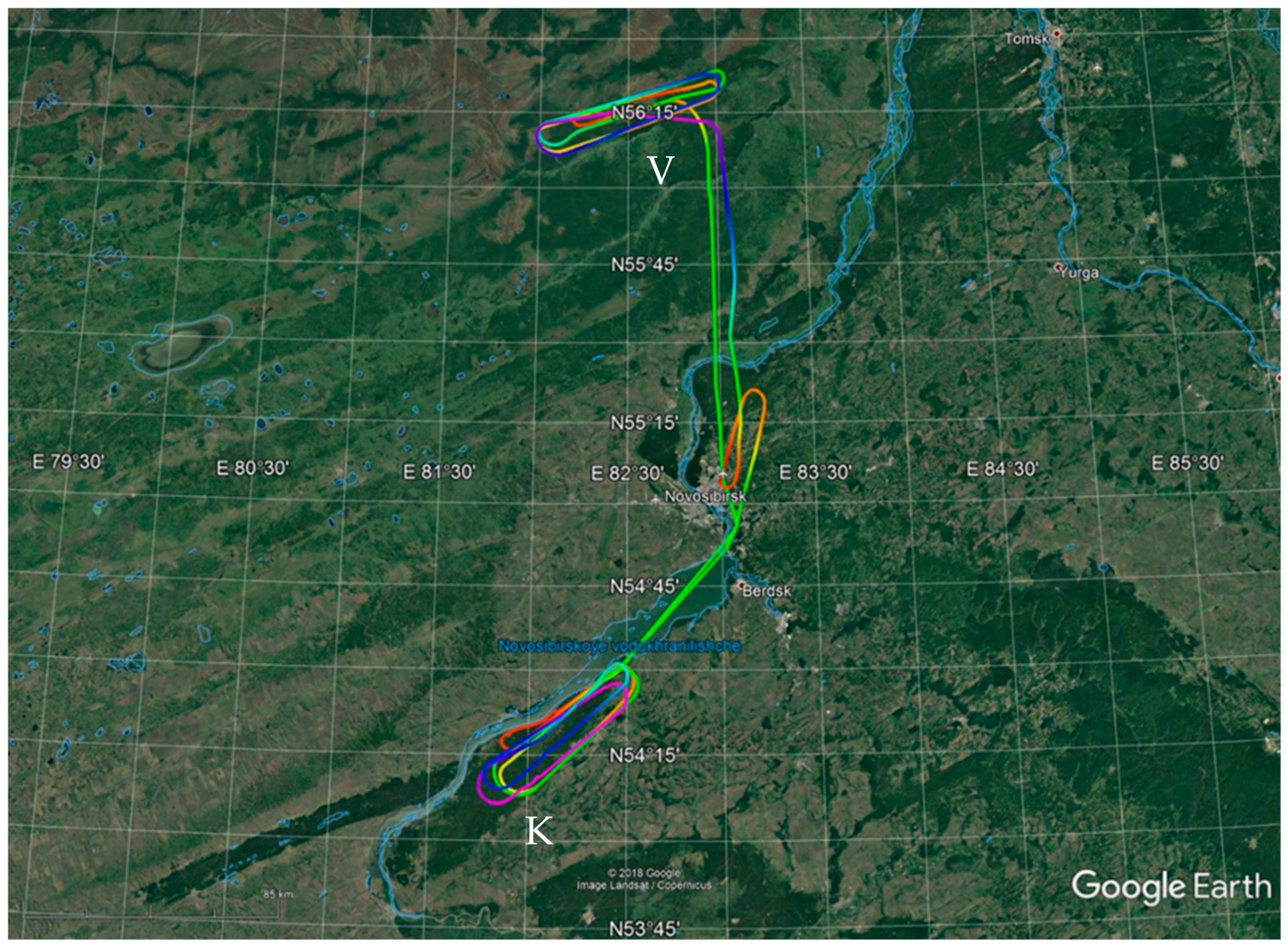
3.1. Backward Trajectories Analysis

An analysis of the backward trajectories of the movement of air masses from which samples were taken shows that all of them were over the northern territories/water areas 10 days before and then moved to the sampling points without descending far to the south (Figure S1–S8). Thus, it can be assumed that the air masses that contribute to the observed concentrations of total protein and culturable microorganisms in the studied samples of atmospheric aerosols were enriched with biocomponents from sources located in the northern regions.

3.2. Total Potein Cocentrations

The results of the analysis of samples for the presence of total protein in each of the samples, along with data on sampling altitude, are summarized in Table 1. Analysis of data on concentrations of total protein revealed two trends. First, at altitudes of 500–2000 m, concentrations in both sampling areas are noticeably higher than at altitudes of 3000–7000 m: <0.001 vs. 0.117 ± 0.064 µg/m3 for the Vasyugan marshes and 0.064 ± 0.091 vs. 0.195 ± 0.140 µg/m3 for the Karakan pine forest.

3.3. Culturable Microorganisms Cocentrations

The data of microbiological analysis obtained by inoculation of aerosol samples on selective media are presented in Table 2. Attention is drawn to the significant difference in the number of culturable microorganisms in the samples over Vasyugan marshes and Karakan pine forest taken at high altitudes (5500 and 3000 m), and the similarity in the samples taken at altitudes of 500 and 1000 m. The total number of microbial isolates isolated at a cultivation temperature of 28–30 °C above the Vasyugan marshes at altitudes of 5500 and 3000 m was 6.1 × 10⁶ and 4.7 × 10⁶ CFU/m3, respectively, while for Karakan pine forest aerosols sampled at the same altitudes, at an altitude of 5500 m, no culturable microorganisms were detected, and for an altitude of 3000 m their concentration was 5.0 × 10³ CFU/m3. Similar data were also obtained during the cultivation of aerosol samples at low temperatures (Table 2). One of the reasons for this phenomenon may be volatile phytoncides produced by coniferous trees and other plants of the pine forest. The relationship between the concentration of protein in aerosols and the number of detected cultivated microorganisms was not found.
In contrast, the samples taken at altitudes of 500 and 1000 m are similar both in the composition of isolated microorganisms and in their abundance, reaching 2.0 × 10⁶ CFU/m3 for the Karakan samples and 1.9 × 10⁶ CFU/m3 for the Vasyugan samples (Table 2); this significantly exceeds the concentrations of cultivated fungi and bacteria in the previously studied aerosols of Western Siberia, which were determined on average as 1–5 × 10³ CFU/m3. The concentration of fungi in this case was about 10–12% of the total number of microorganisms isolated from aerosols, while in the aerosols studied in this work, fungi accounted for about 50% or more of the number of microbial isolates (Table 3).

3.4. Culturable Microorganisms Diversity

With some exceptions, the temperature range of the growth of isolated fungi, in accordance with phenotypic and genomic features, identified as belonging to the genera Penicillium, Aspergillus, Alternaria, Candida, Aureobasidium, Vishniacozyma, Bullera, Meyerozyma, Cystofilobasidium, and Filobasidium (similarities in ITS and NS sequence fragments at the level 98–99%), etc., ranged from 6 to 30 °C (Table S2). The results of the genetic identification of fungal isolates are presented in Table 4, and their raw sequences in Table S3.
It should be noted that situations are quite frequent when in the atmosphere of a certain area there is a sharp change in the concentration and composition of biogenic particles, including microorganisms, which occurs as a result of the horizontal movement of atmospheric flows or the action of some local source [39,40]. Thus, Asian dust storms are known factors contributing to a significant change in the dominant groups in the microbial community of air aerosols [41]. As an example, we can cite a significant excess in the concentration and diversity of spore-forming bacteria in the atmosphere of the Karakan pine forest in the autumn of 2016 as a result of the air masses with dust transported from Central Asia [19].
Among the culturable bacteria isolated from aerosols, representatives of the genera Pseudomonas and Acinetobacter, spore-forming and corynemorphic bacteria, micrococci, and a number of other unidentified non-spore-bearing bacteria were found. In a quantitative ratio, bacteria of the genus Acinetobacter prevailed in the Vasyugan samples at altitudes of 5500, 3000, and 500 m and at a height of 500 m in the Karakan sample, and was represented by gram-negative, aerobic, immobile, non-fermentative, catalase-positive, and oxidase-negative cocco-bacilli, 0.6–0.7 × 0.8–1.5 µm in size, as is typical for representatives of this genus.

3.5. Discussion

Bacteria of the genus Acinetobacter are soil and aquatic saprotrophs, but they are found in the washings of the skin and mucous membranes of healthy people; they have natural and newly acquired resistance to many antibiotics. It is important that Acinetobacter belongs to typical opportunistic microorganisms that cause infectious processes against the background of immunosuppression and are capable of spreading to almost all organs and tissues [42], which makes it possible to consider the abundance of bacteria of the Acinetobacter genus found in atmospheric aerosols as a potentially dangerous factor.
Among the culturable fungal isolates isolated from the Vasyugan and Karakan samples, black yeasts of the genus Aureobasidium prevailed. It is known that peat soils of swamps differ in the ratio of the main components of microbial biomass: fungal mycelium predominates in the upper layers (43–83%), while fungal spores and yeast cells (57–93%) predominate in the lower layers. In the lower horizons, not only the proportion of spores is high, but also the proportion of bacterial cells (7–43%) [43,44]. The yeast population of peat is represented mainly by basidiomycete species with the dominance of Rhodotorula mucilaginosa, Sporobolomyces roseus, and Cryptococcus albidus. A distinctive feature of peat is also the higher occurrence and diversity of yeasts of the genera Pichia, Debaryomyces, Metschnikowia, and Candida than in other soil and plant substrates [45,46]. Yeast fungi Aureobasidium in peat soils are recorded singly [47]. Since a significant part of microorganisms enter the atmosphere from the surface of the swamp as a result of evaporation and then are carried by wind currents, it can be assumed from the data presented that the composition of aerosols formed above the surface of the Vasyugan marshes can mainly contain fungi of the above species, but not representatives of genus Aureobasidium.
Another indirect argument in favor of this thesis can be the data obtained by us during the microbiological analysis of 19 samples of the upper soil horizons and litter from deciduous, coniferous, and herbaceous plants, and lichens and sphagnum moss, obtained during an expedition in the area of the Vasyugan marshes in May 2021. (Tomsk region, Bakcharsky district), where yeasts of the genus Aureobasidium were also not found.
An example showing a non-standard situation in the studied atmospheric aerosols dated 14 September 2018, is also the sowing of samples taken over the Karakan pine forest on 8 September 2022. With a similar sampling and incubation at a temperature of 6–9 °C, a concentration of culturable microorganisms was revealed in atmospheric aerosols, which is characteristic for the atmosphere of this region, which amounted to 5.5 × 10³ CFU/m3; fungi of the genus Aureobasidium were not found (Figure S9).
It is known that the melanin-producing black fungi Aureobasidium are capable of causing diseases; they are increasingly recognized as the causative agents of a wide variety of forms of human mycoses that are difficult to treat [48]; they are included in the list of more than 80 fungal genera associated with allergic reactions; and they are assigned to the 4th pathogenicity group [49]. The range of species of opportunistic fungi and bacteria that can cause human diseases and have polyresistance to antibiotic drugs is constantly expanding. In this regard, it is important to determine the resistance of fungal cultures to drugs that suppress the development of fungal infections. In the example of 20 isolates partially reflecting the diversity of fungi in the studied aerosols, their sensitivity to the six most commonly used antimycotic drugs was determined (Table 5).
The drugs used differ in structure and mechanism of action on ergosterol, the main component of the fungal cell membrane. As a result of their action, the permeability of membranes is disturbed, which leads to cytolysis. Most of the tested cultures of fungi of the genus Aureobasidium (Dr 9-1, Dr 9-30, Dr 11-12, MR-12, etc.) showed the most pronounced sensitivity to nystatin and ketoconazole but were resistant to fluconazole, amphotericin B, and itraconazole, which does not coincide with the data on 150 clinical isolates [48,50], where itraconazole is recommended as an effective drug against Aureobasidium cultures. Unlike other tested isolates, Meyerozyma guilliermondii (MR-58) and Vishniacozyma victoriae (MR-135) strains were additionally sensitive to fluconazole; V. (Cryptococcus) heimaeyensis (Dr 10-13 and Dr 11-13) strains were sensitive to amphotericin B, as is typical for cryptococci. Multi-resistant strains can be attributed to those that have shown resistance to four to five drugs (seven strains). Nine strains were resistant to three drugs in different combinations, three strains were resistant to two antibiotics, and strain MR-135 was resistant only to amphotericin B. The MR-58 strain, identified as Meyerozyma guilliermondii, a species related to the causative agents of infectious diseases such as onychomycosis, invasive candidiasis, and candidal osteomyelitis [51,52], was distinguished not only by its sensitivity to antibiotics but also by the ability to grow at a temperature of 37 °C, as is typical for pathogens (Table S2)

4. Conclusions

The knowledge about the properties of microorganisms potentially dangerous to humans, and about the conditions of infection, is extremely important, as is knowledge about the features of the accumulation and transport of pathogens in the environment, including the atmosphere, and the ways of their penetration into the human habitat. The unique data obtained in this work, which characterizes the atmospheric microbiota of the Vasyugan marshes and Karakan pine forest, are one of the examples useful for understanding the possible transport of biogenic components of atmospheric aerosols and their contribution to the ecology of various territories.
It should be noted that the recorded values of the concentration of total protein in the atmosphere at different heights above the Karakan forest are significantly lower than the average annual concentration of total protein for this region in 2018 [We 2022]. Despite the fact that, as a rule, this concentration in the warm season is higher than the average annual value. The recorded concentrations of total protein in the atmosphere over the Vasyugan marshes were even lower.
In contrast, the measured values of the concentration of culturable microorganisms in the atmosphere at low altitudes above the Karakan pine forest are significantly higher than the average annual concentration in 2018 at these altitudes. The measured values of the concentration of culturable microorganisms in the atmosphere above the Vasyugan marshes at the same heights are close to those for the Karakan pine forest. At the same time, no culturable microorganisms were found at altitudes of 4000 m and above over the Karakan pine forest, in contrast to the same heights above the Vasyugan marshes. Such a situation over the Karakan pine forest was revealed from time to time in more than 20 years of observations. Moreover, sometimes no culturable microorganisms were found in the atmosphere at all sounded altitudes, especially after intense rains or snows. In addition, the data on the diversity of culturable microorganisms in the atmosphere above the Vasyugan marshes were obtained for the first time.
Thus, the data obtained in this work require further research both over the Karakan pine forest and over the Vasyugan marshes.

Supplementary Materials

The following supporting information can be downloaded at: https://www.mdpi.com/article/10.3390/atmos14020301/s1; Figure S1: backward trajectories of the movement of air masses, from which sampling was carried out over the Vasyugan marshes at altitudes of 7000 and 5500 m; Figure S2: backward trajectories of the movement of air masses, from which sampling was carried out over the Vasyugan marshes at altitudes of 4000 and 3000 m; Figure S3: backward trajectories of the movement of air masses, from which sampling was carried out over the Vasyugan marshes at altitudes of 2000 and 1500 m; Figure S4: backward trajectories of the movement of air masses, from which sampling was carried out over the Vasyugan marshes at altitudes of 1000 and 500 m; Figure S5: backward trajectories of the movement of air masses, from which sampling was carried out over the Karakan pine forest at altitudes of 7000 and 5500 m; Figure S6: backward trajectories of the movement of air masses, from which sampling was carried out over the Karakan pine forest at altitudes of 4000 and 3000 m; Figure S7: backward trajectories of the movement of air masses, from which sampling was carried out over the Karakan pine forest at altitudes of 2000 and 1500 m; Figure S8: backward trajectories of the movement of air masses, from which sampling was carried out over the Karakan pine forest at altitudes of 1000 and 500 m; Figure S9: fungal colonies on Sabouraud’s medium, detected by cultivating aerosol samples from Karakan and Vasyugan regions at a temperature of 6–9 °C.; Table S1: information about the culture media used; Table S2: growth of cultures of isolated fungi at different cultivation temperatures; Table S3: Sequencing raw results for some isolates; Table S4: the sensitivity of cultures to antimycotics was considered in accordance with the instructions for using discs with antifungal drugs of the Pasteur Research Institute of Epidemiology and Microbiology.

Author Contributions

Conceptualization, I.S.A. and A.S.S.; methodology, I.S.A. and B.D.B.; formal analysis, I.S.A., G.A.B., V.V.M., S.E.O., I.K.R., and A.S.S.; investigation, I.S.A., V.V.M., N.A.S., L.I.P., I.K.R., S.E.O., O.V.O., E.K.E., D.V.S., and B.D.B.; resources, A.S.S.; data curation, G.A.B. and I.S.A.; writing—original draft preparation, I.S.A.; writing—review and editing, I.S.A. and A.S.S.; visualization, I.S.A. and G.A.B.; project administration, A.S.S. All authors have read and agreed to the published version of the manuscript.

Funding

The work was funded by State orders of Rospotrebnadzor GZ 14/18 and GZ 11/21 and State order No. 121031300043-8.

Informed Consent Statement

Not applicable.

Data Availability Statement

Data are available from the authors on request.

Conflicts of Interest

The authors declare no conflict of interest.

References

  1. Després, V.R.; Huffman, J.A.; Burrows, S.M.; Hoose, C.; Safatov, A.S.; Buryak, G.; Fröhlich-Nowoisky, J.; Elbert, W.; Andreae, M.O.; Pöschl, U.; et al. Primary biological aerosols in the atmosphere: A review. Tellus. B 2012, 64, 15598. [Google Scholar] [CrossRef]
  2. Dommergue, A.; Amato, P.; Tignat-Perrier, R.; Magand, O.; Thollot, A.; Joly, M.; Bouvier, L.; Sellegri, K.; Vogel, T.; Sonke, J.E.; et al. Methods to investigate the global atmospheric microbiome. Front. Microbiol. 2019, 10, 243. [Google Scholar] [CrossRef]
  3. Fröhlich-Nowoisky, J.; Burrows, S.M.; Xie, Z.; Engling, G.; Solomon, P.A.; Fraser, M.P.; Mayol-Bracero, O.L.; Artaxo, P.; Begerow, D.; Conrad, R.; et al. Biogeography in the air: Fungal diversity over land and oceans. Biogeosciences 2012, 9, 1125–1136. [Google Scholar] [CrossRef]
  4. Hyde, P.; Mahalov, A. Contribution of bioaerosols to airborne particulate matter. J. Air Waste Manag. Assoc. 2020, 70, 71–77. [Google Scholar] [CrossRef]
  5. Pearce, D.A.; Alekhina, I.A.; Terauds, A.; Wilmotte, A.; Quesada, A.; Edwards, A.; Dommergue, A.; Sattler, B.; Adams, B.J.; Magalhães, C.; et al. Aerobiology over Antarctica—A new initiative for atmospheric ecology. Front. Microbiol. 2016, 7, 16. [Google Scholar] [CrossRef] [PubMed]
  6. Safatov, A.; Andreeva, I.; Buryak, G.; Olkin, S.; Reznikova, I.; Belan, B.; Panchenko, M.; Simonenkov, D. Long-term studies of biological components of atmospheric aerosol: Trends and variability. Atmosphere 2022, 13, 651. [Google Scholar] [CrossRef]
  7. Andreeva, I.S.; Safatov, A.S.; Puchkova, L.I.; Emelyanova, E.K.; Buryak, G.A.; Olkin, S.E.; Reznikova, I.K.; Ohlopkova, O.V. Culturable microorganisms in high-altitude atmospheric aerosol samples collected above northern Siberia by aircraft sounding. Bull. Nizhnevartovsk. State Univer. 2019, 2, 3–11. (In Russian) [Google Scholar] [CrossRef]
  8. Andreeva, I.S.; Safatov, A.S.; Morozova, V.V.; Tikunova, N.V.; Emelyanova, E.K.; Solovyanova, N.A.; Babkin, I.V.; Buryak, G.A.; Puchkova, L.I. Saprophytic and pathogenic yeasts in atmospheric aerosols of Southwestern Siberia. Atmos. Ocean. Opt. 2020, 33, 505–511. [Google Scholar] [CrossRef]
  9. Inisheva, L.I. Vasyugan Bog Nature Conditions, Structure and Functioning; TsNTI: Tomsk, Russia, 2003. (In Russian) [Google Scholar]
  10. Bodelier, P.L.E.; Dedysh, S.N. Microbiology of wetlands. Front. Microbiol. 2013, 4, 79. [Google Scholar] [CrossRef] [PubMed]
  11. Lin, X.; Tfaily, M.M.; Green, S.J.; Steinweg, J.M.; Chanton, P.; Imvittaya, A.; Chanton, J.P.; Cooper, W.; Schadt, C.; Kostka, J.E. Microbial metabolic potential for carbon degradation and nutrient (nitrogen and phosphorus) acquisition in an ombrotrophic peatland. Appl. Environ. Microbiol. 2014, 80, 3531–3540. [Google Scholar] [CrossRef] [PubMed] [Green Version]
  12. Dobrovolskaya, T.G.; Kukharenko, O.S.; Golovchenko, A.V. Features of the taxonomic composition of bacterial complexes of peatlands of different genesis. In Proceedings of the Wetlands and Biosphere: Collection of Materials of the Fourth Scientific School, Tomsk, Russia, 12–15 September 2005. (In Russian). [Google Scholar]
  13. Grodnitskaya, I.D.; Trusova, M.Y. Microbial communities and transformation of carbon compounds in bog soils of the taiga zone (Tomsk oblast). Eurasian Soil Sci. 2009, 42, 1021–1028. (In Russian) [Google Scholar] [CrossRef]
  14. Dobrovolskaya, T.G.; Golovchenko, A.V.; Kukharenko, O.S.; Yakushev, A.V.; Semenova, T.A.; Inisheva, L.I. The structure of the microbial communities in low-moor and high-moor peat bogs of Tomsk oblast. Eurasian Soil Sci. 2012, 45, 273–281. (In Russian) [Google Scholar] [CrossRef]
  15. Sergeeva, M.A.; Khokhlova, A.M. Microbial biomass and its activity in peat bogs of Siberia. TSPU Bull. 2015, 2, 143–149. (In Russian) [Google Scholar]
  16. Dedysh, S.N. Cultivating uncultured bacteria from Northern wetlands: Knowledge gained and remaining gaps. Front. Microbiol. 2011, 2, 184. [Google Scholar] [CrossRef]
  17. Alekseeva, M.N.; Preis, Y.I.; Dyukarev, E.A. Spatial structure of vegetation cover and top layer of peat of northeastern spurs of the great Vasyugan mire by remote and ground data. Bull. Tomsk. Polytech. Univ. 2015, 326, 81–90. (In Russian) [Google Scholar]
  18. Sergeeva, M.A.; Inisheva, L.I. Biochemical processes in oligotrophic peat deposits of the Vasyugan bog. TSPU Bull. 2008, 4, 57–63. (In Russian) [Google Scholar]
  19. Andreeva, I.S.; Safatov, A.S.; Puchkova, L.I.; Emelyanova, E.K.; Buryak, G.A.; Ternovoi, V.A. Spore forming bacteria isolated from atmospheric aerosols in Southwestern Siberia during atmospheric transfer of air masses. Bull. Nizhnevartovsk State Univer. 2018, 3, 123–130. (In Russian) [Google Scholar]
  20. Shevchenko, V.P.; Vorob′ev, S.N.; Kirpotin, S.N.; Kritskov, I.V.; Manasypov, R.M.; Pokrovsky, O.S.; Politova, N.V. Investigations of insoluble particles in the snow cover of the Western Siberia from Tomsk to the Ob estuary. Atmos. Ocean. Opt. 2015, 28, 499–504. (In Russian) [Google Scholar]
  21. Moskovchenko, D.V.; Babushkin, A.G. Peculiarities of formation of chemical composition of snow waters (on example of Khanty-Mansi autonomous district). Earth′s Cryosphere 2012, XVI, 71–81. (In Russian) [Google Scholar]
  22. Korica, A.; Polis, O.; Spalvis, K.; Bartkevics, V. Quantitative and qualitative seasonal changes of Scots pine and Norway spruce foliage essential oils in Latvia, and the extraction dynamics thereof. Balt. For. 2015, 21, 51–58. [Google Scholar]
  23. Apetrei, L.C.; Tuchiluş, C.; Aprotosoaie, A.C.; Oprea, A.; Malterud, K.E.; Miron, A. Chemical, antioxidant and antimicrobial investigations of Pinus cembra L. bark and needles. Molecules 2011, 16, 7773–7788. [Google Scholar] [CrossRef] [PubMed]
  24. Mimoune, N.A.; Mimoune, D.A.; Yataghene, A. Chemical composition and antimicrobial activity of the essential oils of Pinus pinaster. J. Coast. Life Med. 2013, 1, 55–59. [Google Scholar] [CrossRef]
  25. Vainio-Kaila, T.; Kyyhkynen, A.; Rautkari, L.; Siitonen, A. Antibacterial effects of extracts of Pinus sylvestris and Picea abies against Staphylococcus aureus, Enterococcus faecalis, Escherichia coli, and Streptococcus pneumoniae. BioResources 2015, 10, 7763–7771. [Google Scholar] [CrossRef]
  26. Efremov, A.A.; Zykova, I.D.; Senashova, V.A.; Grodnickaya, I.D.; Pashenova, N.V. Antimicrobial and antiradical activity of individual fractions of pinus sibirica du tour and abies sibirica ledeb., native to the Siberian region. Chem. Plant Mater. 2020, 4, 203–210. [Google Scholar] [CrossRef]
  27. Tarakanov, V.V.; Chindyaeva, L.N.; Tsybulya, N.V.; Tikhonova, I.V. Variability of needle antimicrobic activity in clone plantation of Scots pine Pinus sylvestris L. Sib. J. For. Sci. 2017, 1, 95–104. (In Russian) [Google Scholar] [CrossRef]
  28. Draxler, R.R.; Hess, G.D. An overview of the HYSPLIT_4 modelling system for trajectories, dispersion and deposition. Aust. Meteorol. Mag. 1998, 47, 295–308. [Google Scholar]
  29. Belan, B.D.; Rasskazchikova, T.M.; Simonenkov, D.V.; Tolmachev, G.N.; Fofonov, A.V. Chemical composition of atmospheric aerosols over background areas of the southern part of Western Siberia observed during the IAO complex atmospheric radiation experiment carried out in December 2015. Proc. SPIE 2016, 10035, 1003549. [Google Scholar] [CrossRef]
  30. Freitag, S.; Clarke, A.D.; Howell, S.G.; Kapustin, V.N.; Campos, T.; Brekhovskikh, V.L.; Zhou, J. Combining airborne gas and aerosol measurements with HYSPLIT: A visualization tool for simultaneous evaluation of air mass history and back trajectory consistency. Atmos. Meas. Tech. 2014, 7, 107–128. [Google Scholar] [CrossRef] [Green Version]
  31. Antokhin, P.N.; Arshinov, M.Y.; Belan, B.D.; Davydov, D.K.; Zhidovkin, E.V.; Ivlev, G.A.; Kozlov, A.V.; Kozlov, V.S.; Panchenko, M.V.; Penner, I.E.; et al. Optik-É AN-30 Aircraft Laboratory for Studies of the Atmospheric Composition. J. Atmos. Ocean. Technol. 2012, 29, 64–75. [Google Scholar] [CrossRef]
  32. APA. Filters. Catalogue-Guide; Atomizdat: Moscow, Russia, 1970. (In Russian) [Google Scholar]
  33. You, W.W.; Haugland, R.P.; Ryan, D.K.; Haugland, R.P. 3-(4-Carboxybenzoyl)quinoline-2-carboxaldehyde, a reagent with broad dynamic range for the assay of proteins and lipoproteins in solution. Ann. Biochem. 1997, 244, 277–282. [Google Scholar] [CrossRef]
  34. Ashmarin, I.P.; Vorobyov, A.A. Statistical Methods in Microbiological Studies; Medgiz: Leningrad, Russia, 1962. (In Russian) [Google Scholar]
  35. Babieva, I.P.; Chernov, I.Y. Biology of Yeast; Association of Scientific Publications KMK: Moscow, Russia, 2004. (In Russian) [Google Scholar]
  36. Sutton, D.A.; Fothergill, A.W.; Rinaldi, M.G. Guide to Clinically Significant Fungi; Williams & Wilkins: Philadelphia, PA, USA; A Waverly Company: Baltimore, MD, USA, 1998. [Google Scholar]
  37. Gerhardt, F.; Murray, R.G.E.; Wood, W.A.; Krieg, N.R. Methods of General Bacteriology, 2nd ed.; American Society for Microbiology: Washington, DC, USA, 1994.
  38. Holt, J.G.; Krieg, N.R.; Sneath, P.H.A.; Staley, J.T.; Williams, S.T. Bergey’s Manual of Determinative Bacteriology; Williams & Wilkins: Philadelphia, PA, USA, 1994. [Google Scholar]
  39. Kellogg, C.A.; Griffin, D.W. Aerobiology and the global transport of desert dust. Trends Ecol. Evol. 2006, 21, 638–644. [Google Scholar] [CrossRef]
  40. Hara, K.; Zhang, D. Bacterial abundance and viability in long-range transported dust. Atmos. Environ. 2012, 47, 20–25. [Google Scholar] [CrossRef]
  41. Jeon, E.M.; Kim, H.J.; Jung, K.; Kim, J.H.; Kim, M.Y.; Kim, Y.P.; Ka, J.-O. Impact of Asian dust events on airborne bacterial community assessed by molecular analyses. Atmos. Environ. 2011, 45, 4313–4321. [Google Scholar] [CrossRef]
  42. Visca, P.; Seifert, H.; Towner, K.J. Acinetobacter infection—An emerging threat to human health. IUBMB Life 2011, 63, 1048–1054. [Google Scholar] [CrossRef]
  43. Golovchenko, A.V.; Zvyagintsev, D.G.; Tikhonova, E.Y. Abundance, biomass, structure, and activity of the microbial complexes of minerotrophic and ombrotrophic peatlands. Microbiology 2007, 76, 630–637. [Google Scholar] [CrossRef]
  44. Golovchenko, A.V.; Semenova, T.A.; Polyakova, A.V.; Inisheva, L.I. The structure of the micromycete complexes of oligotrophic peat deposits in the southern taiga subzone of West Siberia. Microbiology 2002, 71, 667–674. (In Russian) [Google Scholar] [CrossRef]
  45. Polyakova, A.V.; Chernov, I.Y.; Panikov, N.S. Yeast diversity in hydromorphic soils with reference to a grass-sphagnum wetland in Western Siberia and a hummocky tundra region at cape Barrow (Alaska). Microbiology 2001, 70, 714–720. (In Russian) [Google Scholar] [CrossRef]
  46. Polyakova, A.V. Yeast in Peat-Bog Soils. Ph.D. Thesis, Institute of Microbiology of the Russian Academy of Sciences, Moscow, Russia, 2002. (In Russian). [Google Scholar]
  47. Yakushev, A.V.; Matyshak, G.V.; Tarkhov, M.O.; Kachalkin, A.V.; Sefilyan, A.R.; Petrov, D.G. Microbiological characteristics of bare peat circles on flat-topped peat mounds in the north of Western Siberia. Eurasian Soil Sci. 2019, 52, 1081–1090. [Google Scholar] [CrossRef]
  48. Bagirova, N.S. Aureobasidium pullulans place among human pathogens opportunistic infections (literature review). Lab. Serv. 2018, 7, 12–18. (In Russian) [Google Scholar] [CrossRef]
  49. Soeria-Atmadja, D.; Önell, A.; Borga, A. IgE sensitization to fungi mirrors fungal phylogenetic systematics. J. Allergy Clin. Immunol. 2010, 125, 1379–1386. [Google Scholar] [CrossRef]
  50. Javad Najafzadeh, M.; Sutton, D.A.; Saradeghi Keisari, M.; Zarrinfar, H.; Sybren de Hoog, G.; Chowdhary, A.; Meise, J.F. In vitro activities of eight antifungal drugs against 104 environmental and clinical isolates of Aureobasidium pullulans. Antimicrob. Agents Chemother. 2014, 58, 5629–5631. [Google Scholar] [CrossRef]
  51. Pfaller, M.A.; Diekema, D.J.; Mendez, M.; Kibbler, C.; Erzsebet, P.; Chang, S.-C.; Gibbs, D.L.; Newell, V.A. Candida guilliermondii, an opportunistic fungal pathogen with decreased susceptibility to fluconazole: Geographic and temporal trends from the ARTEMIS DISK antifungal surveillance program. J. Clin. Microbiol. 2006, 44, 3551–3556. [Google Scholar] [CrossRef]
  52. Gamaletsou, M.N.; Kontoyiannis, D.P.; Sipsas, N.V.; Moriyama, B.; Alexander, E.; Roilides, E.; Brause, B.; Walsh, T.J. Candida osteomyelitis: Analysis of 207 pediatric and adult cases (1970–2011). Clin. Infect. Dis. 2012, 55, 1338–1351. [Google Scholar] [CrossRef] [Green Version]
Figure 1. Route of the aircraft moving during sampling of aerosols over the Vasyugan marshes and Karakansky pine forest. Index V is for the region of the Vasyugan marshes (the starting point coordinates of backward trajectories are 56.00 N; 82.30 E), index K is for the region of the Karakan pine forest (the starting point coordinates of backward trajectories are 54.22 N; 82.15 E).
Atmosphere 14 00301 g001
Figure 2. The flow chart of the investigation.
Atmosphere 14 00301 g002
Table 1. The concentration of total protein in the studied samples of atmospheric aerosol.
Sampling Altitude (m) Total Protein Concentration, μg/m3
Vasyugan MarshesKarakan Pine Forest
7000<0.0010.037
5500<0.001<0.001
4000<0.0010.219
3000<0.001<0.001
20000.1980.130
15000.113<0.001
10000.0200.290
5000.1360.358
Table 2. The concentration of culturable microorganisms in the studied samples of atmospheric aerosols.
Region of SamplingAltitude,
m
Total Number of Isolated
Microorganisms, CFU/m3
Titer of Isolated Microorganisms on of Different
Culture Media, CFU/m3
GFM *LB 1:10SAASASabouraud
Cultivation temperature 28–30 °C
Vasyugan marshes55006.1 × 1064.2 × 1061.5 × 1052.0 × 1063.3 × 1062.3 × 105
30004.7 × 1056.9 × 1041.5 × 1061.4 × 1061.6 × 1061.2 × 105
10002.3 × 10501.1 × 10508.4 × 1043.3 × 104
5004.8 × 1065.7 × 1042.4 × 1052.6 × 1061.7 × 1072.7 × 106
Karakan pine forest5500000000
4000000000
30005.0 × 1030005.0 × 1030
10003.7 × 1054.0 × 1033.0 × 1052.0 × 1033.3 × 1043.2 × 104
5008.3 × 1059.7 × 1042.9 × 1043.0 × 1048.5 × 1043.3 × 105
Cultivation temperature 6–9 °C
Vasyugan marshes55005.4 × 1065.5 × 1053.1 × 1052.0 × 1072.2 × 1073.2 × 106
30002.1 × 10601.1 × 1061.0 × 10607.4 × 103
10003.4 × 1052.0 × 1031.5 × 10502.4 × 1041.6 × 105
5001.9 × 1062.5 × 1048.8 × 10401.5 × 1062.9 × 105
Karakan pine forest5500000000
4000000000
30001.7 × 1030001.7 × 1030
10001.9 × 1062.8 × 1048.3 × 10506.3 × 1053.8 × 105
5002.0 × 1065.2 × 1047.0 × 1051.5 × 1058.4 × 1052.7 × 106
* The names of different culture media are presented in the Materials and Methods section. Its compositions are described in Table S1.
Table 3. The ratio of the number of culturable bacteria and fungi isolated from the studied aerosols.
Region of SamplingAltitude,
m
Titer, CFU/m3, 6–9 °CTiter, CFU/m3, 28–30 °C
BacteriaFungiBacteriaFungi
Vasyugan marshes55003.6 × 1061.8 × 1065.8 × 1063.0 × 105
30002.1 × 1064.6 × 1034.5 × 1062.0 × 105
10009.6 × 1042.4 × 1058.8 × 1041.4 × 105
5001.5 × 1064.1 × 1054.4 × 1064.3 × 105
Karakan pine forest55000000
40000000
300001.7 × 1033.3 × 1031.7 × 103
10002.5 × 1051.6 × 1062.8 × 1059.1 × 104
5001.3 × 1067.7 × 1053.0 × 1055.3 × 105
Table 4. Taxonomic positions of isolated fungal isolates determined by molecular genetic methods.
StrainClassFamilyGenusSpecies
Dr 9-1DothideomycetesSaccotheciaceaeAuerobasidiumA. leucospermi
Dr 9-2TremellomycetesRhynchogastrema-taceaePapiliotremaP. flavescens/P. aurea
Dr 9-5DothideomycetesSaccotheciaceaeAuerobasidiumA. leucospermi
Dr 9-6DothideomycetesSaccotheciaceaeAuerobasidiumA. leucospermi
Dr 9-26DothideomycetesSaccotheciaceaeAuerobasidiumA. leucospermi
Dr 9-30DothideomycetesSaccotheciaceaeAuerobasidiumA. pullulans
Dr 10-13TremellomycetesBulleribasidiaceaeVishniacozymaV. (Cryptococcus) heimaeyensis
Dr 10-15DothideomycetesSaccotheciaceaeAuerobasidiumA. pullulans
Dr 10-16MDothideomycetesSaccotheciaceaeAuerobasidiumA. pullulans
Dr 11-7TremellomycetesBulleraceaeBulleraB. alba
Dr 11-8DothideomycetesSaccotheciaceaeAuerobasidiumA. pullulans
Dr 11-12DothideomycetesSaccotheciaceaeAuerobasidiumA. pullulans
Dr 11-13TremellomycetesBulleribasidiaceaeVishniacozymaV. (Cryptococcus) heimaeyensis
MR 12DothideomycetesSaccotheciaceaeAuerobasidiumA. pullulans
MR-14DothideomycetesSaccotheciaceaeAuerobasidiumA. leucospermi
MR 58SaccharomycetesDebaryomycetaceaeMeyerozymaM. (Pichia) guilliermondii
MR 135TremellomycetesBulleribasidiaceaeVishniacozymaV. victoriae
MR 160TremellomycetesCystofilobasidiaceaeCystofilobasidiumC. macerans
MR 166TremellomycetesCystofilobasidiaceaeCystofilobasidiumC. macerans
MR 189TremellomycetesFilobasidiaceaeFilobasidiumF. (Cryptococcus) magnum
Table 5. Sensitivity of fungi isolated from studied aerosol samples to antimycotic drugs.
StrainAntibiotic, Amount on Disk, μg/Diameter of Zone of Growth Inhibition, mm
Derivatives of
Triazole
Derivatives of
Imidazole
Polyene
Antibiotics
Fluconazole,
40
Itraconazole,
10
Ketoconazole,
20
Clotrimazole,
10
Nystatin,
80
Amphotericin B,
40
Dr 9-1003510288
Dr 9-501012121212
Dr 9-60025112610
Dr 9-2003018250
Dr 9-260028123012
Dr 9-30092813240
Dr 10-130040184023
Dr 10-1500240309
Dr 10-16001815289
Dr 11-713134022259
Dr 11-80142012250
Dr 11-12002212268
Dr 11-130042183922
MR 12092612250
MR 140102515259
MR 5820026171611
MR 135201735253011
MR 1600826122810
MR 16601228123010
MR 189002010209
Note: the sensitivity of cultures to antibiotics was considered in accordance with the instructions for using discs with antifungal drugs of the Pasteur Research Institute of Epidemiology and Microbiology, Table S4.
Disclaimer/Publisher’s Note: The statements, opinions and data contained in all publications are solely those of the individual author(s) and contributor(s) and not of MDPI and/or the editor(s). MDPI and/or the editor(s) disclaim responsibility for any injury to people or property resulting from any ideas, methods, instructions or products referred to in the content.

