Article

Efficient Lung Cancer Molecular Diagnostics by Combining Next Generation Sequencing with Reflex Idylla Genefusion Assay Testing

Department of Pathology and Laboratory Medicine, School of Medicine & Dentistry, University of Rochester, Rochester, NY 14642, USA
*
Author to whom correspondence should be addressed.
Genes 2023, 14(8), 1551; https://doi.org/10.3390/genes14081551
Submission received: 6 July 2023 / Revised: 25 July 2023 / Accepted: 27 July 2023 / Published: 28 July 2023
(This article belongs to the Special Issue Next Generation Sequencing in Clinical Diagnostics)

Abstract

:
Molecular diagnostics for lung cancer is a well-established standard of care, but how to use the available diagnostic tools for optimal and cost-effective patient care remains unresolved. Here, we show that DNA-only, small gene next-generation sequencing (sNGS) panels (<50 genes) combined with ultra-rapid reflex testing for common fusion transcripts using the Idylla Genefusion assay provide a cost-effective and sufficiently comprehensive testing modality for the majority of lung cancer cases. We also demonstrate the need for additional reflex testing capability on larger DNA and fusion panels for a small subset of lung cancers bearing rare single-nucleotide variants, indels and fusion transcripts and secondary, post-treatment resistance mutations. A similar testing workflow could be adopted for other solid tumor types for which extensive gene/fusion variant profiles are available both in the treatment-naïve and post-therapy settings.

1. Introduction

More than 65% of patients diagnosed with advanced non-small cell lung cancer harbor actionable gene mutations, gene amplifications, or oncogenic fusion transcripts [1,2]. Consequentially, NGS-based tumor molecular profiling is now the standard of care for patients with advanced non-small cell lung cancer (NSCLC) [3,4,5,6]. Multiple genetic alterations have been identified as therapeutic targets in lung adenocarcinoma, including mutations in EGFR, ERBB2, BRAF, KRAS, and MET and rearrangements of ALK, RET, and ROS1 [4,5,7,8]. Moreover, several other oncogenes with potential prognostic roles in lung adenocarcinoma, including MET and PIK3CA, have also been described [7,8,9]. Currently, various diagnostic tools and platforms are being used in both academic and commercial pathology laboratory settings to profile lung tumors. The modalities being used include fluorescence in situ hybridization (FISH), immunohistochemistry (IHC), and next-generation sequencing (NGS). However, the optimal combination(s) of the different diagnostic modalities to enhance timely yet cost effective patient care remains to be determined.
Currently, as is the case in most academic laboratories, the University of Rochester Medical Center (URMC) molecular genetic pathology (MGP) unit (molecular diagnostics and cytogenetics laboratories) utilizes both FISH and NGS to profile actionable genomic mutations and rearrangements in lung cancer. In the present URMC workflow, parallel testing for ALK and ROS1 rearrangements is evaluated through FISH and the search for gene mutations and amplifications is conducted using a <50 gene NGS panel at the DNA level only (Oncomine Focus Assay [OFA], see Methods). In addition, the URMC anatomic pathology laboratory performs IHC-based testing for ALK and ROS1 fusions and for NTRK 1/2/3 fusions (using a panNTRK antibody), respectively.
Here, we reevaluate this workflow for lung cancer molecular diagnostics. We first describe the validation of the Biocartis Idylla Genefusion assay for detection of ALK, ROS1, RET fusions, and MET exon 14 skipping mutations, which is performed at the RNA level on tissues obtained directly from formalin-fixed paraffin embedded (FFPE) slides requiring less than 5 min hands-on time from a technologist. We also demonstrate that a DNA-only, less than 50-gene next-generation sequencing (sNGS) panel combined with ultra-rapid reflex testing via Idylla Genefusion assay provides a cost-effective testing schema for most lung cancer cases, thereby largely negating the need for upfront ALK and ROS1 fusion testing by FISH. Lastly, we demonstrate the need for additional reflex testing capability on larger DNA and fusion panels for a small subset of lung tumor specimens bearing rare genetic aberrations.

2. Materials and Methods

2.1. Clinical Specimens

We retrospectively analyzed NGS and ALK/ROS1 FISH results for all patient samples with primary or metastatic lung adenocarcinoma (n = 368) referred to the University of Rochester Medical Center (URMC) molecular diagnostics and cytogenetics laboratories for mutational testing and fusion analysis from July 2021 to June 2022. Clinical data were collected as approved under an Institutional Review Board protocol allowing genomic testing on patients’ tumors.

2.2. Sample Collection for Idylla Gene Fusion Assay Validation

Cases of lung adenocarcinoma with known gene rearrangements involving ALK, ROS1, RET, and MET exon 14 skipping mutations were identified on the basis of standard-of-care testing using NGS and FISH at the URMC MGP laboratories. Each positive sample served as a negative control for the remaining aberrations, and eight NGS/FISH negative cases were also included. Electronic medical records were reviewed to obtain data regarding tumor characteristics.

2.3. Validation of the Idylla Gene Fusion Assay

The Idylla Genefusion Assay (Biocartis, Inc., Mechelen, Belgium) is a cartridge-based, automated multiplex RT-qPCR test designed to detect rearrangements in ALK, ROS1, RET (both for specific translocations and 3′5′ expression imbalance) and NTRK1/2/3 rearrangements (3′5′ expression imbalance only) and MET exon 14 skipping mutations within a span of 3.5 h. Automation is enabled through a cartridge-based system, in which, beginning with the FFPE slide(s), nucleic acid extraction, reverse transcription, amplification, and variant detection occur. Each cartridge contains a single sample and is intended for one-time use. Briefly, following insertion of the scraped FFPE tissue sample into the cartridge, the cartridge is placed into the lysis chamber in the Idylla instrument. Within the lysis chamber, various chemicals and reagents, heat, and high-intensity focused ultrasound enable deparaffinization and cell lysis. Nucleic acids are then purified and concentrated on a solid support, and following elution they are transferred to the PCR chambers for multiplex RT-qPCR using TaqMan PCR and fluorescence-based detection.
Gene fusion detection in the Idylla system is accomplished through detection of fusion-specific amplification and expression imbalances. Proprietary fusion-specific primers amplifying the flanking sequences of the ALK, ROS1, and RET fusion breakpoints are used. MET exon 14 skipping variant and wild-type transcripts are identified with dedicated primer and probe sets. To compare the levels of expression, multiple probes are used to compare wild-type expression to the remaining samples. Notably, the assay is only able to detect the gene rearrangements and is unable to identify the specific fusion partner.
Assessment of the limit of detection (LOD) was accomplished using six different cell lines harboring the different mutations. Cell lines were grown and then mixed to reflect the tested ratios (25%, 20%, 10%, and 5%) for ALK, ROS1, RET, and MET exon 14 skipping mutations. FFPE cytospin pellets were prepared for testing. See the Supplemental Data for a detailed description of the assay validation.

2.4. Performance Comparisons with Reference Methods

NGS and FISH served as reference modalities in all cases. A subset of cases was submitted for testing by a commercial provider (Foundation Medicine, Inc., Cambridge, MA, USA) but the majority of cases were tested using a small, in-house 35-gene NGS (sNGS) assay. Specifically, the URMC solid tumor oncology NGS test uses the DNA-only portion of Oncomine Focus Assay (OFA) (ThermoFisher, Inc., Waltham, MA, USA), which enables single-nucleotide variant (SNV) and small indel detection and interrogates hotspot regions of 35 genes. This panel also possesses an additional, partly overlapping set of 18 genes for copy number variation (CNV) analyses and RNA fusion transcript detection capability for 23 genes. The limit of detection for SNVs and indels is precise and reproducible at 5% allele frequency with 500× coverage. The limit of detection of indels is less than 25 bp. Variants of uncertain origin (germline versus somatic origin) cannot be determined unequivocally based on sequencing tumor samples alone. The data obtained are analyzed with the Ion ReporterTM software for variant classification and the Torrent Suite Analysis plugin for the coverage analysis. The mutation nomenclature is based on a recommendation from the Catalogue of Somatic Mutations in Cancer (COSMIC, http://cancer.sanger.ac.uk/cosmic (accessed on 1 July 2023)). Note that only the SNV/small indel detection portion of the OFA assay has been validated for use by our laboratory. The list of genes of the OFA includes: AKT1 NM_001014431.1, BRAF NM_004333.4, DDR2 NM_006182.2, ERBB3 NM_001982.3, FGFR2 NM_000141.4, GNAQ NM_002072.4, IDH2 NM_002168.2, JAK3 NM_000215.3, MAP2K1 NM_002755.3., MTOR NM_004958.3, PIK3CA NM_006218.3, ROS1 NM_002944.2, AR NM_000044.3, CTNNB1 NM_001904.3, ERBB2 NM_004448.3, ESR1 NM_001122740.1, GNA11 NM_002067.4, IDH1 NM_005896.2, JAK2 NM_004972.3, KRAS NM_033360.3, MET NM_001127500.2, PDGFRA NM_006206.5, RET NM_020975.4, ALK NM_004304.4, CDK4 NM_000075.3, EGFR NM_005228.4, ERBB4 NM_005235, FGFR3 NM_000142.4, HRAS NM_001130442.1, JAK1 NM_002227.2, KIT NM_000222.2, MAP2K2 NM_030662.3, NRAS NM_002524.4, RAF1 NM_002880.3, SMO NM_005631.4.
FISH was carried out using formalin-fixed, paraffin-embedded tissues with 4 μm sections mounted on positively charged slides. Pretreated slides were incubated in an oven at 60 °C for 1 h, and then processed on a VP2000 (Abbott Molecular, Abbott Park, IL, USA), which includes deparaffinization and protease pretreatment. Slides were hybridized using commercial break-apart probes for ALK and ROS1 (Abbott Molecular, Abbott Park, IL, USA) in IntelliFISH hybridization buffer (Abbott Molecular, Abbott Park, IL, USA) for 2 h at 37 °C after being denatured for 5 min at 73 °C. Slides were washed and counterstained with DAPI (1 µg/mL) (catalog#: 06J49-001, Abbott Molecular, IL, USA). A total of 200 cells was analyzed by two technologists, with each one reading 100 cells. Specimens were considered positive for ALK or ROS1 rearrangement if >15% of the cells demonstrated a positive signal pattern (split ALK or ROS1 signal).

2.5. Classification of Mutations

The identified variants were stratified into one of four tiers based on published clinical or laboratory evidence. Tier I includes mutations and fusions that are categorized as variants of strong clinical significance and possess Level A (an FDA-approved therapy and/or included in professional guidelines) or Level B evidence (well-powered studies with expert consensus). Tier II consists of alterations that are classified as variants of potential clinical significance with Level C (FDA-approved therapies for different tumor types or investigational therapies or multiple small published studies with some consensus) or Level D evidence (preclinical trials or a few case reports without consensus). Tier III encompasses mutations that are known as variants of unknown clinical significance, which are not observed at significant allele frequencies in the general population or in specific databases. Finally, Tier IV includes benign or likely benign variants that are observed at significant allele frequencies in the general or specific subpopulation databases.

2.6. Statistical Analysis

Data analysis and figures were created using Microsoft Excel, GraphPad 8.3, and CorelDraw 2019 software programs.

3. Results

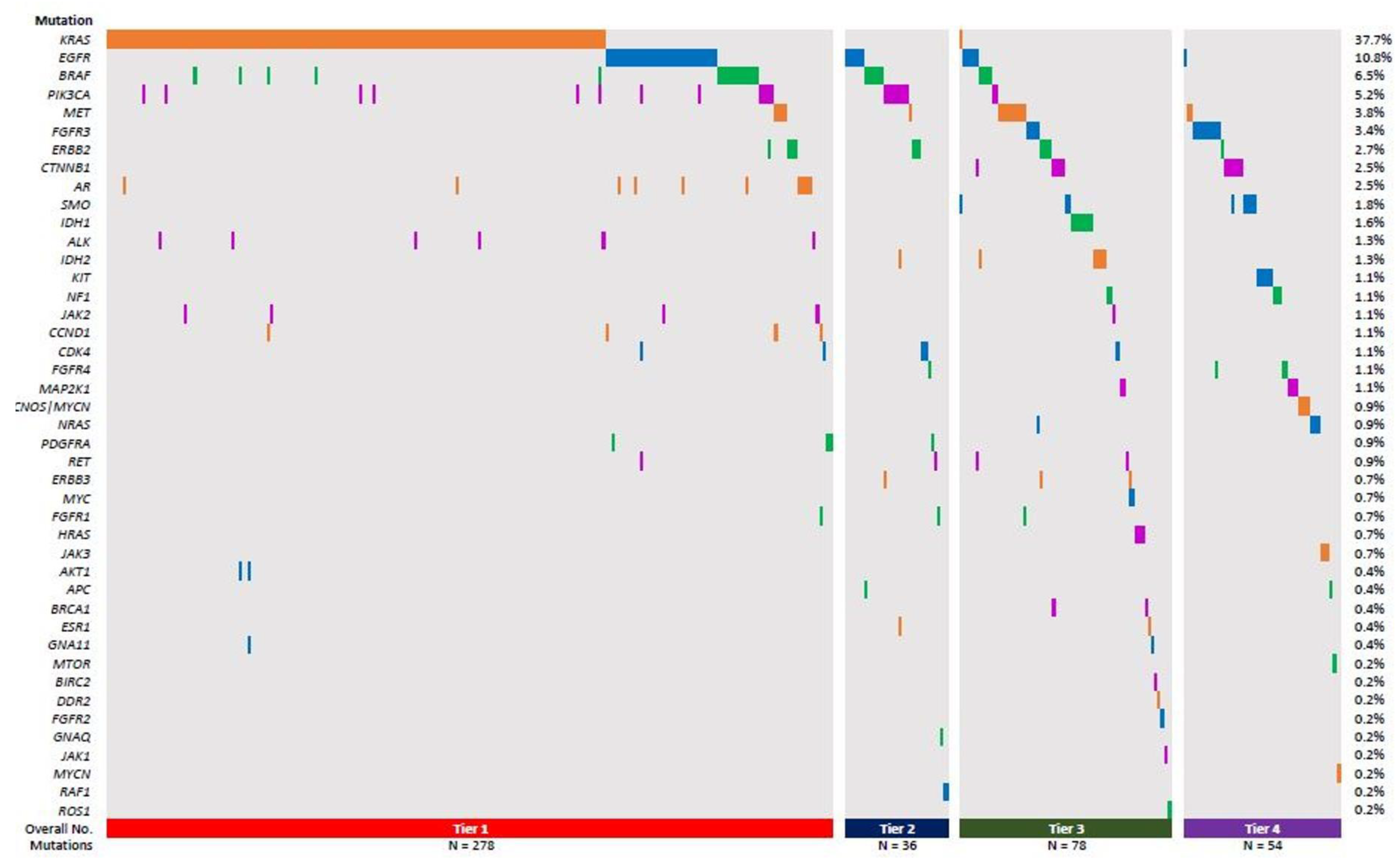
3.1. ALK or ROS1 Fusion-Bearing Lung Cancer Cases Are Rare and Do Not Bear Additional SNV, or Small Indel Variants as Determined by the OFA NGS Test

Currently, standard molecular testing of NSCLC at URMC involves FISH testing for ALK and ROS1 fusions, which is performed concomitantly with sNGS for 35 hotspot genes using the ThermoFisher’s Oncomine Focus Assay (OFA) [10]. If these tests do not identify any mutation, recommendations are made for send-out testing for RET and other rare fusion transcripts (e.g., for BRAF fusions) and for MET exon 14 skipping mutation detection at the RNA level.
To assess the effectiveness of this combination approach, we compiled the results of in-house FISH and sNGS assays for all lung tumors (primary or metastatic) that were submitted for testing during a 1-year time period (July 2021–June 2022), for a total of 368 specimens. We found that only 2.17% (8/368) of lung tumor cases in our cohort harbored an ALK fusion, while a mere 0.54% (2/368) possessed a ROS1 rearrangement caused by FISH, a ratio that is somewhat lower than was reported in a previous study [11]. In contrast, in the ALK and ROS1 fusion-negative specimens, the OFA sNGS panel identified NSCLC-relevant SNVs and indels in nearly 80% (293/368) of cases, indicating that at least one oncogenic or likely oncogenic variant or a variant of uncertain significance (VUS) was identified. One specimen was positive for both a ROS1 fusion and a pathogenic KRAS mutation. In our cohort, lung tumors bearing oncogenic Tier I KRAS (~40%) mutations were found most commonly, followed by Tier I–III EGFR, BRAF, and PIK3CA variants and then a number of less common Tier I–IV variants (Figure 1). Furthermore, about 278 (76%) cases in our cohort harbored at least one Tier 1 mutation or more, 36 cases (9.8%) had at least Tier II mutations, 78 (21.2%) had Tier III, while 54 (14.7%) had Tier IV mutations (Figure 1). These data demonstrate that sNGS is a high-yield frontline testing modality, while FISH for ALK and ROS1 fusion should be reserved for DNA-variant negative cases in the workup of NSCLCs.

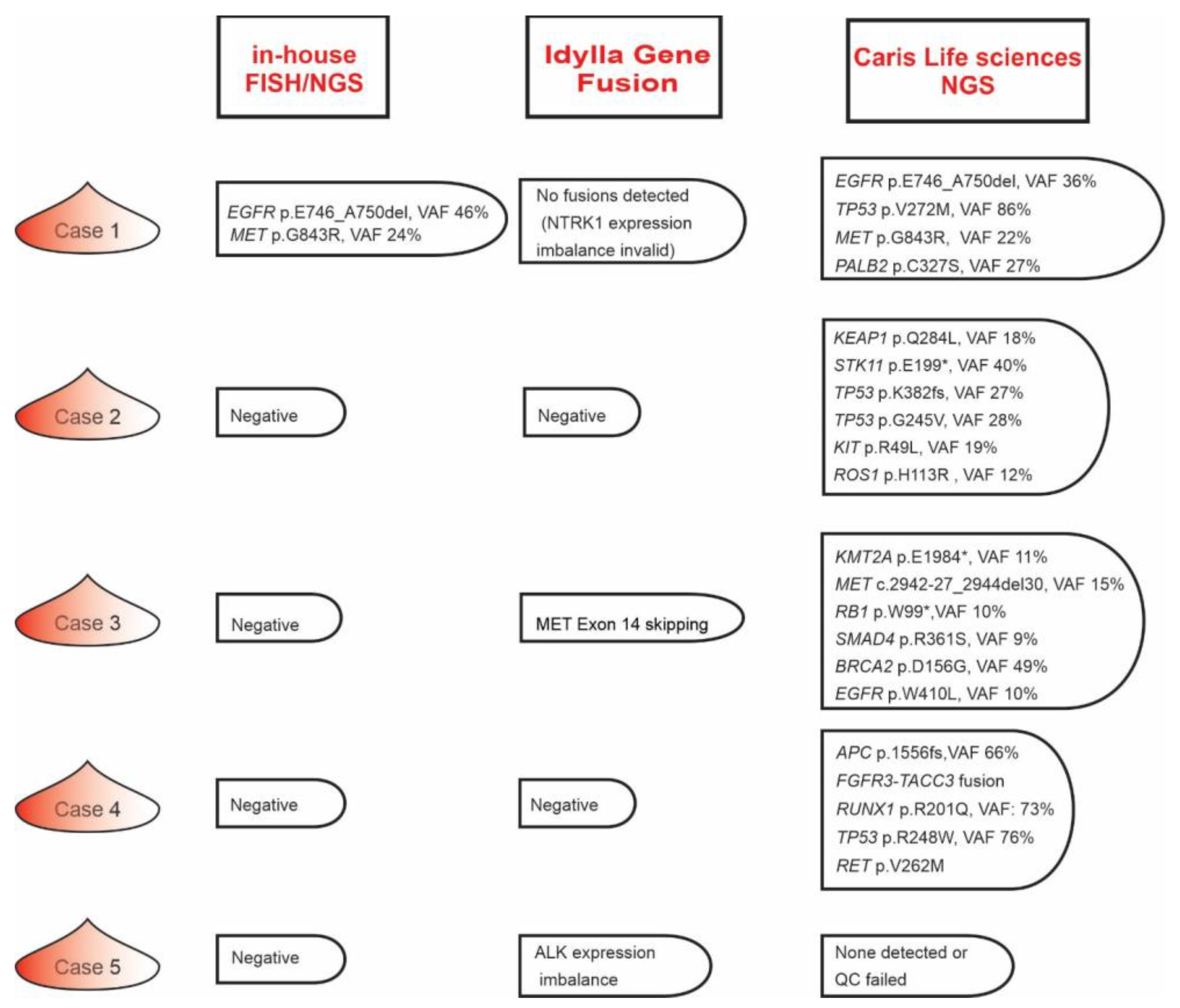
3.2. Validating the Idylla Gene Fusion Assay

Recently, the BioCartis Idylla Genefusion assay, a rapid testing modality with a turn-around time (TAT) of less than 4 h and technologist hands-on time < 5 min has been introduced to identify targetable, NSCLC-related fusion transcripts (ALK, ROS1, RET, NTRK1-3) and MET exon 14 splice mutations [12,13,14]. Therefore, to potentially complement sNGS, we validated the Idylla Genefusion assay for ALK, ROS1, and RET rearrangements and MET exon 14 skipping variant detection using 34 tumor samples that were previously orthogonally tested via FISH and NGS. We assessed 24 surgical resections/biopsies and 10 cytological blocks paraffin-embedded tumor samples with tumor cellularity between 10 and 90% (Table 1 and Supplemental Table S1).
Of the tumor samples, 13 harbored ALK fusion mutations, 5 harbored ROS1 fusion mutations, 2 harbored RET fusion mutations, 6 contained MET exon14 skipping mutations, and 8 were negative controls (Table 2). Overall, concordance was highest when detecting RET fusions (100%, n = 2) and MET exon 14 skipping mutations (100%, n = 6), somewhat lower for ALK fusions (92%, n = 13), and lowest for ROS1 fusions (75%, n = 5). Detection of fusions via fusion-specific (FS) primers and expression imbalance (EI) by the Idylla Genefusion cartridge demonstrated high-level sensitivity (92.3%) and specificity (100%) (Table 2).
These samples were also tested on different instruments by different technologists on different days to demonstrate inter-assay reproducibility (Supplementary Figure S1). Both FS- and EI-based detection demonstrated high reproducibility with a range of specific fusion cycles among triplicate runs of <1.5 and a range of ΔCQ values among triplicate runs of <1 cycle. Using cell lines harboring various targetable kinase gene fusions, the limit of detection was determined to be 10% tumor cell content for ALK, ROS1, and RET rearrangements (Supplementary Figure S1).

3.3. Testing the Diagnostic Utility of Idylla Genefusion Assay

While NGS-based detection of genomic mutations on the OFA sNGS panel identified Tier I-III variants in a high percentage of cases, more than 20% of specimens lacked genetic variants on the OFA panel and were also ALK/ROS1 rearrangement negative according to FISH. Therefore, we wished to assess the Idylla Genefusion assay’s utility in these cases. To this end, we selected four in-house NGS/FISH negative cases (Figure 2, cases 2–5) together with one case (Figure 2, case 1) which harbored an sNGS-detected oncogenic EGFR p.E746_A750del (Tier I) and MET p.G843R (Tier III) VUS with variant allele frequencies (VAF) of 46% and 24%, respectively.
The Idylla Genefusion assay yielded negative results for three of these cases (cases 1, 2, and 4) but identified a MET exon 14 skipping mutation in case 3 (Figure 2). Another case (case 5) showed ALK expression imbalance, potentially uncovering a rare ALK fusion that was not represented on the specific fusion partner portion of the Idylla Genefusion assay and which was also negative according to FISH.

3.4. Fine Tuning the Lung Cancer Molecular Testing Blueprint

While the Idylla Genefusion assay efficiently complemented our in-house OFA sNGS panel, in a small subset of NSCLC cases, we failed to identify any genetic aberrations using the three in-house testing modalities (FISH, sNGS, Idylla Genefusion assays). Thus, to further clarify the mutational profile of these five cases, we subsequently sent out 10–16 FFPE slides for testing by Caris, Inc.’s MI Tumor Seek HybridTM test, which provides combined NGS-based whole-exome (WES) and transcriptome sequencing (WTS) profiling on FFPE samples. Except in case 5, in which the Caris test failed at the WES level, a number of genetic variants were uncovered in the three cases in which the OFA sNGS test identified no variants (Figure 2, cases 2, 3, 4). In case 1, the MI Tumor Seek HybridTM test confirmed the OFA sNGS detected EGFR p.E746_A750del and MET p.G843R variants and identified two additional VUSs. Notably, all sNGS negative cases harbored mutant gene variants with prognostic significance at the WES level but only one specimen (case 4) harbored a potentially actionable FGFR3::TACC3 fusion that defines an unique molecular subtype of NSCLC and that may be amenable to targeted therapy [15,16]. The TAT for the MI Tumor Seek HybridTM test for the five cases varied from 10 to 21 days and required 10–16 FFPE slides. Thus, for OFA sNGS plus Idylla Genefusion negative NSCLC cases, prompt send-out testing for combined WES/WTS testing is warranted if a sufficient amount of material remains available.

4. Discussion

Molecular testing for lung cancer is well established, but the guidelines of how to provide optimal patient care while also providing rapid, cost-effective, insurance-reimbursed testing modalities are yet to be established. In this study, we demonstrate that a small (<50 gene) DNA-only NGS (sNGS) panel combined with ultra-rapid reflex testing for relatively common fusion transcripts using the Idylla Genefusion assay provides one such approach for efficient tumor profiling in most cases of advanced NSCLC.
We found that after obtaining negative results on our in-house sNGS panel, subsequent testing for NSCLC relevant fusion transcripts using the Idylla Genefusion assay was rapid and easy to perform with a TAT of <4 h. Following validation of the assay using cases of lung adenocarcinoma with known gene rearrangements involving ALK, ROS1, RET, and MET exon 14 skipping mutations, we identified a MET exon 14 skipping mutation in one NGS-negative case and ALK expression imbalance in another, of which the MET exon 14 skipping mutation was subsequently confirmed by WES/WTS NGS via a commercial assay. In agreement with this, several studies performed recently have shown that the Idylla Genefusion testing has high sensitivity, specificity, and overall high concordance with the reference methods [13,14,17,18]. This MET exon 14 skipping mutation could not be detected by our in-house DNA-based NGS panel due to the absence of this region on MET amplicons present in the OFA sNGS panel.
We also found that of 368 samples tested for ALK and ROS1 rearrangements via FISH, a mere 2.8% (6/368) were ALK-fusion positive and only 0.5% (2/368) were ROS1-fusion positive. In contrast, we detected Tier I-III mutations by OFA NGS in nearly 80% (294/368) of samples. These data imply that ALK and ROS1 rearrangement detection by FISH should not be offered as frontline testing, and that in lung tumor specimens (lung primary and metastases) sNGS-detectable SNV and indel variants are common, but only rarely co-occur with ALK, ROS1, and RET fusion variants or MET exon 14 skipping mutations.
Our results are comparable to those previously reported by Jordan et al. using MSK-IMPACT. Consistent with the MSK-IMPACT study, mutant EGFR and KRAS were the most commonly identified oncogenic drivers in our cohort and were found to be mutually exclusive. In contrast, ALK, ROS1, RET, and MET exon 14 skipping alterations were infrequent in our cohort, comparable to the 1.7–3.8% frequency reported by Jordan et al. [11]. Taken together, this suggests that fusion transcripts in DNA variant-bearing lung cancers are rare. Therefore, we propose that fusion transcript testing should be reserved for sNGS panel variant-negative cases (Figure 2).
Moreover, reflex Idylla Genefusion assay testing is rapid and feasible and provides an ideal complement for sNGS-negative samples, allowing for direct incorporation of the results into sNGS reports prior to case sign-out. Rarely, FISH-detected ALK or ROS1 fusions were not detected by the Idylla Genefusion assay, perhaps due to an unusual translocation pattern. In such instances and in cases in which the tumor cellularity is below 10%, reflexing OFA sNGS and Idylla Genefusion-negative cases for ALK and ROS1 fusion detection by FISH and/or IHC may be necessary. Finally, the rare sNGS panel plus Idylla Genefusion negative NSCLC cases should be considered for rapid send-out WES/WTS profiling to a reputable commercial vendor with a reasonable TAT.
This study and the diagnostic workflow proposed have some limitations. Detection of expression imbalance only, as was seen in case 5, or no identification of mutation by NGS or Idylla genefusion testing warrants additional testing using an orthogonal method. Though the Caris MI Tumor Seek HybridTM test was able to identify the genetic alterations in almost all the samples, the average turnaround time for the results was about 14 days. Furthermore, as previously demonstrated, the larger NGS panels identify more variants; however, most of them do not have an impact on patient management [19]. Comparative analysis between the smaller NGS panels and larger panels show that the smaller panels detect at least 90% of the mutations and are nearly as informative as the larger panels when identifying actionable genetic alterations [19].
Another important issue to consider is the cost of these different diagnostic tests. Currently, for URMC, the reagent cost per sample for the OFA sNGS assay is approximately USD 380, while that of the Idylla Genefusion assay is approximately USD 640 per cartridge. The send-out testing cost of the Caris MI Tumor Seek HybridTM test (per report) is currently approximately USD 3500. Thus, the suggested testing modality scheme of sNGS > Genefusion > sendout is also price competitive, while avoiding significant upfront expense for large-capacity NGS instruments (and their yearly maintenance fee), which represents a significant issue for smaller and midsize academic molecular laboratories.
Finally, the TAT of the OFA sNGS panel is 10–14 days, making it suboptimal for NSCLC molecular diagnostics. Therefore, we plan to replace our current OFA sNGS assay with the Oncomine Precision Assay (OPA) on the Genexus sNGS platform, which would only require 30–60 min of hands-on technologist time and a much faster TAT of 3–5 days. The reagent cost per sample for this assay would be approximately USD 420. Additionally, the OPA contains both DNA and RNA fusion sequencing portions and allows simultaneous extraction of both DNA and RNA from the submitted specimens. Future studies will evaluate whether the utilization of the OPA in its full DNA/RNA sequencing mode is faster and/or more cost effective for NSCLC molecular diagnostics compared to the current proposed modality of coupled DNA-only sNGS and reflex Idylla Genefusion testing.

Supplementary Materials

The following supporting information can be downloaded at: https://www.mdpi.com/article/10.3390/genes14081551/s1, Figure S1: Reproducibility and limit of detection for the Idylla GeneFusion assay; Table S1: Detailed data on Idylla Genefusion assay validation.

Author Contributions

Conceptualization, D.N., M.J.V. and Z.N.O.; Formal analysis, D.N., G.V.G. and Z.N.O.; Investigation, D.N., H.L. and M.B.; Methodology, H.L.; Supervision, Z.N.O.; Writing—Original draft, D.N., G.V.G., H.L. and Z.N.O.; Writing—Review and editing, M.B. and M.J.V. All authors have read and agreed to the published version of the manuscript.

Funding

This research received no external funding.

Institutional Review Board Statement

This study was approved by the research ethics boards of the University of Rochester Medical center (URMC), Research Subjects Review Board (STUDY00007966).

Informed Consent Statement

All patients signed the institution-approved, standard consent for clinical diagnostic testing by NGS, including an agreement to opt in/out clause for the use of genetic and other diagnostic information for research purposes. This consent mechanism does not allow for genetic and other diagnostic information to be shared beyond that which is clinically relevant and reported in the manuscript.

Data Availability Statement

The consent documentation signed by the patients do not expressly allow submission of full sequencing data (FASTQ, BAM/BAI, VCF) to external data repositories.

Conflicts of Interest

The authors declare no conflict of interest.

References

  1. Tsao, A.S.; Scagliotti, G.V.; Bunn, P.A.; Carbone, D.P.; Warren, G.W.; Bai, C.; de Koning, H.J.; Yousaf-Khan, A.U.; McWilliams, A.; Tsao, M.S.; et al. Scientific Advances in Lung Cancer 2015. J. Thorac. Oncol. 2016, 11, 613–638. [Google Scholar] [CrossRef]
  2. Cheng, Y.; Zhang, T.; Xu, Q. Therapeutic advances in non-small cell lung cancer: Focus on clinical development of targeted therapy and immunotherapy. MedComm 2021, 2, 692–729. [Google Scholar] [CrossRef] [PubMed]
  3. El-Deiry, W.S.; Goldberg, R.M.; Lenz, H.J.; Shields, A.F.; Gibney, G.T.; Tan, A.R.; Brown, J.; Eisenberg, B.; Heath, E.I.; Phuphanich, S.; et al. The current state of molecular testing in the treatment of patients with solid tumors, 2019. CA Cancer J. Clin. 2019, 69, 305–343. [Google Scholar] [CrossRef] [PubMed]
  4. Chevallier, M.; Borgeaud, M.; Addeo, A.; Friedlaender, A. Oncogenic driver mutations in non-small cell lung cancer: Past, present and future. World J. Clin. Oncol. 2021, 12, 217–237. [Google Scholar] [CrossRef] [PubMed]
  5. Planchard, D.; Popat, S.; Kerr, K.; Novello, S.; Smit, E.; Faivre-Finn, C.; Mok, T.; Reck, M.; Van Schil, P.; Hellmann, M. Metastatic non-small cell lung cancer: ESMO Clinical Practice Guidelines for diagnosis, treatment and follow-up. Ann. Oncol. 2018, 29, iv192–iv237. [Google Scholar] [PubMed]
  6. Lindeman, N.I.; Cagle, P.T.; Aisner, D.L.; Arcila, M.E.; Beasley, M.B.; Bernicker, E.H.; Colasacco, C.; Dacic, S.; Hirsch, F.R.; Kerr, K.; et al. Updated Molecular Testing Guideline for the Selection of Lung Cancer Patients for Treatment With Targeted Tyrosine Kinase Inhibitors: Guideline From the College of American Pathologists, the International Association for the Study of Lung Cancer, and the Association for Molecular Pathology. Arch. Pathol. Lab. Med. 2018, 142, 321–346. [Google Scholar] [CrossRef] [PubMed] [Green Version]
  7. Hirsch, F.R.; Scagliotti, G.V.; Mulshine, J.L.; Kwon, R.; Curran, W.J.; Wu, Y.-L.; Paz-Ares, L. Lung cancer: Current therapies and new targeted treatments. Lancet 2017, 389, 299–311. [Google Scholar] [PubMed]
  8. Hirsch, F.R.; Suda, K.; Wiens, J.; Bunn, P.A. New and emerging targeted treatments in advanced non-small-cell lung cancer. Lancet 2016, 388, 1012–1024. [Google Scholar]
  9. Shi, J.; Hua, X.; Zhu, B.; Ravichandran, S.; Wang, M.; Nguyen, C.; Brodie, S.A.; Palleschi, A.; Alloisio, M.; Pariscenti, G. Somatic genomics and clinical features of lung adenocarcinoma: A retrospective study. PLoS Med. 2016, 13, e1002162. [Google Scholar] [CrossRef] [PubMed]
  10. Nkosi, D.; Miller, C.A.; Jajosky, A.N.; Oltvai, Z.N. Incidental discovery of acute myeloid leukemia during liquid biopsy of a lung cancer patient. Cold Spring Harb. Mol. Case Stud. 2022, 8, a006201. [Google Scholar] [CrossRef] [PubMed]
  11. Jordan, E.J.; Kim, H.R.; Arcila, M.E.; Barron, D.; Chakravarty, D.; Gao, J.; Chang, M.T.; Ni, A.; Kundra, R.; Jonsson, P.; et al. Prospective Comprehensive Molecular Characterization of Lung Adenocarcinomas for Efficient Patient Matching to Approved and Emerging Therapies. Cancer Discov. 2017, 7, 596–609. [Google Scholar] [CrossRef] [PubMed] [Green Version]
  12. Chu, Y.-H.; Barbee, J.; Yang, S.-R.; Chang, J.C.; Liang, P.; Mullaney, K.; Chan, R.; Salazar, P.; Benayed, R.; Offin, M.; et al. Clinical Utility and Performance of an Ultrarapid Multiplex RNA-Based Assay for Detection of ALK, ROS1, RET, and NTRK1/2/3 Rearrangements and MET Exon 14 Skipping Alterations. J. Mol. Diagn. 2022, 24, 642–654. [Google Scholar] [CrossRef] [PubMed]
  13. Sorber, L.; Van Dorst, B.; Bellon, E.; Zwaenepoel, K.; Lambin, S.; De Winne, K.; Lardon, F.; Pauwels, P.; Siozopoulou, V. NTRK Gene Fusion Detection in a Pan-Cancer Setting Using the Idylla GeneFusion Assay. J. Mol. Diagn. 2022, 24, 750–759. [Google Scholar] [CrossRef] [PubMed]
  14. Depoilly, T.; Garinet, S.; van Kempen, L.C.; Schuuring, E.; Clavé, S.; Bellosillo, B.; Ercolani, C.; Buglioni, S.; Siemanowski, J.; Merkelbach-Bruse, S.; et al. Multicenter Evaluation of the Idylla GeneFusion in Non–Small-Cell Lung Cancer. J. Mol. Diagn. 2022, 24, 1021–1030. [Google Scholar] [CrossRef]
  15. Wang, R.; Wang, L.; Li, Y.; Hu, H.; Shen, L.; Shen, X.; Pan, Y.; Ye, T.; Zhang, Y.; Luo, X.; et al. FGFR1/3 tyrosine kinase fusions define a unique molecular subtype of non-small cell lung cancer. Clin. Cancer Res. 2014, 20, 4107–4114. [Google Scholar] [CrossRef] [PubMed] [Green Version]
  16. Pham, C.; Lang, D.; Iams, W.T. Successful Treatment and Retreatment With Erdafitinib for a Patient With FGFR3-TACC3 Fusion Squamous NSCLC: A Case Report. JTO Clin. Res. Rep. 2023, 4, 100511. [Google Scholar] [CrossRef] [PubMed]
  17. Matthieu, G.; Charline, C.; Pascale, M.; Jean-Philippe, M.; David, C.; Arnaud, U. Performances of the Idylla GeneFusion Assay: Contribution to a rapid diagnosis of targetable gene fusions in tumour samples. J. Clin. Pathol. 2023, jcp-2023-208798. [Google Scholar] [CrossRef]
  18. Leone, A.; Muscarella, L.A.; Graziano, P.; Tornese, A.; Grillo, L.R.; Di Lorenzo, A.; Bronzini, M.; Scarpino, S.; Sparaneo, A.; Rossi, G. Robust Performance of the Novel Research-Use-Only Idylla GeneFusion Assay Using a Diverse Set of Pathological Samples with a Proposed 1-Day Workflow for Advanced NSCLC Evaluation. Cancers 2023, 15, 292. [Google Scholar] [CrossRef] [PubMed]
  19. Vail, E.; Song, J.; Xu, J.; Frye, J.S.; Kim, J.T.; Pao, A.; Schreck, R.; Aguiluz, A.S.; Zhang, W.; Alkan, S.; et al. Comparison of Large, Medium, and Small Solid Tumor Gene Panels for Detection of Clinically Actionable Mutations in Cancer. Target. Oncol. 2020, 15, 523–530. [Google Scholar] [CrossRef] [PubMed]
Figure 1. Frequency of different mutations identified by in-house NGS grouped according to Tiers. Total percentage frequencies of the different gene-harboring Tier I-IV variants (depicted by the different colors; Red-Tier 1, Blue-Tier 2, Green–Tier 3, Purple-Tier 4) are listed on the right hand side. The specimens harboring the Tier I–IV variants is depicted by the columns.
Genes 14 01551 g001
Figure 2. Results of in-house molecular tests compared to a commercial test result. The results of molecular tests involving in-house FISH, sNGS and Idylla gene fusion assays were compared to Caris, Inc.’s MI Tumor Seek HybridTM test, which provides combined NGS-based whole exome (WES) and transcriptome sequencing (WTS) profiling on FFPE samples, on five selected specimens.
Genes 14 01551 g002
Table 1. Clinicopathologic characteristics of samples evaluated using the Idylla testing.
Sample Type
Surgical resection/biopsy24
Cytology cell blocks10
Primary Site
Lung 15
Lymph node 7
Thyroid 6
Bone1
Ovary 1
Brain1
Pleural fluid 1
Diaphragmatic implant 2
Tumor Cellularity
>70%16
>50–708
>40–503
>30–401
>20–301
>10–204
0–101
Table 2. Idylla Genefusion performance. FS—fusion specific; EI—expression imbalance.
Kinase GeneFISH/NGS
Confirmed
IdyllaOverall Concordance
PositiveNegativePositiveNegativeFSEI
ALK fusions 13812912992% (12/13)
RET fusions282822100% (2/2)
ROS1 fusions 58493280% (4/5)
MET exon 14 skipping68686N/A100% (6/6)
Disclaimer/Publisher’s Note: The statements, opinions and data contained in all publications are solely those of the individual author(s) and contributor(s) and not of MDPI and/or the editor(s). MDPI and/or the editor(s) disclaim responsibility for any injury to people or property resulting from any ideas, methods, instructions or products referred to in the content.

