Article

Lipid-Associated GWAS Loci Predict Antiatherogenic Effects of Rosuvastatin in Patients with Coronary Artery Disease

1
Department of Internal Medicine No. 2, Kursk State Medical University, 3 Karl Marx Street, 305041 Kursk, Russia
2
Department of Biological Chemistry, Kursk State Medical University, 3 Karl Marx Street, 305041 Kursk, Russia
3
Laboratory of Biochemical Genetics and Metabolomics, Research Institute for Genetic and Molecular Epidemiology, Kursk State Medical University, 18 Yamskaya Street, 305041 Kursk, Russia
4
Department of Biology, Medical Genetics and Ecology, Kursk State Medical University, 3 Karl Marx Street, 305041 Kursk, Russia
5
Laboratory of Genomic Research, Research Institute for Genetic and Molecular Epidemiology, Kursk State Medical University, 18 Yamskaya Street, 305041 Kursk, Russia
6
Department of Medical Biological Disciplines, Belgorod State University, 85 Pobedy Street, 308015 Belgorod, Russia
7
Laboratory of Statistical Genetics and Bioinformatics, Research Institute for Genetic and Molecular Epidemiology, Kursk State Medical University, 18 Yamskaya Street, 305041 Kursk, Russia
*
Author to whom correspondence should be addressed.
Genes 2023, 14(6), 1259; https://doi.org/10.3390/genes14061259
Submission received: 31 March 2023 / Revised: 7 June 2023 / Accepted: 12 June 2023 / Published: 13 June 2023
(This article belongs to the Special Issue Pharmacogenomics of Cardiovascular Disease)

Abstract

:
We have shown that lipid-associated loci discovered by genome-wide association studies (GWAS) have pleiotropic effects on lipid metabolism, carotid intima-media thickness (CIMT), and CAD risk. Here, we investigated the impact of lipid-associated GWAS loci on the efficacy of rosuvastatin therapy in terms of changes in plasma lipid levels and CIMT. The study comprised 116 CAD patients with hypercholesterolemia. CIMT, total cholesterol (TC), low-density lipoprotein cholesterol (LDL-C), and triglycerides (TG) were measured at baseline and after 6 and 12 months of follow-up, respectively. Genotyping of fifteen lipid-associated GWAS loci was performed by the MassArray-4 System. Linear regression analysis adjusted for sex, age, body mass index, and rosuvastatin dose was used to estimate the phenotypic effects of polymorphisms, and p-values were calculated through adaptive permutation tests by the PLINK software, v1.9. Over one-year rosuvastatin therapy, a decrease in CIMT was linked to rs1689800, rs4846914, rs12328675, rs55730499, rs9987289, rs11220463, rs16942887, and rs881844 polymorphisms (Pperm < 0.05). TC change was associated with rs55730499, rs11220463, and rs6065906; LDL-C change was linked to the rs55730499, rs1689800, and rs16942887 polymorphisms; and TG change was linked to polymorphisms rs838880 and rs1883025 (Pperm < 0.05). In conclusion, polymorphisms rs1689800, rs55730499, rs11220463, and rs16942887 were found to be predictive markers for multiple antiatherogenic effects of rosuvastatin in CAD patients.

Graphical Abstract

1. Introduction

Atherosclerosis is a complex disease whose pathogenesis includes numerous pathological conditions, such as increased plasma cholesterol, its deposition in the arterial wall, endothelial dysfunction, and vascular remodeling. These processes are influenced both by environmental and genetic factors [1,2]. It is widely agreed that dyslipidemia plays a crucial role in the development of atherosclerosis and coronary artery disease (CAD) [2]. Lipid metabolism abnormalities and coronary heart disease are determined by substantial effects of genetic factors [3,4,5]. According to the GWAS Catalog (https://www.ebi.ac.uk/gwas/home, accessed on 14 February 2019), approximately 1700 genome-wide association studies (GWAS) on over 450 cardiovascular phenotypes have been conducted, yielding substantial insights into the genetic etiology of cardiovascular disease and related traits [6]. Numerous GWAS, including large-scale studies by the Global Lipids Genetics Consortium, have discovered loci associated with plasma lipids [7,8,9,10,11,12]. GWAS were also conducted to find genetic determinants of statin pharmacogenetics, including JUPITER, the largest study of the lipid-lowering effects of rosuvastatin [13,14,15]. Statins are the first line of lipid-lowering drugs widely used in both primary and secondary prevention of CAD. For one of the most popular statins, rosuvastatin, it was shown that it could reduce both blood lipid levels, including total cholesterol (TC), LDL-C (low-density lipoprotein cholesterol), triglycerides (TG), and carotid intima-media thickness (CIMT) [16].
Carotid intima-media thickness is a surrogate marker for the presence and progression of atherosclerosis and is useful for evaluating the risk and incidence of cardiovascular disease [17]. A causal relationship between LDL-C and CIMT has been observed [18], and CIMT is being considered a marker of preclinical atherosclerosis [18,19]. A growing body of evidence has been provided to consider CIMT as a predictive marker for coronary artery atherosclerosis, its severity, and the extent of plaque burden [20,21,22,23]. Importantly, rosuvastatin may reduce not only CIMT but also atherosclerotic plaque growth [24,25]. It is impossible to claim definitively whether the hypolipidemic effects of rosuvastatin are solely responsible for the regression of atherosclerosis because the pleiotropic effects of the drug may contribute to arterial wall changes through inflammation reduction [25,26]. The mechanisms by which rosuvastatin is responsible for vascular wall changes may be unraveled by pharmacogenetic studies; however, a majority of such studies have investigated only the lipid-lowering effects of the drug [13,14,27,28]. Our research team has conducted several studies on the effect of gene polymorphisms on plasma lipids and CIMT, as well as the dynamics of their changes during rosuvastatin lipid-lowering therapy [29,30,31]. It has been discovered that some lipid-associated GWAS loci contribute to the lipid-lowering effects associated with rosuvastatin therapy and determine CIMT regression. Without a doubt, CIMT seems to be a surrogate marker of atherosclerosis in CAD patients, despite the fact that it correlates with disease severity [20,21,22,23]. However, in terms of clinical application, CIMT regression during rosuvastatin therapy may mirror vascular wall changes and be indicative of drug efficacy. The aim of our pharmacogenetic study was to identify the effects of fifteen lipid-associated GWAS loci such as rs4846914 (GALNT2), rs11220463 (ST3GAL4), rs881844 (STARD3), rs1689800 (ZNF648), rs12328675 (COBLL1), rs9987289 (PPP1R3B), rs55730499 (LPA), rs3136441 (F2), rs6065906 (PLTP), rs838880 (SCARB1), rs386000 (LILRA3), rs1883025 (ABCA1), rs3764261(CETP), rs217406 (NPC1L1), and rs16942887 (PSKH1) on plasma lipids and CIMT in CAD patients taking rosuvastatin. These loci have recently been observed to be associated with CAD susceptibility and CIMT [32].

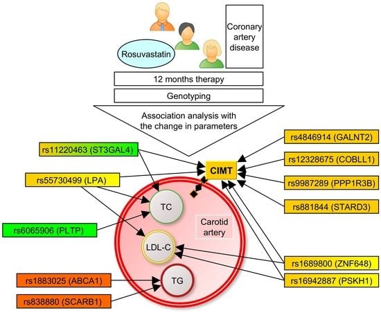
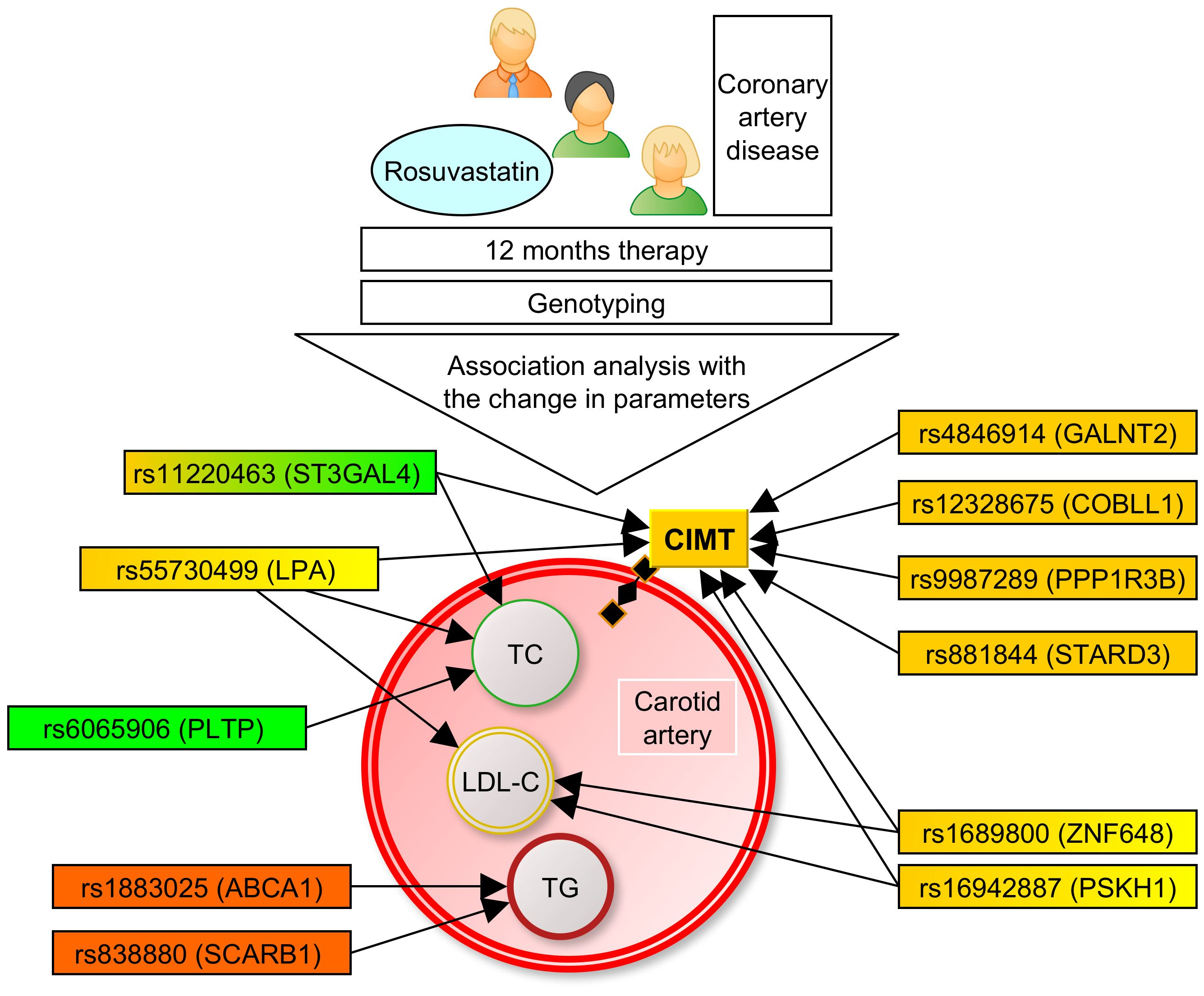
2. Materials and Methods

Study design. The present research was designed as a prospective study, in which the participants received rosuvastatin at an initial dose of 5 mg. Then, in 4 weeks, we controlled the levels of LDL-C. In cases where the levels were below the target level (1.8 mmol/L), patients continued taking the dose, which let them attain the target level. If it was not attained, the dose of rosuvastatin was gradually increased to 10, 20, and finally the maximum dose of 40 mg. The control of LDL-C levels was performed every 4 weeks after the dose increase. For the association analysis, we used the change (delta, Δ) in TC, LDL-C, TG, and CIMT values over the first 6 and 12 months of the study.
Study participants. The patient cohort comprised 116 Russian patients with the diagnosis of CAD stable angina pectoris grade II–III. The study participants were described in detail in our previous work [31]. Characteristics of the study cohort are given in Table 1. In brief, the cohort included 85 men (73%) and 31 postmenopausal women (27%), with a mean age of 61.0 ± 7.25 years. Approximately 58% of CAD patients experienced a myocardial infarction in the past, and 97.5% of study participants had arterial hypertension. The diagnosis of CAD was confirmed by qualified cardiologists according to the Canadian Cardiovascular Society’s grading of angina, ECG stress tests (treadmill test), and 24-h Holter’s ECG monitoring. TC levels higher than 4.0 mmol/L and LDL-C levels higher than 1.8 mmol/L were considered criteria for dyslipidemia.
Biochemical and ultrasound investigations were described in our previous paper [31]. Plasma lipid levels, except LDL-C, were detected using an automatic laboratory analyzer. LDL-C was calculated using Friedewald’s equation. CIMT measurement was performed in B-mode using the standard method at the distal third of the common carotid artery at a distance of 1–1.5 cm proximal to the bifurcation along the posterior wall [33]. The following two parameters of carotid intima-media thickness were used in the study: mean CIMT (the mean value of all measurements on the right and left sides) and maximum CIMT (the maximum value obtained from measurements on both sides with the further assessment of its change on the side where it was the highest).
Genetic analysis. Single nucleotide polymorphisms (SNPs) selected for this study have previously been found to be associated with plasma lipids in several genome-wide association studies [7,8,9,10,11,12] (GWAS Catalog, (https://www.ebi.ac.uk/gwas, accessed on 13 March 2023). As mentioned above, some of the SNPs have been recently found to be associated with CAD susceptibility and CIMT [32], and we were interested in investigating whether these variants determine the lipid- and CIMT-lowering effects of rosuvastatin in CAD patients. The investigated SNPs included rs4846914 (GALNT2), rs11220463 (ST3GAL4), rs881844 (STARD3), rs1689800 (ZNF648), rs12328675 (COBLL1), rs9987289 (PPP1R3B), rs55730499 (LPA), rs3136441 (F2), rs6065906 (PLTP), rs838880 (SCARB1), rs386000 (LILRA3), rs1883025 (ABCA1), rs3764261 (CETP), rs217406 (NPC1L1), and rs16942887 (PSKH1).
Extraction of DNA was performed from venous blood samples using the phenol-chloroform method and precipitation with ethanol. Genotyping was performed using iPLEX technology on the MassARRAY 4 system (Agena Bioscience, San Diego, CA, USA). The software MassARRAY Assay Design Suite was used to select a primer set and to design a multiplex panel for SNP genotyping. The sequences of primers are available on request.
Statistical analysis. The normality of the distribution of lipid and CIMT values was determined using the Kolmogorov–Smirnov and Shapiro–Wilk tests. As the trait distributions deviated from the normal one, the values were expressed as the median and interquartile range (Me, Q1; Q3). The statistical package STATISTICA v13.0 (Statsoft, Tulsa, OK, USA) was utilized for descriptive statistics, distribution analysis, and determining the significance of lipid and CIMT changes. The significance of lipid and CIMT changes during rosuvastatin therapy was tested by Wilcoxon’s matched pairs test. The distribution of genotype frequencies according to Hardy–Weinberg equilibrium was assessed with Fisher’s exact test. For the association analysis, we used linear regression with adjustments for sex, age, body mass index (calculated as body mass in kilograms divided by height in meters in a square), and rosuvastatin dose, which allowed the participants to attain the target LDL-C level. The change in lipid levels was calculated as the difference between the natural log-transformed on- and off-treatment levels divided by the natural log-transformed off-treatment level, as described by Postmus et al. [13]. We applied the same approach for the CIMT change, but before the logarithmic transformation, the CIMT data were multiplied by 10. Empirical p-values (Pperm) were calculated through the adaptive permutation procedure. We tested three genetic models for SNP-phenotype associations, such as additive, dominant, and recessive. The PLINK v1.92 software [34] was used for all genetic calculations, including estimation of minor allele frequencies (MAF), tests for Hardy-Weinberg equilibrium (HWE), multiple regression analysis, and permutation procedures. When the p-value was less than 0.05, all of the results were declared statistically significant.

3. Results

The majority of the patients (N = 110, 94.8%) attained the target LDL-C level during rosuvastatin therapy. The change in lipid and CIMT values was assessed during 6 and 12 months of observation. The reduction of all lipid parameters (p < 1 × 10⁶ for TC, LDL-C, and TG changes during 6- and 12-month periods) was significant, except HDL-C levels (p = 0.16 and 0.87, respectively). Reductions in the maximum (p < 1 × 10⁶) and mean CIMT (p = 0.025 and <1 × 10⁵ for the change in 6 and 12 months, respectively) were also significant (Supplementary Table S1).

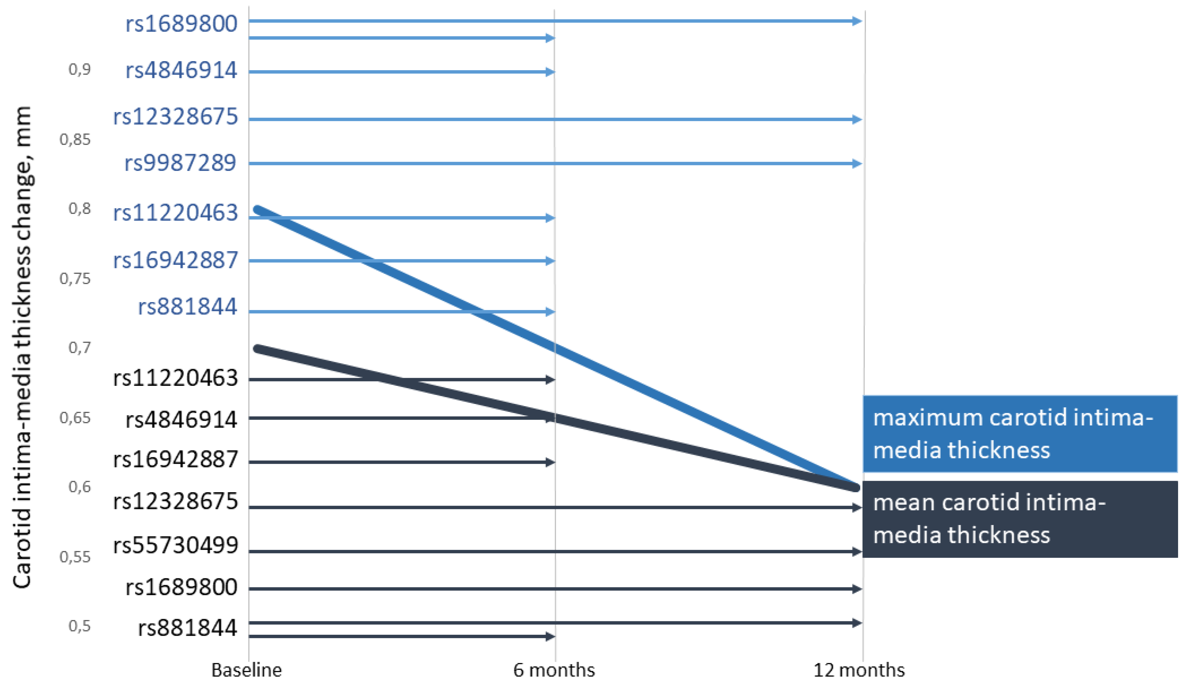
3.1. Associations of the SNPs with Lipid and CIMT Reduction during the 6-Month Therapy by Rosuvastatin

The distribution of genotypes for the majority of SNPs (except for rs16942887) was in Hardy-Weinberg equilibrium. The observed effects of SNPs on the lipid-lowering effect and CIMT regression in CAD patients taking rosuvastatin for the 6-month period are shown in Table 2. We found that plasma TC reduction was associated with two SNPs, such as rs55730499 of LPA (beta = 0.323, Pperm = 0.0022, recessive effect) and rs6065906 of PLTP (beta = 0.140, Pperm = 0.0135, recessive effect). Based on the beta coefficients, it can be estimated that the carriage of homozygous genotypes for the minor alleles of the above polymorphisms was associated with 32.3% and 14.0% worse TC reduction, respectively, compared with the carriers of alternative genotypes (the positive beta indicates a worse lipid reduction and consequently a worse statin response). LDL-C reduction was found to be associated with rs55730499 of the LPA gene (Pperm = 0.0224, recessive effect) and rs1689800 of the ZNF648 gene (Pperm = 0.0493, additive effect). The rs1689800G allele was associated with a 4.6 percent worse reduction in plasma LDL-C levels after 6 months of rosuvastatin therapy. The effect size for the rs55730499 polymorphism of LPA was greater in the TT homozygotes and characterized by a 50.4% worse decrease in LDL-C concentration than in carriers of alternative genotypes.
As can be seen from Table 2, four lipid-associated GWAS loci were associated with both maximum and mean CIMT change: rs4846914 in GALNT2 (Pperm = 0.0133 and 0.0344 for the association with maximum and mean CIMT change, respectively), rs11220463 in ST3GAL4 (Pperm = 0.0159 and 0.0243, respectively), rs16942887 in PSKH1 (Pperm = 0.0421 and 0.0483, respectively), and rs881844 in STARD3 (Pperm = 0.0086 and 0.0033, respectively). SNP rs1689800 in the ZNF648 gene (Pperm = 0.0234) was associated with a change in the maximum CIMT. The most significant association with TC and LDL-C reduction during 6 months of rosuvastatin therapy was found for SNP rs55730499 in LPA, whereas the rs881844 polymorphism of the STARD3 gene showed the most substantial association with the regression of CIMT in CAD patients.
Table 3 shows associations between the SNPs and plasma triglyceride reduction. It has been revealed that the only SNP rs838880 of the SCARB1 gene was associated with TG reduction (beta = 1.736, Pperm = 0.0478, recessive effect) after 6 months of lipid-lowering therapy.

3.2. Associations of SNPs with Lipid and CIMT Reduction during 12-Month Therapy by Rosuvastatin

Table 4 shows the associations of studied SNPs with the 12-month lipid-lowering effect and carotid intima-media thickness change in CAD patients receiving rosuvastatin.
We observed that two polymorphisms contributed to TC reduction: rs55730499 in LPA (beta = 0.364, Pperm = 0.0001, recessive effect) and rs11220463 in ST3GAL4 (beta = −0.181, Pperm = 0.0273, recessive effect) after one year of lipid-lowering therapy. For the rs11220463 polymorphism, the homozygous genotype for the minor allele T was associated with an 18.1% more pronounced TC-lowering effect than in carriers of alternative genotypes. The reduction in plasma LDL-C levels was associated with rs55730499 of LPA (Pperm = 0.0415, recessive effect) and rs16942887 of PSKH1 (Pperm = 0.0175, dominant effect).
Three SNPs were associated with both maximum and mean CIMT change: rs1689800 of ZNF648 (better statin response; Pperm = 0.0105 and 0.0282 for the association with maximum and mean CIMT change, respectively); rs12328675 of COBLL1 (worse statin response; Pperm = 0.0213 and 0.0056, respectively), and rs9987289 of PPP1R3B (worse statin response; Pperm = 0.0359 and 0.0109, respectively). Two polymorphisms, such as rs55730499 of LPA (Pperm = 0.0146) and rs881844 of STARD3 (Pperm = 0.0223), were associated with the mean CIMT change.
During 12 months of lipid-lowering therapy with rosuvastatin, the most significant effect on the TC reduction was found for SNP rs55730499 of LPA, whereas the most significant effects of rs16942887 of PSKH1 and rs12328675 of COBLL1 were found on the reduction of LDL-C and CIMT, respectively. Associations of TG reduction (Table 3) with genotype in a 12-month period were found for rs1883025 of the ABCA1 gene (beta = −0.7246, Pperm = 0.0160, dominant effect) and for rs11220463 in ST3GAL4 (beta = 3.624, Pperm = 0.05, recessive effect).

4. Discussion

The present study demonstrated, for the first time, the impact of lipid-associated GWAS loci on CIMT (Figure 1) and plasma levels of TC, LDL-C, and TG in CAD patients during rosuvastatin therapy (Figure 2).
In particular, we found associations between SNPs located at ZNF648, LPA, ST3GAL4, PSKH1, GALNT2, COBLL1, PPP1R3B, and STARD3 genes and reductions in CIMT. Pharmacogenetic associations of the variants in ZNF648, LPA, ST3GAL4, PSKH1, PLTP, SCARB1 with the change in lipid levels were also found for the first time. Thus, lipid-associated GWAS loci are associated not only with lipids and the risk of coronary artery disease [32] but are also responsible for the antiatherogenic effects of rosuvastatin after 6 and 12 months of lipid-lowering therapy in patients suffering from CAD.
Potential mechanisms by which polymorphic variants of the investigated lipid metabolism genes can impact the antiatherogenic effects of rosuvastatin therapy are of interest. The ZNF648 gene encodes a zinc finger protein 648, which may be involved in DNA-templated transcription [35]. In the present study, the rs1689800 polymorphism was associated with CIMT change. The possible mechanism of the effect of the variant on the vascular wall may include its influence on oxidative stress (GLUL catabolizes glutamate, an amino acid required for glutathione synthesis) because such a mechanism is described for the nearest SNP, rs10911021, located in the same genomic region [36].
The GALNT2 gene encodes N-acetylgalactosaminyltransferase 2, an enzyme involved in O-linked glycosylation of substrates, including those regulating lipid metabolisms such as apolipoprotein C-III (APOC-III), angiopoietin-related protein 3 (ANGPTL3), and phospholipid transfer protein (PLTP); in these ways, it influences HDL-C and TG metabolism [37,38]. The rs4846914G allele has been linked to atherogenic changes in plasma lipid metabolism, such as an increase in TG and a decrease in HDL-C levels [9,10]. We found no associations of this variant with changes in any plasma lipid, but this SNP was significantly associated with a better CIMT regression during a 6-month period of rosuvastatin therapy. The presence of the link with CIMT change and the absence of any association with lipid level change suggest that the pharmacogenetic effect on CIMT change might be mediated through non-lipid-related mechanisms, taking into account that SNP rs4846914 is known to be associated with endothelial function, serum levels of insulin and glucose [39], as well as hypertension [40]. In particular, the latter study demonstrated higher promoter methylation of the GALNT2 gene, higher levels of ApoB, and lower levels of ApoA1 in hypertensives with the GG genotype [40].
The COBLL1 gene encodes a cordon-bleu WH2 repeat protein such as 1, which has actin monomer and cadherin binding activity [41]. In the present study, the variant rs12328675 was associated with worse CIMT regression (for the carriers of the C-allele, dominant effect) during 12 months of observation. This association was the most significant among all variants studied over a 12-month period. There were no associations with lipid levels changing. The link between this polymorphism and CIMT change can be explained by the ability of the risk allele C, associated with the higher CAD risk, to form transcription factor binding sites for the factors involved in the processes of vascular inflammation regulation and angiogenesis (AP-1 (syn. JUN), SMAD2, SMAD3, SMAD4, and E2F8) [42].
The LPA gene encodes lipoprotein (a), which is a well-known and independent risk factor for coronary artery disease [11]. The studied rs55730499 variant is associated with Lp(a) concentrations [11], coronary artery disease risk [8], myocardial infarction risk [43], and stroke risk [44]. The T-allele is associated with higher TC levels out of therapy [43], and in our study, the TT genotype (homozygous for the minor allele) was associated with reduced drug response in terms of TC, LDL-C, and CIMT change. The association with TC change had the highest significance among all associations with TC change, and moreover, rs55730499 was the only variant studied that was associated with both TC, LDL-C, and CIMT change in plasma at the same period of observation (12 months). Based on these findings, the studied LPA variant can be considered the first of all studied variants to be used as a predictor for rosuvastatin therapy personalization.
The PPP1R3B gene encodes protein phosphatase 1 regulatory subunit 3B, a key protein in hepatic glycogen metabolism [12]. The rs9987289 variant is associated with plasma TC and LDL-C levels [9,12] but was not tested for carotid atherosclerosis traits or the pharmacogenetics of rosuvastatin. In our study, the presence of the minor A-allele was associated with a worse CIMT response to rosuvastatin. Such an effect could be explained by the association of the studied SNP with inflammatory markers, such as C-reactive protein [45], known for its influence on cardiovascular risk and atherosclerosis progression [46], and also by the link of the variant with metabolic syndrome [47] and glucose levels [48], taking into account the known role of hyperglycemia in the dysfunction of the endothelium [49,50].
The ABCA1 gene encodes the phospholipid-transporting ATPase ABCA1, which provides the efflux of intracellular cholesterol to apolipoproteins and the formation of nascent high-density lipoproteins [51]. The minor T-allele of the rs1883025 SNP is associated in GWAS with an anti-atherogenic phenotype: lower TC [9], LDL-C [52], TG [53], and HDL-C levels [52], and with a better response to statin therapy ([54], the particular drug and lipid are not specified). In the present study, we confirmed a better response to statin therapy in terms of triglyceride reduction in carriers of the minor T-allele (the dominant effect of the SNP).
The ST3GAL4 gene encodes CMP-N-acetylneuraminate-beta-galactosamide-alpha-2,3-sialyltransferase 4, an enzyme involved in the terminal sialylation of glycoproteins and glycolipids. A beta-galactoside alpha2-3 sialyltransferase takes part in hemostasis (sialylation of plasma von Willebrand factor) and the inflammatory process (selectin-mediated rolling and adhesion of leukocytes during extravasation) [55]. The studied SNP in this gene, rs11220463, is associated with both blood lipids (TC and LDL-C) [53] and inflammation (CRP levels) [56]. In the present study, we found pharmacogenetic associations between the studied variant and TC and CIMT reduction during therapy. The association with CIMT can possibly be explained by the involvement of the product of ST3GAL4 in inflammatory processes, taking into account the known association of systemic inflammation (assessed by the systemic immune-inflammatory index) with CIMT [57]. For rs11220463, the association with inflammation was found in terms of CRP levels [56], and CRP is known to be associated with CIMT [58,59], but this association is not proven to be causal [58].
The PSKH1 gene encodes serine/threonine-protein kinase H1, involved in intracellular protein trafficking and pre-mRNA processing [60]. For the rs16942887 variant, we found associations with LDL-C and CIMT changes on rosuvastatin therapy. There are no reported associations in the literature between this variation and changes in LDL-C and CIMT when taking rosuvastatin. The possible mechanism of the effect of SNP is difficult to predict because of the lack of information on the mechanisms of lipid and vascular influence of PSKH1.
The STARD3 gene encodes StAR-related lipid transfer protein 3, which mediates cholesterol transport from the endoplasmic reticulum to endosomes [61]. In the literature, the rs881844 polymorphism in STARD3 was known to be associated with plasma lipids, including TC and HDL-C [43,53]. In the present study, we did not find any associations with changes in plasma lipid levels on rosuvastatin therapy; however, this SNP showed the strongest effects on CIMT changes in both 6- and 12-month periods. The effect on the vascular wall can be explained by the involvement of STARD3 in the regulation of cholesterol-dependent inflammation and sensitivity to proinflammatory cytokines [62].
The PLTP gene encodes phospholipid transfer protein, which is involved in the transfer of phospholipids and free cholesterol from LDLs and VLDLs into HDLs [63], as well as the uptake of cholesterol from peripheral cells [64]. Taking into account the function of the gene product, it was rather logical to find the influence of rs6065906 in PLTP on the lipid-lowering effect of rosuvastatin in terms of TC reduction in the present study, which was not reported before.
Thus, the molecular mechanisms underlying the effects of the studied loci in terms of lipid and CIMT reduction remain unknown, but taking into account the biological functions of the proteins, these genes possess pleiotropic effects on biological and pathological processes including lipid metabolism, inflammation, leukocyte adhesion to the endothelium, endothelial dysfunction, and glucose metabolism. Further experimental studies are needed to explain the functional effects of polymorphisms.
It can be noticed that the association between SNPs and the change in plasma cholesterol level and CIMT was different between 6 months and 12 months. This could be explained by two reasons. The first explanation is a weak effect of polymorphisms that reached a borderline significance level at the one-time point but were not seen at another time point. However, it could be explained according to the physiology of lipid metabolism, the function of genes, and the effects of their polymorphisms. For the variant in LPA, the influence on CIMT can be delayed: firstly, there is drug response from lipids (because lipoprotein (a) particles contain LDLs), and then from the vascular wall (which needs more time to respond). In the same manner, rs1689800 (ZNF648) initially influenced blood lipids and maximum CIMT, and in time the effect on the vessels became more significant (both mean and maximum CIMT with lower p-value). Some genetic polymorphisms influence CIMT change independently of lipids (e.g., loci at COBLL1 and STARD3). The COBLL1 gene is known to influence angiogenesis and the regulation of inflammation in vessels [42], but its effect on plasma lipids is unknown. The delayed effect of this SNP (CIMT change over a 12-month period) can be explained by the longer time needed for vascular changes (compared with plasma lipids) as a pleiotropic effect of rosuvastatin. The STARD3 variant showed the vascular effect faster—in a 6-month period, and it lasted for 12-months. We believe it could be a fast response to lipid concentration changes in the cells of the vascular wall because the STARD3 gene regulates the inflammatory response to cholesterol and cholesterol transport within membrane contacts [61] (that is why it possibly did not influence plasma lipid concentration in our study). The variant at ST3GAL4 showed first an effect on CIMT and then on lipids. According to the function of the gene, it regulates inflammation, selectin-mediated leukocyte adhesion, and arterial calcification [55], so the effect on the vessels (due to inflammation) can be realized faster. The effect size of the association with LDL-C change in the first 6 months was rather low, which is why the association did not reach the targeted significance level.
The fifteen loci selected for the current study have been discovered in GWAS as genetic variants associated with blood lipids, the risk of cardiovascular disease (angina pectoris, myocardial infarction, heart failure, and stroke), and the effectiveness of statin therapy in terms of lipid reduction [7,8,9,10,11,12,43,54]. The present study showed that lipid-associated GWAS loci are linked to the lipid-lowering effects of rosuvastatin, and these effects were observed in the reduction of CIMT. This finding provides some insights into the mechanisms of drug action, such as: (1) only CIMT decrease was associated with genotype (for example, based on the link with COBLL1 and STARD3 variants and no association with lipid reduction); or (2) both CIMT decrease and lipid reduction were associated with genotype (for example, the variant in LPA). Thus, it can be concluded that there could be diverse mechanisms of the drug effect on the vascular wall, governed by genetic factors that are dependent or independent of lipid change. Confirmation of rosuvastatin’s effect on the vascular wall using genetic association analysis supports the clinical use of the drug in CAD patients and explains inter-individual variability in the drug effect.
Study limitations. Due to the non-significant HDL-C change in the therapy observed in the present study, we did not test the SNPs for associations with HDL-C change. This is a limitation of our study; however, we cannot say it is a big omission because one of the main phenotypes studied here was CIMT, which is known to have a causal relationship with LDL-C and with other factors but not with HDL-C itself [18]. Furthermore, the use of a surrogate endpoint for the vascular wall, CIMT change, was a limitation of our study. Further studies using other endpoints, such as coronary plaque characteristics, could provide a more precise genetic impact on the changes in the vascular wall in CAD patients taking statins. As for CIMT change, the effect size could not be so high since the prominent change in the vascular wall could not occur in a rather short period of time. The majority of the observed associations were significant but at a borderline level, except for the variant in the LPA gene. This is because of both the relatively moderate effects of SNPs and/or the small sample size investigated with respect to rosuvastatin pharmacogenetics. Therefore, the study’s findings should be interpreted with caution, and further investigation with a larger sample size is required to reproduce the observed associations.

5. Conclusions

In the present pharmacogenetic study of rosuvastatin effects in CAD patients, associations between both TC (and/or LDL-C) and CIMT changes during the same follow-up period (6 or 12 months) were observed for the polymorphisms rs55730499 of LPA (after 12 months of therapy) and rs1689800 of ZNF648 (after 6 months of therapy). In general, a decrease in both atherogenic lipids and CIMT was associated with the effects of four polymorphisms: rs1689800 (LDL-C and CIMT), rs55730499 (TC, LDL-C, and CIMT), rs11220463 (TC and CIMT), and rs16942887 (LDL-C and CIMT). Half of the eight investigated lipid-associated GWAS loci, such as rs4846914, rs12328675, rs9987289, and rs881844, were linked to CIMT changes but were not associated with changes in plasma lipids during rosuvastatin therapy. The rs881844 and rs12328675 polymorphisms were most strongly associated with CIMT regression, whereas rs55730499 and rs16942887 showed associations with the reduction of plasma TC and LDL-C. Thus, the rs1689800, rs55730499, rs11220463, and rs16942887 polymorphisms were found to be predictive markers for multiple antiatherogenic effects of rosuvastatin in CAD patients. Undoubtedly, the observed associations should be replicated in independent pharmacogenetic studies with a larger number of CAD patients. The successful validation of these associations may provide novel genetic markers for personalized hypolipidemic therapy with rosuvastatin and monitoring of drug effectiveness in patients with atherogenic changes in lipid metabolism.

Supplementary Materials

The following supporting information can be downloaded at: https://www.mdpi.com/article/10.3390/genes14061259/s1, Table S1: Change in lipid and carotid intima-media thickness values during rosuvastatin therapy.

Author Contributions

Conceptualization, A.P.; methodology, A.P., S.K., M.S. and M.C.; software, S.K. and A.P.; validation, S.K. and A.P.; formal analysis, S.K. and M.S.; genetic investigation, I.A., E.K. and M.B.; resources, A.P.; data curation, S.K. and A.P.; writing—original draft preparation, S.K.; writing—review and editing, A.P.; visualization, S.K.; supervision, A.P.; project administration, A.P. All authors have read and agreed to the published version of the manuscript.

Funding

This research received funding from Kursk State Medical University.

Institutional Review Board Statement

The study was conducted in accordance with the Declaration of Helsinki and approved by the Institutional Ethics Committee of Kursk State Medical University (11 May 2015).

Informed Consent Statement

Informed consent was obtained from all subjects involved in the study.

Data Availability Statement

Data supporting reported results are available upon request.

Conflicts of Interest

The authors declare no conflict of interest.

References

  1. Gupta, R.M.; Schnitzler, G.; Fang, S.; Lee-Kim, V.S.; Barry, A. Multiomic Analysis and CRISPR Perturbation Screens Identify Endothelial Cell Programs and Novel Therapeutic Targets for Coronary Artery Disease. Arterioscler. Thromb. Vasc. Biol. 2023, 43, 600–608. [Google Scholar] [CrossRef] [PubMed]
  2. Blackburn, H. Invited commentary: 30-year perspective on the seven countries study. Am. J. Epidemiol. 2017, 185, 1143–1147. [Google Scholar] [CrossRef]
  3. McPherson, R.; Tybjaerg-Hansen, A. Genetics of Coronary Artery Disease. Circ. Res. 2016, 118, 564–578. [Google Scholar] [CrossRef] [PubMed]
  4. Churilin, M.I.; Kononov, S.I.; Mal, G.S.; Polonikov, A.V.; Lazarenko, V.A. Lipid metabolism genes and predisposition to ischemic heart disease. Med. News North Cauc. 2019, 14, 401–407. [Google Scholar] [CrossRef]
  5. Mutlu, A.S.; Duffy, J.; Wang, M.C. Lipid metabolism and lipid signals in aging and longevity. Dev. Cell. 2021, 56, 1394–1407. [Google Scholar] [CrossRef]
  6. Walsh, R.; Jurgens, S.J.; Erdmann, J.; Bezzina, C.R. Genome-wide association studies of cardiovascular disease. Physiol. Rev. 2023, 103, 2039–2055. [Google Scholar] [CrossRef] [PubMed]
  7. Willer, C.J.; Schmidt, E.M.; Sengupta, S.; Peloso, G.M.; Gustafsson, S.; Kanoni, S.; Ganna, A.; Chen, J.; Buchkovich, M.L.; Mora, S.; et al. Discovery and refinement of loci associated with lipid levels. Nat. Genet. 2013, 45, 1274–1283. [Google Scholar]
  8. Nikpay, M.; Goel, A.; Won, H.H.; Hall, L.M.; Willenborg, C.; Kanoni, S.; Saleheen, D.; Kyriakou, T.; Nelson, C.P.; Hopewell, J.C.; et al. A comprehensive 1000 Genomes–based genome-wide association meta-analysis of coronary artery disease. Nat. Genet. 2015, 47, 1121–1130. [Google Scholar] [PubMed] [Green Version]
  9. Teslovich, T.M.; Musunuru, K.; Smith, A.V.; Edmondson, A.C.; Stylianou, I.M.; Koseki, M.; Pirruccello, J.P.; Ripatti, S.; Chasman, D.I.; Willer, C.J.; et al. Biological, clinical and population relevance of 95 loci for blood lipids. Nature 2010, 466, 707–713. [Google Scholar] [CrossRef] [Green Version]
  10. Kathiresan, S.; Melander, O.; Guiducci, C.; Surti, A.; Burtt, N.P.; Rieder, M.J.; Cooper, G.M.; Roos, C.; Voight, B.F.; Havulinna, A.S.; et al. Six new loci associated with blood low-density lipoprotein cholesterol, high-density lipoprotein cholesterol or triglycerides in humans. Nat. Genet. 2008, 40, 189–197. [Google Scholar] [CrossRef]
  11. Mack, S.; Coassin, S.; Rueedi, R.; Yousri, N.A.; Seppälä, I.; Gieger, C.; Schönherr, S.; Forer, L.; Erhart, G.; Marques-Vidal, P.; et al. A genome-wide association meta-analysis on lipoprotein (a) concentrations adjusted for apolipoprotein (a) isoforms. J. Lipid Res. 2017, 58, 1834–1844. [Google Scholar] [CrossRef] [PubMed] [Green Version]
  12. Zhang, Y.; Gan, W.; Tian, C.; Li, H.; Lin, X.; Chen, Y. Association of PPP1R3B polymorphisms with blood lipid and C-reactive protein levels in a Chinese population. J. Diabetes 2013, 5, 275–281. [Google Scholar] [CrossRef]
  13. Postmus, I.; Trompet, S.; Deshmukh, H.A.; Arsenault, B.J.; Avery, C.L.; Bis, J.C.; Chasman, D.I.; de Keyser, C.E.; Deshmukh, H.A.; Evans, D.S.; et al. Pharmacogenetic meta-analysis of genome-wide association studies of LDL cholesterol response to statins. Nat. Commun. 2014, 5, 5068. [Google Scholar] [CrossRef] [PubMed] [Green Version]
  14. Chasman, D.I.; Giulianini, F.; MacFadyen, J.; Barratt, B.J.; Nyberg, F.; Ridker, P.M. Genetic determinants of statin-induced low-density lipoprotein cholesterol reduction: The Justification for the Use of Statins in Prevention: An Intervention Trial Evaluating Rosuvastatin (JUPITER) trial. Circ. Cardiovasc. Genet. 2012, 5, 257–264. [Google Scholar] [CrossRef] [Green Version]
  15. Chasman, D.I.; Giulianini, F.; Demler, O.V.; Udler, M.S. Pleiotropy-Based Decomposition of Genetic Risk Scores: Association and Interaction Analysis for Type 2 Diabetes and CAD. Am. J. Hum. Genet. 2020, 106, 646–658. [Google Scholar] [CrossRef]
  16. Crouse, J.R., 3rd; Raichlen, J.S.; Riley, W.A.; Evans, G.W.; Palmer, M.K.; O’Leary, D.H.; Grobbee, D.E.; Bots, M.L.; METEOR Study Group. Effect of rosuvastatin on progression of carotid intima-media thickness in low-risk individuals with subclinical atherosclerosis: The METEOR Trial. JAMA 2007, 297, 1344–1353. [Google Scholar] [CrossRef] [Green Version]
  17. Nezu, T.; Hosomi, N.; Aoki, S.; Matsumoto, M. Carotid Intima-Media Thickness for Atherosclerosis. J. Atheroscler. Thromb. 2016, 23, 18–31. [Google Scholar] [CrossRef] [Green Version]
  18. Shah, S.; Casas, J.P.; Drenos, F.; Whittaker, J.; Deanfield, J.; Swerdlow, D.I.; Holmes, M.V.; Kivimaki, M.; Langenberg, C.; Wareham, N.; et al. Causal relevance of blood lipid fractions in the development of carotid atherosclerosis: Mendelian randomization analysis. Circ. Cardiovasc. Genet. 2013, 6, 63–72. [Google Scholar] [CrossRef]
  19. Vargas, J.D.; Manichaikul, A.; Wang, X.Q.; Rich, S.S.; Rotter, J.I.; Post, W.S.; Polak, J.F.; Budoff, M.J.; Bluemke, D.A. Detailed analysis of association between common single nucleotide polymorphisms and subclinical atherosclerosis: The Multi-ethnic Study of Atherosclerosis. Data Brief. 2016, 7, 229–242. [Google Scholar] [CrossRef] [Green Version]
  20. Guaricci, A.I.; Arcadi, T.; Brunetti, N.D.; Maffei, E.; Montrone, D.; Martini, C.; De Luca, M.; De Rosa, F.; Cocco, D.; Midiri, M.; et al. Carotid intima media thickness and coronary atherosclerosis linkage in symptomatic intermediate risk patients evaluated by coronary computed tomography angiography. Int. J. Cardiol. 2014, 176, 988–993. [Google Scholar] [CrossRef]
  21. Amato, M.; Montorsi, P.; Ravani, A.; Oldani, E.; Galli, S.; Ravagnani, P.M.; Tremoli, E.; Baldassarre, D. Carotid intima-media thickness by B-mode ultrasound as surrogate of coronary atherosclerosis: Correlation with quantitative coronary angiography and coronary intravascular ultrasound findings. Eur. Heart J. 2007, 28, 2094–2101. [Google Scholar] [CrossRef] [Green Version]
  22. Granér, M.; Varpula, M.; Kahri, J.; Salonen, R.M.; Nyyssönen, K.; Nieminen, M.S.; Taskinen, M.R.; Syvänne, M. Association of carotid intima-media thickness with angiographic severity and extent of coronary artery disease. Am. J. Cardiol. 2006, 97, 624–629. [Google Scholar] [CrossRef] [PubMed]
  23. Matsushima, Y.; Kawano, H.; Koide, Y.; Baba, T.; Toda, G.; Seto, S.; Yano, K. Relationship of carotid intima-media thickness, pulse wave velocity, and ankle brachial index to the severity of coronary artery atherosclerosis. Clin Cardiol. 2004, 27, 629–634. [Google Scholar] [CrossRef]
  24. Ballantyne, C.M.; Raichlen, J.S.; Nicholls, S.J.; Erbel, R.; Tardif, J.C.; Brener, S.J.; Cain, V.A.; Nissen, S.E.; ASTEROID Investigators. Effect of rosuvastatin therapy on coronary artery stenoses assessed by quantitative coronary angiography: A study to evaluate the effect of rosuvastatin on intravascular ultrasound-derived coronary atheroma burden. Circulation 2008, 117, 2458–2466. [Google Scholar] [CrossRef] [Green Version]
  25. Puri, R.; Libby, P.; Nissen, S.E.; Wolski, K.; Ballantyne, C.M.; Barter, P.J.; Chapman, M.J.; Erbel, R.; Raichlen, J.S.; Uno, K.; et al. Long-term effects of maximally intensive statin therapy on changes in coronary atheroma composition: Insights from SATURN. Eur. Heart J. Cardiovasc. Imaging 2014, 15, 380–388. [Google Scholar] [CrossRef] [Green Version]
  26. Rumyantsev, N.A.; Kukes, V.G.; Kazakov, R.E.; Rumyantsev, A.A.; Sychev, D.A. Ispol’zovanie farmakogeneticheskogo testirovaniia dlia predotvrashcheniia nezhelatel’nykh lekarstvennykh reaktsiĭ pri terapii statinami [Use of pharmacogenetic testing to prevent adverse drug reactions during statin therapy]. Ter Arkh. 2017, 89, 82–87. (In Russian) [Google Scholar] [CrossRef]
  27. Soko, N.D.; Masimirembwa, C.; Dandara, C. Pharmacogenomics of Rosuvastatin: A Glocal (Global + Local) African Perspective and Expert Review on a Statin Drug. OMICS A J. Integr. Biol. 2016, 20, 498–509. [Google Scholar] [CrossRef]
  28. Alfonsi, J.E.; Hegele, R.A.; Gryn, S.E. Pharmacogenetics of lipid-lowering agents: Precision or indecision medicine? Curr. Atheroscler. Rep. 2016, 18, 24. [Google Scholar] [CrossRef]
  29. Churilin, M.I.; Kononov, S.I.; Luneva, Y.V.; Kazanov, V.A.; Azarova, I.E.; Klyosova, E.Y.; Bykanova, M.A.; Paschoalini, G.; Kharchenko, A.V.; Zhabin, S.N.; et al. Polymorphisms of Intracellular Cholesterol Transporters Genes: Relationship to Blood Lipid Levels, Carotid Intima-Media Thickness, and the Development of Coronary Heart Disease. Russ. J. Genet. 2020, 56, 234–241. [Google Scholar] [CrossRef]
  30. Churilin, M.I.; Kononov, S.I.; Luneva, Y.V.; Azarova, Y.E.; Klesova, E.Y.; Kharchenko, A.V.; Zhabin, S.N.; Bushueva, O.Y.; Povetkin, S.V.; Mal, G.S.; et al. Apolipoprotein E gene polymorphisms: A relationship with the risk of coronary artery disease and the effectiveness of lipid-lowering therapy with rosuvastatin. Cardiovasc. Therapy Prev. Russ. Fed. 2020, 19, 17–23. [Google Scholar] [CrossRef] [Green Version]
  31. Kononov, S.; Mal, G.; Azarova, I.; Klyosova, E.; Bykanova, M.; Churnosov, M.; Polonikov, A. Pharmacogenetic loci for rosuvastatin are associated with intima-media thickness change and coronary artery disease risk. Pharmacogenomics 2022, 23, 15–34. [Google Scholar] [CrossRef]
  32. Lazarenko, V.; Churilin, M.; Azarova, I.; Klyosova, E.; Bykanova, M.; Ob’edkova, N.; Churnosov, M.; Bushueva, O.; Mal, G.; Povetkin, S.; et al. Comprehensive Statistical and Bioinformatics Analysis in the Deciphering of Putative Mechanisms by Which Lipid-Associated GWAS Loci Contribute to Coronary Artery Disease. Biomedicines 2022, 10, 259. [Google Scholar] [CrossRef] [PubMed]
  33. Pignoli, P.; Tremoli, E.; Poli, A.; Oreste, P.; Paoletti, R. Intimal plus medial thickness of the arterial wall: A direct measurement with ultrasound imaging. Circulation 1986, 74, 1399–1406. [Google Scholar] [CrossRef] [Green Version]
  34. Purcell, S.; Neale, B.; Todd-Brown, K.; Thomas, L.; Ferreira, M.A.; Bender, D.; Maller, J.; Sklar, P.; de Bakker, P.I.; Daly, M.J.; et al. PLINK: A tool set for whole-genome association and population-based linkage analyses. Am. J. Hum. Genet. 2007, 81, 559–575. [Google Scholar] [CrossRef] [PubMed] [Green Version]
  35. Gaudet, P.; Livstone, M.S.; Lewis, S.E.; Thomas, P.D. Phylogenetic-based propagation of functional annotations within the Gene Ontology consortium. Brief Bioinform. 2011, 12, 449–462. [Google Scholar] [CrossRef] [Green Version]
  36. Shahid, S.U.; Shabana Humphries, S. The SNP rs10911021 is associated with oxidative stress in coronary heart disease patients from Pakistan. Lipids Health Dis. 2018, 17, 6. [Google Scholar] [CrossRef] [PubMed] [Green Version]
  37. Khetarpal, S.A.; Schjoldager, K.T.; Christoffersen, C.; Raghavan, A.; Edmondson, A.C.; Reutter, H.M.; Ahmed, B.; Ouazzani, R.; Peloso, G.M.; Vitali, C.; et al. Loss of Function of GALNT2 Lowers High-Density Lipoproteins in Humans, Nonhuman Primates, and Rodents. Cell Metab. 2016, 24, 234–245. [Google Scholar] [CrossRef] [Green Version]
  38. Zilmer, M.; Edmondson, A.C.; Khetarpal, S.A.; Alesi, V.; Zaki, M.S.; Rostasy, K.; Madsen, C.G.; Lepri, F.R.; Sinibaldi, L.; Cusmai, R.; et al. Novel congenital disorder of O-linked glycosylation caused by GALNT2 loss of function. Brain 2020, 143, 1114–1126. [Google Scholar] [CrossRef]
  39. Chen, J.; Guan, L.; Liu, H.; Liu, Q.; Fan, P.; Bai, H. GALNT2 Gene Variant rs4846914 Is Associated with Insulin and Insulin Resistance Depending on BMI in PCOS Patients: A Case-Control Study. Reprod Sci. 2021, 28, 1122–1132. [Google Scholar] [CrossRef]
  40. Zhang, X.; Zhao, H.; Zhang, J.; Han, D.; Zheng, Y.; Guo, X.; He, D.; Guo, J.; Wang, Y. Gene environment interaction of GALNT2 and APOE gene with hypertension in the Chinese Han Population. Biomed. Mater. Eng. 2015, 26 (Suppl. S1), S1977–S2083. [Google Scholar] [CrossRef] [Green Version]
  41. Guo, Z.; Neilson, L.J.; Zhong, H.; Murray, P.S.; Zanivan, S.; Zaidel-Bar, R. E-cadherin interactome complexity and robustness resolved by quantitative proteomics. Sci. Signal. 2014, 7, rs7. [Google Scholar] [CrossRef] [Green Version]
  42. Churilin, M.I. Association of RS12328675 COBLL1 polymorphism with coronary heart disease and intermediate phenotypes of atherosclerosis: Validation study in Central Russia. Res. Results Biomed. 2020, 6, 209–218. [Google Scholar] [CrossRef]
  43. Sakaue, S.; Kanai, M.; Tanigawa, Y.; Karjalainen, J.; Kurki, M.; Koshiba, S.; Narita, A.; Konuma, T.; Yamamoto, K.; Akiyama, M.; et al. A cross-population atlas of genetic associations for 220 human phenotypes. Nat. Genet. 2021, 53, 1415–1424. [Google Scholar] [CrossRef]
  44. Mishra, A.; Malik, R.; Hachiya, T.; Jürgenson, T.; Namba, S.; Posner, D.C.; Kamanu, F.K.; Koido, M.; Le Grand, Q.; Shi, M.; et al. Stroke genetics informs drug discovery and risk prediction across ancestries. Nature 2022, 611, 115–123, Erratum in Nature 2022, 612, E7. [Google Scholar] [CrossRef] [PubMed]
  45. Wojcik, G.L.; Graff, M.; Nishimura, K.K.; Tao, R.; Haessler, J.; Gignoux, C.R.; Highland, H.M.; Patel, Y.M.; Sorokin, E.P.; Avery, C.L.; et al. Genetic analyses of diverse populations improves discovery for complex traits. Nature 2019, 570, 514–518. [Google Scholar] [CrossRef]
  46. Sethwala, A.M.; Goh, I.; Amerena, J.V. Combating Inflammation in Cardiovascular Disease. Heart Lung Circ. 2021, 30, 197–206. [Google Scholar] [CrossRef]
  47. Lind, L. Genome-Wide Association Study of the Metabolic Syndrome in UK Biobank. Metab. Syndr. Relat. Disord. 2019, 17, 505–511. [Google Scholar] [CrossRef] [PubMed]
  48. Chen, J.; Spracklen, C.N.; Marenne, G.; Varshney, A.; Corbin, L.J.; Luan, J.; Willems, S.M.; Wu, Y.; Zhang, X.; Horikoshi, M.; et al. The trans-ancestral genomic architecture of glycemic traits. Nat. Genet. 2021, 53, 840–860. [Google Scholar] [CrossRef] [PubMed]
  49. Meza, C.A.; La Favor, J.D.; Kim, D.H.; Hickner, R.C. Endothelial Dysfunction: Is There a Hyperglycemia-Induced Imbalance of NOX and NOS? Int. J. Mol. Sci. 2019, 20, 3775. [Google Scholar] [CrossRef] [Green Version]
  50. Clyne, A.M. Endothelial response to glucose: Dysfunction, metabolism, and transport. Biochem. Soc. Trans. 2021, 49, 313–325. [Google Scholar] [CrossRef]
  51. Krimbou, L.; Denis, M.; Haidar, B.; Carrier, M.; Marcil, M.; Genest, J., Jr. Molecular interactions between apoE and ABCA1: Impact on apoE lipidation. J. Lipid Res. 2004, 45, 839–848. [Google Scholar] [CrossRef] [PubMed] [Green Version]
  52. Huang, Q.Q.; Sallah, N.; Dunca, D.; Trivedi, B.; Hunt, K.A.; Hodgson, S.; Lambert, S.A.; Arciero, E.; Wright, J.; Griffiths, C.; et al. Transferability of genetic loci and polygenic scores for cardiometabolic traits in British Pakistani and Bangladeshi individuals. Nat. Commun. 2022, 13, 4664. [Google Scholar] [CrossRef]
  53. Hoffmann, T.J.; Theusch, E.; Haldar, T.; Ranatunga, D.K.; Jorgenson, E.; Medina, M.W.; Kvale, M.N.; Kwok, P.Y.; Schaefer, C.; Krauss, R.M.; et al. A large electronic-health-record-based genome-wide study of serum lipids. Nat. Genet. 2018, 50, 401–413. [Google Scholar] [CrossRef] [PubMed]
  54. Wu, Y.; Byrne, E.M.; Zheng, Z.; Kemper, K.E.; Yengo, L.; Mallett, A.J.; Yang, J.; Visscher, P.M.; Wray, N.R. Genome-wide association study of medication-use and associated disease in the UK Biobank. Nat. Commun. 2019, 10, 1891. [Google Scholar] [CrossRef] [Green Version]
  55. Mondal, N.; Buffone, A., Jr.; Stolfa, G.; Antonopoulos, A.; Lau, J.T.; Haslam, S.M.; Dell, A.; Neelamegham, S. ST3Gal-4 is the primary sialyltransferase regulating the synthesis of E-, P-, and L-selectin ligands on human myeloid leukocytes. Blood 2015, 125, 687–696. [Google Scholar] [CrossRef] [Green Version]
  56. Ligthart, S.; Vaez, A.; Hsu, Y.H.; Inflammation Working Group of the CHARGE Consortium; PMI-WG-XCP; LifeLines Cohort Study; Stolk, R.; Uitterlinden, A.G.; Hofman, A.; Alizadeh, B.Z.; et al. Bivariate genome-wide association study identifies novel pleiotropic loci for lipids and inflammation. BMC Genom. 2016, 17, 443. [Google Scholar] [CrossRef] [Green Version]
  57. Ozbeyaz, N.B.; Gokalp, G.; Algul, E.; Kilic, P.; Saricam, O.; Aydinyilmaz, F.; Guliyev, I. Could Systemic Inflammation in Healthy Individuals with Obesity Indicate Subclinical Atherosclerosis? Angiology 2023, 74, 62–69. [Google Scholar] [CrossRef]
  58. Kivimäki, M.; Lawlor, D.A.; Smith, G.D.; Kumari, M.; Donald, A.; Britton, A.; Casas, J.P.; Shah, T.; Brunner, E.; Timpson, N.J.; et al. Does high C-reactive protein concentration increase atherosclerosis? The Whitehall II Study. PLoS ONE 2008, 3, e3013. [Google Scholar] [CrossRef] [Green Version]
  59. Moran, C.A.; Sheth, A.N.; Mehta, C.C.; Hanna, D.B.; Gustafson, D.R.; Plankey, M.W.; Mack, W.J.; Tien, P.C.; French, A.L.; Golub, E.T.; et al. The association of C-reactive protein with subclinical cardiovascular disease in HIV-infected and HIV-uninfected women. AIDS 2018, 32, 999–1006. [Google Scholar] [CrossRef]
  60. Brede, G.; Solheim, J.; Prydz, H. PSKH1, a novel splice factor compartment-associated serine kinase. Nucleic Acids Res. 2002, 30, 5301–5309. [Google Scholar] [CrossRef] [PubMed] [Green Version]
  61. Wilhelm, L.P.; Wendling, C.; Védie, B.; Kobayashi, T.; Chenard, M.P.; Tomasetto, C.; Drin, G.; Alpy, F. STARD3 mediates endoplasmic reticulum-to-endosome cholesterol transport at membrane contact sites. EMBO J. 2017, 36, 1412–1433. [Google Scholar] [CrossRef] [PubMed]
  62. Li, L.; Liu, Y.; Liu, X.; Zheng, N.; Gu, Y.; Song, Y.; Wang, X. Regulatory roles of external cholesterol in human airway epithelial mitochondrial function through STARD3 signalling. Clin. Transl. Med. 2022, 12, e902. [Google Scholar] [CrossRef] [PubMed]
  63. Zhang, M.; Zhai, X.; Li, J.; Albers, J.J.; Vuletic, S.; Ren, G. Structural basis of the lipid transfer mechanism of phospholipid transfer protein (PLTP). Biochim. Biophys. Acta Mol. Cell Biol. Lipids 2018, 1863, 1082–1094. [Google Scholar] [CrossRef]
  64. Albers, J.J.; Vuletic, S.; Cheung, M.C. Role of plasma phospholipid transfer protein in lipid and lipoprotein metabolism. Biochim. Biophys. Acta 2012, 1821, 345–357. [Google Scholar] [CrossRef] [PubMed] [Green Version]
Figure 1. Associations of single nucleotide polymorphisms with carotid intima-media thickness (CIMT) change in coronary artery disease patients taking rosuvastatin at 6 and 12 months of therapy. The bold lines show CIMT reduction (mm), and the length of the arrows depicts the effects of particular single nucleotide polymorphisms that were observed in 6- or 12-month CIMT reduction. The color of the arrows shows the influence of the SNPs on maximum (blue) or mean (dark blue) CIMT reduction.
Genes 14 01259 g001
Figure 2. Associations of single nucleotide polymorphisms with lipid-lowering effect of rosuvastatin in coronary artery disease patients at 6 and 12 months of therapy. TC, total cholesterol; LDL-C, low-density lipoprotein cholesterol; TG, triglyceride levels. The bold lines show lipid fraction change (mmol/L), and the length of the arrows depicts the effects of single nucleotide polymorphisms, observed at 6- and 12 months. The color of the arrows shows the influence of the SNPs on TC (green), LDL-C (brown), or TG (red) reduction.
Genes 14 01259 g002
Table 1. Baseline parameters of the study patients.
Baseline ParameterMean ± Standard Deviation/
Median (Q1; Q3)
Age (years)61.0 ± 7.25
Body mass index (kg/m2)28.77 ± 4.18
Hypertension (%)97.5
Past myocardial infarction (%)57.6
Systolic blood pressure (mmHg)131.1 ± 8.1
Diastolic blood pressure (mmHg)74.9 ± 4.4
Total cholesterol (mmol/L)5.28 (4.60; 6.06)
LDL-C (mmol/L)3.27 (2.70; 4.08)
HDL-C (mmol/L)1.06 (0.97; 1.29)
TG (mmol/L)1.71 (1.22; 2.37)
CIMT, maximum (mm)0.80 (0.60; 1.00)
CIMT, mean (mm)0.70 (0.55; 0.85)
LDL-C indicates low-density lipoprotein cholesterol; HDL-C, high-density lipoprotein cholesterol; TG, triglycerides; CIMT, carotid intima-media thickness.
Table 2. Associations of single nucleotide polymorphisms with the 6-month lipid-lowering effect (total cholesterol, low density lipoprotein cholesterol) and carotid intima-media thickness change in coronary artery disease patients taking rosuvastatin.
ChrGene (SNP ID)Effect AlleleEAFNTotal CholesterolLDL-CCIMT, MaximumCIMT, Mean
Beta *Pperm #Beta *Pperm #Beta *Pperm #Beta *Pperm #
1ZNF648 (rs1689800)G0.3921150.0200.17860.0460.0493 A−0.0840.0234 R−0.0210.2308
1GALNT2 (rs4846914)G0.3881150.0040.7778−0.0050.8571−0.0450.0133 A−0.0380.0344 A
2COBLL1 (rs12328675)C0.170111−0.0030.8571−0.0031.00000.0350.26470.0410.1000
6LPA (rs55730499)T0.0561150.3230.0022 R0.5040.0224 R−0.0100.85710.0280.8571
7NPC1L1 (rs217406)G0.2031150.0170.23080.0330.3556−0.0011.0000−0.0031.0000
8PPP1R3B (rs9987289)A0.0861150.0190.3404−0.0130.85710.0410.34040.0510.1550
9ABCA1 (rs1883025)T0.2631150.0011.0000−0.0040.8571−0.0240.2982−0.0250.2535
11F2 (rs3136441)C0.1801020.0110.64290.0250.77780.0210.69230.0230.2982
11ST3GAL4 (rs11220463)T0.190115−0.0310.1280−0.0680.08030.0660.0159 D0.0610.0243 A
12SCARB1 (rs838880)C0.3361150.0021.00000.0051.0000−0.0140.7273−0.0150.6923
16CETP (rs3764261)A0.1471150.0070.62500.0270.8571−0.0450.2466−0.0420.1900
16PSKH1 (rs16942887)A0.1161150.0090.64290.0250.46430.0660.0421 D0.0680.0483 D
17STARD3 (rs881844)C0.3101150.0030.8571−0.0380.16670.0480.0086 A0.0570.0033 A
19LILRA3 (rs386000)C0.2031150.0060.5455−0.0011.00000.0150.77780.0030.8571
20PLTP (rs6065906)C0.1601150.1400.0135 R−0.0081.0000−0.0220.6923−0.0220.5455
Chr, chromosome; SNP, single nucleotide polymorphism; EAF, the effect allele frequency; LDL-C, low-density lipoprotein cholesterol; CIMT, carotid intima-media thickness. Significant Pperm-values are highlighted in bold. * Beta the for difference between the natural log-transformed on- and off-treatment values adjusted for natural log-transformed off-treatment level. The beta reflects the fraction of differential lipid- or CIMT-lowering effect in carriers versus non-carriers of the minor allele (for additive model), in carriers of the genotype according to the model (dominant, recessive) versus the carriers of alternative genotypes; a negative beta indicates a better statin response (stronger lipid or CIMT reduction), a positive beta—a worse statin response. Betas were generated using linear regression analysis with age, sex, body mass index, and rosuvastatin dose as covariates. # p-values were generated using the permutation procedure. Superscript indicates genetic model: R, recessive; A, additive; D, dominant.
Table 3. Associations of single nucleotide polymorphisms with triglyceride-lowering effect in coronary artery disease patients taking rosuvastatin.
ChrGene (SNP ID)Effect AlleleEAFN6-Month Period12-Month Period
Beta *Pperm #Beta *Pperm #
1ZNF648 (rs1689800)G0.392114−0.64950.1148−0.19460.4643
1GALNT2 (rs4846914)G0.3881140.48160.19190.050460.7778
2COBLL1 (rs12328675)C0.170110−0.29480.6250−0.14310.7778
6LPA (rs55730499)T0.056114−0.59230.42420.18250.6923
7NPC1L1 (rs217406)G0.2031140.41060.4118−0.12080.6429
8PPP1R3B (rs9987289)A0.086114−0.91970.1887−0.79770.0756
9ABCA1 (rs1883025)T0.263114−0.1770.5789−0.72460.0160 D
11F2 (rs3136441)C0.180101−0.1850.57890.025121.0000
11ST3GAL4 (rs11220463)T0.190114−0.7740.15873.6240.0503 R
12SCARB1 (rs838880)C0.3361141.7360.0478 R0.10630.6429
16CETP (rs3764261)A0.1471140.6950.29310.023381.0000
16PSKH1 (rs16942887)A0.116114−0.63060.1439−0.26260.3947
17STARD3 (rs881844)C0.3101140.40750.4815−0.23370.2647
19LILRA3 (rs386000)C0.203114−0.18470.8571−0.14480.6923
20PLTP (rs6065906)C0.160114−0.46540.5789−0.07180.8571
Chr, chromosome; SNP, single nucleotide polymorphism; EAF, effect allele frequency. Significant Pperm-values are highlighted in bold. * Beta for the difference between the natural log-transformed on- and off-treatment values adjusted for natural log-transformed off-treatment level. Betas were generated using linear regression analysis with age, sex, body mass index, and rosuvastatin dose as covariates. # p-values were generated using the permutation procedure. Superscript indicates genetic model: R, recessive; D, dominant.
Table 4. Associations of single nucleotide polymorphisms with the 12-month lipid-lowering effect and carotid intima-media thickness change in coronary artery disease patients taking rosuvastatin.
ChrGene (SNP ID)Effect AlleleEAFNTotal CholesterolLDL-CCIMT, MaximumCIMT, Mean
Beta *Pperm #Beta *Pperm #Beta *Pperm #Beta *Pperm #
1ZNF648 (rs1689800)G0.3921130.0100.30910.0240.2043−0.0930.0105 R−0.0360.0282 A
1GALNT2 (rs4846914)G0.388113−0.0040.7273−0.0160.41180.0020.8571−0.0020.8571
2COBLL1 (rs12328675)C0.170109−0.0110.4643−0.0180.46430.0590.0213 A0.0750.0056 D
6LPA (rs55730499)T0.0561130.3640.0001 R0.3670.0415 R0.0180.85710.4140.0146 R
7NPC1L1 (rs217406)G0.203113−0.0021.00000.0030.20430.0190.36360.0180.4516
8PPP1R3B (rs9987289)A0.0861130.0040.8571−0.0680.41180.0720.0359 D0.0790.0109 D
9ABCA1 (rs1883025)T0.2631130.0100.31480.0010.46430.0110.85710.0190.2687
11F2 (rs3136441)C0.1801000.0130.34780.0180.2043−0.0110.6429−0.0051.0000
11ST3GAL4 (rs11220463)T0.190113−0.1810.0273 R−0.0270.41180.0011.00000.0320.1709
12SCARB1 (rs838880)C0.336113−0.0011.0000−0.0050.4643−0.0031.0000−0.0041.0000
16CETP (rs3764261)A0.1471130.0170.34780.0210.2043−0.0530.0833−0.0430.0880
16PSKH1 (rs16942887)A0.1161130.0060.57890.0860.0175 D−0.0110.5217−0.0040.7778
17STARD3 (rs881844)C0.3101130.0090.43750.0100.85710.0280.18520.0400.0223 A
19LILRA3 (rs386000)C0.2031130.0190.24000.0150.52000.0260.29820.0100.7778
20PLTP (rs6065906)C0.1601130.0130.48150.0310.2571−0.0290.3478−0.0061.0000
Chr, chromosome; SNP, single nucleotide polymorphism; EAF, the effect allele frequency; LDL-C, low-density lipoprotein cholesterol; CIMT, carotid intima-media thickness. Significant Pperm-values are highlighted in bold. * Beta for the difference between the natural log-transformed on- and off-treatment values adjusted for natural log-transformed off-treatment level. The beta reflects the fraction of differential lipid- or CIMT-lowering effect in carriers versus non-carriers of the minor allele (for additive model), in carriers of the genotype according to the model (dominant, recessive) versus the carriers of alternative genotypes; a negative beta indicates a better statin response (stronger lipid or CIMT reduction), a positive beta—a worse statin response. Betas were generated using linear regression analysis with age, sex, body mass index, and rosuvastatin dose as covariates. # p-values were generated using the permutation procedure. Superscript indicates genetic model: R, recessive; A, additive; D, dominant.
Disclaimer/Publisher’s Note: The statements, opinions and data contained in all publications are solely those of the individual author(s) and contributor(s) and not of MDPI and/or the editor(s). MDPI and/or the editor(s) disclaim responsibility for any injury to people or property resulting from any ideas, methods, instructions or products referred to in the content.

