Article

Coronin 1C, Regulated by Multiple microRNAs, Facilitates Cancer Cell Aggressiveness in Pancreatic Ductal Adenocarcinoma

1
Department of Digestive Surgery, Breast and Thyroid Surgery, Graduate School of Medical and Dental Sciences, Kagoshima University, Kagoshima 890-8520, Japan
2
Department of Functional Genomics, Graduate School of Medicine, Chiba University, Chiba 260-8670, Japan
*
Author to whom correspondence should be addressed.
Genes 2023, 14(5), 995; https://doi.org/10.3390/genes14050995
Submission received: 28 March 2023 / Revised: 18 April 2023 / Accepted: 25 April 2023 / Published: 27 April 2023
(This article belongs to the Special Issue Genomics and Epigenomics of Gastrointestinal Disorders)

Abstract

:
Coronin proteins are actin-related proteins containing WD repeat domains encoded by seven genes (CORO1A, CORO1B, CORO1C, CORO2A, CORO2B, CORO6, and CORO7) in the human genome. Analysis of large cohort data from The Cancer Genome Atlas revealed that expression of CORO1A, CORO1B, CORO1C, CORO2A, and CORO7 was significantly upregulated in pancreatic ductal adenocarcinoma (PDAC) tissues (p < 0.05). Moreover, high expression of CORO1C and CORO2A significantly predicted the 5 year survival rate of patients with PDAC (p = 0.0071 and p = 0.0389, respectively). In this study, we focused on CORO1C and investigated its functional significance and epigenetic regulation in PDAC cells. Knockdown assays using siRNAs targeting CORO1C were performed in PDAC cells. Aggressive cancer cell phenotypes, especially cancer cell migration and invasion, were inhibited by CORO1C knockdown. The involvement of microRNAs (miRNAs) is a molecular mechanism underlying the aberrant expression of cancer-related genes in cancer cells. Our in silico analysis revealed that five miRNAs (miR-26a-5p, miR-29c-3p, miR-130b-5p, miR-148a-5p, and miR-217) are putative candidate miRNAs regulating CORO1C expression in PDAC cells. Importantly, all five miRNAs exhibited tumor-suppressive functions and four miRNAs except miR-130b-5p negatively regulated CORO1C expression in PDAC cells. CORO1C and its downstream signaling molecules are potential therapeutic targets in PDAC.

1. Introduction

Pancreatic ductal adenocarcinoma (PDAC) is one of the deadliest human cancers, with a 5 year survival rate of approximately 10%, regardless of the stage [1,2,3]. In Japan, the annual number of deaths from PDAC is approximately 38,000 among both men and women, ranking as the fourth deadliest cancer after lung cancer, colorectal cancer, and gastric cancer [4].
Long-term survival is expected in patients with early-stage PDAC and tumors that can be surgically resected. However, only about 20% of all pancreatic cancers can be surgically resected. Furthermore, approximately 30% of patients already have locally advanced PDAC at diagnosis, and 50% of patients have metastatic disease [2,3,5]. Chemotherapy regimens, e.g., FOLFIRINOX (5-fluorouracil, folinic acid, irinotecan, and oxaliplatin), gemcitabine-containing combination treatments, and gemcitabine treatments, are indicated for patients who are ineligible for surgery [2,5,6]. Despite these treatments, the survival time of patients with advanced-stage PDAC is less than 1 year [2,3,6].
Previous studies have shown that four major genetic mutations (KRAS, TP53, CDKN2A, and SMAD4) are closely involved in the oncogenic process of PDAC [6,7,8]. Recent advances in sequencing technology have enabled whole-genome mutation analyses in individual cancer cells, thereby revealing genomic differences among individual cancer cells. The molecular classification of PDAC (the Know Your Tumor program) has revealed that approximately 25% of PDAC patients have actionable molecular alterations [9]. Recently, several therapeutic strategies that target actionable driver gene mutations in cancer cells have been developed. For example, PARP inhibitors target PDAC cells with BRCA1 or BRCA2 mutations, and immune checkpoint inhibitors target cells with mismatch repair deficiencies [3,5,7,10].
PDAC patients frequently develop recurrence or metastasis even after curative resection [11]. To improve the prognosis of PDAC, it is essential to clarify the molecular mechanisms of recurrence and metastasis of PDAC cells. To date, no driver molecules causing recurrence or metastasis have been identified. A unique feature of PDAC is the abundant fibrous stroma composed of extracellular matrix (ECM) proteins surrounding the cancer cells [12,13]. Cell invasion through the ECM of cancer cells is the first step in metastasis. The formation of various structures, such as invadopodia and pseudopodia, is required for PDAC cells to degrade and migrate through the ECM. These structural changes require actin assembly regulated by specialized actin nucleation factors. Reorganization of the actin cytoskeleton is essential for invasive cell migration and is initiated by various actin nucleation factors [14]. Actin nucleators are now emerging as promising targets to control cancer cell metastasis.
In this study, we focused on coronin proteins, which bind to filamentous actin and the Arp2/3 complex and contribute to modulating actin dynamics [15]. In humans, the coronin family consists of seven genes (CORO1A, CORO1B, CORO1C, CORO2A, CORO2B, CORO6, and CORO7), all of which possess a conserved basic N-terminal motif and several WD repeats clustered in the core domains [16]. Analysis of The Cancer Genome Atlas (TCGA) data revealed that overexpression of CORO1C and CORO2A is closely involved in PDAC molecular pathogenesis. Functional assays of CORO1C revealed that aberrant expression of CORO1C facilitates PDAC cell migration and invasion abilities.
We analyzed the aberrant expression of CORO1C in PDAC cells from an epigenomic viewpoint. A vast number of studies have shown that microRNAs (miRNAs) negatively regulate gene expression [17,18]. Downregulation of tumor-suppressive miRNAs induces overexpression of cancer-promoting genes in cancer cells [19,20]. Our analysis revealed that four miRNAs (miR-26a-5p, miR-29c-3p, miR-148a-5p, and miR-217) regulated CORO1C expression in PDAC cells. All these miRNAs acted as tumor-suppressive miRNAs in PDAC cells.

2. Materials and Methods

2.1. Analysis of the Expression and Clinical Significance of Coronin Genes in PDAC

The expression levels of the coronin genes CORO1A, CORO1B, CORO1C, CORO2A, CORO1B, CORO6, and CORO7 in PDAC clinical specimens were evaluated using the GEPIA2 platform (http://gepia2.cancer-pku.cn/#index; accessed on 31 May 2022). For the Kaplan–Meier plots and log-rank test, we used the OncoLnc database (http://www.oncolnc.org, accessed on 13 June 2022).

2.2. Gene Set Enrichment Analysis (GSEA)

GSEA was performed to investigate the CORO1C-mediated molecular pathways. The TCGA–PDAC data were divided into high- and low-expression groups according to the Z-score of the CORO1C expression level. The expression levels of each gene were compared in the high- and low-CORO1C expression groups, and the genes were ranked according to the log2 ratio. We uploaded the resultant ranked gene lists into GSEA software [21,22] and applied the Hallmark gene set in the Molecular Signatures Database [23].

2.3. Human PDAC Cell Lines

Two PDAC cell lines, PANC-1 and SW1990, were used in this study. The cell lines were purchased from the American Type Culture Collection (Manassas, VA, USA) and the RIKEN Cell Bank (Tsukuba, Japan), respectively.

2.4. Transfection of Small Interfering RNAs (siRNAs) and miRNAs into PDAC Cells and Quantitative Reverse-Transcription PCR (qRT-PCR)

Transfection of siRNAs and miRNAs into PDAC cell lines was performed using Lipofectamine RNAiMAX reagent (Invitrogen, Carlsbad, CA, USA) according to our previous studies [24,25,26]. In this study, we define “mock” and “control” as follows: mock: transfection reagent was added to the medium; control: scrambled RNA different from the siRNA or miRNA sequence was added to the medium. The reagents used in this study are listed in Table S1. We performed qRT-PCR using the StepOnePlus™ Real-Time PCR System (Applied Biosystems, Waltham, MA, USA), and gene expression levels were normalized to those of GUSB as the internal control. The sequences of the SYBR Green primers are listed in Table S2. The procedures for qRT-PCR assays in PDAC cells have been described previously [24,25,26].

2.5. Functional Assays (Cell Proliferation, Migration, and Invasion) in PDAC Cells

The procedures for the functional assays (cell proliferation, migration, and invasion assays) in PDAC cells have been described previously [24,25,26]. Cell proliferation was examined by the XTT assay (Sigma–Aldrich, St. Louis, MO, USA) at 72 h after miRNA or siRNA transfection. Cell migration and invasion assays were conducted using the BioCoatTM cell culture chamber and the BioCoat Matrigel Invasion Chamber, both from Corning (Corning, NY, USA). Forty-eight hours after transfection, the cells at the bottom of the chamber were counted and analyzed.

2.6. Identification of the miRNAs Regulating CORO1C Expression in PDAC Cells

The Target Scan Human 8.0 database (http://www.targetscan.org/vert_80, accessed on 21 September 2022) was used to select miRNAs that have putative binding sites in the 3′UTR of CORO1C. The miRNA expression signature of PDAC was used to screen miRNAs [27]. Expression of miRNAs in PDAC clinical specimens was assessed using the GEO database (GSE24279 and GSE71533).

2.7. Western Blotting

The Western blotting procedure has been described previously [24,25,26]. The anti-CORO1C antibody (Proteintech Group, Inc., Rosemont, IL, USA) was diluted 1:2500, and the anti-GAPDH antibody (Wako, Osaka, Japan), used as the internal control, was diluted 1:1600. The antibodies used are listed in Table S1.

2.8. Statistical Analysis

JMP Pro 15 software (SAS Institute Inc., Cary, NC, USA) was used for the statistical analyses. Differences between the two groups were evaluated using Welch’s t-test, and differences among multiple groups were evaluated using Dunnett’s test. To analyze prognosis based on CORO1C expression, the patients were divided into two groups according to the expression level of CORO1C, and differences in survival were evaluated. A p-value less than 0.05 was considered statistically significant.

3. Results

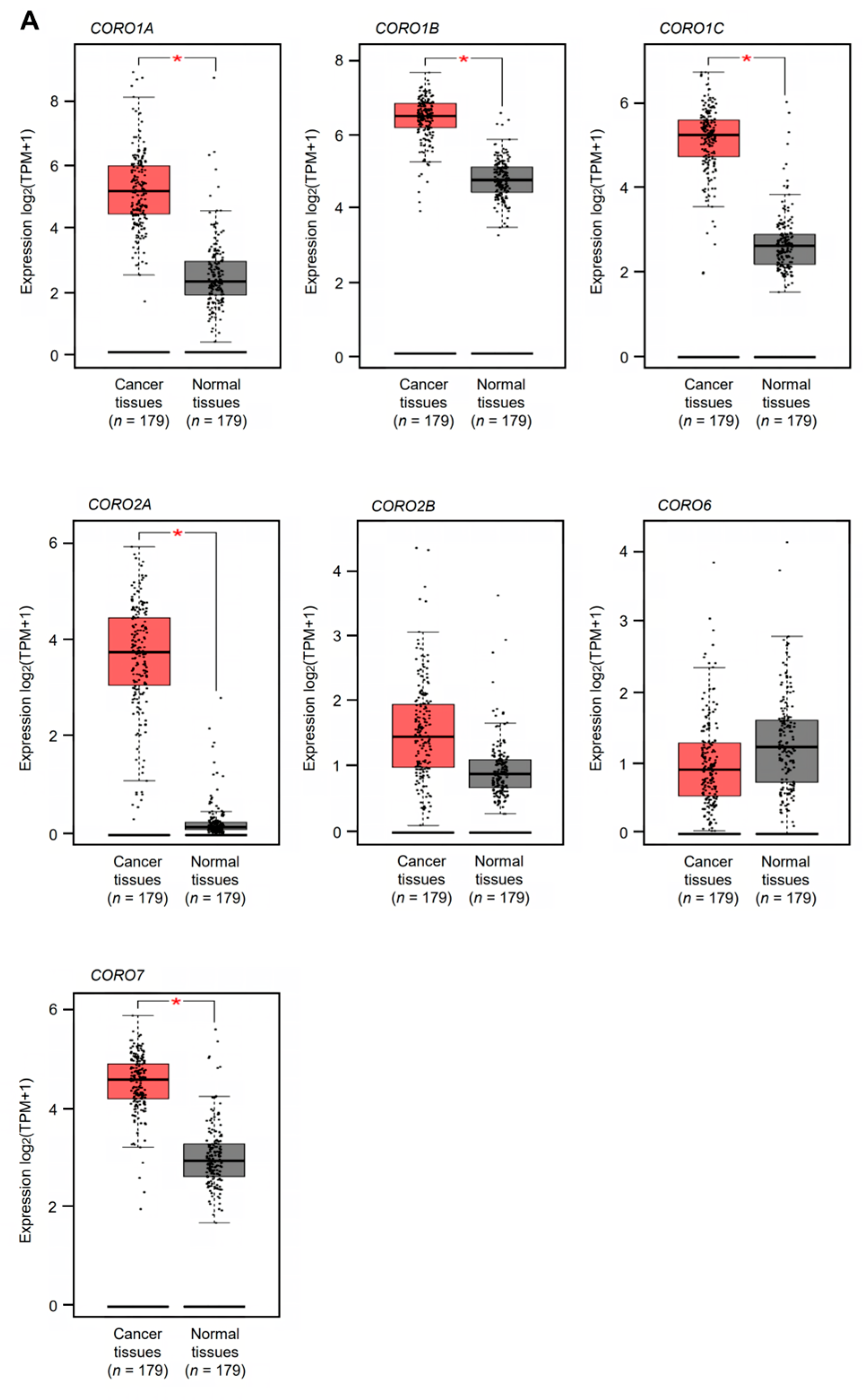
3.1. Expression and Clinical Significance of Coronin Family Members in Patients with PDAC

Based on the TCGA and GEPIA2 databases, the expression levels of all members of the coronin family (CORO1A, CORO1B, CORO1C, CORO2A, CORO2B, CORO6, and CORO7) were analyzed. Among the coronin genes, the expression levels of five (CORO1A, CORO1B, CORO1C, CORO2A, and CORO7) were significantly upregulated in PDAC tissues (n = 179) compared with normal pancreatic tissues (n = 179) (p < 0.05, Figure 1A). Two genes (CORO2B and CORO6) did not show significant expression in PDAC tissues (Figure 1A).
Next, we investigated whether high expression of these genes affected patient prognosis using TCGA PDAC data. The prognosis was significantly worse in patients with high expression of CORO1C and CORO2A versus low expression of these genes (p = 0.0071 and p = 0.0389, respectively) (Figure 1B). Notably, the prognosis was significantly worse in patients with low versus high expression of CORO2B (p = 0.0144, Figure 1B). As a result of the in silico analysis, two genes (CORO1C and CORO2A) were selected as candidate cancer-promoting genes that affect the prognosis of PDAC patients. Molecular analysis of these two genes is essential for elucidating the malignant transformation of PDAC cells. By comparing the significant difference in the 5 year survival rate, this study focused on CORO1C and performed functional analysis.

3.2. Molecular Pathways Associated with High CORO1C Expression in PDAC Cells

Using TCGA PDAC data, we performed GSEA to identify the molecular pathways activated in patients with high CORO1C expression. We found that “epithelial–mesenchymal transition”, “inflammatory response”, and “KRAS signaling” were pathways enriched in the high CORO1C expression group (Figure 2). Activation of these pathways could potentially accelerate the malignant transformation of PDAC cells.

3.3. Effects of CORO1C Knockdown on PDAC Cell Proliferation, Migration, and Invasion

To assess the oncogenic function of CORO1C in PDAC cells, we performed knockdown assays using siRNAs. The inhibitory effect of two different siRNAs targeting CORO1C (siCORO1C-1 and siCORO1C-2) on CORO1C expression was examined. The CORO1C mRNA and protein levels were effectively suppressed by transfection of both siRNAs into two PDAC cell lines, PANC-1 and SW1990 (Figure S1).
Then, functional assays using these siRNAs were performed. The knockdown of CORO1C had little effect on cell proliferation in PANC-1 and SW1990 cells (Figure 3A).
Cell migration and invasion were significantly suppressed after transfection of the CORO1C siRNAs into PANC-1 and SW1990 cells (Figure 3B,C). Typical images of cells during the migration and invasion assays after siCORO1C transfection are shown in Figure 3D,E.
The antitumor effects (cell proliferation and migration) of the two siRNAs, siCORO1C-1 and siCORO1C-2, differed (Figure 3A,B). Under the analysis conditions of this study, the antitumor effects of siCORO1C-1 were insufficient compared to siCORO1C-2 in SW1990 cells.

3.4. Identification of miRNAs That Regulate CORO1C Expression in PDAC Cells

A vast number of studies have revealed that gene expression is regulated by various functional RNAs, e.g., lncRNAs, miRNAs, and circRNAs. Among them, it has been clarified that dysregulation of miRNAs causes aberrant expression of cancer-related genes and is closely involved in the malignant transformation of cancer cells. Therefore, we hypothesized that downregulation of some miRNAs might cause overexpression of CORO1C in PDAC cells. We searched for miRNAs that negatively regulate CORO1C expression in PDAC cells.
The strategy for identifying miRNAs that regulate CORO1C expression is shown in Figure 4. The combination of TargetScan data (release 8.0) and our miRNA signature of PDAC revealed 35 candidate miRNAs that regulate CORO1C expression in PDAC cells (Table 1). The expression levels of the 35 miRNAs were evaluated in GEO datasets (GSE 24279 and GSE 71533). Five miRNAs (miR-26a-5p, miR-29c-3p, miR-130b-5p, miR-148a-5p, and miR-217) were commonly downregulated in two datasets. We confirmed that expression levels of five miRNAs were downregulated in PDAC tissues (n = 136) compared with normal tissues (n = 22) by using GSE 24279 data (Figure 5A). Analysis using another dataset (GSE71533) also confirmed that the expression levels of five miRNAs were suppressed in PDAC tissues (n = 36) compared with normal tissues (n = 16) (Figure 5B).
The correlations of expression levels between five miRNAs (miR-26a-5p, miR-29c-3p, miR-130b-5p, miR-148a-5p, and miR-217) and CORO1C were evaluated using TCGA-PDAC data (Figure S2). A Spearman’s correlation coefficient rank test indicated that a negative correlation was detected in the expression levels of miR-29c-3p and CORO1C in PDAC clinical specimens (p < 0.05, r = −0.3666). No inverse correlation was observed between the expression levels of other miRNAs and CORO1C.
Next, we examined whether these miRNAs control the expression of CORO1C in PDAC cells. CORO1C mRNA expression after miRNA transfection. CORO1C mRNA expression was reduced at 48 h after transfection of miR-26a-5p, miR-29c-3p, miR-148a-5p, or miR-217 in PDAC cell lines (PANC-1 and SW1990). On the other hand, transfection with miR-130b-5p did not show remarkable regulation of CORO1C expression in PDAC cells (Figure 6A). CORO1C protein expression was reduced at 72 h after transfection of miR-26a-5p, miR-29c-3p, miR-148a-5p, or miR-217 in PDAC cells (Figure 6B). The intensity of the bands for the Western blotting was analyzed using ImageJ software. The results of the ImageJ analyses are shown in Figure S3.

3.5. Tumor-Suppressive Function of miR-26a-5p and miR-29c-3p in PDAC Cells

Our previous studies revealed that miR-130b-5p, miR-148a-5p, and miR-217 act as tumor-suppressive miRNAs in PDAC cells by targeting several oncogenes [28,29,30]. Therefore, in this study, we analyzed whether the two miRNAs, miR-26a-5p and miR-29c-3p, have tumor-suppressive functions in PDAC cells.
The tumor-suppressive activities of miR-26a-5p and miR-29c-3p were assessed by ectopic expression of mature miRNAs in PANC-1 and SW1990 cells. The inhibitory effect of miR-26a-5p on cell proliferation was observed in SW1990 cells but not in PANC-1 cells (Figure 7A). Cancer cell invasion and migration abilities were markedly suppressed by ectopic expression of miR-26a-5p in PDAC cells (Figure 7B,C). Typical images of Figure 7B (cell migration) and Figure 7C (cell invasion) are shown in Figure 7D,E, respectively.
Similar to miR-26a-5p, ectopic expression of miR-29c-3p attenuated the malignant phenotype, i.e., cell proliferation, invasion, and migration abilities, of PDAC cells (Figure 8A–C). Typical images of Figure 8B (cell migration) and Figure 8C (cell invasion) are shown in Figure 8D,E, respectively.

4. Discussion

The main cause of death in cancer patients is malignant transformation and metastasis of cancer cells rather than the surgically removable primary tumor. To improve the prognosis of PDAC patients, it is essential to clarify the molecular mechanism of pancreatic cancer metastasis.
In this study, we analyzed the coronin family of WD-repeat actin-binding proteins. Previous studies showed that coronin proteins regulate actin-dependent processes via F-actin assembly [15,16]. TCGA analysis revealed that overexpression of CORO1C is closely involved in the molecular pathogenesis of PDAC. Aberrant expression of CORO1C has been reported in several types of human cancers, e.g., glioblastoma, hepatocellular cancer, breast cancer, non-small cell lung cancer, gastric cancer, and colorectal cancer [31,32,33,34,35,36,37,38]. In addition, high expression of CORO1C is closely associated with a worse prognosis and aggressive pathological parameters in hepatocellular carcinoma, gastric cancer, and colorectal cancer [32,37,38]. Furthermore, we investigated the CORO1C-mediated molecular pathways in PDAC. GSEA analysis revealed that “epithelial–mesenchymal transition (EMT)” was the most enriched pathway in the CORO1C high-expression group. EMT has widely been accepted as a critical step in metastatic dissemination in PDAC cells, and various functional RNAs are closely involved in this process [39,40]. However, EMT-independent pathways have also been reported to be involved in distant metastases of PDAC [41]. Exploring the molecular pathways mediated by CORO1C will provide hints for elucidating the invasion and metastasis mechanisms of PDAC cells.
Previous functional analyses of CORO1C have shown that it functions as a cancer-promoting gene in several cancer cells [32,33,34,37,38]. In breast cancer cells, CORO1C was found to be involved in invadopodia formation and MT1-MMP surface trafficking and to promote invasiveness [35]. In gastric cancer cells, CORO1C expression facilitated cancer cell aggressiveness (e.g., cell viability, colony formation, and metastasis) by positively regulating cyclin D1 and vimentin expression [37]. In colorectal cancer cells, CORO1C interacted with trophoblast cell surface protein 2, and CORO1C overexpression enhanced cancer phenotypes via the PI3K/AKT signaling pathway [38]. The multifaceted functions of CORO1C enable it to activate various molecular pathways, which may accelerate the malignant transformation of cancer cells. Aberrant activation of the PI3K/AKT/mTOR pathway strongly contributed to the malignant transformation of various cancers, including PDAC [42,43]. Therefore, various inhibitors have been developed to block this oncogenic pathway [44]. Currently, PI3K/AKT/mTOR inhibitors are being tested in vitro and in vivo with promising results in PDAC patients [45].
Furthermore, in this study, we investigated the molecular mechanism of aberrant expression of CORO1C in PDAC cells from an epigenomic viewpoint. miRNAs are small RNA molecules that act as fine tuners of gene expression, and aberrant expression of miRNAs is involved in malignant transformation, drug resistance, and metastasis of cancer cells [19]. Our research group revealed the miRNA expression signature of PDAC based on RNA sequencing, and we are continuing to identify tumor-suppressive miRNAs and their oncogenic targets [24,27,28,29,30,46,47,48,49,50].
In this study, our analysis revealed that four miRNAs (miR-26a-5p, miR-29c-3p, miR-148a-5p, and miR-217) negatively regulate CORO1C expression in PDAC cells. Previous reports showed that miR-26a expression was reduced in PDAC tissues and that overexpression of miR-26a attenuated PDAC cell proliferation [51,52]. Another study showed that expression of miR-26a-5p blocked cancer cell malignant phenotypes by targeting the aryl hydrocarbon receptor nuclear translocator like 2, which plays crucial roles in the oncogenesis of multiple cancers [53].
Regarding miR-29c, overexpression of miR-29c in PDAC cells was found to enhance gemcitabine sensitivity by inhibiting autophagy activation [54]. Downregulation of miR-29c was detected in clinical PDAC tissues, and this miRNA showed an antitumor function by inhibiting integrin subunit β1 [55]. A recent study showed that miR-29c targets MAPK1 to suppress activation of the ERK/MAPK pathway [56].
According to the traditional concept of miRNA biogenesis, the miRNA guide strands derived from pre-miRNAs are incorporated into the RNA-induced silencing complex and suppress the expression of their target genes. In contrast, the miRNA passenger strands are degraded in the cytoplasm and appear to have no function [57]. Our recent studies demonstrated that some passenger strands of miRNAs act as tumor suppressors by targeting several cancer-related genes and oncogenic pathways in several types of cancer, including PDAC [26,28,29,58,59,60]. We also demonstrated that overexpression of the passenger strands miR-130b-5p and miR-148a-5p attenuated PDAC cell malignant phenotypes by regulating several genes involved in PDAC molecular pathogenesis [28,29].
Numerous studies have reported that miR-217 is downregulated in multiple types of cancers and that expression of miR-217 blocks cancer cell proliferation, metastasis, epithelial–mesenchymal transition, and drug resistance [61]. Several miR-217-regulated genes are strongly associated with cancer diagnosis and prognosis [58]. Our miRNA signature in PDAC indicated that miR-217 was one of the most downregulated miRNAs, and its expression blocked PDAC cell aggressiveness by targeting the actin-binding protein Anillin [30].
Thus, it is a very interesting finding that CORO1C expression is regulated by multiple tumor-suppressive miRNAs. Downregulation of these tumor-suppressive miRNAs may be a key factor in the molecular mechanism leading to overexpression of CORO1C in PDAC cells.

5. Conclusions

Analysis of TCGA and GEPIA2 data revealed that expression of CORO1C is closely involved in PDAC molecular pathogenesis. Functional assays showed that overexpression of CORO1C facilitated PDAC cell aggressiveness. Four miRNAs (miR-26a-5p, miR-29c-3p, miR-148a-5p, and miR-217) negatively regulated CORO1C expression in PDAC cells. These miRNAs were downregulated in PDAC tissues, and they acted as tumor-suppressive miRNAs in PDAC cells. In summary, CORO1C may be a therapeutic target for PDAC, and control of this molecule will lead to improved prognosis in patients with PDAC.

Supplementary Materials

The following supporting information can be downloaded at: https://www.mdpi.com/article/10.3390/genes14050995/s1, Figure S1: Effects of CORO1C knockdown by siRNAs in PDAC cell lines (PANC-1 and SW1990); Figure S2: The correlations of expression levels between five miRNAs (miR-26a-5p, miR-29c-3p, miR-130b-5p, miR-148a-5p, and miR-217) and CORO1C according to TCGA-PDAC data; Figure S3: CORO1C protein expression in PDAC cells lines (PANC-1 and SW1990) by transfection of miR-26a-5p, miR-29c-3p, miR-130b-5p, miR-148a-5p, and miR-217; Table S1: The reagents used in this study; Table S2: The sequences of primers used for SYBR green assays.

Author Contributions

Conceptualization, K.F. and N.S.; data curation, K.F., R.Y. and R.M.; formal analysis, K.F., R.Y. and R.M.; funding acquisition, N.S., T.I. and T.O.; investigation, K.F., R.Y. and R.M.; methodology, N.S.; project administration, N.S.; resources, N.S., T.I. and T.O.; supervision, T.O.; validation, S.A. and M.K.; visualization, K.F. and N.S.; writing—original draft preparation, K.F. and N.S.; writing—review and editing, H.K. and N.S. All authors have read and agreed to the published version of the manuscript.

Funding

The present study was supported by KAKENHI grant numbers 20H03753, 21K09577, and 21K16426.

Institutional Review Board Statement

The study was conducted according to the guidelines of the Declaration of Helsinki and approved by the Ethics Committee of Kagoshima University (approval no. 160038 28-65, date of approval: 19 March 2021).

Data Availability Statement

The data presented in this study are available on request from the corresponding author.

Conflicts of Interest

The authors declare no conflict of interest.

References

  1. Siegel, R.L.; Miller, K.D.; Wagle, N.S.; Jemal, A. Cancer statistics, 2023. CA A Cancer J. Clin. 2023, 73, 17–48. [Google Scholar] [CrossRef] [PubMed]
  2. McGuigan, A.; Kelly, P.; Turkington, R.C.; Jones, C.; Coleman, H.G.; McCain, R.S. Pancreatic cancer: A review of clinical diagnosis, epidemiology, treatment and outcomes. World J. Gastroenterol. 2018, 24, 4846–4861. [Google Scholar] [CrossRef] [PubMed]
  3. Mizrahi, J.D.; Surana, R.; Valle, J.W.; Shroff, R.T. Pancreatic cancer. Lancet 2020, 395, 2008–2020. [Google Scholar] [CrossRef] [PubMed]
  4. Cancer Statistics. Cancer Information Service, National Cancer Center, Japan (Vital Statistics of Japan, Ministry of Health, Labour and Welfare). Available online: https://ganjoho.jp/reg_stat/statistics/data/dl/index.html#mortality (accessed on 7 March 2023).
  5. Park, W.; Chawla, A.; O’Reilly, E.M. Pancreatic cancer: A review. JAMA 2021, 326, 851–862. [Google Scholar] [CrossRef]
  6. Rémond, M.S.; Pellat, A.; Brezault, C.; Dhooge, M.; Coriat, R. Are targeted therapies or immunotherapies effective in metastatic pancreatic adenocarcinoma? ESMO Open 2022, 7, 100638. [Google Scholar] [CrossRef]
  7. Qian, Y.; Gong, Y.; Fan, Z.; Luo, G.; Huang, Q.; Deng, S.; Cheng, H.; Jin, K.; Ni, Q.; Yu, X.; et al. Molecular alterations and targeted therapy in pancreatic ductal adenocarcinoma. J. Hematol. Oncol. 2020, 13, 130. [Google Scholar] [CrossRef]
  8. Cicenas, J.; Kvederaviciute, K.; Meskinyte, I.; Meskinyte-Kausiliene, E.; Skeberdyte, A.; Cicenas, J. KRAS, TP53, CDKN2A, SMAD4, BRCA1, and BRCA2 mutations in pancreatic cancer. Cancers 2017, 9, 42. [Google Scholar] [CrossRef]
  9. Pishvaian, M.J.; Blais, E.M.; Brody, J.R.; Lyons, E.; DeArbeloa, P.; Hendifar, A.; Mikhail, S.; Chung, V.; Sahai, V.; Sohal, D.P.S.; et al. Overall survival in patients with pancreatic cancer receiving matched therapies following molecular profiling: A retrospective analysis of the Know Your Tumor registry trial. Lancet Oncol. 2020, 21, 508–518. [Google Scholar] [CrossRef]
  10. Tempero, M.A. NCCN guidelines updates: Pancreatic cancer. J. Natl. Compr. Cancer Netw. JNCCN 2019, 17, 603–605. [Google Scholar]
  11. Jones, R.P.; Psarelli, E.E.; Jackson, R.; Ghaneh, P.; Halloran, C.M.; Palmer, D.H.; Campbell, F.; Valle, J.W.; Faluyi, O.; O’Reilly, D.A.; et al. Patterns of recurrence after resection of pancreatic ductal adenocarcinoma: A secondary analysis of the ESPAC-4 randomized adjuvant chemotherapy trial. JAMA Surg. 2019, 154, 1038–1048. [Google Scholar] [CrossRef]
  12. Winkler, J.; Abisoye-Ogunniyan, A.; Metcalf, K.J.; Werb, Z. Concepts of extracellular matrix remodelling in tumour progression and metastasis. Nat. Commun. 2020, 11, 5120. [Google Scholar] [CrossRef] [PubMed]
  13. Wang, D.; Li, Y.; Ge, H.; Ghadban, T.; Reeh, M.; Güngör, C. The extracellular matrix: A key accomplice of cancer stem cell migration, metastasis formation, and drug resistance in PDAC. Cancers 2022, 14, 3998. [Google Scholar] [CrossRef]
  14. Gross, S.R. Actin binding proteins: Their ups and downs in metastatic life. Cell Adhes. Migr. 2013, 7, 199–213. [Google Scholar] [CrossRef] [PubMed]
  15. Chan, K.T.; Creed, S.J.; Bear, J.E. Unraveling the enigma: Progress towards understanding the coronin family of actin regulators. Trends Cell Biol. 2011, 21, 481–488. [Google Scholar] [CrossRef] [PubMed]
  16. Uetrecht, A.C.; Bear, J.E. Coronins: The return of the crown. Trends Cell Biol. 2006, 16, 421–426. [Google Scholar] [CrossRef]
  17. Bartel, D.P. MicroRNAs: Target recognition and regulatory functions. Cell 2009, 136, 215–233. [Google Scholar] [CrossRef]
  18. Friedman, R.C.; Farh, K.K.; Burge, C.B.; Bartel, D.P. Most mammalian mRNAs are conserved targets of microRNAs. Genome Res. 2009, 19, 92–105. [Google Scholar] [CrossRef]
  19. Kong, Y.W.; Ferland-McCollough, D.; Jackson, T.J.; Bushell, M. MicroRNAs in cancer management. Lancet Oncol. 2012, 13, e249–e258. [Google Scholar] [CrossRef]
  20. Lin, S.; Gregory, R.I. MicroRNA biogenesis pathways in cancer. Nat. Rev. Cancer 2015, 15, 321–333. [Google Scholar] [CrossRef]
  21. Mootha, V.K.; Lindgren, C.M.; Eriksson, K.F.; Subramanian, A.; Sihag, S.; Lehar, J.; Puigserver, P.; Carlsson, E.; Ridderstråle, M.; Laurila, E.; et al. PGC-1alpha-responsive genes involved in oxidative phosphorylation are coordinately downregulated in human diabetes. Nat. Genet. 2003, 34, 267–273. [Google Scholar] [CrossRef]
  22. Subramanian, A.; Tamayo, P.; Mootha, V.K.; Mukherjee, S.; Ebert, B.L.; Gillette, M.A.; Paulovich, A.; Pomeroy, S.L.; Golub, T.R.; Lander, E.S.; et al. Gene set enrichment analysis: A knowledge-based approach for interpreting genome-wide expression profiles. Proc. Natl. Acad. Sci. USA 2005, 102, 15545–15550. [Google Scholar] [CrossRef] [PubMed]
  23. Liberzon, A.; Subramanian, A.; Pinchback, R.; Thorvaldsdóttir, H.; Tamayo, P.; Mesirov, J.P. Molecular signatures database (MSigDB) 3.0. Bioinformatics 2011, 27, 1739–1740. [Google Scholar] [CrossRef] [PubMed]
  24. Idichi, T.; Seki, N.; Kurahara, H.; Fukuhisa, H.; Toda, H.; Shimonosono, M.; Yamada, Y.; Arai, T.; Kita, Y.; Kijima, Y.; et al. Involvement of anti-tumor miR-124-3p and its targets in the pathogenesis of pancreatic ductal adenocarcinoma: Direct regulation of ITGA3 and ITGB1 by miR-124-3p. Oncotarget 2018, 9, 28849–28865. [Google Scholar] [CrossRef] [PubMed]
  25. Hozaka, Y.; Kita, Y.; Yasudome, R.; Tanaka, T.; Wada, M.; Idichi, T.; Tanabe, K.; Asai, S.; Moriya, S.; Toda, H.; et al. RNA-sequencing based microRNA expression signature of colorectal cancer: The impact of oncogenic targets regulated by miR-490-3p. Int. J. Mol. Sci. 2021, 22, 9876. [Google Scholar] [CrossRef] [PubMed]
  26. Yasudome, R.; Seki, N.; Asai, S.; Goto, Y.; Kita, Y.; Hozaka, Y.; Wada, M.; Tanabe, K.; Idichi, T.; Mori, S.; et al. Molecular pathogenesis of colorectal cancer: Impact of oncogenic targets regulated by tumor suppressive miR-139-3p. Int. J. Mol. Sci. 2022, 23, 11616. [Google Scholar] [CrossRef] [PubMed]
  27. Yonemori, K.; Seki, N.; Idichi, T.; Kurahara, H.; Osako, Y.; Koshizuka, K.; Arai, T.; Okato, A.; Kita, Y.; Arigami, T.; et al. The microRNA expression signature of pancreatic ductal adenocarcinoma by RNA sequencing: Anti-tumour functions of the microRNA-216 cluster. Oncotarget 2017, 8, 70097–70115. [Google Scholar] [CrossRef] [PubMed]
  28. Fukuhisa, H.; Seki, N.; Idichi, T.; Kurahara, H.; Yamada, Y.; Toda, H.; Kita, Y.; Kawasaki, Y.; Tanoue, K.; Mataki, Y.; et al. Gene regulation by antitumor miR-130b-5p in pancreatic ductal adenocarcinoma: The clinical significance of oncogenic EPS8. J. Hum. Genet. 2019, 64, 521–534. [Google Scholar] [CrossRef]
  29. Idichi, T.; Seki, N.; Kurahara, H.; Fukuhisa, H.; Toda, H.; Shimonosono, M.; Okato, A.; Arai, T.; Kita, Y.; Mataki, Y.; et al. Molecular pathogenesis of pancreatic ductal adenocarcinoma: Impact of passenger strand of pre-miR-148a on gene regulation. Cancer Sci. 2018, 109, 2013–2026. [Google Scholar] [CrossRef]
  30. Idichi, T.; Seki, N.; Kurahara, H.; Yonemori, K.; Osako, Y.; Arai, T.; Okato, A.; Kita, Y.; Arigami, T.; Mataki, Y.; et al. Regulation of actin-binding protein ANLN by antitumor miR-217 inhibits cancer cell aggressiveness in pancreatic ductal adenocarcinoma. Oncotarget 2017, 8, 53180–53193. [Google Scholar] [CrossRef]
  31. Thal, D.; Xavier, C.P.; Rosentreter, A.; Linder, S.; Friedrichs, B.; Waha, A.; Pietsch, T.; Stumpf, M.; Noegel, A.; Clemen, C. Expression of coronin-3 (coronin-1C) in diffuse gliomas is related to malignancy. J. Pathol. 2008, 214, 415–424. [Google Scholar] [CrossRef]
  32. Han, S.; Ding, X.; Wang, S.; Xu, L.; Li, W.; Sun, W. MiR-133a-3p regulates hepatocellular carcinoma progression through targeting CORO1C. Cancer Manag. Res. 2020, 12, 8685–8693. [Google Scholar] [CrossRef] [PubMed]
  33. Wang, J.; Tsouko, E.; Jonsson, P.; Bergh, J.; Hartman, J.; Aydogdu, E.; Williams, C. MiR-206 inhibits cell migration through direct targeting of the actin-binding protein coronin 1C in triple-negative breast cancer. Mol. Oncol. 2014, 8, 1690–1702. [Google Scholar] [CrossRef]
  34. Lim, J.P.; Shyamasundar, S.; Gunaratne, J.; Scully, O.J.; Matsumoto, K.; Bay, B.H. YBX1 gene silencing inhibits migratory and invasive potential via CORO1C in breast cancer in vitro. BMC Cancer 2017, 17, 201. [Google Scholar] [CrossRef] [PubMed]
  35. Castagnino, A.; Castro-Castro, A.; Irondelle, M.; Guichard, A.; Lodillinsky, C.; Fuhrmann, L.; Vacher, S.; Aguera-Gonzalez, S.; Zagryazhskaya-Masson, A.; Romao, M.; et al. Coronin 1C promotes triple-negative breast cancer invasiveness through regulation of MT1-MMP traffic and invadopodia function. Oncogene 2018, 37, 6425–6441. [Google Scholar] [CrossRef]
  36. Fujii, K.; Miyata, Y.; Takahashi, I.; Koizumi, H.; Saji, H.; Hoshikawa, M.; Takagi, M.; Nishimura, T.; Nakamura, H. Differential proteomic analysis between small cell lung carcinoma (SCLC) and pulmonary carcinoid tumors reveals molecular signatures for malignancy in lung cancer. Proteom. Clin. Appl. 2018, 12, e1800015. [Google Scholar] [CrossRef] [PubMed]
  37. Cheng, X.; Wang, X.; Wu, Z.; Tan, S.; Zhu, T.; Ding, K. CORO1C expression is associated with poor survival rates in gastric cancer and promotes metastasis in vitro. FEBS Open Bio 2019, 9, 1097–1108. [Google Scholar] [CrossRef] [PubMed]
  38. Wang, Z.; Jia, L.; Sun, Y.; Li, C.; Zhang, L.; Wang, X.; Chen, H. CORO1C is associated with poor prognosis and promotes metastasis through PI3K/AKT pathway in colorectal cancer. Front. Mol. Biosci 2021, 8, 682594. [Google Scholar] [CrossRef]
  39. Greco, L.; Rubbino, F.; Laghi, L. Epithelial to mesenchymal transition as mechanism of progression of pancreatic cancer: From mice to men. Cancers 2022, 14, 5797. [Google Scholar] [CrossRef]
  40. Joshi, V.B.; Gutierrez Ruiz, O.L.; Razidlo, G.L. The cell biology of metastatic invasion in pancreatic cancer: Updates and Mechanistic Insights. Cancers 2023, 15, 2169. [Google Scholar] [CrossRef]
  41. Zheng, X.; Carstens, J.L.; Kim, J.; Scheible, M.; Kaye, J.; Sugimoto, H.; Wu, C.C.; LeBleu, V.S.; Kalluri, R. Epithelial-to-mesenchymal transition is dispensable for metastasis but induces chemoresistance in pancreatic cancer. Nature 2015, 527, 525–530. [Google Scholar] [CrossRef]
  42. Alzahrani, A.S. PI3K/Akt/mTOR inhibitors in cancer: At the bench and bedside. Semin. Cancer Biol. 2019, 59, 125–132. [Google Scholar] [CrossRef] [PubMed]
  43. Mortazavi, M.; Moosavi, F.; Martini, M.; Giovannetti, E.; Firuzi, O. Prospects of targeting PI3K/AKT/mTOR pathway in pancreatic cancer. Crit. Rev. Oncol./Hematol. 2022, 176, 103749. [Google Scholar] [CrossRef] [PubMed]
  44. Asati, V.; Mahapatra, D.K.; Bharti, S.K. PI3K/Akt/mTOR and Ras/Raf/MEK/ERK signaling pathways inhibitors as anticancer agents: Structural and pharmacological perspectives. Eur. J. Med. Chem. 2016, 109, 314–341. [Google Scholar] [CrossRef] [PubMed]
  45. Stanciu, S.; Ionita-Radu, F.; Stefani, C.; Miricescu, D.; Stanescu, S., II; Greabu, M.; Ripszky Totan, A.; Jinga, M. Targeting PI3K/AKT/mTOR signaling pathway in pancreatic cancer: From molecular to clinical aspects. Int. J. Mol. Sci. 2022, 23, 10132. [Google Scholar] [CrossRef]
  46. Yonemori, K.; Seki, N.; Kurahara, H.; Osako, Y.; Idichi, T.; Arai, T.; Koshizuka, K.; Kita, Y.; Maemura, K.; Natsugoe, S. ZFP36L2 promotes cancer cell aggressiveness and is regulated by antitumor microRNA-375 in pancreatic ductal adenocarcinoma. Cancer Sci. 2017, 108, 124–135. [Google Scholar] [CrossRef]
  47. Khalid, M.; Idichi, T.; Seki, N.; Wada, M.; Yamada, Y.; Fukuhisa, H.; Toda, H.; Kita, Y.; Kawasaki, Y.; Tanoue, K.; et al. Gene Regulation by antitumor miR-204-5p in pancreatic ductal adenocarcinoma: The clinical significance of direct RACGAP1 regulation. Cancers 2019, 11, 327. [Google Scholar] [CrossRef]
  48. Shimomura, H.; Okada, R.; Tanaka, T.; Hozaka, Y.; Wada, M.; Moriya, S.; Idichi, T.; Kita, Y.; Kurahara, H.; Ohtsuka, T.; et al. Role of miR-30a-3p regulation of oncogenic targets in pancreatic ductal adenocarcinoma pathogenesis. Int. J. Mol. Sci. 2020, 21, 6459. [Google Scholar] [CrossRef]
  49. Tanaka, T.; Okada, R.; Hozaka, Y.; Wada, M.; Moriya, S.; Satake, S.; Idichi, T.; Kurahara, H.; Ohtsuka, T.; Seki, N. Molecular pathogenesis of pancreatic ductal adenocarcinoma: Impact of miR-30c-5p and miR-30c-2-3p regulation on oncogenic genes. Cancers 2020, 12, 2731. [Google Scholar] [CrossRef]
  50. Nepal, P.; Hozaka, Y.; Tanaka, T.; Wada, M.; Asai, S.; Minemura, C.; Idichi, T.; Arigami, T.; Kurahara, H.; Seki, N.; et al. Impact of oncogenic targets controlled by tumor-suppressive miR-30a-5p in pancreatic ductal adenocarcinoma. Anticancer Res. 2021, 41, 4821–4836. [Google Scholar] [CrossRef]
  51. Deng, J.; He, M.; Chen, L.; Chen, C.; Zheng, J.; Cai, Z. The loss of miR-26a-mediated post-transcriptional regulation of cyclin E2 in pancreatic cancer cell proliferation and decreased patient survival. PLoS ONE 2013, 8, e76450. [Google Scholar] [CrossRef]
  52. Wang, L.; Li, M.; Chen, F. MicroRNA-26a represses pancreatic cancer cell malignant behaviors by targeting E2F7. Discov. Oncol. 2021, 12, 55. [Google Scholar] [CrossRef] [PubMed]
  53. Wang, Z.; Liu, T.; Xue, W.; Fang, Y.; Chen, X.; Xu, L.; Zhang, L.; Guan, K.; Pan, J.; Zheng, L.; et al. ARNTL2 promotes pancreatic ductal adenocarcinoma progression through TGF/BETA pathway and is regulated by miR-26a-5p. Cell Death Dis. 2020, 11, 692. [Google Scholar] [CrossRef] [PubMed]
  54. Huang, L.; Hu, C.; Cao, H.; Wu, X.; Wang, R.; Lu, H.; Li, H.; Chen, H. MicroRNA-29c Increases the chemosensitivity of pancreatic cancer cells by inhibiting USP22 mediated autophagy. Cell Physiol. Biochem. 2018, 47, 747–758. [Google Scholar] [CrossRef] [PubMed]
  55. Lu, Y.; Hu, J.; Sun, W.; Li, S.; Deng, S.; Li, M. MiR-29c inhibits cell growth, invasion, and migration of pancreatic cancer by targeting ITGB1. OncoTargets Ther. 2016, 9, 99–109. [Google Scholar] [CrossRef]
  56. Si, H.; Zhang, N.; Shi, C.; Luo, Z.; Hou, S. Tumor-suppressive miR-29c binds to MAPK1 inhibiting the ERK/MAPK pathway in pancreatic cancer. Clin. Transl. Oncol. 2022, 25, 803–816. [Google Scholar] [CrossRef]
  57. Ha, M.; Kim, V.N. Regulation of microRNA biogenesis. Nat. Rev. Mol. Cell Biol. 2014, 15, 509–524. [Google Scholar] [CrossRef]
  58. Kawagoe, K.; Wada, M.; Idichi, T.; Okada, R.; Yamada, Y.; Moriya, S.; Okubo, K.; Matsushita, D.; Arigami, T.; Kurahara, H.; et al. Regulation of aberrantly expressed SERPINH1 by antitumor miR-148a-5p inhibits cancer cell aggressiveness in gastric cancer. J. Hum. Genet. 2020, 65, 647–656. [Google Scholar] [CrossRef]
  59. Wada, M.; Goto, Y.; Tanaka, T.; Okada, R.; Moriya, S.; Idichi, T.; Noda, M.; Sasaki, K.; Kita, Y.; Kurahara, H.; et al. RNA sequencing-based microRNA expression signature in esophageal squamous cell carcinoma: Oncogenic targets by antitumor miR-143-5p and miR-143-3p regulation. J. Hum. Genet. 2020, 65, 1019–1034. [Google Scholar] [CrossRef]
  60. Shinden, Y.; Hirashima, T.; Nohata, N.; Toda, H.; Okada, R.; Asai, S.; Tanaka, T.; Hozaka, Y.; Ohtsuka, T.; Kijima, Y.; et al. Molecular pathogenesis of breast cancer: Impact of miR-99a-5p and miR-99a-3p regulation on oncogenic genes. J. Hum. Genet. 2021, 66, 519–534. [Google Scholar] [CrossRef]
  61. Zhou, S.; Zhu, C.; Pang, Q.; Liu, H.C. MicroRNA-217: A regulator of human cancer. Biomed. Pharm. 2021, 133, 110943. [Google Scholar] [CrossRef]
Figure 1. Expression and clinical significance of all coronin family members based on TCGA analysis. (A) Expression levels of CORO1A, CORO1B, CORO1C, CORO2A, CORO2B, CORO6, and CORO7 in PDAC tissues. A total of 179 PDAC tissues and 179 normal pancreatic tissues were analyzed (* p < 0.05). (B) Kaplan–Meier survival analysis of patients with PDAC using the TCGA PDAC dataset. The patients were divided into high- and low-expression groups according to miRNA expression (based on a median expression level). The red line represents the high-expression group, and the blue line represents the low-expression group.
Genes 14 00995 g001aGenes 14 00995 g001b
Figure 2. CORO1C-mediated pathways identified by gene set enrichment analysis. The top six enrichment plots in the high CORO1C expression group.
Genes 14 00995 g002
Figure 3. Functional assays of cell proliferation, migration, and invasion following transient transfection of siRNAs (siCORO1C-1 and siCORO1C-2) in two PDAC cell lines (PANC-1 and SW1990). (A) Cell proliferation is assessed by the XTT assay 72 h after siRNA transfection. (B) Cell migration is assessed by a membrane culture system 48 h after seeding siRNA-transfected cells into the chambers. (C) Cell invasion is assessed by Matrigel invasion assays 48 h after seeding siRNA-transfected cells into the chambers. (D,E) Photographs of typical results from the migration (D) and invasion (E) assays.
Genes 14 00995 g003
Figure 4. Flowchart of the strategy used to identify candidate miRNAs controlling CORO1C expression in PDAC cells.
Genes 14 00995 g004
Figure 5. Expression of miR-26a-5p, miR-29c-3p, miR-130b-5p, miR-148a-5p, and miR-217 in PDAC tissues. (A,B) The expression levels of five miRNAs were evaluated using GEO databases GSE24279 (A) and GSE71533 (B).
Genes 14 00995 g005
Figure 6. Regulation of CORO1C expression by five miRNAs (miR-26a-5p, miR-29c-3p, miR-130b-5p, miR-148a-5p, and miR-217) in PDAC cells. (A) CORO1C mRNA expression after five miRNA transfections. (B) CORO1C protein expression according to Western blotting after five miRNA transfections.
Genes 14 00995 g006
Figure 7. Tumor-suppressive function of miR-26a-5p in PDAC cells. (A) Cell proliferation is assessed by the XTT assay 72 h after transfection of mature miRNAs. (B) Cell migration is assessed by a membrane culture system 48 h after seeding miRNA-transfected cells into the chambers. (C) Cell invasion is assessed by Matrigel invasion assays 48 h after seeding miRNA-transfected cells into the chambers. (D,E) Photographs of typical results from the migration (D) and invasion (E) assays.
Genes 14 00995 g007
Figure 8. Tumor-suppressive function of miR-29c-3p in PDAC cells. (A) Cell proliferation is assessed by the XTT assay 72 h after transfection of mature miRNAs. (B) Cell migration is assessed by a membrane culture system 48 h after seeding miRNA-transfected cells into the chambers. (C) Cell invasion is assessed by Matrigel invasion assays 48 h after seeding miRNA-transfected cells into the chambers. (D,E) Photographs of typical results from the migration (D) and invasion (E) assays.
Genes 14 00995 g008
Table 1. Candidate miRNAs that regulate CORO1C expression in PDAC cells.
miRNAmiRbase
Accession
ChromosomeFC (log2)p ValueFDR
hsa-miR-217MIMAT00002742−3.13330.0033945910.460235049
hsa-miR-216a-3pMIMAT00228442−2.57920.0007627940.172184571
hsa-miR-129-1-3pMIMAT00045487−2.40450.0011315030.208196466
hsa-miR-148a-5pMIMAT00045497−2.30430.0008570680.172184571
hsa-miR-211-5pMIMAT000026815−2.10270.0083949370.554496371
hsa-miR-129-2-3pMIMAT000460511−2.00920.0089921120.564967844
hsa-miR-2114-3pMIMAT0011157X−1.96200.0256649490.920037824
hsa-miR-7-2-3pMIMAT000455415−1.92940.1184400351
hsa-miR-4780MIMAT00199392−1.79040.0637872841
hsa-miR-129-5pMIMAT00002427−1.66700.0632433261
hsa-miR-204-5pMIMAT00002659−1.63200.0289680950.969114438
hsa-miR-130b-5pMIMAT000468022−1.59260.0069115160.502959718
hsa-miR-19a-3pMIMAT000007313−1.35650.0721635181
hsa-miR-7855-5pMIMAT003043014−1.33710.2198713231
hsa-miR-135a-5pMIMAT00004283−1.32060.0497280491
hsa-miR-4507MIMAT001904414−1.31760.2841650561
hsa-miR-16-1-3pMIMAT000448913−1.30470.2973036111
hsa-miR-4732-5pMIMAT001985517−1.30260.1786808451
hsa-miR-30c-2-3pMIMAT00045506−1.29930.0064923180.502959718
hsa-miR-576-5pMIMAT00032414−1.29620.0364295581
hsa-miR-3938MIMAT00183533−1.28990.2955498651
hsa-miR-5589-3pMIMAT002229819−1.27300.3054620441
hsa-miR-323a-3pMIMAT000075514−1.25190.0673193671
hsa-miR-5189-5pMIMAT002112016−1.18950.3025018691
hsa-miR-3133MIMAT00149982−1.18830.3462697631
hsa-miR-9-3pMIMAT00004421−1.17220.2650801411
hsa-miR-3178MIMAT001505516−1.17220.2650801411
hsa-miR-494-3pMIMAT000281614−1.13290.0691701561
hsa-miR-382-3pMIMAT002269714−1.11120.1868772131
hsa-miR-19b-3pMIMAT000007413−1.09400.0730804091
hsa-miR-4772-3pMIMAT00199272−1.05690.3439046311
hsa-miR-5193MIMAT00211243−1.05330.2895835691
hsa-miR-29c-3pMIMAT00006811−1.02480.0414384181
hsa-miR-7152-5pMIMAT002821410−1.02030.3097638741
hsa-miR-26a-5pMIMAT00000823−1.01530.0050380890.502959718
Disclaimer/Publisher’s Note: The statements, opinions and data contained in all publications are solely those of the individual author(s) and contributor(s) and not of MDPI and/or the editor(s). MDPI and/or the editor(s) disclaim responsibility for any injury to people or property resulting from any ideas, methods, instructions or products referred to in the content.

