Article

Identification of GLI1 and KIAA0825 Variants in Two Families with Postaxial Polydactyly

1
Gomal Center of Biochemistry and Biotechnology, Gomal University, Dera Ismail Khan 29050, Pakistan
2
Research Program for Clinical and Molecular Metabolism, Faculty of Medicine, University of Helsinki, 00290 Helsinki, Finland
3
Children’s Hospital, University of Helsinki and Helsinki University Hospital, 00290 Helsinki, Finland
4
Folkhälsan Research Center, Folkhälsan Institute of Genetics, 00290 Helsinki, Finland
*
Authors to whom correspondence should be addressed.
Genes 2023, 14(4), 869; https://doi.org/10.3390/genes14040869
Submission received: 6 March 2023 / Revised: 27 March 2023 / Accepted: 29 March 2023 / Published: 5 April 2023
(This article belongs to the Special Issue Diagnosis of Rare Genetic Disorders)

Abstract

:
Polydactyly is a rare autosomal dominant or recessive appendicular patterning defect of the hands and feet, phenotypically characterized by the duplication of digits. Postaxial polydactyly (PAP) is the most common form and includes two main types: PAP type A (PAPA) and PAP type B (PAPB). Type A involves a well-established extra digit articulated with the fifth or sixth metacarpal, while type B presents a rudimentary or poorly developed superfluous digit. Pathogenic variants in several genes have been identified in isolated and syndromic forms of polydactyly. The current study presents two Pakistani families with autosomal recessive PAPA with intra- and inter-familial phenotype variability. Whole-exome sequencing and Sanger analysis revealed a novel missense variant in KIAA0825 (c.3572C>T: p.Pro1191Leu) in family A and a known nonsense variant in GLI1 (c.337C>T: p.Arg113*) in family B. In silico studies of mutant KIAA0825 and GLI1 proteins revealed considerable structural and interactional modifications that suggest an abnormal function of the proteins leading to the disease phenotype. The present study broadens the mutational spectrum of KIAA0825 and demonstrates the second case of a previously identified GLI1 variant with variable phenotypes. These findings facilitate genetic counseling in Pakistani families with a polydactyly-related phenotype.

1. Introduction

Polydactyly (PD) or hyperdactyly is one of the most common congenital limb anomalies, phenotypically characterized by the duplication of digits, observed prenatally or instantly after birth [1]. It can be inherited as an isolated limb abnormality or as a syndromic feature. Polydactyly mainly results from inaccurate patterning along the anterior to posterior axis of the developing limb [2]. Non-syndromic polydactyly has been classified into three main types based on the position of the superfluous digit [3]. The presence of an extra digit outside the little finger and/or toe is referred to as postaxial polydactyly (PAP). The presence of an extra digit outside the thumb and/or big toe is termed preaxial polydactyly (PPD). Mesoaxial or central polydactyly is defined as the duplication of the index, long, or ring finger. Mesoaxial polydactyly is very rare and is typically associated with complex syndactyly or cleft hand [4].
PAP is categorized into two forms that include type A (PAPA) and type B (PAPB) [5]. PAPA involves a completely developed supernumerary digit articulated with either the fifth or the sixth metacarpal, while PAPB involves a rudimentary, non-functional extra digit or poorly developed soft digit with or without a defective bony element [3]. A Pakistan-based clinical and descriptive genetic study reported that PAP accounts for 52% of non-syndromic polydactyly [6]. In the literature, both syndromic and non-syndromic PAPs have been reported, segregating as an autosomal recessive or autosomal dominant trait [7].
To date, pathogenic variants in 12 genes (GLI3, GLI1, ZNF141, IQCE, KIA0825, FAM92A1, DACH1, SMO, STKLD1, PITX1, MIPOL1 and LMBR1) and five loci (13q21-32, 13q13.3-21.2, 7q21-q34, 19p13.1-13.2, and 2q31.1-31.2) have been associated with isolated polydactyly [4].
The molecular processes involved in limb development are complex and still not fully understood in vertebrates and invertebrates [8]. Several cellular pathways, including Sonic Hedgehog (SHH), Transforming Growth Factor Β (TGFβ), Fibroblast Growth Factor (FGF), Wingless (WNT), Bone Morphogenetic Protein (BMP), and Notch regulate the limb development in humans [2]. A deficiency or defect in any of the regulators in these pathways can result in several limb anomalies [5,9]. The molecular etiology of polydactyly has been associated with genes involved in anterior-posterior patterning. The most important pathway in regulating anterior-posterior patterning is the SHH-GLI pathway [10].
The SHH-GLI signaling pathway is a highly conserved signaling pathway that controls cell specification, cell–cell interaction, and tissue patterning during embryonic development [11]. Components of the SHH pathway are primarily located within the cilia, which coordinate and control signaling/trafficking events. Advances in mouse genetics and genome-wide CRISPR-based research have explained the connection between SHH and cilia signaling. SHH signaling is mostly generalized within the cilia’s specialized domains, such as the transition zone that regulates cilia receptor entry, the tip region that regulates the GLI protein, and the EVC zone that scaffolds SMO signaling [4].
The role of GLI in limb development is well-explained in the literature [11]. GLI3 is primarily responsible for transcription repression, while GLI2 is responsible for transcription activation. GLI3 and GLI2 bind to GLI1 and act as transcriptional activators, increasing the overall HH activity [12]. So far, the function of KIAA0825 has not been characterized. Some studies have been performed on the KIAA0825 mouse orthologous gene 2210408I21Rik, which shows its expression in the developing limbs and forelimb buds. A homozygous knock-out mouse (2210408I21Riktm1b (EUCOMM) Wtsi) demonstrated several skeletal irregularities affecting growth, body size, and bone mineral density [13].
In this study, we present two unrelated consanguineous Pakistani families presenting with autosomal recessive PAPA with intra-familial phenotypic variations. Whole exome sequencing (WES), followed by Sanger sequencing, revealed a novel missense variant in KIAA0825 (c.3572C>T: p.Pro1191Leu) in family A. In silico analysis of the variant demonstrated major structural and interactional changes in the mutant KIAA0825 protein. WES and Sanger analyses of family B revealed a nonsense variant in GLI1 (c.337C>T: p.Arg113*). This is the second Pakistani family with this nonsense GLI1 variant with variable phenotypes. In silico analysis showed significant structural and interactional changes in the mutant GLI1 protein.

2. Materials and Methods

2.1. Study Approval and Subjects

The institutional review boards (IRBs) of Gomal University, Pakistan, approved the current genetic study. Written informed consent was obtained from the study participants and guardians of the family for the research experiments, molecular analysis, and publication of the data. In this study, two unrelated families from different regions of Pakistan were recruited. Family A was recruited from Dera Ismail Khan, a district of Khyber Pakhtunkhwa, and family B from Bhakkhar, a district of Punjab. Both families presented non-syndromic PAPA. Detailed family histories and pedigree analyses revealed an autosomal recessive inheritance pattern of the disease in both families. Clinical examinations of all the patients were carried out in the local government hospital. Blood samples from all participants were collected, and genomic DNA extraction was performed by standard laboratory protocol.

2.2. Whole-Exome Sequencing and Data Analysis

Genomic DNA samples of one affected member from each family underwent whole-exome sequencing. An Agilent SureSelect Exome V6 Capture Library kit was used to prepare the exome library. Barcoded libraries were pooled, and sequencing was performed on an Illumina HiSeq with an average on-target coverage of 87×. Read alignment with human assembly hg19 (GRCh37) was done via Burrows-Wheeler Aligner (BWA v0.7.5), and variant calling was performed by different tools, including Samtools (v0.1.18), Picard (v2.26.10), and GATK (v4.2.0.0). The VCF file of SNVs and small indels was annotated by an offline version of ANNOVAR (July 2017). The variants were filtered with a minor allele frequency (MAF) ≥0.005 in gnomAD, a CADD_PHRED score of ≥20, and a Kaviar allele count of ≤10. The list of variants was further restricted to the genes in OMIM, which are associated with polydactyly. Since there was a clear autosomal recessive pattern of inheritance by pedigree analysis, we focused on rare homozygous and compound heterozygous variants.

2.3. Pathogenicity Index and Protein Stability

The biological pathogenicity of the variants was determined using several online bioinformatics tools, such as MutationTaster2021 https://www.genecascade.org/MutationTaster2021/ (accessed on 1 March 2023) [14], Combined Annotation Dependent Depletion (CADD) https://cadd.gs.washington.edu/ (accessed on 1 March 2023) [15], Polymorphism Phenotyping V2 (PolyPhen-2) http://genetics.bwh.harvard.edu/pph2/ (accessed on 1 March 2023) [16], Sorting Intolerant From Tolerant (SIFT) http://sift.bii.a-star.edu.sg/ (accessed on 1 March 2023) [17], fathmm http://fathmm.biocompute.org.uk/ (accessed on 1 March 2023) [18], and some others. The MAF of the variants was determined using gnomAD, ExAC and 1000 genomes, and dbSNP. The evolutionary rate of an amino (or nucleic) acid position is strongly dependent on its structural and functional importance. Clustal Omega https://www.ebi.ac.uk/Tools/msa/clustalo/ (accessed on 1 March 2023) [19] was used for the conservational analysis of the variants. As web-based tools, I-Mutant3.0 http://gpcr2.biocomp.unibo.it/cgi/predictors/I-Mutant3.0/I-Mutant3.0.cgi (accessed on 1 March 2023) [20] and MUpro http://mupro.proteomics.ics.uci.edu (accessed on 1 March 2023) [21] were used to predict the stability of the mutant KIAA0825 protein.

2.4. Sanger Sequencing

Sanger DNA sequencing was used to determine the segregation of the KIAA0825 and GLI1 variants in the affected families. Primer3web (version: 4.1.0) https://primer3.ut.ee/ (accessed on 15 August 2022) was used to design the primers [22]. KIAA0825 and GLI1 were bidirectionally sequenced using the di-deoxy chain termination method. Sequence analysis was done with UGENE (version 41.0) and BioEdit (version 7.0.5) software [23,24].

2.5. Modeling and Interaction Studies

Wildtype and mutant 3-D models of KIAA0825 and GLI1 were obtained using I-TASSER https://zhanggroup.org/I-TASSER/ (accessed on 10 December 2022) [25], and the models with the highest confidence score (C-score) were selected for further interaction analysis. STRING https://string-db.org/ (accessed on 10 December 2022) [26] was used to predict the close interactors of KIAA0825 and GLI1. The docking analysis of wildtype and mutant KIAA0825 and GLI1 proteins with their close interacting proteins was performed using ClusPro 2.0 https://cluspro.bu.edu/login.php (accessed on 18 December 2022) [27]. Chimera 1.13.1 [28] and LigPlot+ (Version 2.1) [29] were used for molecular visualization.

3. Results

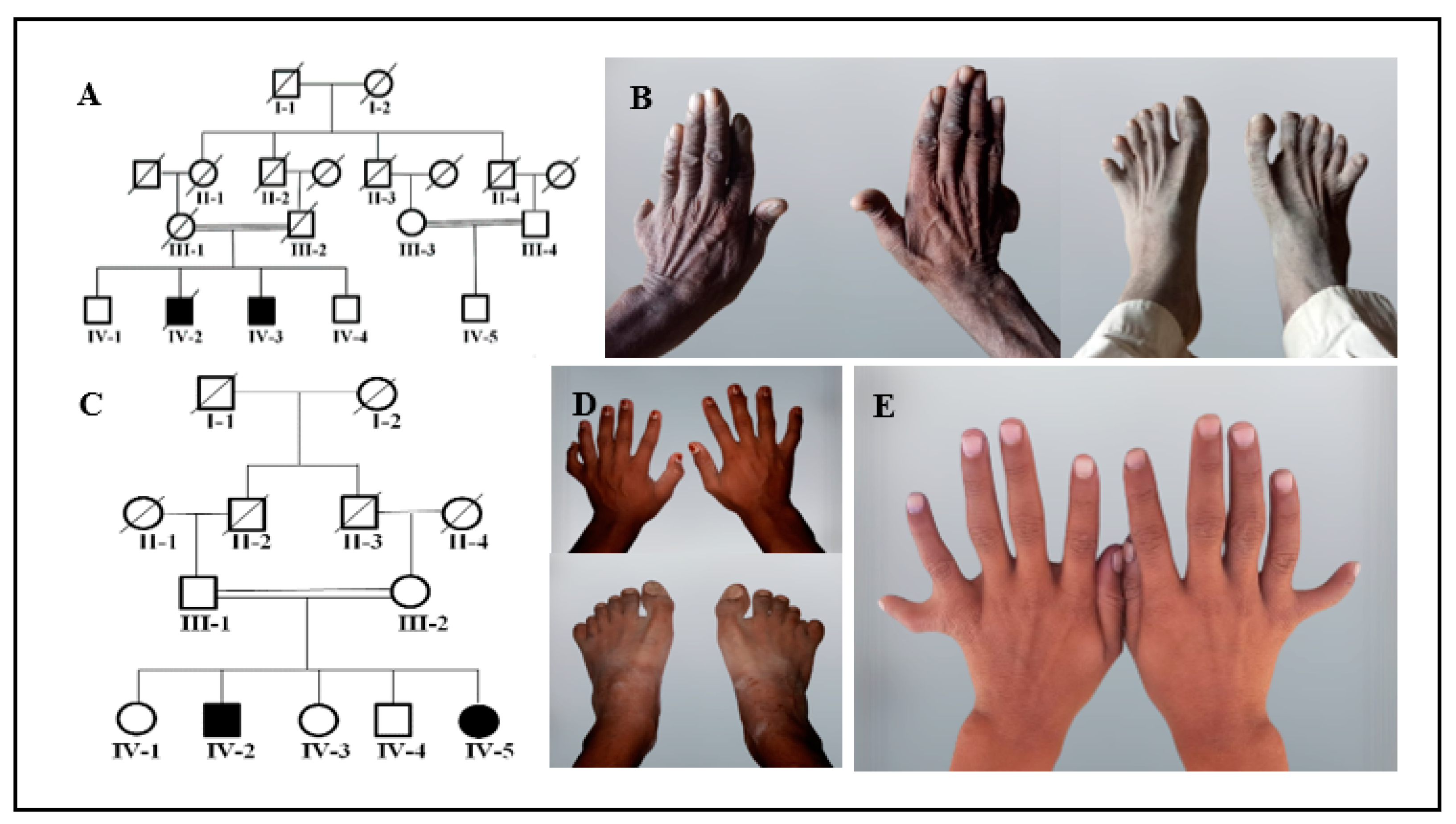
3.1. Clinical Description

Family A includes two affected members (IV-2 and IV-3) with PAP, syndactyly, and clinodactyly in a recessive pattern (Figure 1A). Family A demonstrated intra-familial phenotypic differences. The affected individual IV-2 presented PAPA in both hands and feet, fused second and third digits in both feet and clinodactyly in both hands (Figure 1B) (Table 1). The other affected individual, IV-3 (not shown in the figure), had only PAPA in both hands. Family B also comprises two affected members (IV-2 and IV-5) presenting with autosomal recessive PAPA with an intra-familial phenotypic difference (Figure 1C). The affected member IV-2 presented PAPA in the left hand and feet, clinodactyly of the sixth finger, 5/6 cutaneous syndactyly in the right foot, a sandal gap, and an abnormal shape of the big toe (Figure 1D). The affected individual IV-5 demonstrated a bilateral PAPA in the hands (Figure 1D) and had normal feet (Table 1). No facial dysmorphism, dental abnormalities, hearing problems, or heart defects were observed in any of the family members. Developmental delays were not observed, and all of the affected members from each family had a normal skull, nails, eyesight, weight, and height.

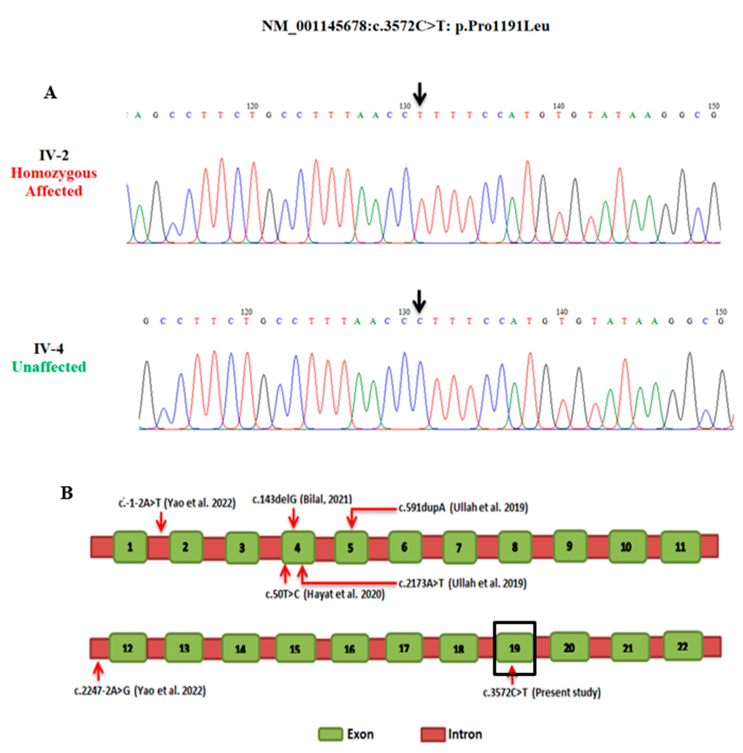
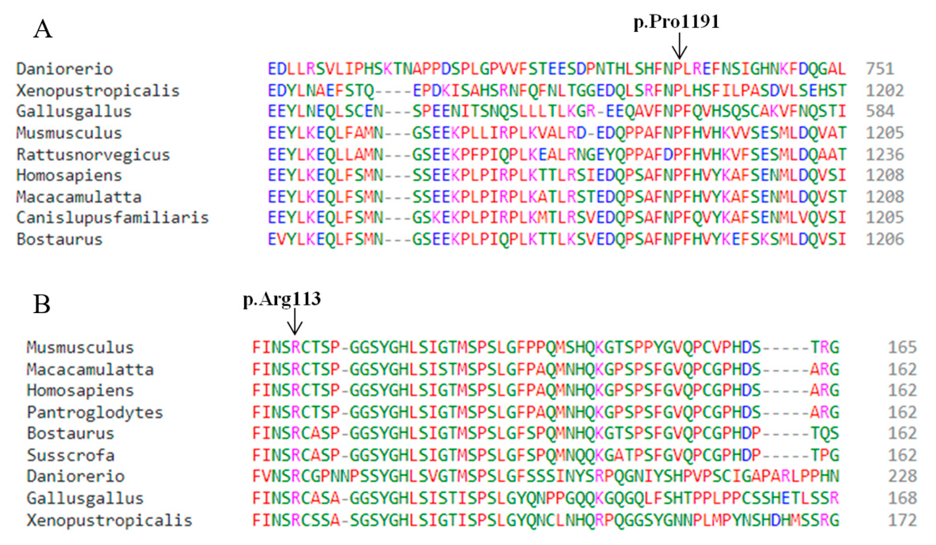
3.2. Molecular Findings

DNA samples from affected individuals IV-2 (family A) and IV-2 (family B) were subjected to whole-exome sequencing. Variant filtering in family A revealed a novel missense variant (c.3572C>T: p.Pro1191Leu) in exon 19 of the KIAA0825 gene (Table 1 and Table 2). According to ACMG classification, the identified KIAA0825 variant is classified as a variant of uncertain significance. In gnomAD, there are nine heterozygotes of this variant found in the South Asian region. However, the variant is absent in South Asian population databases like GenomeAsia100k, IndiGenomes, and South Asian Genome and Exome (SAGE). The segregation analysis of family A confirmed the co-segregation of the missense KIAA0825 variant with the disease phenotype. The mutation analysis demonstrated IV-2 as being mutation homozygous affected and IV-4 as wildtype homozygous unaffected in family A (Figure 2A). The mutation was novel and had not previously been described.
In family B, a variant analysis revealed a previously reported nonsense variant (c.337C>T: p.Arg113*) in exon 4 of the GLI1 gene (Table 1 and Table 2) [30]. The Sanger analysis confirmed the co-segregation of the nonsense GLI1 variant with the disease phenotype. The mutation analysis demonstrated IV-2 and IV-5 as mutation homozygous affected and IV-4 as wildtype homozygous unaffected (Figure 3A).

3.3. Pathogenicity Validation and Protein Stability

The predicted biological pathogenicity of the identified variants is presented in Table 2. Multiple sequence alignments through Clustal Omega revealed high conservation of Pro1191 (KIAA0825) and Arg113 (GLI1) across several species (Figure 4). I-Mutant3.0. predicted the decreased stability of the mutant (p.Pro1191Leu) KIAA0825 protein with a reliability index of 2 and a DGG value prediction of 0.16Kcal/mol. Mupro also predicted the mutant KIAA0825 protein with decreased stability, having a confidence score of −0.5.

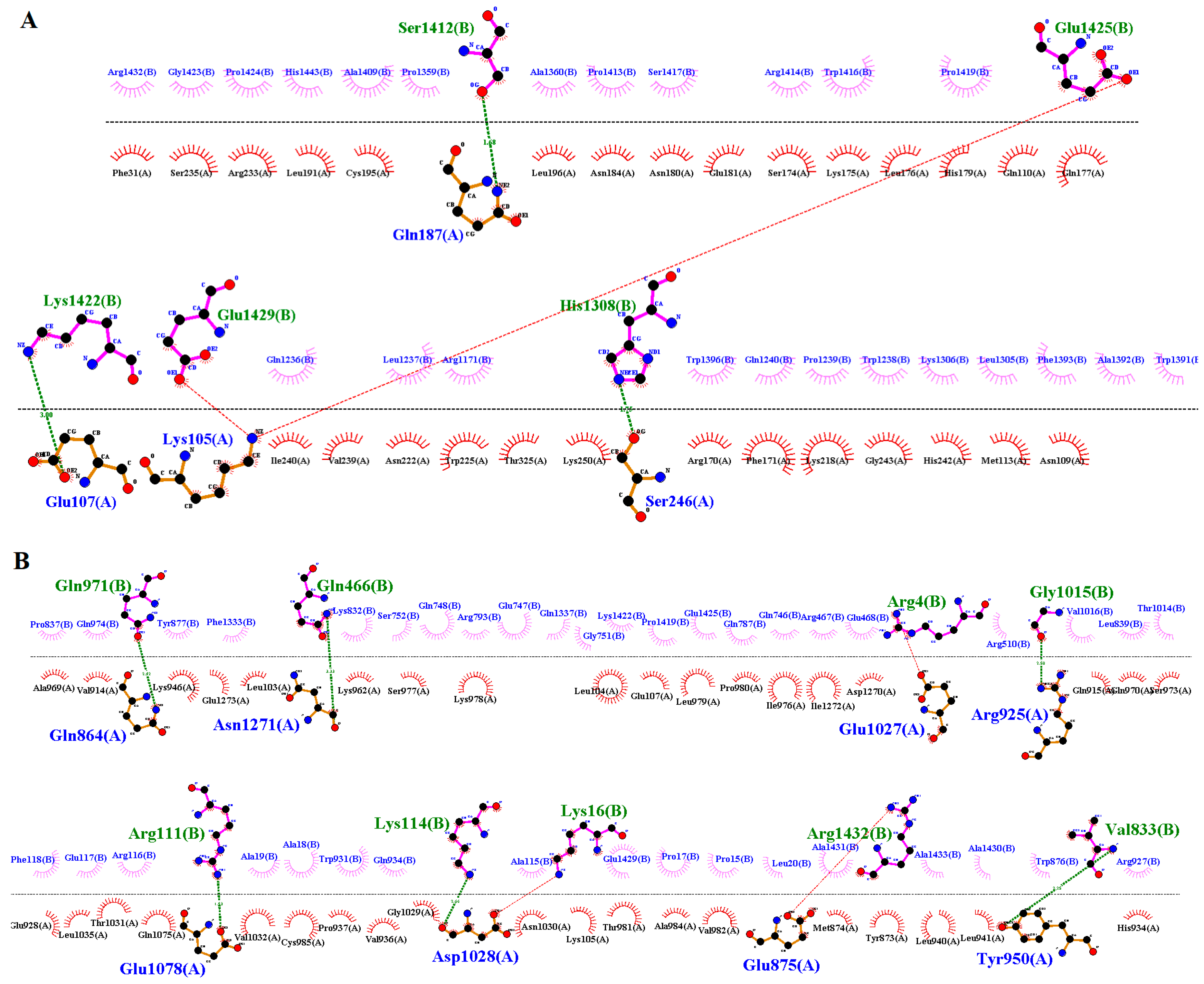
3.4. Modeling and Docking Analysis

Three-dimensional structures of the wildtype and mutant KIAA0825 were modeled by I-TASSER (Figure 5A,B). By structure comparison, the wildtype and mutant KIAA0825 showed 0.71 percent identity and failed to completely superimpose on each other (Figure 5C). The docking analysis revealed a significant interaction alteration between the wildtype and mutant KIAA0825 proteins with the close interactor C1orf16. Four residues of the wildtype KIAA0825 made an interaction with five residues of C1orf167 via three hydrogen bonds and two salt bridges, while eight residues of the mutant KIAA0825 interacted with nine residues of C1orf167 via seven hydrogen bonds and two salt bridges (Figure 6A,B) (Table S1).
The wildtype and mutant 3-D models of GLI1 were also generated by I-TASSER (Figure 7A,B). The wildtype and mutant GLI1 failed to superimpose on each other (Figure 7C) and showed 9.82 percent identity. Interactions of the wildtype and mutant GLI1 proteins with close interactor (SUFU) demonstrated interactional changes. Five residues of the wildtype GLI1 interacted with six residues of SUFU via three hydrogen bonds and three salt bridges, while four residues of each mutant GLI1 and SUFU interacted via four hydrogen bonds (Figure 8A,B) (Table S1).

4. Discussion

The present study reports two unrelated consanguineous Pakistani families with autosomal recessive PAPA with intra- and inter-familial phenotypic variations. Family A included two affected members who inherited PAPA with a recessive inheritance pattern. The individual IV-2 of family A presented PAPA in both hands and feet, cutaneous syndactyly in both feet, and clinodactyly in both hands, while individual IV-3 of family A had PAPA in both hands but no other hand or feet abnormalities. WES of family A revealed a novel missense variant (c.3572C>T: p.Pro1191Leu) in the KIAA0825 gene. The identified variant co-segregated with the disease phenotype. The KIAA0825 gene is mapped on chromosome 5q15 and encodes a 553 amino acids protein. So far, the function of the KIAA0825 protein has not been characterized. A few studies have been performed on the mouse orthologous gene 2210408I21Rik. The expression of 2210408I21Rik was observed in the developing limb buds from E11.5 to E15, and a homozygous knock-out mouse (2210408I21Riktm1b(EUCOMM)Wtsi) demonstrated several skeletal irregularities affecting growth, body size, and reduction in bone mineral density [13]. To date, a total of six other pathogenic variants in KIAA0825 have been reported in subjects with PAP. Four pathogenic variants [p.(Gln198Thrfs*21), p.(Lys725*), p.(Leu17Ser), and p.(Cys48Serfs*28)] were reported in Pakistani families [7,8,31] as well as two splice site variants [c.-1-2A>T and c.2247-2A>G] in a Chinese fetus (Table 3) [32].
A phenotypic comparison between the present study and the previous reports of KIAA0825 is summarized in Table 4. KIAA0825 consists of a long isoform that includes 1275 amino acids and a small isoform that includes 324 amino acids. The variant identified in our study affects only the long isoform.
Family B is the second Pakistani family with PAP, having a previously reported nonsense variant (c.337C>T: p.Arg113*) in the GLI1 gene. The WES of family B revealed a known nonsense variant (c.337C>T: p.Arg113*) in the GLI1 gene, and the identified variant was co-segregated with the disease phenotype. We are unable to assess the segregation of the variant in the parents. In the presence of two homozygous siblings in the family, parents can be considered obligate carriers of the variant. GLI1 acts as a pathway mediator for hedgehog signaling. When the hedgehog molecule binds to its receptor, the GLI proteins become active, resulting in the transcription of target genes involved in bone formation and patterning. GLI1 is mapped on chromosome 12q13.3 and encodes an 1106 amino acid protein [33]. Several domains are present in the GLI1 protein, including degron degradation signals (amino acids Dn 77–116 and Dc 464–469), SUFU binding domains (amino acids SU 111–125 and C-terminus), a zinc finger domain (ZF amino acids 235–387), nuclear localization signal (NLS 380–420 amino acids), and the transactivation domain (1020–1091 amino acid) [33]. Previously, Palencia-Campos et al. (2017) reported the same stop gain GLI1 variant (p.Arg113*) in a Pakistani family with non-syndromic PAPA [30]. Their affected individuals had presented bilateral PAPA in feet, and one individual (patient 7) with additional PAP in the left hand. The affected member (IV-2) of the present Family B displayed the same phenotype as patient 7 of the Palencia report. However, additionally, clinodactyly of the sixth finger and 5/6 cutaneous syndactyly in the right foot were also observed in the same individual (IV-2) of the present study. Individual IV-5 presented PAPA only in the hands, while the feet were normal. This shows a considerable phenotypic difference in the present study in comparison to the previous findings by Palencia. So far, four Pakistani and two Turkish families showing intra-and inter-familial phenotypic variations in polydactyly due to mutation in GLI1 (Table 5) have been reported. A phenotypic comparison between the present study and the previous reports of GLI1 is summarized in Table 6. The identified variant p.(Arg113*) in family B, located in the degron degradation signals domain, results in the truncation of the protein before the Zinc finger domains. Previously, two variants, p. (Thr355Asn) and p.(Ser378Leu), were reported in the ZF motif region, and three others [p.(Leu506Gln); p.(Gln644*); p.(Trp780*)] were mapped between the transactivation domain and the nuclear localization signal.

5. Conclusions

We present two unrelated Pakistani families having PAPA with intra- and inter-familial phenotypic variability. These two different families harbor the two different genetic variants in KIAA0825 and GLI1 underlying the disease. We have identified the seventh novel variant in KIAA0825 causing polysyndactyly and clinodactyly. The novel variant in KIAA0825 expands the mutational spectrum and demonstrates new molecular signaling cascades required for proper limb orientation and growth. A known variant of GLI1 was found in a second Pakistani family in a different region with phenotypic variability with respect to previous findings. As a transcription factor, the presence of a truncated/abnormal GLI1 protein can disrupt the function of the downstream genes responsible for limb development. The present study will also aid in genetic counseling of affected families with polydactyly.

Supplementary Materials

The following supporting information can be downloaded at: https://www.mdpi.com/article/10.3390/genes14040869/s1, Table S1: Protein-Protein interaction of the wildtype and mutant KIAA0825 with C1ORF167 and the wildtype and mutant GLI1 with SUFU.

Author Contributions

S.A., A.U.K. and M.I. were involved in patient recruitment, conducting experiments, and data analysis. S.A., M.Z.A. and M.M. (Muhammad Muzammal) performed bioinformatic analysis and carried out the sequence data analysis. S.A., M.M. (Mari Muurinen) S.H. and P.L. were involved in the manuscript drafting. M.A.K. and O.M. conceptualized and supervised the study and were also involved in funding acquisition and manuscript drafting. All authors have read and agreed to the published version of the manuscript.

Funding

This research was funded by Finnish National Agency for Education (EDUFI fellowship, grant number: OPH-363-2022). The APC was funded by the University of Helsinki.

Institutional Review Board Statement

The study was conducted in accordance with the Declaration of Helsinki, and approved by the institutional ethical review board of Gomal University D.I.Khan (IRB# 04/ERB/GU), KPK, Pakistan.

Informed Consent Statement

Informed consent was obtained from all subjects involved in the study.

Data Availability Statement

The data presented in this study are available on request from the corresponding author. The data are not publicly available due to privacy.

Acknowledgments

We thank all study participants for their cooperation. We also thank 3billion, South Korea, for their generous help in performing the whole exome sequencing. Open access funding provided by University of Helsinki.

Conflicts of Interest

The authors declare no conflict of interest.

References

  1. Xiang, Y.; Bian, J.; Wang, Z.; Xu, Y.; Fu, Q. Clinical study of 459 polydactyly cases in China, 2010 to 2014. Congenit. Anom. 2016, 56, 226–232. [Google Scholar] [CrossRef]
  2. Ahmad, S.; Ali, M.Z.; Muzammal, M.; Mir, F.A.; Khan, M.A. The molecular genetics of human appendicular skeleton. Mol. Genet. Genom. 2022, 297, 1195–1214. [Google Scholar] [CrossRef]
  3. Umair, M.; Ahmad, F.; Bilal, M.; Ahmad, W.; Alfadhel, M. Clinical genetics of polydactyly: An updated review. Front. Genet. 2018, 9, 447. [Google Scholar] [CrossRef] [Green Version]
  4. Ahmad, Z.; Liaqat, R.; Palander, O.; Bilal, M.; Zeb, S.; Ahmad, F.; Jawad Khan, M.; Umair, M. Genetic overview of postaxial polydactyly: Updated classification. Clin. Genet. 2022, 103, 3–15. [Google Scholar] [CrossRef]
  5. Biesecker, L.G. Polydactyly: How many disorders and how many genes? 2010 update. Dev. Dyn. 2011, 240, 931–942. [Google Scholar] [CrossRef] [Green Version]
  6. Malik, S. Polydactyly: Phenotypes, genetics and classification. Clin. Genet. 2014, 85, 203–212. [Google Scholar] [CrossRef]
  7. Hayat, A.; Umair, M.; Abbas, S.; Rauf, A.; Ahmad, F.; Ullah, S.; Ahmad, W.; Khan, B. Identification of a novel biallelic missense variant in the KIAA0825 underlies postaxial polydactyly type A. Genomics 2020, 112, 2729–2733. [Google Scholar] [CrossRef]
  8. Bilal, M.; Ahmad, W. A Frameshift Variant in KIAA0825 Causes Postaxial Polydactyly. Mol. Syndromol. 2021, 12, 20–24. [Google Scholar] [CrossRef]
  9. Stewart, T.A.; Bhat, R.; Newman, S.A. The evolutionary origin of digit patterning. EvoDevo 2017, 8, 1–7. [Google Scholar] [CrossRef] [Green Version]
  10. Verma, P.K.; El-Harouni, A.A. Review of literature: Genes related to postaxial polydactyly. Front. Pediatr. 2015, 3, 8. [Google Scholar] [CrossRef] [Green Version]
  11. Tickle, C.; Towers, M. Sonic hedgehog signaling in limb development. Front. Cell Dev. Biol. 2017, 5, 14. [Google Scholar] [CrossRef] [PubMed] [Green Version]
  12. Lopez-Rios, J. The many lives of SHH in limb development and evolution. Semin. Cell Dev. Biol. 2016, 49, 116–124. [Google Scholar] [CrossRef] [PubMed]
  13. Koscielny, G.; Yaikhom, G.; Iyer, V.; Meehan, T.F.; Morgan, H.; Atienza-Herrero, J.; Blake, A.; Chen, C.-K.; Easty, R.; Di Fenza, A. The International Mouse Phenotyping Consortium Web Portal, a unified point of access for knockout mice and related phenotyping data. Nucleic Acids Res. 2014, 42, D802–D809. [Google Scholar] [CrossRef] [Green Version]
  14. Steinhaus, R.; Proft, S.; Schuelke, M.; Cooper, D.N.; Schwarz, J.M.; Seelow, D. MutationTaster2021. Nucleic Acids Res. 2021, 49, W446–W451. [Google Scholar] [CrossRef]
  15. Rentzsch, P.; Schubach, M.; Shendure, J.; Kircher, M. CADD-Splice—Improving genome-wide variant effect prediction using deep learning-derived splice scores. Genome Med. 2021, 13, 1–12. [Google Scholar] [CrossRef]
  16. Adzhubei, I.A.; Schmidt, S.; Peshkin, L.; Ramensky, V.E.; Gerasimova, A.; Bork, P.; Kondrashov, A.S.; Sunyaev, S.R. A method and server for predicting damaging missense mutations. Nat. Methods 2010, 7, 248–249. [Google Scholar] [CrossRef] [Green Version]
  17. Ng, P.C.; Henikoff, S. SIFT: Predicting amino acid changes that affect protein function. Nucleic Acids Res. 2003, 31, 3812–3814. [Google Scholar] [CrossRef] [PubMed] [Green Version]
  18. Shihab, H.A.; Gough, J.; Cooper, D.N.; Stenson, P.D.; Barker, G.L.; Edwards, K.J.; Day, I.N.; Gaunt, T.R. Predicting the functional, molecular, and phenotypic consequences of amino acid substitutions using hidden Markov models. Hum. Mutat. 2013, 34, 57–65. [Google Scholar] [CrossRef] [Green Version]
  19. Sievers, F.; Higgins, D.G. Clustal Omega for making accurate alignments of many protein sequences. Protein Sci. 2018, 27, 135–145. [Google Scholar] [CrossRef] [Green Version]
  20. Capriotti, E.; Fariselli, P.; Casadio, R. I-Mutant2. 0: Predicting stability changes upon mutation from the protein sequence or structure. Nucleic Acids Res. 2005, 33, W306–W310. [Google Scholar] [CrossRef] [Green Version]
  21. Cheng, J.; Randall, A.; Baldi, P. Prediction of protein stability changes for single-site mutations using support vector machines. Proteins Struct. Funct. Bioinform. 2006, 62, 1125–1132. [Google Scholar] [CrossRef]
  22. Untergasser, A.; Cutcutache, I.; Koressaar, T.; Ye, J.; Faircloth, B.C.; Remm, M.; Rozen, S.G. Primer3—New capabilities and interfaces. Nucleic Acids Res. 2012, 40, e115. [Google Scholar] [CrossRef] [PubMed] [Green Version]
  23. Okonechnikov, K.; Golosova, O.; Fursov, M.; Ugene Team. Unipro UGENE: A unified bioinformatics toolkit. Bioinformatics 2012, 28, 1166–1167. Available online: http://ugene.net/ (accessed on 5 September 2022).
  24. Kent, W.J.; Sugnet, C.W.; Furey, T.S.; Roskin, K.M.; Pringle, T.H.; Zahler, A.M.; Haussler, D. The human genome browser at UCSC. Genome Res. 2002, 12, 996–1006. [Google Scholar] [CrossRef] [Green Version]
  25. Yang, J.; Zhang, Y. I-TASSER server: New development for protein structure and function predictions. Nucleic Acids Res. 2015, 43, W174–W181. [Google Scholar] [CrossRef] [PubMed] [Green Version]
  26. Szklarczyk, D.; Gable, A.L.; Lyon, D.; Junge, A.; Wyder, S.; Huerta-Cepas, J.; Simonovic, M.; Doncheva, N.T.; Morris, J.H.; Bork, P. STRING v11: Protein–protein association networks with increased coverage, supporting functional discovery in genome-wide experimental datasets. Nucleic Acids Res. 2019, 47, D607–D613. [Google Scholar] [CrossRef] [Green Version]
  27. Vajda, S.; Yueh, C.; Beglov, D.; Bohnuud, T.; Mottarella, S.E.; Xia, B.; Hall, D.R.; Kozakov, D. New additions to the C lus P ro server motivated by CAPRI. Proteins: Struct. Funct. Bioinform. 2017, 85, 435–444. [Google Scholar] [CrossRef] [Green Version]
  28. Pettersen, E.F.; Goddard, T.D.; Huang, C.C.; Couch, G.S.; Greenblatt, D.M.; Meng, E.C.; Ferrin, T.E. UCSF Chimera—A visualization system for exploratory research and analysis. J. Comput. Chem. 2004, 25, 1605–1612. [Google Scholar] [CrossRef] [Green Version]
  29. Laskowski, R.A.; Swindells, M.B. LigPlot+: Multiple ligand–protein interaction diagrams for drug discovery. J. Chem. Inf. Model. 2011, 51, 2778–2786. [Google Scholar] [CrossRef]
  30. Palencia-Campos, A.; Ullah, A.; Nevado, J.; Yıldırım, R.; Unal, E.; Ciorraga, M.; Barruz, P.; Chico, L.; Piceci-Sparascio, F.; Guida, V. GLI1 inactivation is associated with developmental phenotypes overlapping with Ellis–van Creveld syndrome. Hum. Mol. Genet. 2017, 26, 4556–4571. [Google Scholar] [CrossRef] [Green Version]
  31. Ullah, I.; Kakar, N.; Schrauwen, I.; Hussain, S.; Chakchouk, I.; Liaqat, K.; Acharya, A.; Wasif, N.; Santos-Cortez, R.L.P.; Khan, S. Variants in KIAA0825 underlie autosomal recessive postaxial polydactyly. Hum. Genet. 2019, 138, 593–600. [Google Scholar] [CrossRef]
  32. Yao, Y.; Deng, S.; Zhu, F. Prenatal Detection of Novel Compound Heterozygous Splice Site Variants of the KIAA0825 Gene in a Fetus with Postaxial Polydactyly Type A. Genes 2022, 13, 1230. [Google Scholar] [CrossRef] [PubMed]
  33. Bakar, A.; Ullah, A.; Bibi, N.; Khan, H.; ur Rahman, A.; Ahmad, W.; Khan, B. A novel homozygous variant in the GLI1 underlies postaxial polydactyly in a large consanguineous family with intra familial variable phenotypes. Eur. J. Med. Genet. 2022, 65, 104599. [Google Scholar] [CrossRef] [PubMed]
  34. Yousaf, M.; Ullah, A.; Azeem, Z.; Isani Majeed, A.; Memon, M.I.; Ghous, T.; Basit, S.; Ahmad, W. Novel heterozygous sequence variant in the GLI1 underlies postaxial polydactyly. Congenit. Anom. 2020, 60, 115–119. [Google Scholar] [CrossRef] [PubMed]
  35. Ullah, A.; Umair, M.; Majeed, A.I.; Jan, A.; Ahmad, W. A novel homozygous sequence variant in GLI1 underlies first case of autosomal recessive pre-axial polydactyly. Clin. Genet. 2019, 95, 540–541. [Google Scholar] [CrossRef] [PubMed]
Figure 1. Pedigrees and images of family A and family B. (A) Pedigree of family A showing autosomal recessive inheritance. Blank “Square” and “circle” presents normal males and females while shaded “square” and “circle” presents affected males and females, respectively. A diagonal line over the “square” and “circle” indicates deceased individuals. Double lines between “square” and “circle” shows the consanguineous marriage. (B) Images of affected member IV-2 of family A displaying PAPA in both hands and feet, 2/3 cutaneous syndactyly in both feet and clinodactyly in both hands. (C) Pedigree of family B presenting autosomal recessive inheritance. Blank “Square” and “circle” presents normal males and females while shaded “square” and “circle” presents affected males and females, respectively. A diagonal line over the “square” and “circle” indicates deceased individuals. Double lines between “square” and “circle” shows the consanguineous marriage. (D) Images of the hands and feet of individual IV-2 of family B showing PAPA in the left hand and both feet, sixth finger clinodactyly in the right hand, 5/6 cutaneous syndactyly in the right foot, sandal gap, and abnormal big toe shape. (E) Image of individual IV-5 from family B demonstrating bilateral PAPA in hands.
Genes 14 00869 g001
Figure 2. Sequence chromatogram of exon 19 of KIAA0825. Panel (A) presents individual IV-2 as homozygous affected and IV-4 as homozygous wild type. Arrow highlights the position of the substitution mutation (c.3572C>T: p.Pro1191Leu). Panel (B) presents a schematic diagram of the KIAA0825 gene structure showing exons. Red arrows indicate the position of mutations reported in polydactyly to date. Variant found in the present study is marked in black square.
Genes 14 00869 g002
Figure 3. Sequence chromatogram of exon 4 of GLI1. Panel (A) presents individuals IV-2 and IV-5 as homozygous affected and IV-4 as homozygous wild type. Arrow highlights the position of the substitution mutation (c.337C>T: p.Arg113*). Panel (B) presents the coding areas of GLI1 and anticipated domains, and the arrows indicate the reported mutations in polydactyly to date. (DN/DC: degron degradation signals, SU: SUFU binding domains, ZN: a zinc finger domain, NLS: nuclear localization signal, TAD: transactivation domain).
Genes 14 00869 g003
Figure 4. Conservation profile of KIAA0825 and GLI1 across several species predicted by Clustal Omega. (A) Conservation profile of Pro1191 (KIAA0825). (B) Conservation profile of Arg113 (GLI1).
Genes 14 00869 g004
Figure 5. Three-dimensional structural presentations of KIAA0825 predicted by I-Tasser (A) Wildtype KIAA0825 (B) Mutant KIAA0825 (C) Superimposed Wildtype and mutant KIAA0825.
Genes 14 00869 g005
Figure 6. Interactions of wildtype and mutant KIAA0825 with close interactor C1orf167. (A) Four residues (chain A) of the wildtype KIAA0825 interacting with five residues (chain B) of C1orf167 via three hydrogen bonds and two salt bridges. (B) Eight residues (chain A) of the mutant KIAA0825 interacting with nine residues (chain B) of C1orf167 via 7 hydrogen bonds and two salt bridges.
Genes 14 00869 g006
Figure 7. Three-dimensional structural presentations of GLI1 predicted by I-TASSER (A) Wildtype GLI1 (B) Mutant KIAA0825 (C) Superimposed wildtype and mutant GLI1.
Genes 14 00869 g007
Figure 8. Interactions of the wildtype and mutant GLI1 with their close interactor SUFU. (A) Five residues (chain A) of wild type GLI1 interacting with six residues (chain B) of SUFU via three hydrogen bonds and three salt bridges. (B) Four residues (chain A) of mutant GLI1 interacting with four residues (chain B) of SUFU via four hydrogen bonds.
Genes 14 00869 g008
Table 1. Clinical presentation of the affected members in two unrelated families.
FamilyClinical ExaminationsGene/
Mutation
SubjectPolydactylySyndactylyClinodactylyBrachydactylyOther Anomaly
AIV-2
(Male)
Bilateral PAPA of hands and bilateral PAPA of feetBilateral 2/3 cutaneous syndactyly of feetBilateral clinodactyly of handsSandal gap
abnormal big toe shape
KIAA0825
c.3572C>T
p.Pro1191Leu
IV-3
(Male)
Bilateral PAPA of hands
BIV-2
(Male)
Unilateral PAPA of hand and bilateral PAPA of feet GLI1
c.337C>T
p.Arg113*
IV-5 (Female)Bilateral PAPA of hands
Table 2. Detail of the variants and in silico pathogenicity predictions.
Gene ZygosityGenomic Position (hg19)mRNA TranscriptcDNA ChangeAmino Acid ChangegnomAD Allele CountgnomAD AllgnomAD South Asian
KIAA0825 Homozygous5:93,721,994NM_001145678c.3572C>Tp.Pro1191Leu9 Heterozygote0.000057310.0003953
ToolsCADD_phredMutation TasterMutation AssessorM-capSiftPolyphen-2FATHMMProvean
Predictions33Disease causingMediumDamagingDamagingDamagingDamagingDamaging
GLI1 ZygosityGenomic position (hg19)mRNA TranscriptcDNA changeAmino acid changegnomAD allele countgnomAD AllgnomAD South Asian
Homozygous12:57,858,599NM_005269c.337C>Tp.Arg113*1 Heterozygote0.000003970.0000326
ToolsCADD_phredMutation TasterFATHMM
Predictions36Disease causingDamaging
Table 3. Description of previously reported mutations in KIAA0825 in association with PAP.
StudyPhenotypeMutation TypeNaturecDNA Amino Acid ChangeEthnicity
Yao et al., 2022 [32]PAPASplice siteHETc.-1-2A>T Chinese
Yao et al., 2022 [32]PAPASplice siteHETc.2247-2A>GChinese
Bilal and Ahmad, 2021 [8]PAPAFrameshiftHZc.143delGp. 28Cys>Ser48fs*Pakistani
Hayat el at., 2020 [7]PAPAMissenseHZc.50T>Cp. 17Leu>SerPakistani
Ullah et al., 2019 [31]PAPA/BFrameshiftHZc.591dupA p. 21Gln>Thr198fs*Pakistani
Ullah et al., 2019 [31]PAPA/BNonsenseHZc. 2173A>T p. Lys 725*Pakistani
Present studyPAPA with cutaneous syndactylyMissesnseHZc. 3572 C >Tp.1191Pro >LeuPakistani
PAP: Postaxial polydactyly, HZ: Homozygous, HET: Heterozygous, *: Terminated.
Table 4. Phenotypic comparison of the patients in the present study with previously reported patients with pathogenic variants in KIAA0825.
StudySubjectPhenotypes
PAPAPAPBSyndactylyCamptodactylyClinodactyly
HandsFeetHandsFeetHandsFeetHandsFeetHandsFeet
Present studyIV-2++++++++
IV-3+
Yao et al., 2022 [32]Fetus II-2++++
Bilal, 2021 [8]IV-2++++++
IV-3+++
Hayat et at., 2020 [7]IV-1++++++
Ullah et al., 2019 [31]V-1++++++
V-2++++++
++: bilateral, +: unilateral, −: Absent, PAPA: Postaxial polydactyly type A, PAPB: Postaxial polydactyly type B.
Table 5. Description of previously reported pathogenic GLI1 variants associated with PAP.
StudyPhenotypeMutation TypeNaturecDNA Amino Acid ChangeEthnicity
Bakar et al., 2022 [33]PAPA/BMissense HETc.1133C>Tp.378Ser>LeuPakistani
Yousaf et al., 2020 [34] PAPA/BMissense HETc.1064C>Ap.355Thr>AsnPakistani
Ullah et al., 2019 [35]PPDMissense HZc.1517T>Ap.506Leu>GlnPakistani
Palencia et al., 2017 [30]PAPANonsense HZc.337C>Tp.Arg113*Pakistani
Palencia et al., 2017 [30]Ranging from simple PAP to EVC syndromeNonsenseHZc.2340G>A p.Trp780*Turkish
Palencia et al., 2017 [30]Ranging from simple PAP to EVC syndromeNonsenseHZc.1930C>T p.Gln644*Turkish
Present studyPAPANonsense HZc.337C>Tp.Arg113*Pakistani
PAP: Postaxial polydactyly, PPD: Preaxial polydactyly, HZ: Homozygous, HET: Heterozygous, *: Terminated.
Table 6. Phenotypic comparison of the patients in the present study with previously reported patients with pathogenic variants in GLI1.
StudyMemberPhenotypes
PPDPAPAPAPB SyndactylyCamptodactyly Clinodactyly
HandsFeetHandsFeetHandsFeetHandsFeetHandsFeetHandsFeet
Present studyIV-2+++++
IV-5++
Bakar et al., 2022 [33]III-2++++
III-9++
IV-3++++
IV-4++++
IV-14++
Yousaf et al., 2020 [34]III-4+
IV-2++++
IV-3+
IV-6++
IV-8++
Ullah et al., 2019 [35]VI-4+
VI-5++
Palencia et al., 2017 [30]Patient 1With EVC ++++
Patient 2++
Patient 3++
Patient 4No confirmed phenotype
Patient 5 With EVC++++
Patient 6 With EVC++++
Patient 7+++
Patient 8++
Patient 9++
Patient 10++
++: bilateral, +: unilateral, −: Absent, PPD: Preaxial polydactyly, PAPA: Postaxial polydactyly type A, PAPB: Postaxial polydactyly type B, EVC: Ellis-van Creveld syndrome.
Disclaimer/Publisher’s Note: The statements, opinions and data contained in all publications are solely those of the individual author(s) and contributor(s) and not of MDPI and/or the editor(s). MDPI and/or the editor(s) disclaim responsibility for any injury to people or property resulting from any ideas, methods, instructions or products referred to in the content.

