Review

The Developmental Origins of Cancer: A Review of the Genes Expressed in Embryonic Cells with Implications for Tumorigenesis

by
Savitha Balachandran
and
Aru Narendran
*
Departments of Pediatrics, Oncology, Biochemistry and Molecular Biology, Physiology and Pharmacology, Cumming School of Medicine, University of Calgary, 2500 University Dr. NW, Calgary, AB T2N 1N4, Canada
*
Author to whom correspondence should be addressed.
Genes 2023, 14(3), 604; https://doi.org/10.3390/genes14030604
Submission received: 26 November 2022 / Revised: 19 January 2023 / Accepted: 22 February 2023 / Published: 28 February 2023
(This article belongs to the Section Human Genomics and Genetic Diseases)

Abstract

:
Tumorigenesis, which involves the uncontrolled proliferation and differentiation of cells, has been observed to imitate a variety of pathways vital to embryonic development, motivating cancer researchers to explore the genetic origins of these pathways. The pluripotency gene regulatory network is an established collection of genes that induces stemness in embryonic cells. Dysregulation in the expression genes of the pluripotency gene networks including OCT4, SOX2, NANOG and REX1 have been implicated in tumor development, and have been observed to result in poorer patient outcomes. The p53 pathway is a highly important regulatory process in a multitude of cell types, including embryonic, and the tumor suppressor gene TP53 is widely regarded as being one of the most important genes involved in tumorigenesis. Dysregulations in TP53 expression, along with altered expression of developmentally originating p53 regulators such as MDM2 and MDM4 have been implicated in various cancers, leading to poorer prognosis. Epithelial–mesenchymal transition (EMT), the process allowing epithelial cells to undergo biochemical changes to mesenchymal phenotypes, also plays a vital role in the fate of both embryonic and neoplastic cells. Genes that regulate EMT such as Twist1, SOX9 and REX1 have been associated with an increased occurrence of EMT in cancer cells, leading to enhanced cell stemness, proliferation and metastasis. The class of RNA that does not encode for proteins, known as non-coding RNA, has been implicated in a variety of cellular processes and emerging research has shown that its dysregulation can lead to uncontrolled cell proliferation and differentiation. Genes that have been shown to play a role in this dysregulation include PIWIL1, LIN28A and LIN28B, and have been associated with poorer patient outcomes and more aggressive cancer subtypes. The identification of these developmentally regulated genes in tumorigenesis has proved to play an advantageous role in cancer diagnosis and prognosis, and has provided researchers with a multitude of new target mechanisms for novel chemotherapeutic research.

1. Introduction

The development, proliferation and migration of cancer cells have been observed mimicking critical pathways of embryonic development, providing a rationale for targeting these pathways in novel therapeutics’ development for cancer therapies. Over the last few years, various collections of developmentally vital genes, which remain relatively quiescent in normal tissues, have been identified in preclinical and clinical studies of cancer. This is a promising step forward for cancer therapy research, as these identified genes carry the potential to inform molecular pathways and regulatory factors that can be targeted to inhibit cancer progression. Normally, embryonic stem cells (ESCs), which are derived from the inner cell mass of the pre-implanted blastocyst-stage embryo, drive the proliferation of cells to facilitate embryonic development by their ability to be both self-renewing and pluripotent [1,2]. The diverse patterns of gene expression that promote these properties have also been observed in cancer cells, specifically in cancer stem cells (CSCs). The CSCs are the fraction of the tumor mass that are distinct in their capacity to promote self-renewal and tumor development. Such cells have also been shown to mediate cancer cell migration and invasion to facilitate subsequent tumor metastasis [3]. Thus, it has been proposed that the dysregulation of critical developmentally regulated genes may contribute to the initiation, development and metastasis of certain cancers. This review aims to provide an overview of the recent literature on a selection of genes that have been studied in relation to cancer, focusing primarily on the oncogenic mechanisms and cellular pathways they influence (Figure 1).

2. Pluripotency Gene Regulatory Network

It is well established that the pluripotent state of embryonic cells is controlled by a network of genes that encode for critical pluripotency transcription factors which promote an intricate network of regulatory events [3]. Genes commonly associated with this pluripotency network are: OCT4 (also known as POU51), SOX2, NANOG, REX1, STAT3 and KLF4 [3]. Though determined to be the gatekeeper genes of the pluripotent state, recent studies have observed the specific processes by which disruptions in this network in progenitor cells may contribute to early tumor development. Disruptions in these circuits lead to the dysregulated expression of such transcription factors, inducing pluripotency-disrupting normal cell maturation and promoting the uncontrolled differentiation into cancer stem cells.
The most commonly associated gene is OCT4, or octamer-binding transcription factor 4 [4]. Expressed in unfertilized oocytes, OCT4 stimulates the initiation and preservation of pluripotency in the inner cell mass of blastocysts and epiblasts. It prevents the differentiation of blastomeres into extraembryonic trophectoderm cells, thus controlling pluripotency. Via epigenetic modifications such as DNA methylation and histone modifications, OCT4 is silenced when differentiation is necessary. Embryonic stem cells contain hypomethylated OCT3 gene loci which are rich with active histone marks to ensure pluripotency. In somatic cells, OCT4 is silenced via DNA methylation in the promoter and enhancer regions, thus it is not expressed in normal adult cells. In cancer cells, OCT4 has been established as a driver of neoplastic growth in various types of cancer, and has also been associated with poorer prognosis and shorter survival rates in clinical situations [4]. A recent study found that active histone methylation and acetylation marks, such as H3K4me3 and H3K9acS10p in the promoter region, were responsible for the overexpression of OCT4 in breast cancer [4]. This study confirmed not only that neoplastic tissue contained a higher than normal expression of OCT4, but also demonstrated the functional importance of OCT4 in tumor bulk. Silencing of OCT4 lowered the rate of proliferation and increased apoptotic activity—indicating that OCT4 plays a role in the anti-apoptotic characteristics of cancer cells. Furthermore, this study concluded that OCT4 plays a role in promoting cell migration, and enhances motility to support metastasis [4]. Enhanced expression of OCT4 has also been observed contributing to resistance to chemotherapeutic drugs and tumor recurrence [5,6].
SOX2 is another important gene in the regulatory network controlling pluripotency. SOX2 is a part of the SRY-related gene family, and the expression of SOX2 begins at the morula stage of the embryo [7]. It is expressed in both embryonic and extraembryonic cells during development and is observed in various tissues after birth, as opposed to OCT4 which is silenced in adult tissue. SOX2 appears to be essential for regeneration in adult tissue, though dysregulations of this gene have been observed in neoplastic cells [7]. Studies have uncovered the important role that SOX2 has in cancer cell proliferation, anti-apoptotic properties and cell invasion in a variety of cancers [7]. Recently, it has been reported that SOX2 is associated with rapid onset of metastasis and decreased survival rates [8]. Interestingly, SOX2 has also been observed in association with changes in cancer cell metabolism such as enhanced oxidative phosphorylation, glycolysis and fatty acid metabolism, along with increased mitochondrial quantity, which is thought to contribute to the cells’ ability to metastasize [8].
NANOG, another gene that is associated with the pluripotency network, exhibits similar properties and clinical outcomes in cancer as the genes previously described [9]. During embryogenesis, it controls pluripotency in the epiblast and prevents the differentiation into the primitive endoderm. Elevated levels of NANOG have been observed in a variety of cancers, and are observed to be regulated by post translational modifications, specifically phosphorylation [9], Recent studies have observed distinct mechanisms by which high expressions of NANOG promotes tumorigenesis, including the ability for immune evasion [10].
REX1 (reduced expression 1) is another gene involved in embryonic stem cells that promotes pluripotency [11]. Studies found that the hypermethylation of the promoter region of this gene contributed to reduced expression. This manifested clinically through increased cancer progression and advanced tumor stage [11].
Signal transducer and activator of transcription 3 (STAT3) has been shown to play a crucial role in the maintenance of pluripotency and somatic cell reprogramming through a number of mechanisms [12,13,14,15]. For example, in ESCs Leukemia Inhibitory factor (LIF) helps to maintain the undifferentiated state enabled by the activated STAT3 pathway [12]. However, Humphrey et al. have reported that in certain experimental conditions the maintenance of hES stemness may occur through STAT3 independent mechanisms [16].
The transcription factor KLF4 (Krüppel-like factor 4) has been implicated as functioning in the regulation of the gene expression programs involved in the maintenance of naïve pluripotency [17,18].

3. The p53 Pathway

The p53 pathway is a highly important physiological pathway that plays a vital role in genomic integrity and cell fate in embryonic, somatic and neoplastic cells [19,20]. The pathway is activated by stress signals, which initiates cell cycle arrest—allowing for the repair of damaged DNA; meanwhile, continued activation can result in senescence and/or apoptosis [19,20]. The tumor suppressor gene TP53, encoding for the p53 protein, is widely accepted as being one of the most important genes in tumor development, and is observed to be mutated or deleted in up to 50% of cancers [19,20]. The p53 protein, once activated, functions primarily as a transcription factor to regulate a large network of downstream processes that control cell fate [19,20]. A multitude of single nucleotide polymorphisms and nonsynonymous polymorphisms have been identified in the TP53 gene (a vast majority in the DNA-binding segment region) which give rise to modified p53 function [21]. These mutations have the ability to render the protein incapable of regulating the downstream expression of genes, resulting in dysregulated occurrences of cell cycle arrest and apoptosis [21]. Mutations in this gene have also been observed to confer cancer-associated abilities to the mutated protein (referred to as “gain-of-function” activities) which can promote tumor development, metastases and increased resistance to anticancer therapies [21]. Studies have shown that the p53 protein promotes cell programmed death/apoptosis, allowing them to enter apoptosis in response to certain chemotherapeutics [22]. Specifically, certain mutations in p53 were observed to suppress the activation of the downstream p53 upregulated modulator of apoptosis (PUMA) protein, which is typically activated by chemotherapeutic agents to promote apoptosis of cancer cells. Other apoptosis-initiating downstream genes that are impacted by mutations in p53 include BAX (Bcl-2-associated X protein) and NOXA (Phorbol-12-myristate-13-acetate-induced protein 1), which are both essential for the apoptotic activity of the p53 pathway [22]. MDM2 and MDM4 are both genes that encode proteins that regulate the function of p53. MDM2, which is vital for embryonic development, controls low p53 levels in normal cell types by forming a negative feedback loop with the p53 protein (whereby increased p53 levels induce expression of MDM2, which leads to the degradation of p53). Overamplification of MDM2-encoded proteins has been observed in more than 17% of tumors [23]. Single nucleotide polymorphisms have been observed in the MDM2 gene, correlating with tumor development and progression and an earlier onset of cancer occurrence [20,23]. The MDM4 protein functions alongside MDM2 to regulate p53, and its overexpression is also observed in cancers [23]. MDM4 binds to and inactivates p53 (as opposed to MDM2, which promotes its degradation) [23]. In addition, several of the single nucleotide polymorphisms identified in MDM4 have been linked to various cancers and their aggressiveness [20,23].

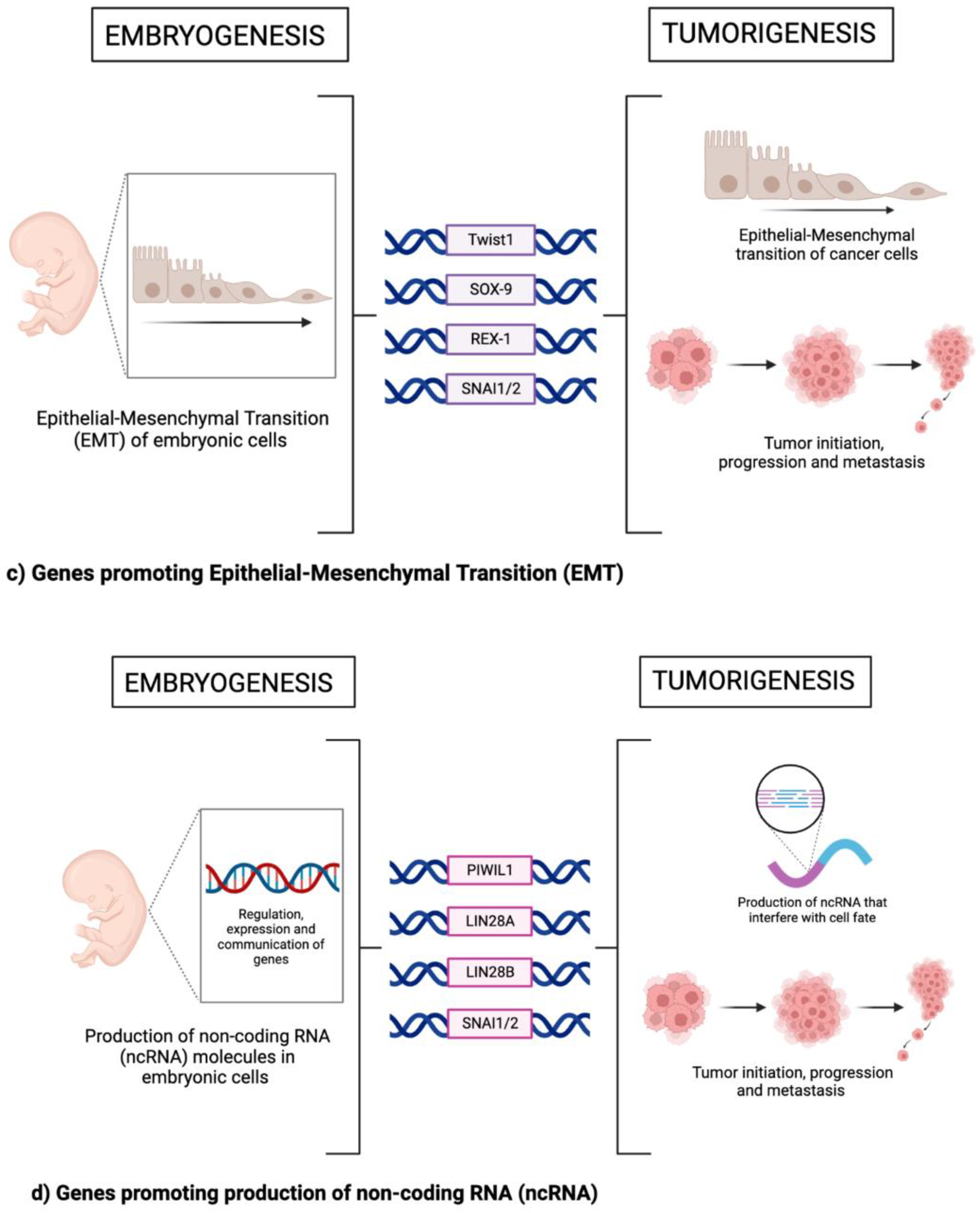
4. Epithelial Mesenchymal Transition (EMT)

Epithelial-mesenchymal transition (EMT) is the process by which polarized epithelial cells of the basement membrane sustain various biochemical modifications to become phenotypically mesenchymal [24,25,26]. Mesenchymal cells are capable of cellular processes such as migration and invasion, and become more resistant to apoptosis, thus making it an important development in associated physiological processes.
EMT is involved in three primary processes [24,25,26]. The first is during embryonic gastrulation and organ development, known as Type 1 EMT, in which the primitive epithelium (epiblast) transitions into primary mesenchyme, giving rise to the mesoderm, endoderm and mobile neural crest cells. The second process, or Type 2 EMT, is wound healing, tissue regeneration and/or organ fibrosis. Type 3 EMT is associated with neoplastic cells, specifically those with enhanced cancer progression and metastasis. Several studies have found that carcinoma cells adopt a mesenchymal phenotype by observing the expression of various mesenchymal markers (such as desmin, vimentin, α-SMA and FSP1), and it is these cells that enter into the invasion–metastasis cascade, establishing secondary colonies at distant sites in the body [24,25,26].
Various genes have been identified as promoters of EMT. Twist1, a transcription regulator during embryonic development, is an important driver of EMT and a key regulator of metastasis in cancer. A landmark study found that the suppression of Twist1 prevented the spreading of breast cancer cells to the lungs [27]. This has since led to the discovery of Twist1 as a key component in promoting metastasis in a number of other cancers [28].
Another important gene associated with EMT and cancer metastasis is SOX9, which specifically activates the HIPPO-YaP pathway [29]. The Hippo signaling pathway plays a central role in regulating cell proliferation and cell fate. This pathway is considered a tumor suppressive pathway which controls the expression of two proteins: YAP and TAZ—both of which are vital for tissue regeneration and organ development but which are also observed in tumorigenesis [29]. The YAP/TAZ proteins have been observed to promote cancer stem cell phenotypes, cell proliferation and metastasis and are often overexpressed in tumors [29]. Recent studies have found that the activation of this pathway can also promote EMT in carcinoma cells [30]. The silencing of SOX9 resulted in reduced invasion and proliferation of carcinoma cells [30]. Increased expression of epithelial markers and the downregulated expression of mesenchymal markers have also been observed, indicating reduced occurrences of EMT [30]. Similar findings were also observed in esophageal squamous cell carcinoma, where the downregulation of both YAP and SOX9 resulted in the suppression of malignant phenotypes in tumors [30]. REX1, previously mentioned as a driver of pluripotency, has also been found to function as a promoter of EMT as a result of overexpression. In addition, studies have found that cells with overexpressed REX1 downregulated E-cadherin levels and activated the JAK2/STAT3 signaling pathway, which could play a role in neoplastic cell invasion and metastasis [31].
Another very important gene involved in the promotion of EMT is SNAI1, which encodes for the Snail1 zinc finger transcriptional repressor, a member of the Snail superfamily of transcription factors. The Snail superfamily of transcription factors, which are encoded by genes such as SNAI1 (snail family transcriptional repressor 1) and SNAI2 (snail family transcriptional repressor 2), is a group of proteins that is very important in EMT and needed for proper gastrulation and mesoderm formation in the developing embryo but in recent years has also been heavily implicated in tumorigenesis [32]. SNAI1 induces EMT by binding to the epithelial-tumor suppressing gene CDH1, amongst other epithelial genes, repressing E-cadherin and claudins. It has been associated with elevated levels in a variety of aggressive cancers, and in chemotherapeutic resistance. For example, in an analysis of triple negative breast cancer cell models, the SNAI1 repressor was associated with as many as 180 different genes, including genes that control cell differentiation and signaling. The suppression of SNAI1 expression was observed to result in the altered regulation and expression of several hundred additional genes, resulting in a significant repression of cell motility in breast cancer cells [32]. Despite SNAI1 being a major transcription factor involved in the epithelial–mesenchymal transition, its silencing was not sufficient in the full reversion of the cells to a more epithelial phenotype, indicating the existence of an intermediary phenotype between epithelial and mesenchymal [32]. SNAI1 has also been observed to be upregulated in hepatocellular carcinoma [33] and ovarian carcinoma [34] amongst several other cancer types, indicating its vital role in cancer progression, and its significance for prognostic and diagnostic tools in cancer research.
SNAI2, occasionally referred to as SLUG, is another member of the Snail transcriptional superfamily that is associated with both embryogenesis and tumor development [35]. SNAI2 upregulation has been associated with altered expression EMT in breast cancer cell models and has been shown to contribute to enhanced incidence of metastasis and lower survival rates in aggressive cancer phenotypes [35]. Higher than normal SNAI2 expression is associated with poorer prognosis, as observed in cases of pancreatic cancer [36], colorectal cancer [37] and ovarian cancer [38], indicating its important role in cancer development and migration.

5. Non-Coding RNAs

Historically, genetic research on cancer cell physiology focused primarily on genes that encode proteins. However, recent discoveries of non-coding RNA (ncRNA) have shown that the class of RNA molecules that do not encode for proteins may also play a vital role in a variety of biological processes, including embryogenesis and tumorigenesis. Emerging research shows that ncRNAs play a vital role in the regulation, expression and communication of genes. Conversely, the dysregulation of the genes encoding RNA have been implicated in uncontrolled cell proliferation and differentiation [39,40].
An important gene that controls the ncRNA-mediated cell proliferation of both embryo and tumor development is the PIWI-like RNA-mediated gene silencing 1 (PIWIL1). PIWIL1 is highly expressed in embryos at seven weeks of development, but is downregulated in subsequent weeks [41]. Expression of this gene leads to the production of piwi-interacting RNA (piRNA), a class of small ncRNAs that bind to PIWI proteins to form a piRNA/PIWI complex which contributes to silencing of gene expressions, genomic rearrangements and stem cell pluripotency maintenance [41]. In recent studies, PIWIL1 has been associated with enhanced cell proliferation and migration [42,43]. Clinical studies comparing PIWIL1 levels in malignant lung tissue to levels in normal lung tissue found that PIWIL1 was detected distinctively in tumor cells, whereas it was absent in normal tissue. The overexpression of PIWIL1 contributes to increased proliferation rates, colony formation, cell migration and invasion [42,43]. Clinically, these effects manifested in a shorter time to relapse and lower overall survival rate. PIWIL1 overexpression was also detected in breast cancer, and interestingly, it was found that the levels at which the gene was expressed can be used to classify different breast cancer subtypes [44]. Breast cancers that were negative for PIWIL1 expression were more likely to be classified as Luminal A subtypes, whereas breast cancers with a high expression of PIWIL1 were more often classified as Luminal B or Triple Negative subtypes [44]. Similar findings have been observed in a variety of other cancers including gastric, colorectal, ovarian, urothelial, nasopharyngeal and renal tumors [45,46,47,48,49].
Another important set of genes that drive the ncRNA-influenced development of embryonic cells and cancer cells includes LIN28A and LIN28B [49,50]. The expression of these genes promotes the downregulation of an important tumor suppressor, a microRNA (miRNA) known as let-7 [49,50]. In embryonic stem cells, LIN28A post-transcriptionally regulates OCT4, an aforementioned driver of pluripotency, by binding to a coding region of its mRNA. Both LIN28A and LIN28B are found to be vital to embryonic survival [49,50]. Let-7 represses a variety of oncogenes, and the loss of let-7 function contributes to tumorigenesis [51]. Increased levels of LIN28A/B and low levels of let-7 are found in a variety of human cancers, such as in liver, lung, ovarian, breast, colorectal and brain cancers [50,52,53,54].
The expression of SNAI1, a gene previously described in the EMT portion of this review and which is a part of the Snail superfamily of transcription factors, is also heavily implicated in and influenced by ncRNAs [55]. Several different miRNAs were observed binding to the 3′UTR region of the snail family transcriptional repressor 1 (SNAI1), resulting from the complementary binding sites between miRNA and these regions of snail. One prominent family of miRNAs which serve as key regulators of SNAI1 is the miR-30 family. These miRNAs have been observed to target SNAI1 mRNA in a variety of cancers, resulting in its inhibition. miR-22 has been observed to target SNAI1 and inhibit EMT in tumor cells and their migration/invasion in a variety of cancers [55]. miR-153 is another miRNA that has been heavily implicated with SNAI1. Studies have reported that the downregulation of SNAI1 by miR-153 results in the suppression of cancer phenotypes, allowing for miR-153 to serve as a potential prognostic marker [56]. Consequently, the downregulation of miR-153 has been associated with increased EMT and thus enhanced metastasis [56]. Furthermore, studies have found that miR-153 has the potential to enhance the effects of chemotherapeutics. The downregulation of miR-153 decreased pancreatic cancer cells’ sensitivity to chemotherapy drugs, while the transfection of miR-153 significantly inhibited tumor cell metastasis and increased apoptosis in chemotherapeutic resistant cancer cells [56]. This indicates that miR-153 and its influence on the Snail pathway may serve as a novel therapeutic target for chemotherapy resistance and as a preventative mechanism of reducing cancer metastasis [56] in a variety of cancers including laryngeal squamous carcinoma [57], malignant melanoma [58], lung cancer [59] and breast cancer [60].
Snail family transcriptional repressor 2 (SNAI2), another member of the Snail superfamily of transcription factors, is also heavily influenced by non-coding RNAs [55]. In recent studies, the epigenetic silencing of the microRNA miR-203 has been observed to upregulate SNAI2 expression. This overexpression of SNAI2 is associated with the increased invasiveness of malignant breast cancers, suggesting that in cases of aggressive and invasive cancers, miR-203 may be epigenetically downregulated or silenced [61]. On the contrary, the expression of the miRNA-181a has been observed to inhibit cell migration, invasion and metastasis by directly targeting the SNAI2 pathway in cases of salivary adenoid cystic carcinoma [62], suggesting that this miRNA could be a potential therapeutic target in the prevention of metastasis in cases of aggressive cancers. In summary, non-coding RNA plays a highly important role in the regulation of genes implicated in both embryogenesis and tumorigenesis, and the emergence of research surrounding non-coding RNA is providing a plethora of new avenues in the discovery of novel cancer biomarkers and therapeutic targets.

6. Clinical Relevance

The identification of developmentally regulated genes in tumor development and metastasis plays a promising role in the clinical diagnosis and prognosis of cancers. Understanding the unique genetic makeup of the tumor allows for a more precise and individualized approach to diagnosis, and a more targeted approach to therapies. In recent advancements, research surrounding the specific genetic origins of cancers has allowed for the development of novel artificial intelligence diagnostic tools to guide medical professionals in their observations of cancer cases and diagnoses. These tools effectively supplement routine histopathological testing [63], providing a more molecularly-guided approach to tumor observation, and alleviating uncertainty in the diagnosis process, allowing for the generation of more effective treatment plans [63]. Furthermore, the advancement in knowledge surrounding developmentally regulated genes in tumorigenesis has opened the door to a multitude of potential chemotherapeutic targets for pharmaceutical research. In vivo administration of let-7 miRNA, which was previously described to be downregulated in tumors through the control of developmentally regulated genes, was found to be effective against animal models of lung and breast cancer, suggesting that it could be the basis of a potential novel therapy [64]. Additionally, further research into the implications of EMT in cancer has identified significant metabolic pathways that are involved in the maintenance and promotion of EMT [64]. Thus, therapeutically targeting the metabolic utility between the epithelial and mesenchymal cell type states through the use of repurposed metabolic inhibitors shows promise in the reduction in metastasis incidence and improved patient outcomes [64]. As previously described, research surrounding the pluripotency gene network, a group of established inducers of pluripotency in embryonic cells and tumor cells, has found that its dysregulated expression has led to increased cellular resistance to chemotherapeutic agents. Recent studies have found that the inhibition of certain molecular targets involved in the pluripotency gene network has led to decreased chemoresistance in certain cancer types. For example, the inhibition of HDAC6, a histone deacetylase enzyme that is involved in the epigenetic regulation of pluripotency genes, has been proposed to increase therapeutic sensitivity in oral squamous cell carcinoma [65]. Knockout of CHD1L, a gene associated with tumor progression, has also been found to downregulate pluripotency factors, leading to decreased drug resistance, along with increased apoptosis of cancer cells and reduced cell proliferation, proving to be a promising target for novel therapeutics [66]. Ongoing research will continue to prove the important role of developmentally regulated genes in tumorigenesis, with the outlook of discovering and testing novel chemotherapeutic targets that will minimize the burden and improve outcomes for those living with cancer.

Author Contributions

Conceptualization, S.B. and A.N.; writing—original draft preparation, S.B.; writing—review and editing; S.B. and A.N.; visualization, S.B.; supervision, A.N. All authors have read and agreed to the published version of the manuscript.

Funding

This research was funded by the Kids Cancer Care Foundation (KCC) Chair in Clinical and Translational Research (grant number 10024168) and the Alberta Children’s Hospital Foundation (ACHF).

Institutional Review Board Statement

Not applicable.

Informed Consent Statement

Informed consent was obtained from all subjects involved in the study.

Data Availability Statement

No new data were created or analyzed in this study. Data sharing is not applicable to this article.

Acknowledgments

This work has been supported in part by a grant from the Alberta Children’s Hospital Foundation (ACHF) and the Kids Cancer care (KCC) (AN). We thank Andy Tran for the critical review of the manuscript.

Conflicts of Interest

The authors declare no conflict of interest.

References

  1. Biswas, A.; Hutchins, R. Embryonic Stem Cells. Stem Cells Dev. 2007, 16, 213–222. [Google Scholar] [CrossRef]
  2. Kim, J.; Orkin, S.H. Embryonic stem cell-specific signatures in cancer: Insights into genomic regulatory networks and implications for medicine. Genome Med. 2011, 3, 75–78. [Google Scholar] [CrossRef] [Green Version]
  3. Manzo, G. Similarities Between Embryo Development and Cancer Process Suggest New Strategies for Research and Therapy of Tumors: A New Point of View. Front. Cell Dev. Biol. 2019, 7, 20. [Google Scholar] [CrossRef]
  4. Kar, S.; Patra, S.K. Overexpression of OCT4 induced by modulation of histone marks plays crucial role in breast cancer progression. Gene 2018, 643, 35–45. [Google Scholar] [CrossRef]
  5. Lu, C.-S.; Shieh, G.-S.; Wang, C.-T.; Su, B.-H.; Su, Y.-C.; Chen, Y.-C.; Su, W.-C.; Wu, P.; Yang, W.-H.; Shiau, A.-L.; et al. Chemotherapeutics-induced Oct4 expression contributes to drug resistance and tumor recurrence in bladder cancer. Oncotarget 2016, 8, 30844–30858. [Google Scholar] [CrossRef] [Green Version]
  6. Villodre, E.S.; Kipper, F.C.; Pereira, M.B.; Lenz, G. Roles of OCT4 in tumorigenesis, cancer therapy resistance and prognosis. Cancer Treat. Rev. 2016, 51, 1–9. [Google Scholar] [CrossRef] [PubMed]
  7. Rizzino, A.; Wuebben, E.L. Sox2/Oct4: A delicately balanced partnership in pluripotent stem cells and embryogenesis. Biochim. Biophys. Acta (BBA)-Gene Regul. Mech. 2016, 1859, 780–791. [Google Scholar] [CrossRef] [PubMed]
  8. de Wet, L.; Williams, A.; Gillard, M.; Kregel, S.; Lamperis, S.; Gutgesell, L.C.; Vellky, J.E.; Brown, R.; Conger, K.; Paner, G.P.; et al. SOX2 mediates metabolic reprogramming of prostate cancer cells. Oncogene 2022, 41, 1190–1202. [Google Scholar] [CrossRef]
  9. Xie, X.; Piao, L.; Cavey, G.S.; Old, M.; Teknos, T.N.; Mapp, A.K.; Pan, Q. Phosphorylation of Nanog is essential to regulate Bmi1 and promote tumorigenesis. Oncogene 2013, 33, 2040–2052. [Google Scholar] [CrossRef] [Green Version]
  10. Saga, K.; Park, J.; Nimura, K.; Kawamura, N.; Ishibashi, A.; Nonomura, N.; Kaneda, Y. NANOG helps cancer cells escape NK cell attack by downregulating ICAM1 during tumorigenesis. J. Exp. Clin. Cancer Res. 2019, 38, 416. [Google Scholar] [CrossRef] [PubMed]
  11. Luk, S.T.; Ng, K.; Zhou, L.; Tong, M.; Wong, T.; Yu, H.; Lo, C.; Man, K.; Guan, X.; Lee, T.K.; et al. Deficiency in embryonic stem cell marker reduced expression 1 activates mitogen-activated protein kinase kinase 6–dependent p38 mitogen-activated protein kinase signaling to drive hepatocarcinogenesis. Hepatology 2020, 72, 183–197. [Google Scholar] [CrossRef] [PubMed]
  12. Zhang, Y.; Wang, D.; Xu, J.; Wang, Y.; Ma, F.; Li, Z.; Liu, N. Stat3 activation is critical for pluripotency maintenance. J. Cell. Physiol. 2018, 234, 1044–1051. [Google Scholar] [CrossRef]
  13. Yang, J.; van Oosten, A.L.; Theunissen, T.W.; Guo, G.; Silva, J.C.; Smith, A. Stat3 Activation Is Limiting for Reprogramming to Ground State Pluripotency. Cell Stem Cell 2010, 7, 319–328. [Google Scholar] [CrossRef] [PubMed] [Green Version]
  14. van Oosten, A.L.; Costa, Y.; Smith, A.; Silva, J.C. JAK/STAT3 signalling is sufficient and dominant over antagonistic cues for the establishment of naive pluripotency. Nat. Commun. 2012, 3, 817. [Google Scholar] [CrossRef] [PubMed] [Green Version]
  15. Raz, R.; Lee, C.-K.; Cannizzaro, L.A.; D’Eustachio, P.; Levy, D.E. Essential role of STAT3 for embryonic stem cell pluripotency. Proc. Natl. Acad. Sci. USA 1999, 96, 2846–2851. [Google Scholar] [CrossRef] [Green Version]
  16. Humphrey, R.K.; Beattie, G.M.; Lopez, A.D.; Bucay, N.; King, C.C.; Firpo, M.T.; Rose-John, S.; Hayek, A. Maintenance of Pluripotency in Human Embryonic Stem Cells Is STAT3 Independent. Stem Cells 2004, 22, 522–530. [Google Scholar] [CrossRef]
  17. Takahashi, K.; Yamanaka, S. Induction of pluripotent stem cells from mouse embryonic and adult fibroblast cultures by defined factors. Cell 2006, 126, 663–676. [Google Scholar] [CrossRef] [PubMed] [Green Version]
  18. Xie, L.; Torigoe, S.E.; Xiao, J.; Mai, D.H.; Li, L.; Davis, F.P.; Dong, P.; Marie-Nelly, H.; Grimm, J.; Lavis, L.; et al. A dynamic interplay of enhancer elements regulates Klf4 expression in naïve pluripotency. Genes Dev. 2017, 31, 1795–1808. [Google Scholar] [CrossRef] [PubMed] [Green Version]
  19. Timmerman, D.; Remmers, T.; Hillenius, S.; Looijenga, L. Mechanisms of TP53 pathway inactivation in embryonic and somatic cells—Relevance for understanding (germ cell) tumorigenesis. Int. J. Mol. Sci. 2021, 22, 5377. [Google Scholar] [CrossRef] [PubMed]
  20. Basu, S.; Murphy, M.E. Genetic modifiers of the p53 pathway. Cold Spring Harb. Perspect. Med. 2016, 6, a026302. [Google Scholar] [CrossRef]
  21. Mogi, A.; Kuwano, H. TP53 mutations in nonsmall cell lung cancer. J. Biomed. Biotechnol. 2011, 2011, 583929. [Google Scholar] [CrossRef] [PubMed] [Green Version]
  22. Huang, Y.; Liu, N.; Liu, J.; Liu, Y.; Zhang, C.; Long, S.; Luo, G.; Zhang, L.; Zhang, Y. Mutant p53 drives cancer chemotherapy resistance due to loss of function on activating transcription of PUMA. Cell Cycle 2019, 18, 3442–3455. [Google Scholar] [CrossRef] [PubMed]
  23. Subhasree, N.; Jiangjiang, Q.; Kalkunte, S.; Minghai, W.; Ruiwen, Z. The MDM2-p53 pathway revisited. J. Biomed. Res. 2013, 27, 254–271. [Google Scholar] [CrossRef] [PubMed]
  24. Kalluri, R.; Weinberg, R.A. The basics of epithelial-mesenchymal transition. J. Clin. Investig. 2009, 119, 1420–1428. [Google Scholar] [CrossRef] [Green Version]
  25. Ramos, F.S.; Wons, L.; Cavalli, I.J.; Ribeiro, E.M. Epithelial-mesenchymal transition in cancer: An overview. Integr. Cancer Sci. Ther. 2017, 4, 1–5. [Google Scholar] [CrossRef] [Green Version]
  26. Ribatti, D.; Tamma, R.; Annese, T. Epithelial-Mesenchymal Transition in Cancer: A Historical Overview. Transl. Oncol. 2020, 13, 100773. [Google Scholar] [CrossRef]
  27. Yang, J.; Mani, S.A.; Donaher, J.L.; Ramaswamy, S.; Itzykson, R.A.; Come, C.; Savagner, P.; Gitelman, I.; Richardson, A.; Weinberg, R.A. Twist, a master regulator of morphogenesis, plays an essential role in tumor metastasis. Cell 2004, 117, 927–939. [Google Scholar] [CrossRef] [Green Version]
  28. Zhao, Z.; Rahman, M.A.; Chen, Z.G.; Shin, D.M. Multiple biological functions of Twist1 in various cancers. Oncotarget 2017, 8, 20380–20393. [Google Scholar] [CrossRef] [Green Version]
  29. Zhou, H.; Li, G.; Huang, S.; Feng, Y.; Zhou, A. SOX9 promotes epithelial-mesenchymal transition via the Hippo-YAP signaling pathway in gastric carcinoma cells. Oncol. Lett. 2019, 18, 599–608. [Google Scholar] [CrossRef] [Green Version]
  30. Wang, L.; Zhang, Z.; Yu, X.; Huang, X.; Liu, Z.; Chai, Y.; Yang, L.; Wang, Q.; Li, M.; Zhao, J.; et al. Unbalanced YAP–SOX9 circuit drives stemness and malignant progression in esophageal squamous cell carcinoma. Oncogene 2018, 38, 2042–2055. [Google Scholar] [CrossRef] [Green Version]
  31. Zeng, Y.-T.; Liu, X.-F.; Yang, W.-T.; Zheng, P.-S. REX1 promotes EMT-induced cell metastasis by activating the JAK2/STAT3-signaling pathway by targeting SOCS1 in cervical cancer. Oncogene 2019, 38, 6940–6957. [Google Scholar] [CrossRef] [PubMed] [Green Version]
  32. Maturi, V.; Morén, A.; Enroth, S.; Heldin, C.; Moustakas, A. Genomewide binding of transcription factor Snail1 in triple-negative breast cancer cells. Mol. Oncol. 2018, 12, 1153–1174. [Google Scholar] [CrossRef] [PubMed] [Green Version]
  33. Jiao, W.; Miyazaki, K.; Kitajima, Y. Inverse correlation between E-cadherin and Snail expression in hepatocellular carcinoma cell lines in vitro and in vivo. Br. J. Cancer 2002, 86, 98–101. [Google Scholar] [CrossRef] [PubMed] [Green Version]
  34. Elloul, S.; Elstrand, M.B.; Nesland, J.M.; Tropé, C.G.; Kvalheim, G.; Goldberg, I.; Reich, R.; Davidson, B. Snail, Slug, and Smad-interacting protein 1 as novel parameters of disease aggressiveness in metastatic ovarian and breast carcinoma. Cancer 2005, 103, 1631–1643. [Google Scholar] [CrossRef]
  35. Alves, C.L.; Elias, D.; Lyng, M.B.; Bak, M.; Ditzel, H.J. SNAI2 upregulation is associated with an aggressive phenotype in fulvestrant-resistant breast cancer cells and is an indicator of poor response to endocrine therapy in estrogen receptor-positive metastatic breast cancer. Breast Cancer Res. 2018, 20, 60. [Google Scholar] [CrossRef] [Green Version]
  36. Masuo, K.; Chen, R.; Yogo, A.; Sugiyama, A.; Fukuda, A.; Masui, T.; Uemoto, S.; Seno, H.; Takaishi, S. SNAIL2 contributes to tumorigenicity and chemotherapy resistance in pancreatic cancer by regulating IGFBP2. Cancer Sci. 2021, 112, 4987–4999. [Google Scholar] [CrossRef]
  37. Hu, Y.; Dai, M.; Zheng, Y.; Wu, J.; Yu, B.; Zhang, H.; Kong, W.; Wu, H.; Yu, X. Epigenetic suppression of E-cadherin expression by Snail2 during the metastasis of colorectal cancer. Clin. Epigenetics 2018, 10, 154. [Google Scholar] [CrossRef]
  38. Taki, M.; Abiko, K.; Baba, T.; Hamanishi, J.; Yamaguchi, K.; Murakami, R.; Yamanoi, K.; Horikawa, N.; Hosoe, Y.; Nakamura, E.; et al. Snail promotes ovarian cancer progression by recruiting myeloid-derived suppressor cells via CXCR2 ligand upregulation. Nat. Commun. 2018, 9, 1685. [Google Scholar] [CrossRef] [Green Version]
  39. Slack, F.J.; Chinnaiyan, A.M. The Role of Non-coding RNAs in Oncology. Cell 2019, 179, 1033–1055. [Google Scholar] [CrossRef]
  40. Pauli, A.; Rinn, J.; Schier, A.F. Non-coding RNAs as regulators of embryogenesis. Nat. Rev. Genet. 2011, 12, 136–149. [Google Scholar] [CrossRef]
  41. Han, Y.-N.; Li, Y.; Xia, S.-Q.; Zhang, Y.-Y.; Zheng, J.-H.; Li, W. Piwi proteins and piwi-interacting RNA: Emerging roles in cancer. Cell. Physiol. Biochem. 2017, 44, 1–20. [Google Scholar] [CrossRef] [PubMed] [Green Version]
  42. Navarro, A.; Tejero, R.; Viñolas, N.; Cordeiro, A.; Marrades, R.M.; Fuster, D.; Caritg, O.; Moises, J.; Muñoz, C.; Molins, L.; et al. The significance of PIWI family expression in human lung embryogenesis and non-small cell lung cancer. Oncotarget 2015, 6, 31544–31556. [Google Scholar] [CrossRef] [PubMed] [Green Version]
  43. Xie, K.; Zhang, K.; Kong, J.; Wang, C.; Gu, Y.; Liang, C.; Jiang, T.; Qin, N.; Liu, J.; Guo, X.; et al. Cancer-testis gene PIWIL1 promotes cell proliferation, migration, and invasion in lung adenocarcinoma. Cancer Med. 2017, 7, 157–166. [Google Scholar] [CrossRef] [PubMed] [Green Version]
  44. Erber, R.; Meyer, J.; Taubert, H.; Fasching, P.A.; Wach, S.; Häberle, L.; Gaß, P.; Schulz-Wendtland, R.; Landgraf, L.; Olbricht, S.; et al. PIWI-Like 1 and PIWI-Like 2 Expression in Breast Cancer. Cancers 2020, 12, 2742. [Google Scholar] [CrossRef] [PubMed]
  45. Shi, S.; Yang, Z.-Z.; Liu, S.; Yang, F.; Lin, H. PIWIL1 promotes gastric cancer via a piRNA-independent mechanism. Proc. Natl. Acad. Sci. USA 2020, 117, 22390–22401. [Google Scholar] [CrossRef]
  46. Chu, H.; Xia, L.; Qiu, X.; Gu, D.; Zhu, L.; Jin, J.; Hui, G.; Hua, Q.; Du, M.; Tong, N.; et al. Genetic variants in noncoding PIWI-interacting RNA and colorectal cancer risk. Cancer 2015, 121, 2044–2052. [Google Scholar] [CrossRef]
  47. Chen, C.; Liu, J.; Xu, G. Overexpression of PIWI proteins in human stage III epithelial ovarian cancer with lymph node metastasis. Cancer Biomark. 2013, 13, 315–321. [Google Scholar] [CrossRef]
  48. Eckstein, M.; Jung, R.; Weigelt, K.; Sikic, D.; Stöhr, R.; Geppert, C.; Agaimy, A.; Lieb, V.; Hartmann, A.; Wullich, B.; et al. Piwi-like 1 and -2 protein expression levels are prognostic factors for muscle invasive urothelial bladder cancer patients. Sci. Rep. 2018, 8, 17693. [Google Scholar] [CrossRef] [Green Version]
  49. Zhuang, K.; Wu, Q.; Jin, C.-S.; Yuan, H.-J.; Cheng, J.-Z. Long non-coding RNA HNF1A-AS is upregulated and promotes cell proliferation and metastasis in nasopharyngeal carcinoma. Cancer Biomark. 2016, 16, 291–300. [Google Scholar] [CrossRef]
  50. Middelkamp, M.; Ruck, L.; Krisp, C.; Sumisławski, P.; Mohammadi, B.; Dottermusch, M.; Meister, V.; Küster, L.; Schlüter, H.; Windhorst, S.; et al. Overexpression of Lin28A in neural progenitor cells in vivo does not lead to brain tumor formation but results in reduced spine density. Acta Neuropathol. Commun. 2021, 9, 185. [Google Scholar] [CrossRef]
  51. Qiu, C.; Ma, Y.; Wang, J.; Peng, S.; Huang, Y. Lin28-mediated post-transcriptional regulation of Oct4 expression in human embryonic stem cells. Nucleic Acids Res. 2009, 38, 1240–1248. [Google Scholar] [CrossRef] [PubMed] [Green Version]
  52. Balzeau, J.; Menezes, M.R.; Cao, S.; Hagan, J.P. The LIN28/let-7 Pathway in Cancer. Front. Genet. 2017, 8, 31. [Google Scholar] [CrossRef] [Green Version]
  53. Pretzsch, E.; Max, N.; Kirchner, T.; Engel, J.; Werner, J.; Klauschen, F.; Angele, M.; Neumann, J. LIN28 promotes tumorigenesis in colorectal cancer but is not associated with metastatic spread. Pathol.—Res. Pract. 2021, 228, 153669. [Google Scholar] [CrossRef] [PubMed]
  54. Guilharducci, R.L.; Xavier, P.L.P.; da Silveira, J.C.; Cordeiro, Y.D.G.; Biondi, L.R.; Strefezzi, R.D.F.; Fukumasu, H. Expression of LIN28A/B and Let-7 miRNAs in canine mammary carcinomas. Cienc. Rural. 2022, 52, e20210171. [Google Scholar] [CrossRef]
  55. Skrzypek, K.; Majka, M. Interplay among snail transcription factor, MicroRNAs, long non-coding RNAS, and circular RNAS in the regulation of tumor growth and metastasis. Cancers 2020, 12, 209. [Google Scholar] [CrossRef] [PubMed] [Green Version]
  56. Xu, Q.; Sun, Q.; Zhang, J.; Yu, J.; Chen, W.; Zhang, Z. Downregulation of miR-153 contributes to epithelial-mesenchymal transition and tumor metastasis in human epithelial cancer. Carcinog. 2012, 34, 539–549. [Google Scholar] [CrossRef] [PubMed] [Green Version]
  57. Zhang, B.; Fu, T.; Zhang, L. MicroRNA-153 suppresses human laryngeal squamous cell carcinoma migration and invasion by targeting the SNAI1 gene. Oncol. Lett. 2018, 16, 5075–5083. [Google Scholar] [CrossRef]
  58. Luan, W.; Shi, Y.; Zhou, Z.; Xia, Y.; Wang, J. circRNA_0084043 promote malignant melanoma progression via miR-153-3p/Snail axis. Biochem. Biophys. Res. Commun. 2018, 502, 22–29. [Google Scholar] [CrossRef]
  59. Zhao, G.; Zhang, Y.; Zhao, Z.; Cai, H.; Zhao, X.; Yang, T.; Chen, W.; Yao, C.; Wang, Z.; Wang, Z.; et al. MiR-153 reduces stem cell-like phenotype and tumor growth of lung adenocarcinoma by targeting Jagged1. Stem Cell Res. Ther. 2020, 11, 170. [Google Scholar] [CrossRef]
  60. Zuo, Z.; Ye, F.; Liu, Z.; Huang, J.; Gong, Y. MicroRNA-153 inhibits cell proliferation, migration, invasion and epithelial-mesenchymal transition in breast cancer via direct targeting of RUNX2. Exp. Ther. Med. 2019, 17, 4693–4702. [Google Scholar] [CrossRef]
  61. Zhang, Z.; Zhang, B.; Li, W.; Fu, L.; Zhu, Z.; Dong, J.-T. Epigenetic silencing of mir-203 upregulates snai2 and contributes to the invasiveness of malignant breast cancer cells. Genes Cancer 2011, 2, 782–791. [Google Scholar] [CrossRef] [PubMed]
  62. Nguyen, L.H.; Robinton, D.A.; Seligson, M.T.; Wu, L.; Li, L.; Rakheja, D.; Comerford, S.A.; Ramezani, S.; Sun, X.; Parikh, M.S.; et al. LIN28B is sufficient to drive liver cancer and necessary for its maintenance in murine models. Cancer Cell 2014, 26, 248–261. [Google Scholar] [CrossRef] [PubMed] [Green Version]
  63. Penson, A.; Camacho, N.; Zheng, Y.; Varghese, A.M.; Al-Ahmadie, H.; Razavi, P.; Chandarlapaty, S.; Vallejo, C.E.; Vakiani, E.; Gilewski, T.; et al. Development of genome-derived tumor type prediction to inform clinical cancer care. JAMA Oncol. 2020, 6, 84–91. [Google Scholar] [CrossRef] [PubMed]
  64. Barh, D.; Malhotra, R.; Ravi, B.; Sindhurani, P. Microrna let-7: An emerging next-generation cancer therapeutic. Curr. Oncol. 2010, 17, 70–80. [Google Scholar] [CrossRef] [PubMed] [Green Version]
  65. Tavares, M.O.; Milan, T.M.; Bighetti-Trevisan, R.L.; Leopoldino, A.M.; de Almeida, L.O. Pharmacological inhibition of HDAC6 overcomes cisplatin chemoresistance by targeting cancer stem cells in oral squamous cell carcinoma. J. Oral Pathol. Med. 2022, 51, 529–537. [Google Scholar] [CrossRef]
  66. Fan, G.-T.; Ling, Z.-H.; He, Z.-W.; Wu, S.-J.; Zhou, G.-X. Suppressing CHD1L reduces the proliferation and chemoresistance in osteosarcoma. Biochem. Biophys. Res. Commun. 2021, 554, 214–221. [Google Scholar] [CrossRef]
Figure 1. Key genes with regulatory implications for both embryogenesis and tumorigenesis. Functions and physiological outcomes of genes that target (a) the pluripotency gene regulatory network, (b) the p53 pathway, (c) that promote epithelial–mesenchymal transitioning and (d) the production of non-coding RNA.
Genes 14 00604 g001aGenes 14 00604 g001b
Disclaimer/Publisher’s Note: The statements, opinions and data contained in all publications are solely those of the individual author(s) and contributor(s) and not of MDPI and/or the editor(s). MDPI and/or the editor(s) disclaim responsibility for any injury to people or property resulting from any ideas, methods, instructions or products referred to in the content.

